passport todownloads.lww.com/wolterskluwer_vitalstream_com/sample-content… · note the three...
TRANSCRIPT

After careful study of this chapter, you should be able to:
1. Describe the function of the sensory system2. Differentiate between the special and general senses and give
examples of each3. Describe the structure of the eye4. List and describe the structures that protect the eye5. Define refraction and list the refractive parts of the eye6. Differentiate between the rods and the cones of the eye7. Compare the functions of the extrinsic and intrinsic muscles of
the eye8. Describe the nerve supply to the eye9. Describe the three divisions of the ear
10. Describe the receptor for hearing and explain how it functions11. Compare static and dynamic equilibrium and describe the
location and function of these receptors12. Explain the function of proprioceptors13. List several methods for treatment of pain14. Describe sensory adaptation and explain its value15. Show how word parts are used to build words related to the
sensory system (see Word Anatomy at the end of the chapter)
The following terms andother boldface terms inthe chapter are definedin the Glossary
accommodation
choroid
cochlea
conjunctiva
convergence
cornea
gustation
lacrimal apparatus
lens (crystalline lens)
olfaction
organ of Corti
ossicle
proprioceptor
refraction
retina
sclera
semicircular canal
sensory adaptation
sensory receptor
tympanic membrane
vestibule
vitreous body
PASSportto Success
Visit thePoint or see theStudent Resource CD inthe back of this book fordefinitions and pronun-
ciations of key terms as well as a pretest
for this chapter.

äPaul’s Second Case: Seeing More of the Sun’s Effects
187
condition, and it does have a hereditary fac-tor.” The doctor dilated Paul’s eyes with dropsand examined the fundus of each eye with anophthalmoscope. In answer to Paul’s query, heexplained that in this way he could examinethe health of the retina and the optic nerveand also look at the vessels at the back of theeye for any signs of diabetes or circulatoryproblems. In addition, he used a tonometer totest for glaucoma, a condition that can dam-age the eye with high fluid pressure. “I need tocheck on another patient,” he told Paul. “ThenI’ll be back to explain what I’ve found. Just sitin the waiting room for a few minutes, please.”
Dr. Gilbert uses his knowledge of thestructure and function of the eye to diagnosemedical conditions. In this chapter, you willlearn about the eye and other sensory systemorgans. As well, we’ll examine the conse-quences of Paul’s sun-loving youth on hiseyes and vision.
Paul glanced once again at the postcardsitting on his entranceway table as he ar-rived home in the evening. He knew it
said he was due for a checkup with his oph-thalmologist, but after his run-in with skincancer 6 months earlier, he was in no hurry tosee the inside of a medical office. Well, it’s justroutine, and I may need a slight change in my pre-scription, so I’ll make the call, he thought.
At the office, Dr. Gilbert greeted Paul andasked how he was feeling in general and if hethought there had been any change in his vi-sion. “My vision sometimes seems a littleblurry, especially when my eyes are tired, butno major changes,” Paul replied. Dr. Gilbertproceeded with the eye exam, testing his vi-sual acuity and checking on his astigmatism.
“You’re right; not much change,” said theophthalmologist. “But, we’ll give you a new pre-scription for your glasses. At 42, you’re a littleyoung for presbyopia—far-sightedness that de-velops with age—but we’ll do the routine checkfor glaucoma. I know your father developed that
188 Unit III Coordination and Control
The SensesThe sensory system protects a person by detecting changesin the environment. An environmental change becomes astimulus when it initiates a nerve impulse, which then travels to the central nervous system (CNS) by way of asensory (afferent) neuron. A stimulus becomes a sensa-tion—something we experience—only when a specializedarea of the cerebral cortex interprets the nerve impulse re-ceived. Many stimuli arrive from the external environmentand are detected at or near the body surface. Others, suchas stimuli from the viscera, originate internally and help tomaintain homeostasis.
Sensory ReceptorsThe part of the nervous system that detects a stimulus isthe sensory receptor. In structure, a sensory receptor maybe one of the following:
n The free dendrite of a sensory neuron, such as the re-ceptors for pain and temperature
n A modified ending, or end-organ, on the dendrite ofan afferent neuron, such as those for touch
n A specialized cell associated with an afferent neuron,such as the rods and cones of the eye’s retina and thereceptors in the other special sense organs
Receptors can be classified according to the type ofstimulus to which they respond:
n Chemoreceptors, such as receptors for taste and smell,detect chemicals in solution.
n Photoreceptors, located in the retina of the eye, re-spond to light.
n Thermoreceptors detect change in temperature. Manyof these receptors are located in the skin.
n Mechanoreceptors respond to movement, such asstretch, pressure, or vibration. These include pressurereceptors in the skin, receptors that monitor body po-sition, and the receptors of hearing and equilibrium inthe ear, which are activated by the movement of ciliaon specialized receptor cells.
Any receptor must receive a stimulus of adequate in-tensity, that is, at least a threshold stimulus, in order to re-spond and generate a nerve impulse.
Special and General SensesAnother way of classifying the senses is according to thedistribution of their receptors. A special sense is localizedin a special sense organ; a general sense is widely distrib-uted throughout the body.
n Special senses
> Vision from receptors in the eye
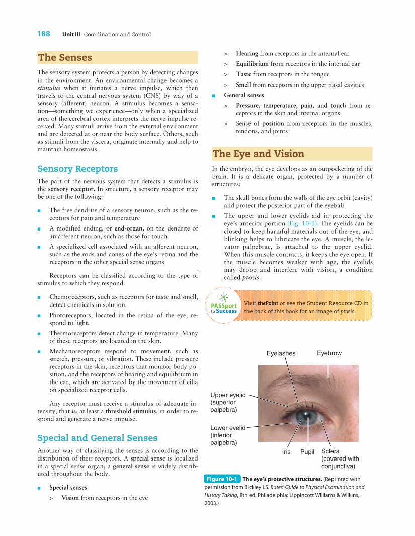
Eyelashes Eyebrow
Upper eyelid (superior palpebra)
Lower eyelid (inferior palpebra)
Iris Pupil Sclera (covered with conjunctiva)
Figure 10-1 The eye’s protective structures. (Reprinted with
permission from Bickley LS. Bates’ Guide to Physical Examination and
History Taking, 8th ed. Philadelphia: Lippincott Williams & Wilkins,
2003.)
PASSportto Success
> Hearing from receptors in the internal ear
> Equilibrium from receptors in the internal ear
> Taste from receptors in the tongue
> Smell from receptors in the upper nasal cavities
n General senses
> Pressure, temperature, pain, and touch from re-ceptors in the skin and internal organs
> Sense of position from receptors in the muscles,tendons, and joints
The Eye and VisionIn the embryo, the eye develops as an outpocketing of thebrain. It is a delicate organ, protected by a number ofstructures:
n The skull bones form the walls of the eye orbit (cavity)and protect the posterior part of the eyeball.
n The upper and lower eyelids aid in protecting theeye’s anterior portion (Fig. 10-1). The eyelids can beclosed to keep harmful materials out of the eye, andblinking helps to lubricate the eye. A muscle, the le-vator palpebrae, is attached to the upper eyelid.When this muscle contracts, it keeps the eye open. Ifthe muscle becomes weaker with age, the eyelidsmay droop and interfere with vision, a conditioncalled ptosis.
Visit thePoint or see the Student Resource CD inthe back of this book for an image of ptosis.

Chapter 10 The Sensory System 189
10
n The eyelashes and eyebrow help to keep foreign mat-ter out of the eye.
n A thin membrane, the conjunctiva (kon-junk-TI-vah),lines the inner surface of the eyelids and covers the vis-ible portion of the white of the eye (sclera). Cellswithin the conjunctiva produce mucus that aids in lu-bricating the eye. Where the conjunctiva folds backfrom the eyelid to the eye’s anterior surface, a sac isformed. The lower portion of the conjunctival sac canbe used to instill medication drops. With age, the con-junctiva often thins and dries, resulting in inflamma-tion and enlarged blood vessels.
n Tears, produced by the lacrimal (LAK-rih-mal) glands(Fig. 10-2), lubricate the eye and contain an enzymethat protects against infection. As tears flow across
Lacrimal gland
Ducts of lacrimal gland
Inferior canal
Superior canal
Lacrimal sac
Nasolacrimal duct
Opening of duct (in nose)
Figure 10-2 The lacrimal apparatus. The lacrimal (tear) gland
and its associated ducts are shown.
the eye from the lacrimal gland, located in the orbit’supper lateral part, they carry away small particles thatmay have entered the eye. The tears then flow intoducts near the eye’s nasal corner where they drain intothe nose by way of the nasolacrimal (na-zo-LAK-rih-mal) duct (see Fig. 10-2). An excess of tears causes a“runny nose”; a greater overproduction of them re-sults in the tears spilling onto the cheeks. With age,the lacrimal glands produce less secretion, but tearsstill may overflow onto the cheek if the nasolacrimalducts become plugged.
CHECKPOINT ä What are some structures thatprotect the eye?
Coats of the EyeballThe eyeball has three separate coats, or tunics (Fig. 10-3).
1. The outermost tunic, called the sclera (SKLE-rah), ismade of tough connective tissue. It is commonly re-ferred to as the white of the eye. It appears white be-cause of the collagen it contains and because it has noblood vessels to add color. (Reddened or “bloodshot”eyes result from inflammation and swelling of bloodvessels in the conjunctiva).
2. The second tunic of the eyeball is the choroid (KO-royd). This coat is composed of a delicate network ofconnective tissue interlaced with many blood vessels.It also contains much dark brown pigment. Thechoroid may be compared to the dull black lining of acamera in that it prevents incoming light rays from
scattering and reflecting off the eye’sinner surface. The blood vessels atthe posterior, or fundus, of the eyecan reveal signs of disease, and visu-alization of these vessels with anophthalmoscope (of-THAL-mo-skope) is an important part of amedical examination.
3. The innermost tunic, the retina(RET-ih-nah), is the eye’s actual receptor layer. It contains light-sensitive cells known as rods andcones, which generate the nerve im-pulses associated with vision.
CHECKPOINT ä What are thenames of thetunics of theeyeball?
Pathway of Light Raysand RefractionAs light rays pass through the eye to-ward the retina, they travel through a
10-2
10-1
Suspensoryligaments
Vitreous body
Sclera
ChoroidTunics
Retina
Foveacentralis
Optic disk(blind spot)
Optic nerve
Iris
Pupil
Cornea
Lens
Aqueoushumor
Ciliarymuscle
Conjunctivalsac
Figure 10-3 The eye. Note the three tunics, the refractive parts of the eye (cornea,
aqueous humor, lens, vitreous body), and other structures involved in vision.
190 Unit III Coordination and Control
series of transparent, colorless parts described below andseen in Figure 10-3. On the way, they undergo a processknown as refraction, which is the bending of light rays asthey pass from one substance to another substance of dif-ferent density. (For a simple demonstration of refraction,place a spoon into a glass of water and observe how thehandle appears to bend at the surface of the water.)Because of refraction, light from a very large area can befocused on a very small area of the retina. The eye’s trans-parent refracting parts are listed here, according to thepathway of light traveling from exterior to interior:
1. The cornea (KOR-ne-ah) is an anterior continuationof the sclera, but it is transparent and colorless,whereas the rest of the sclera is opaque and white. Thecornea is referred to frequently as the window of theeye. It bulges forward slightly and is the main refract-ing structure of the eye. The cornea has no blood ves-sels; it is nourished by the fluids that constantly washover it.
2. The aqueous (A-kwe-us) humor, a watery fluid that fillsmuch of the eyeball anterior to the lens, helps maintainthe cornea’s slight forward curve. The aqueous humoris constantly produced and drained from the eye.
3. The lens, technically called the crystalline lens, is aclear, circular structure made of a firm, elastic mate-rial. The lens has two bulging surfaces and is thus de-
scribed as biconvex. The lens is important in light re-fraction because it is elastic and its thickness can beadjusted to focus light for near or far vision.
4. The vitreous (VIT-re-us) body is a soft jellylike sub-stance that fills the entire space posterior to the lens(the adjective vitreous means “glasslike”). Like theaqueous humor, it is important in maintaining theshape of the eyeball as well as in aiding in refraction.
CHECKPOINT ä What are the structures that refractlight as it passes through the eye?
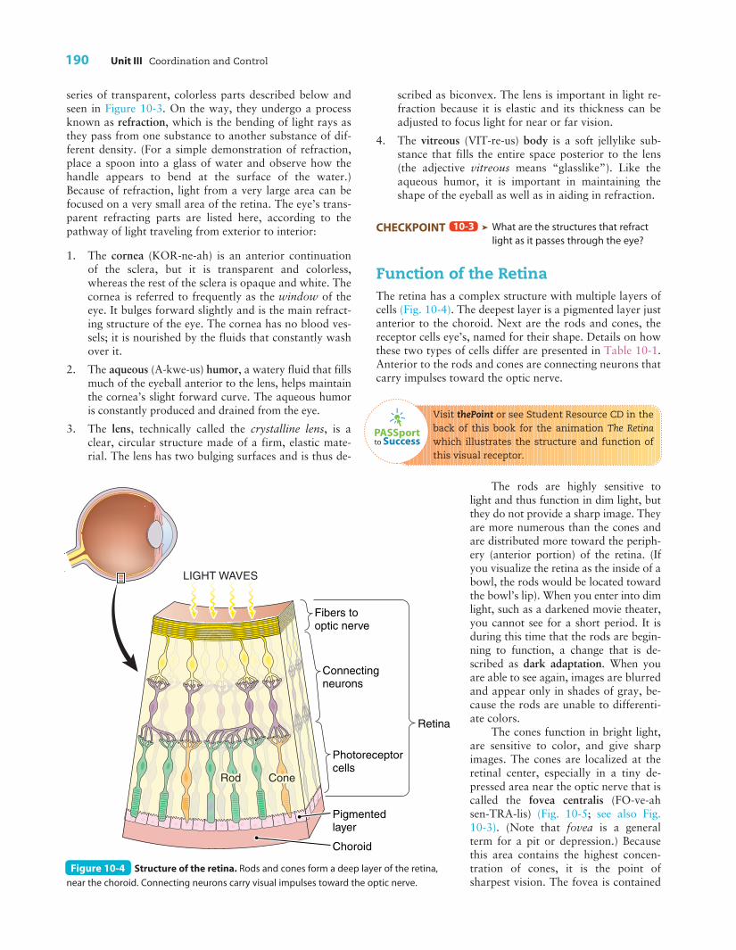
Function of the RetinaThe retina has a complex structure with multiple layers ofcells (Fig. 10-4). The deepest layer is a pigmented layer justanterior to the choroid. Next are the rods and cones, thereceptor cells eye’s, named for their shape. Details on howthese two types of cells differ are presented in Table 10-1.Anterior to the rods and cones are connecting neurons thatcarry impulses toward the optic nerve.
Visit thePoint or see Student Resource CD in theback of this book for the animation The Retinawhich illustrates the structure and function ofthis visual receptor.
The rods are highly sensitive tolight and thus function in dim light, butthey do not provide a sharp image. Theyare more numerous than the cones andare distributed more toward the periph-ery (anterior portion) of the retina. (Ifyou visualize the retina as the inside of abowl, the rods would be located towardthe bowl’s lip). When you enter into dimlight, such as a darkened movie theater,you cannot see for a short period. It isduring this time that the rods are begin-ning to function, a change that is de-scribed as dark adaptation. When youare able to see again, images are blurredand appear only in shades of gray, be-cause the rods are unable to differenti-ate colors.
The cones function in bright light,are sensitive to color, and give sharpimages. The cones are localized at theretinal center, especially in a tiny de-pressed area near the optic nerve that iscalled the fovea centralis (FO-ve-ahsen-TRA-lis) (Fig. 10-5; see also Fig.10-3). (Note that fovea is a generalterm for a pit or depression.) Becausethis area contains the highest concen-tration of cones, it is the point ofsharpest vision. The fovea is contained
10-3
LIGHT WAVES
Fibers to optic nerve
Retina
Pigmented layer
Photoreceptor cells
Connecting neurons
Choroid
Figure 10-4 Structure of the retina. Rods and cones form a deep layer of the retina,
near the choroid. Connecting neurons carry visual impulses toward the optic nerve.
PASSportto Success

Chapter 10 The Sensory System 191
within a yellowish spot, the macula lutea (MAK-u-lah LU-te-ah), an area that may show degenerative changes withage.
There are three types of cones, each sensitive to eitherred, green, or blue light. Color blindness results from alack of retinal cones. People who completely lack cones aretotally colorblind; those who lack one type of cone arepartially color blind. This disorder, because of its patternof inheritance, occurs almost exclusively in males.
The rods and cones function by means of pigmentsthat are sensitive to light. The rod pigment is rhodopsin(ro-DOP-sin), or visual purple. Vitamin A is needed formanufacture of these pigments. If a person is lacking in vi-tamin A, and thus rhodopsin, he or she may have difficultyseeing in dim light because the light is inadequate to acti-vate the rods; this condition is termed night blindness.Nerve impulses from the rods and cones flow into sensoryneurons that eventually merge to form the optic nerve (cra-nial nerve II) at the eye’s posterior (see Figs. 10-3 and 10-5). The impulses travel to the visual center in the brain’soccipital cortex.
When an ophthalmologist (of-thal-MOL-o-jist), aphysician who specializes in treatment of the eye, exam-ines the retina with an ophthalmoscope, he or she can seeabnormalities in the retina and in the retinal blood vessels.Some of these changes may signal more widespread dis-eases that affect the eye, such as diabetes and high bloodpressure (hypertension).
CHECKPOINT ä What are the receptor cells of theretina?
Muscles of the EyeTwo groups of muscles are associated with the eye. Bothgroups are important in adjusting the eye so that a clearimage can form on the retina.
THE EXTRINSIC MUSCLES The voluntary muscles at-tached to the eyeball’s outer surface are the extrinsic (eks-TRIN-sik) muscles. The six ribbonlike extrinsic musclesconnected with each eye originate on the orbital bones and
10-4
10
Characteristic Rods Cones
Table 10-1 Comparison of the Rods and Cones of the Retina
Shape Cylindrical Flask shaped
Number About 120 million in each retina About 6 million in each retina
Distribution Toward the periphery (anterior) of the retina Concentrated at the center of the retina
Stimulus Dim light Bright light
Visual acuity (sharpness) Low High
Pigments Rhodopsin (visual purple) Pigments sensitive to red, green, or blue
Color perception None; shades of gray Respond to color
Fovea centralis (in macula lutea)
Blood vessels
Optic disk
Retina
Figure 10-5 The fundus (back) of the eye as seen through an ophthalmoscope. (Reprinted with permission from Moore KL, Dalley AF.
Clinically Oriented Anatomy, 4th ed. Baltimore: Lippincott Williams & Wilkins, 1999.)
192 Unit III Coordination and Control
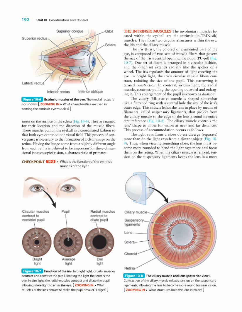
insert on the surface of the sclera (Fig. 10-6). They are namedfor their location and the direction of the muscle fibers.These muscles pull on the eyeball in a coordinated fashion sothat both eyes center on one visual field. This process of con-vergence is necessary to the formation of a clear image on theretina. Having the image come from a slightly different anglefrom each retina is believed to be important for three-dimen-sional (stereoscopic) vision, a characteristic of primates.
CHECKPOINT ä What is the function of the extrinsicmuscles of the eye?
10-5
THE INTRINSIC MUSCLES The involuntary muscles lo-cated within the eyeball are the intrinsic (in-TRIN-sik)muscles. They form two circular structures within the eye,the iris and the ciliary muscle.
The iris (I-ris), the colored or pigmented part of theeye, is composed of two sets of muscle fibers that governthe size of the iris’s central opening, the pupil (PU-pil) (Fig.10-7). One set of fibers is arranged in a circular fashion,and the other set extends radially like the spokes of awheel. The iris regulates the amount of light entering theeye. In bright light, the iris’s circular muscle fibers con-tract, reducing the size of the pupil. This narrowing istermed constriction. In contrast, in dim light, the radialmuscles contract, pulling the opening outward and enlarg-ing it. This enlargement of the pupil is known as dilation.
The ciliary (SIL-e-ar-e) muscle is shaped somewhatlike a flattened ring with a central hole the size of the iris’souter edge. This muscle holds the lens in place by means offilaments, called suspensory ligaments, that project fromthe ciliary muscle to the edge of the lens around its entirecircumference (Fig. 10-8). The ciliary muscle controls thelens’ shape to allow for vision at near and far distances.This process of accommodation occurs as follows.
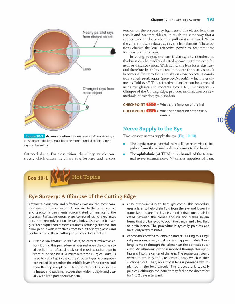
The light rays from a close object diverge (separate)more than do the light rays from a distant object (Fig. 10-9). Thus, when viewing something close, the lens must be-come more rounded to bend the light rays more and focusthem on the retina. When the ciliary muscle is relaxed, ten-sion on the suspensory ligaments keeps the lens in a more
Figure 10-6 Extrinsic muscles of the eye. The medial rectus is
not shown. [ ZOOMING IN ä What characteristics are used in
naming the extrinsic eye muscles? ]
Figure 10-7 Function of the iris. In bright light, circular muscles
contract and constrict the pupil, limiting the light that enters the
eye. In dim light, the radial muscles contract and dilate the pupil,
allowing more light to enter the eye. [ ZOOMING IN ä What
muscles of the iris contract to make the pupil smaller? Larger? ]
Figure 10-8 The ciliary muscle and lens (posterior view).
Contraction of the ciliary muscle relaxes tension on the suspensory
ligaments, allowing the lens to become more round for near vision.
[ ZOOMING IN ä What structures hold the lens in place? ]
Chapter 10 The Sensory System 193
flattened shape. For close vision, the ciliary muscle con-tracts, which draws the ciliary ring forward and relaxes
tension on the suspensory ligaments. The elastic lens thenrecoils and becomes thicker, in much the same way that arubber band thickens when the pull on it is released. Whenthe ciliary muscle relaxes again, the lens flattens. These ac-tions change the lens’ refractive power to accommodatefor near and far vision.
In young people, the lens is elastic, and therefore itsthickness can be readily adjusted according to the need fornear or distance vision. With aging, the lens loses elasticityand therefore its ability to accommodate for near vision. Itbecomes difficult to focus clearly on close objects, a condi-tion called presbyopia (pres-be-O-pe-ah), which literallymeans “old eye.” This refractive disorder can be correctedusing eye glasses and contacts. Box 10-1, Eye Surgery: AGlimpse of the Cutting Edge, provides information on newmethods of treating eye disorders.
CHECKPOINT ä What is the function of the iris?
CHECKPOINT ä What is the function of the ciliarymuscle?
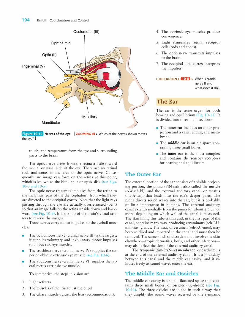
Nerve Supply to the EyeTwo sensory nerves supply the eye (Fig. 10-10):
n The optic nerve (cranial nerve II) carries visual im-pulses from the retinal rods and cones to the brain.
n The ophthalmic (of-THAL-mik) branch of the trigem-inal nerve (cranial nerve V) carries impulses of pain,
10-7
10-6
10
Figure 10-9 Accommodation for near vision. When viewing a
close object, the lens must become more rounded to focus light
rays on the retina.
Box 10-1
Eye Surgery: A Glimpse of the Cutting EdgeCataracts, glaucoma, and refractive errors are the most com-mon eye disorders affecting Americans. In the past, cataractand glaucoma treatments concentrated on managing thediseases. Refractive errors were corrected using eyeglassesand, more recently, contact lenses. Today, laser and microsur-gical techniques can remove cataracts, reduce glaucoma, andallow people with refractive errors to put their eyeglasses andcontacts away. These cutting-edge procedures include:
n Laser in situ keratomileusis (LASIK) to correct refractive er-rors. During this procedure, a laser reshapes the cornea toallow light to refract directly on the retina, rather than infront of or behind it. A microkeratome (surgical knife) isused to cut a flap in the cornea’s outer layer. A computer-controlled laser sculpts the middle layer of the cornea andthen the flap is replaced. The procedure takes only a fewminutes and patients recover their vision quickly and usu-ally with little postoperative pain.
n Laser trabeculoplasty to treat glaucoma. This procedureuses a laser to help drain fluid from the eye and lower in-traocular pressure. The laser is aimed at drainage canals lo-cated between the cornea and iris and makes severalburns that are believed to open the canals and allow fluidto drain better. The procedure is typically painless andtakes only a few minutes.
n Phacoemulsification to remove cataracts. During this surgi-cal procedure, a very small incision (approximately 3 mmlong) is made through the sclera near the cornea’s outeredge. An ultrasonic probe is inserted through this open-ing and into the center of the lens. The probe uses soundwaves to emulsify the lens’ central core, which is thensuctioned out. Then, an artificial lens is permanently im-planted in the lens capsule. The procedure is typicallypainless, although the patient may feel some discomfortfor 1 to 2 days afterward.
194 Unit III Coordination and Control
touch, and temperature from the eye and surroundingparts to the brain.
The optic nerve arises from the retina a little towardthe medial or nasal side of the eye. There are no retinalrods and cones in the area of the optic nerve. Conse-quently, no image can form on the retina at this point,which is known as the blind spot or optic disk (see Figs.10-3 and 10-5).
The optic nerve transmits impulses from the retina tothe thalamus (part of the diencephalon), from which theyare directed to the occipital cortex. Note that the light rayspassing through the eye are actually overrefracted (bent)so that an image falls on the retina upside down and back-ward (see Fig. 10-9). It is the job of the brain’s visual cen-ters to reverse the images.
Three nerves carry motor impulses to the eyeball mus-cles:
n The oculomotor nerve (cranial nerve III) is the largest;it supplies voluntary and involuntary motor impulsesto all but two eye muscles.
n The trochlear nerve (cranial nerve IV) supplies the su-perior oblique extrinsic eye muscle (see Fig. 10-6).
n The abducens nerve (cranial nerve VI) supplies the lat-eral rectus extrinsic eye muscle.
To summarize, the steps in vision are:
1. Light refracts.
2. The muscles of the iris adjust the pupil.
3. The ciliary muscle adjusts the lens (accommodation).
4. The extrinsic eye muscles produceconvergence.
5. Light stimulates retinal receptorcells (rods and cones).
6. The optic nerve transmits impulsesto the brain.
7. The occipital lobe cortex interpretsthe impulses.
CHECKPOINT ä What is cranialnerve II andwhat does it do?
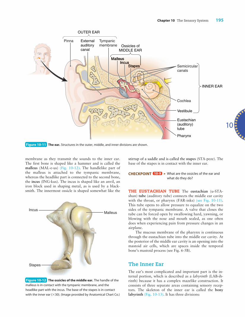
The EarThe ear is the sense organ for bothhearing and equilibrium (Fig. 10-11). Itis divided into three main sections:
n The outer ear includes an outer pro-jection and a canal ending at a mem-brane.
n The middle ear is an air space con-taining three small bones.
n The inner ear is the most complexand contains the sensory receptorsfor hearing and equilibrium.
The Outer EarThe external portion of the ear consists of a visible project-ing portion, the pinna (PIN-nah), also called the auricle(AW-rih-kl), and the external auditory canal, or meatus(me-A-tus), that leads into the ear’s deeper parts. Thepinna directs sound waves into the ear, but it is probablyof little importance in humans. The external auditorycanal extends medially from the pinna for about 2.5 cm ormore, depending on which wall of the canal is measured.The skin lining this tube is thin and, in the first part of thecanal, contains many wax-producing ceruminous (seh-RU-mih-nus) glands. The wax, or cerumen (seh-RU-men), maybecome dried and impacted in the canal and must then beremoved. The same kinds of disorders that involve the skinelsewhere—atopic dermatitis, boils, and other infections—may also affect the skin of the external auditory canal.
The tympanic (tim-PAN-ik) membrane, or eardrum, isat the end of the external auditory canal. It is a boundarybetween this canal and the middle ear cavity, and it vi-brates freely as sound waves enter the ear.
The Middle Ear and OssiclesThe middle ear cavity is a small, flattened space that con-tains three small bones, or ossicles (OS-ih-klz) (see Fig. 10-11). The three ossicles are joined in such a way thatthey amplify the sound waves received by the tympanic
10-8
Trigeminal (V)
Optic (II)
Ophthalmic
Oculomotor (III)
Mandibular
Maxillary
Figure 10-10 Nerves of the eye. [ ZOOMING IN ä Which of the nerves shown moves
the eye? ]
Chapter 10 The Sensory System 195
membrane as they transmit the sounds to the inner ear.The first bone is shaped like a hammer and is called themalleus (MAL-e-us) (Fig. 10-12). The handlelike part ofthe malleus is attached to the tympanic membrane,whereas the headlike part is connected to the second bone,the incus (ING-kus). The incus is shaped like an anvil, aniron block used in shaping metal, as is used by a black-smith. The innermost ossicle is shaped somewhat like the
10
stirrup of a saddle and is called the stapes (STA-peze). Thebase of the stapes is in contact with the inner ear.
CHECKPOINT ä What are the ossicles of the ear andwhat do they do?
THE EUSTACHIAN TUBE The eustachian (u-STA-shun) tube (auditory tube) connects the middle ear cavitywith the throat, or pharynx (FAR-inks) (see Fig. 10-11).This tube opens to allow pressure to equalize on the twosides of the tympanic membrane. A valve that closes thetube can be forced open by swallowing hard, yawning, orblowing with the nose and mouth sealed, as one oftendoes when experiencing pain from pressure changes in anairplane.
The mucous membrane of the pharynx is continuousthrough the eustachian tube into the middle ear cavity. Atthe posterior of the middle ear cavity is an opening into themastoid air cells, which are spaces inside the temporalbone’s mastoid process (see Fig. 6-5B).
The Inner EarThe ear’s most complicated and important part is the in-ternal portion, which is described as a labyrinth (LAB-ih-rinth) because it has a complex mazelike construction. Itconsists of three separate areas containing sensory recep-tors. The skeleton of the inner ear is called the bonylabyrinth (Fig. 10-13). It has three divisions:
10-9
Figure 10-11 The ear. Structures in the outer, middle, and inner divisions are shown.
IncusMalleus
Stapes
Figure 10-12 The ossicles of the middle ear. The handle of the
malleus is in contact with the tympanic membrane, and the
headlike part with the incus. The base of the stapes is in contact
with the inner ear (330). (Image provided by Anatomical Chart Co.)

196 Unit III Coordination and Control
impulses that travel to the brain in the cochlear nerve, abranch of the eighth cranial nerve (formerly called the au-ditory or acoustic nerve). Sound waves ultimately leave theear through another membrane-covered space in the bonylabyrinth, the round window.
Hearing receptors respond to both the pitch (tone) ofsound and its intensity (loudness). The various pitchesstimulate different regions of the organ of Corti. Receptorsdetect higher-pitched sounds near the base of the cochleaand lower-pitched sounds near the top. Loud sounds stim-ulate more cells and produce more vibrations, sendingmore nerve impulses to the brain. Exposure to loud noises,such as very loud music, jet plane noise, or industrialnoises, can damage the receptors for particular pitches ofsound and lead to hearing loss for those tones.
The steps in hearing are:
1. Sound waves enter the external auditory canal.
2. The tympanic membrane vibrates.
3. The ossicles transmit vibrations across the middle earcavity.
4. The stapes transmits the vibrations to the inner ear fluid.
5. Vibrations move cilia on hair cells of the organ ofCorti in the cochlear duct.
6. Movement against the tectorial membrane generatesnerve impulses.
7. Impulses travel to the brain in cranial nerve VIII.
8. The temporal lobe cortex interprets the impulses.
CHECKPOINT ä What is the name of the organ ofhearing, and where is it located?
10-10
n The vestibule consists of two bony chambers that con-tain some of the receptors for equilibrium.
n The semicircular canals are three projecting bonytubes located toward the posterior. Areas at the basesof the semicircular canals also contain receptors forequilibrium.
n The cochlea (KOK-le-ah) is coiled like a snail shell andis located toward the anterior. It contains the recep-tors for hearing.
All three divisions of the bony labyrinth contain afluid called perilymph (PER-e-limf).
Within the bony labyrinth is an exact replica of thisbony shell made of membrane, much like an inner tubewithin a tire. The tubes and chambers of this membranouslabyrinth are filled with a fluid called endolymph (EN-do-limf) (see Fig. 10-13). The endolymph is within the membra-nous labyrinth, and the perilymph surrounds it. These fluidsare important to the sensory functions of the inner ear.
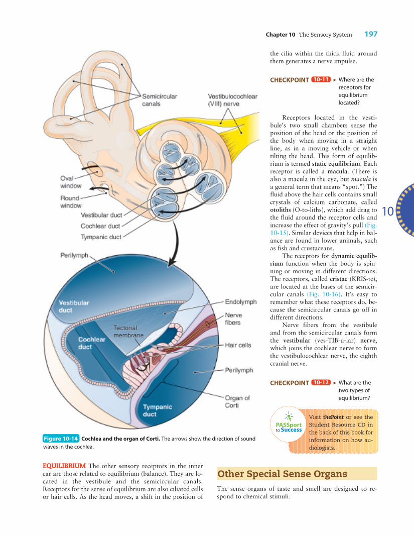
HEARING The organ of hearing, called the organ of Corti(KOR-te), consists of ciliated receptor cells located insidethe membranous cochlea, or cochlear duct (Fig. 10-14).Sound waves enter the external auditory canal and causevibrations in the tympanic membrane. The ossicles amplifythese vibrations and finally transmit them from the stapesto a membrane covering the oval window of the inner ear.
As the sound waves move through the fluids in thesechambers, they set up vibrations in the cochlear duct. As aresult, the tiny, hairlike cilia on the receptor cells (haircells) begin to move back and forth against the tectorialmembrane above them. (The membrane is named from aLatin word that means “roof.”) This motion sets up nerve
Figure 10-13 The inner ear. The vestibule, semicircular canals, and cochlea are made of a bony shell (labyrinth) with an interior
membranous labyrinth. Endolymph fills the membranous labyrinth, and perilymph is around it in the bony labyrinth. The cochlea is the organ
of hearing. The semicircular canals and vestibule are concerned with equilibrium.
Chapter 10 The Sensory System 197
EQUILIBRIUM The other sensory receptors in the innerear are those related to equilibrium (balance). They are lo-cated in the vestibule and the semicircular canals.Receptors for the sense of equilibrium are also ciliated cellsor hair cells. As the head moves, a shift in the position of
10
PASSportto Success
the cilia within the thick fluid aroundthem generates a nerve impulse.
CHECKPOINT ä Where are thereceptors forequilibriumlocated?
Receptors located in the vesti-bule’s two small chambers sense theposition of the head or the position ofthe body when moving in a straightline, as in a moving vehicle or whentilting the head. This form of equilib-rium is termed static equilibrium. Eachreceptor is called a macula. (There isalso a macula in the eye, but macula isa general term that means “spot.”) Thefluid above the hair cells contains smallcrystals of calcium carbonate, calledotoliths (O-to-liths), which add drag tothe fluid around the receptor cells andincrease the effect of gravity’s pull (Fig.10-15). Similar devices that help in bal-ance are found in lower animals, suchas fish and crustaceans.
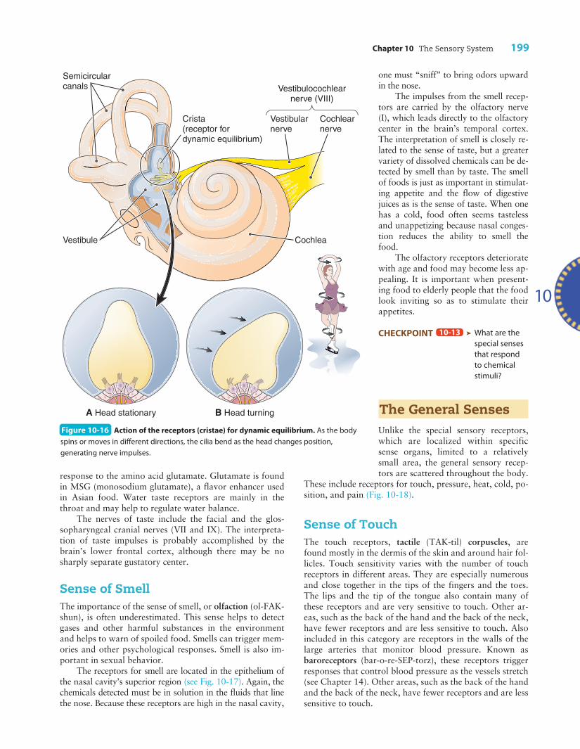
The receptors for dynamic equilib-rium function when the body is spin-ning or moving in different directions.The receptors, called cristae (KRIS-te),are located at the bases of the semicir-cular canals (Fig. 10-16). It’s easy toremember what these receptors do, be-cause the semicircular canals go off indifferent directions.
Nerve fibers from the vestibuleand from the semicircular canals formthe vestibular (ves-TIB-u-lar) nerve,which joins the cochlear nerve to formthe vestibulocochlear nerve, the eighthcranial nerve.
CHECKPOINT ä What are thetwo types ofequilibrium?
Visit thePoint or see theStudent Resource CD inthe back of this book forinformation on how au-diologists.
Other Special Sense OrgansThe sense organs of taste and smell are designed to re-spond to chemical stimuli.
10-12
10-11
Figure 10-14 Cochlea and the organ of Corti. The arrows show the direction of sound
waves in the cochlea.

198 Unit III Coordination and Control
Sense of TasteThe sense of taste, or gustation (gus-TA-shun), involves re-ceptors in the tongue and two different nerves that carrytaste impulses to the brain (Fig. 10-17). The taste receptors,known as taste buds, are located along the edges of small,depressed areas called fissures. Taste buds are stimulatedonly if the substance to be tasted is in solution or dissolvesin the fluids of the mouth. Receptors for four basic tastes arelocalized in different regions, forming a “taste map” of thetongue (see Fig. 10-17B):
n Sweet tastes are most acutely experienced at the tip ofthe tongue (hence the popularity of lollipops and icecream cones).
n Salty tastes are most acute at the anterior sides of thetongue.
n Sour tastes are most effectively detected by the tastebuds located laterally on the tongue.
n Bitter tastes are detected at the tongue’s posteriorpart.
Taste maps vary among people, but in each personcertain tongue regions are more sensitive to a specific basic taste. Other tastes are a combination of these fourwith additional smell sensations. More recently, re-searchers have identified some other tastes besides thesebasic four: water, alkaline (basic), and metallic. Another isumami (u-MOM-e), a pungent or savory taste based on a
Figure 10-15 Action of the receptors (maculae) for static equilibrium. As the head moves, the thick fluid above the receptor cells (hair
cells), weighted with otoliths, pulls on the cells’ cilia, generating a nerve impulse. [ ZOOMING IN ä What happens to the cilia on the receptor
cells when the fluid around them moves? ]
Chapter 10 The Sensory System 199
response to the amino acid glutamate. Glutamate is foundin MSG (monosodium glutamate), a flavor enhancer usedin Asian food. Water taste receptors are mainly in thethroat and may help to regulate water balance.
The nerves of taste include the facial and the glos-sopharyngeal cranial nerves (VII and IX). The interpreta-tion of taste impulses is probably accomplished by thebrain’s lower frontal cortex, although there may be nosharply separate gustatory center.
Sense of SmellThe importance of the sense of smell, or olfaction (ol-FAK-shun), is often underestimated. This sense helps to detectgases and other harmful substances in the environmentand helps to warn of spoiled food. Smells can trigger mem-ories and other psychological responses. Smell is also im-portant in sexual behavior.
The receptors for smell are located in the epithelium ofthe nasal cavity’s superior region (see Fig. 10-17). Again, thechemicals detected must be in solution in the fluids that linethe nose. Because these receptors are high in the nasal cavity,
one must “sniff” to bring odors upwardin the nose.
The impulses from the smell recep-tors are carried by the olfactory nerve(I), which leads directly to the olfactorycenter in the brain’s temporal cortex.The interpretation of smell is closely re-lated to the sense of taste, but a greatervariety of dissolved chemicals can be de-tected by smell than by taste. The smellof foods is just as important in stimulat-ing appetite and the flow of digestivejuices as is the sense of taste. When onehas a cold, food often seems tastelessand unappetizing because nasal conges-tion reduces the ability to smell thefood.
The olfactory receptors deterioratewith age and food may become less ap-pealing. It is important when present-ing food to elderly people that the foodlook inviting so as to stimulate theirappetites.
CHECKPOINT ä What are thespecial sensesthat respondto chemicalstimuli?
The General SensesUnlike the special sensory receptors,which are localized within specificsense organs, limited to a relativelysmall area, the general sensory recep-tors are scattered throughout the body.
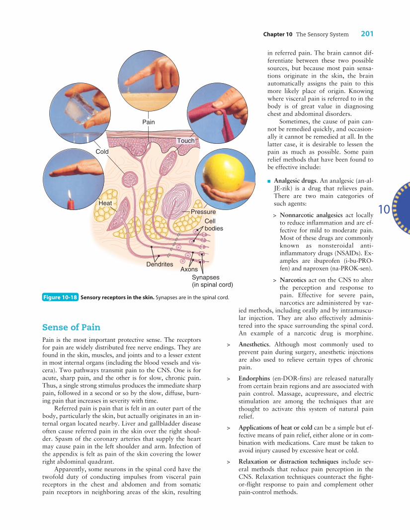
These include receptors for touch, pressure, heat, cold, po-sition, and pain (Fig. 10-18).
Sense of TouchThe touch receptors, tactile (TAK-til) corpuscles, arefound mostly in the dermis of the skin and around hair fol-licles. Touch sensitivity varies with the number of touchreceptors in different areas. They are especially numerousand close together in the tips of the fingers and the toes.The lips and the tip of the tongue also contain many ofthese receptors and are very sensitive to touch. Other ar-eas, such as the back of the hand and the back of the neck,have fewer receptors and are less sensitive to touch. Alsoincluded in this category are receptors in the walls of thelarge arteries that monitor blood pressure. Known asbaroreceptors (bar-o-re-SEP-torz), these receptors triggerresponses that control blood pressure as the vessels stretch(see Chapter 14). Other areas, such as the back of the handand the back of the neck, have fewer receptors and are lesssensitive to touch.
10-13
10
Figure 10-16 Action of the receptors (cristae) for dynamic equilibrium. As the body
spins or moves in different directions, the cilia bend as the head changes position,
generating nerve impulses.

200 Unit III Coordination and Control
Sense of PressureEven when the skin is anesthetized, it can still respond topressure stimuli. These sensory end-organs for deep pres-sure are located in the subcutaneous tissues beneath theskin and also near joints, muscles, and other deep tissues.They are sometimes referred to as receptors for deeptouch.
Sense of TemperatureThe temperature receptors are freenerve endings, receptors that are notenclosed in capsules but are merelybranchings of nerve fibers. Temper-ature receptors are widely distrib-uted in the skin, and there areseparate receptors for heat and cold.A warm object stimulates only theheat receptors, and a cool object af-fects only the cold receptors. In-ternally, there are temperature re-ceptors in the brain’s hypothalamus,which help to adjust body tempera-ture according to the temperature ofthe circulating blood.
Sense of PositionReceptors located in muscles, ten-dons, and joints relay impulses thataid in judging one’s position andchanges in the locations of bodyparts in relation to each other. Theyalso inform the brain of the amountof muscle contraction and tendontension. These rather widespread re-ceptors, known as proprioceptors(pro-pre-o-SEP-tors), are aided inthis function by the internal ear’sequilibrium receptors.
Information received by thesereceptors is needed for the coordina-tion of muscles and is important insuch activities as walking, running,and many more complicated skills,such as playing a musical instru-ment. They help to provide a senseof body movement, known as kines-thesia (kin-es-THE-ze-ah). Proprio-ceptors play an important part inmaintaining muscle tone and goodposture. They also help to assess theweight of an object to be lifted sothat the right amount of muscleforce is used.
The nerve fibers that carry im-pulses from these receptors enter the spinal cord and ascendto the brain in the posterior part of the cord. The cerebellumis a main coordinating center for these impulses.
CHECKPOINT ä What are examples of generalsenses?
CHECKPOINT ä What are proprioceptors, and whereare they located?
10-15
10-14
A
B
Olfactory bulb
Olfactory receptors
Nostril
Tongue with taste receptors
TASTE ZONES:
BitterSaltySweet Sour
Facial nerve (VII)
Glossopharyngeal nerve (IX)
Papillae
Figure 10-17 Special senses that respond to chemicals. (A) Organs of taste (gustation)
and smell (olfaction). (B) A taste map of the tongue.
Chapter 10 The Sensory System 201
in referred pain. The brain cannot dif-ferentiate between these two possiblesources, but because most pain sensa-tions originate in the skin, the brainautomatically assigns the pain to thismore likely place of origin. Knowingwhere visceral pain is referred to in thebody is of great value in diagnosingchest and abdominal disorders.
Sometimes, the cause of pain can-not be remedied quickly, and occasion-ally it cannot be remedied at all. In thelatter case, it is desirable to lessen thepain as much as possible. Some painrelief methods that have been found tobe effective include:
n Analgesic drugs. An analgesic (an-al-JE-zik) is a drug that relieves pain.There are two main categories ofsuch agents:
> Nonnarcotic analgesics act locallyto reduce inflammation and are ef-fective for mild to moderate pain.Most of these drugs are commonlyknown as nonsteroidal anti-inflammatory drugs (NSAIDs). Ex-amples are ibuprofen (i-bu-PRO-fen) and naproxen (na-PROK-sen).
> Narcotics act on the CNS to alterthe perception and response topain. Effective for severe pain,narcotics are administered by var-
ied methods, including orally and by intramuscu-lar injection. They are also effectively adminis-tered into the space surrounding the spinal cord.An example of a narcotic drug is morphine.
> Anesthetics. Although most commonly used toprevent pain during surgery, anesthetic injectionsare also used to relieve certain types of chronicpain.
> Endorphins (en-DOR-fins) are released naturallyfrom certain brain regions and are associated withpain control. Massage, acupressure, and electricstimulation are among the techniques that arethought to activate this system of natural pain relief.
> Applications of heat or cold can be a simple but ef-fective means of pain relief, either alone or in com-bination with medications. Care must be taken toavoid injury caused by excessive heat or cold.
> Relaxation or distraction techniques include sev-eral methods that reduce pain perception in theCNS. Relaxation techniques counteract the fight-or-flight response to pain and complement otherpain-control methods.
10
Sense of PainPain is the most important protective sense. The receptorsfor pain are widely distributed free nerve endings. They arefound in the skin, muscles, and joints and to a lesser extentin most internal organs (including the blood vessels and vis-cera). Two pathways transmit pain to the CNS. One is foracute, sharp pain, and the other is for slow, chronic pain.Thus, a single strong stimulus produces the immediate sharppain, followed in a second or so by the slow, diffuse, burn-ing pain that increases in severity with time.
Referred pain is pain that is felt in an outer part of thebody, particularly the skin, but actually originates in an in-ternal organ located nearby. Liver and gallbladder diseaseoften cause referred pain in the skin over the right shoul-der. Spasm of the coronary arteries that supply the heartmay cause pain in the left shoulder and arm. Infection ofthe appendix is felt as pain of the skin covering the lowerright abdominal quadrant.
Apparently, some neurons in the spinal cord have thetwofold duty of conducting impulses from visceral painreceptors in the chest and abdomen and from somaticpain receptors in neighboring areas of the skin, resulting
Pain
Touch
DendritesAxons
HeatPressure
Cold
Cell bodies
Synapses (in spinal cord)
Figure 10-18 Sensory receptors in the skin. Synapses are in the spinal cord.
202 Unit III Coordination and Control
Sensory AdaptationWhen sensory receptors are exposed to a continuous stim-ulus, receptors often adjust themselves so that the sensa-tion becomes less acute. The term for this phenomenon issensory adaptation. For example, if you immerse yourhand in very warm water, it may be uncomfortable; how-ever, if you leave your hand there, soon the water will feelless hot (even if it has not cooled appreciably).
Receptors adapt at different rates. Those for warmth,cold, and light pressure adapt rapidly. In contrast, thosefor pain do not adapt. In fact, the sensations from the slowpain fibers tend to increase over time. This variation in re-ceptors allows us to save energy by not responding tounimportant stimuli while always heeding the warnings ofpain.
äEarly Signs of Cataract
Dr. Gilbert’s assistant called Paul back to theconsultation room after his eye examina-tion. “You seem to be doing fine, Paul,” the
ophthalmologist reported. “No signs of glaucoma.I am a little concerned about some early signs ofcataract, though. These opacities of the lens de-velop with age in most people, and they’re nothereditary, but there’s firm evidence that theyare influenced by sun exposure.”
“Yikes!” Paul exploded. “Another run-in withthe sun! I’ve already heard about the sun’s un-friendly rays with my skin cancer, and I thoughtthat’s where it would end.”
“Cataracts can be dealt with pretty effectively,”said Dr. Gilbert, “but there’s more. Sunlight isalso a factor in a more serious condition, maculardegeneration, which affects central vision andcan lead to blindness. That’s not as easily treat-able at present. I don’t see any signs of that right
now, but you need to wear good quality sunglasseswith a UV filter. Wearing dark glasses withoutthe filter is worse than nothing, because they di-late your pupils and allow more harmful UV raysto enter your eyes.”
“Great! One more thing to worry about as Iget older,” Paul complained.
“Not a worry if you take precautions,” Dr.Gilbert responded. “Pick up your glasses prescrip-tion at the desk, and Paul, also take a pair of thedark disposable glasses we have there to protectyour dilated eyes. They will be extra-sensitive tothe sun for a while.”
During this case, we learned that structuraldamage leads to functional changes. We alsolearned that some changes develop with age.Later chapters will include information on age-related changes that affect other systems.

Chapter 10 The Sensory System 203
10
Word Anatomy
Medical terms are built from standardized word parts (prefixes, roots, and suffixes). Learning the meanings of these parts can help you remember words and interpret unfamiliar terms.
WORD PART MEANING EXAMPLE
The Eye and Vision
ophthalm/o eye An ophthalmologist is a physician who specializes in treat-ment of the eye.
-scope instrument for examination An ophthalmoscope is an instrument used to examine the posterior of the eye.
lute/o yellow The macula lutea is a yellowish spot in the retina that con-tains the fovea centralis.
presby- old Presbyopia is farsightedness that occurs with age.
The Ear
tympan/o drum The tympanic membrane is the eardrum.
equi- equal Equilibrium is balance (equi- combined with the Latin word libra meaning “balance”).
ot/o ear Otology is the study of the ear.
lith stone Otoliths are small crystals in the inner ear that aid in static equilibrium.
-cusis hearing Presbycusis is hearing loss associated with age.
The General Senses
propri/o- own Proprioception is perception of one’s own body position.
kine movement Kinesthesia is a sense of body movement.
-esthesia sensation Anesthesia is loss of sensation, as of pain.
-alges/i pain An analgesic is a drug that relieves pain.
narc/o stupor A narcotic is a drug that alters the perception of pain.
I. THE SENSESA. Protect by detecting changes (stimuli) in the environ-
mentB. Sensory receptors—detect stimuli
1. Structural typesa. Free dendriteb. End-organ—modified dendritec. Specialized cell—in special sense organs
2. Types based on stimulusa. Chemoreceptors—respond to chemicalsb. Thermoreceptors—respond to temperaturec. Photoreceptors—respond to lightd. Mechanoreceptors—respond to movement
C. Special and general senses1. Special senses—vision, hearing, equilibrium,
taste, smell2. General senses—touch, pressure, temperature,
position, painII. THE EYE AND VISION
A. Protection of the eyeball—bony orbit, eyelid, eye-lashes, conjunctiva, lacrimal glands (produce tears)
B. Coats of the eyeball1. Sclera—white of the eye
a. Cornea—anterior2. Choroid—pigmented; contains blood vessels3. Retina—receptor layer
Summary

204 Unit III Coordination and Control
C. Pathway of light rays and refraction1. Refraction—bending of light rays as they pass
through substances of different density2. Refracting parts—cornea, aqueous humor, lens,
vitreous bodyD. Function of the retina
1. Cellsa. Rods—cannot detect color; function in
dim lightb. Cones—detect color; function in bright
light2. Pigments—sensitive to light; rod pigment is
rhodopsinE. Muscles of the eye
1. Extrinsic muscles—six move each eyeball2. Intrinsic muscles
a. Iris—colored ring around pupil; regulatesthe amount of light entering the eye
b. Ciliary muscle—regulates the thickness ofthe lens to accommodate for near vision
F. Nerve supply to the eye1. Sensory nerves
a. Optic nerve (II)—carries impulses fromretina to brain
b. Ophthalmic branch of trigeminal (V)2. Motor nerves—move eyeball
a. Oculomotor (III), trochlear (IV), abducens(VI)
III. THE EARA. Outer ear—pinna, auditory canal (meatus), tym-
panic membrane (eardrum)B. Middle ear and ossicles
1. Ossicles—malleus, incus, stapes2. Eustachian tube—connects middle ear with
pharynx to equalize pressureC. Inner ear
1. Bony labyrinth—contains perilymph2. Membranous labyrinth—contains endolymph
3. Divisionsa. Cochlea—contains receptors for hearing
(organ of Corti)b. Vestibule—contains receptors for static
equilibrium (maculae)c. Semicircular canals—contain receptors for
dynamic equilibrium (cristae)4. Receptor (hair) cells function by movement of
cilia5. Nerve—vestibulocochlear (auditory) nerve (VIII)
IV. OTHER SPECIAL SENSE ORGANSA. Sense of taste (gustation)
1. Receptors—taste buds on tongue2. Basic tastes—sweet, salty, sour, bitter3. Nerves—facial (VII) and glossopharyngeal (IX)
B. Sense of smell (olfaction)1. Receptors—in upper part of nasal cavity2. Nerve—olfactory nerve (I)
V. GENERAL SENSESA. Sense of touch—tactile corpusclesB. Sense of pressure—receptors in skin deep tissue,
large arteriesC. Sense of temperature—receptors are free nerve
endingsD. Sense of position (proprioception)—receptors are
proprioceptors in muscles, tendons, joints1. Kinesthesia—sense of movement
E. Sense of pain—receptors are free nerve endings1. Referred pain—originates internally but felt at
surface2. Relief of pain—analgesic drugs, anesthetics,
endorphins, heat, cold, relaxation and distrac-tion techniques
VI. SENSORY ADAPTATIONA. Adjustment of receptors so that sensation becomes
less acuteB. Receptors adapt at different rates; pain receptors do
not adapt
Questions for Study and Review
BUILDING UNDERSTANDING
Fill in the blanks
1. The part of the nervous system that detects a stimulus isthe ______.
2. The bending of light rays as they pass from air to fluid iscalled ______.
3. Nerve impulses are carried from the ear to the brain bythe ______ nerve.
4. Information about the position of the knee joint is pro-vided by ______.
5. A receptor’s ability to decrease its sensitivity to a continu-ous stimulus is called ______.

Chapter 10 The Sensory System 205
10
Matching > Match each numbered item with the most closely related lettered item.
___ 6. Consists of ciliated receptor cells sensitive to vibration
___ 7. The actual receptor layer of the eye
___ 8. The receptor that senses static equilibrium
___ 9. The receptor that senses touch
___ 10. The receptor sensitive to temperature and pain
a. retina
b. free nerve endings
c. macula
d. organ of Corti
e. tactile corpuscle
UNDERSTANDING CONCEPTS
16. Differentiate between the terms in each of the followingpairs:
a. special sense and general senseb. aqueous humor and vitreous bodyc. rods and conesd. endolymph and perilymphe. static and dynamic equilibrium
17. Trace the path of a light ray from the outside of the eyeto the retina.
18. Define convergence and accommodation.
19. List in order the structures that sound waves pass throughin traveling through the ear to the receptors for hearing.
20. Name the four basic tastes. Where are the taste recep-tors? Name the nerves of taste.
21. Trace the pathway of a nerve impulse from the olfactoryreceptors to the olfactory center in the brain.
22. Name several types of pain-relieving drugs. Describe sev-eral methods for relieving pain that do not involve drugs.
Multiple Choice
___ 11. All of the following are special senses except
a. smellb. tastec. equilibriumd. pain
___ 12. From superficial to deep, the order of the eyeball’stunics is
a. retina, choroid, and sclerab. sclera, retina, and choroidc. choroid, retina, and sclerad. sclera, choroid, and retina
___ 13. The part of the eye most responsible for light refrac-tion is the
a. corneab. lensc. vitreous bodyd. retina
___ 14. Information from the retina is carried to the brain bythe
a. ophthalmic nerveb. optic nervec. oculomotor nerved. abducens nerve
___ 15. Receptors in the vestibule sense
a. muscle tensionb. soundc. lightd. equilibrium
CONCEPTUAL THINKING
23. Why do you taste eyedrops after applying them to youreyeball?
24. You and a friend have just finished riding the rollercoaster at the amusement park. As you walk away fromthe ride, your friend stumbles and comments that the ridehas affected her balance. How do you explain this?
25. In the case story, Paul discovered he might be developingan age-related eye disorder. What is a cataract? What aresome other age-related disorders of the sensory system?

206 Unit III Coordination and Control