pathogenesis, diagnosis and therapy of disseminated ... · pathogenesis, diagnosis and therapy of...
TRANSCRIPT
Pathogenesis, Diagnosis and Therapy of Disseminated Intravascular
Proceedings of the Workshop on Disseminated Intravascular Coagulation, Castle of Rauischholzhausen, Germany, 12-14 May, 1993
Gert Müller-Berghaus Division of Hemostasis and Transfusion Medicine Kerckhoff-Klinik, Max-PIanck-Institut für physiologische und klinische Forschung, Bad Nauheim, Germany
Katharina Madiener Division of Hemostasis and Transfusion Medicine Kerckhoff-Klinik, Max-Planck-Institut für physiologische und klinische Forschung, Bad Nauheim, Germany
Margareta Blombäck Department of Laboratory Medicine and Blood Coagulation Karolinska Hospital Stockholm, Sweden
Jan W. ten Cate Center for Hemostasis, Thrombosis, Atherosclerosis and Inflammation Research Academic Medical Center Amsterdam, The Netherlands
Fibrin Formation
Editors:
1993
Excerpta Medica, Amsterdam - London - New York - Tokyo
vii
Contents
Preface v Acknowledgements vi
Definition of disseminated intravascular coagulation
Attempts to define disseminated intravascular coagulation G. Müller-Berghaus, M. Blombäck a n d J. W. ten Cate 3
Pathogenesis
Bacteria, cells and disseminated intravascular coagulation in man P . B r a n d t z a e g a n d P . Kierulf 11
Role of tissue factor in the coagulant and inflammatory response to L D 1 0 0 E . c o l i sepsis and in the early diagnosis of DIC in the baboon
F . B . T a y l o r Jr 19 Leukocytes, adhesive proteins and disseminated intravascular coagulation: treatment of DIC with plasma/leukapheresis?
B . 0sterud 32 Tissue factor gene regulation
D . v o n der A h e , B . Jost a n d G. Müller-Berghaus 41 Tissue factor expression in endotoxin-activated monocytes is suppressed by agents which increase intracellular cyclic AMP concentration
D . de P r o s t a n d V. O l l i v i e r 47 Involvement of the vascular endothelium and activation of the coagulation System in human malaria
C. J. Hemmer, A . B i e r h a u s , P . K e r n , R. E g b r i n g , R. Seitz, P . P . N a w r o t h a n d M. D i e t r i c h 51
Effect of peripheral blood lymphocytes on procoagulant activity of human endothelial cells pretreated with lipopolysaccharide
K . B i n d e r , E . S c h m i d a n d T. H. Müller 55 Freshly formed thrombi induce procoagulant activity in human vascular endothelial cells
E . Schmid, K . B i n d e r a n d T. H. Müller 63 Hemostasis in orthotopic liver transplantation — a model for studying DIC development?
H . Riess, G. H i m m e l r e i c h , M. J o c h u m , W. M a c h l e i d t , K. H u n d t , P . Neuhaus a n d D . H u h n 11
viii
Clinical aspects of DIC
Markers of hemostatic activation in experimental endotoxemia: implications for therapy
H . ten C a t e , M. L e v i y B . J. B i e m o n d , S. J. H. v a n D e v e n t e r , T. v a n der P o l l H . R . Büller a n d J.W. ten Cate 81
Quantitative evaluation of hypercoagulability by fibrin monomer ELISA C.-E. Dempfle, M. D o l l m a n , S. Pfitzner, H. U l i , W. Mühlhofen A . Dessauer a n d D . L . Heene 89
Determination of plasma fibrin monomers by a newly developed ELISA method in patients with disseminated intravascular coagulation
K . Okajima, M. Uchiba, K . M u r a k a m l H. O k a b e a n d K. T a k a t s u k i 99 Soluble fibrin — a predictor for development and outcome of multiple organ failure
S. B r e d b a c k a a n d M. Blombäck 111 Monitoring the anticoagulant treatment of DIC and recurrent thrombosis in patients with malignancies using the measurement of "soluble fibrin" (FM-test, Chromogenix), Fl+2 and TAT complexes
J . S t i b b e 113 Degradation of fibrinogen and its derivatives in the course of disseminated intravascular coagulation
W. N i e u w e n h u i z e n 117 Evidence that heparin cofactor II participates in the control of disseminated intravascular coagulation
T.R. A n d e r s s o n , P . M . Sandset, K . N u s t a d a n d U. A b i l d g a a r d 125 Cytokine release in leukocytopenic patients with septic shock is associated with coagulation activation and consumption of inhibitors
H . O s t e r m a n n , R. M e s t e r s a n d J. K i e n a s t 129 Increased activated protein C — its inhibitor complex levels in patients with disseminated intravascular coagulation
H . Wada, K . M i n a m i k a w a , S. S h i r a k a w a , T. H a y a s h i , J. N i s h i o k a a n d K . Suzuki 131
Activation of hemostasis and the release of cytokines following the injection of bacterial lysates: fibrinolysis precedes fibrinemia
J. U. W i e d i n g , H.-J. Veltman, K . - F . Kölmel a n d M. Köhler 139 Alterations of coagulation and fibrinolysis during the preclinical phase after Polytrauma
E . Seifried, L . L a m p l , C. S e i d l , M. H e l m , B . M a i e r a n d K . - H . B o c k 147 Alterations of coagulation inhibitors in consumption coagulopathy
B . Kemkes-Matthes and H . - G . U i s c h 159 Hypercoagulation predicts outcome after isolated brain trauma
S. B r e d b a c k a a n d G. E d n e r 163
ix
Sepsis and septic shock: hemostasis-fibrinolysis equilibrium as a prognostic factor and an indicator during therapy
N . M a u r i n a n d D . Frank 165 Influence of blood sampling from venipunctures and catheter Systems on serial determinations of Prothrombin activation fragment 1+2 and thrombin-antithrombin III complex
G. Hafner, H. Schinzel, W. E h r e n t h a l , C. Wagner, U. K o n h e i s e r , R. Zotz, J . Lötz, R. B l a n k , L . S . W e i l e m a n n and W. P r e l l w i t z 171
Hypercoagulable State and DIC in neonatal asphyxia S. Suzuki 175
New therapeutic approaches
New developments in the field of benzamidine-derived thrombin inhibitors J. Stürzebecher, D . P r a s a , E . B r e t s c h n e i d e r , W. B o d e , M. B a u e r , H. B r a n d s t e t t e r , P . Wikström a n d H. Vieweg 183
Investigation of in vitro antithrombotic potency of a r-hirudin (HBW 023) and a synthetic analogue
J. Römisch, W. Stüber a n d E . - P . Päques 191 Heparin treatment in thrombin-induced disseminated intravascular coagulation (DIC) in the baboon
H J . D u Toit, A . R . Coetzee and D . O. C h a l t o n 197 Clinical evaluation of low-molecular-weight heparin (FR-860) on disseminated intravascular coagulation (DIC). A multicenter cooperative double-blind trial in comparison with heparin
N . S a k u r a g a w a , H . H a s e g a w a , M. M a k i , M. N a k a g a w a a n d M . N a k a s h i m a 203
Protection of DIC-induced mortality in K l e b s i e l l a pneumoniae-infected and LPS-treated rats by antithrombin III
G. D i c k n e i t e and E . - P . Päques 215 Results of a double-blind, placebo-controlled trial of antithrombin III concentrates in septic shock with DIC
F . F o u r r i e r , J.-J. H u a r t , I. Runge, C. C a r o n , J. G o u d e m a n d a n d C. C h o p i n 221
Antithrombin concentrate dose to patients with acquired antithrombin deficiency
J. A l b e r t a n d M. Blombäck 227 Effect of two-domain tissue factor pathway inhibitor (2D-TFPI) and inactivated factor Vl la on endotoxin-induced DIC in rabbits
C. B r e g e n g a a r d , O. Nordfang, P . Wildgoose and V. D i n e s s 2 2 9 Antithrombotic effects of recombinant soluble human thrombomodulin analogs in animal models of tissue-factor and thrombin induced DIC
W. Witt, M . F r e d r i c h , R. Wydro a n d J. M o r s e r 233
X
The xanthine derivatives HWA 138 and A 80 attenuate activation of coagulation in porcine endotoxin shock
M . S p a n n a g l , G. Frank, M. Goedde a n d H . Hoffmann 239 Effect of nafamostat mesilate (NM), a synthetic protease inhibitor, on the extrinsic pathway of coagulation
M. U c h i b a , K . Okajima, H A b e , H O k a b e a n d K. T a k a t s u k i 243 The use of Octaplas in patients undergoing open heart surgery
B . G. S o l h e i m , J . L . Svennevig, B . M o h r , M. D r a g s u n d , H. N o d d e l a n d , S. Töllefsrud, F. B r o s s t a d , H. R o l l a g a n d T.E. M o l i n e s 253
Index of authors 263
Keyword index 265

©1993 Elsevier Science Publishers B.V. All rights reserved. DIC: Pathogenesis, Diagnosis and Therapy of Disseminated Intravascular Fibrin Formation. G. Müller-Berghaus et al., editors.
71
Hemostasis in orthotopic liver transplantation — a model for studying DIC development?
Hanno Riess1*, Gabriele Himmelreich1, Marianne Jochum3, Werner Machleidt4, Karoline Hundt1, Peter Neuhaus2 and Dieter Huhn1
Departments of1 I n t e r n a l M e d i c i n e and 2Surgery, University H o s p i t a l R u d o l f V i r c h o w , Augustenburger Platz 1, 1 3 3 4 4 B e r l i n ; Department of C l i n i c a l Chemistry and Biochemistry, University of M u n i c h , Nußbaum-Straße 2 0 , 8 0 3 3 6 M u n i c h ; 4Department of Physiological Chemistry, University of M u n i c h , Pettenkoferstraße 1 0 , 8 0 3 3 6 M u n i c h , Germany
Introduction
In orthotopic liver transplantation (OLT) hyperfibrinolysis is most prominent im-mediately before reperfusion — characterized by increased formation of plasmin-antiplasmin (PAP) complexes — and enhanced Prothrombin activation thereafter — characterized by rising thrombin-antithrombin III (TAT) complexes — have been described and accepted to occur regularly [1—4]. Paradoxically, the increased Prothrombin activation after revascularisation of the graft may be paralleled in the clinical Situation by oozing in the Operation field, a Situation often observed during DIC.
We investigated parameters of coagulation, e.g., fibrinogen, factor XIII (F XIII), antithrombin III (AT III), free protein S antigen (protein S) and TAT, parameters of fibrinolysis, e.g., tissue-type Plasminogen activator (t-PA), urokinase-type Plasminogen activator (u-PA), Plasminogen activator inhibitor (PAI), Cl-inhibitor and PAP, mediators of leucocyte activation, e.g., tumor necrosis factor alpha (TNF-a), neopterin, cathepsin B, neutrophile elastase in complex with protease inhibitor (EPI), platelet count and platelet aggregability as well as the endothelium-derived soluble thrombomodulin (sTM)) in the course of 20 consecutive liver transplantations in order to understand more clearly the pathophysiological changes occurring during OLT, especially with reperfusion of the graft liver.
The impaired hemostasis due to preexisting liver failure is aggravated during OLT in these patients, resulting in a DIC-like constellation that may be considered to represent a human model for studying DIC.
*Address for correspondence: Professor Dr. med. Hanno Riess, Universitätsklinikum Rudolf Virchow, Augustenburger Platz 1, 13344 Berlin, Germany.
72
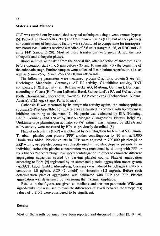
Materials and Methods
OLT was carried out by established surgical techniques using a veno-venous bypass [5]. Packed red blood cells (RBC) and fresh frozen plasma (FFP) but neither platelets nor concentrates of hemostatic factors were substituted to compensate for intraoperative blood loss. Patients received a median of 8.6 units (ränge: 2—26) of RBC and 7.8 units FFP (ränge: 2—28). Most of these transfusions were given during the pre-anhepatic and anhepatic phases.
Blood samples were taken from the arterial line, after induction of anaesthesia and before Operation statt <1>, 5 min before <2> and 10 min after <3> the beginning of the anhepatic stage. Further samples were collected 5 min before reperfusion <4>, as well as 5 min <5>, 15 min <6> and 60 min afterwards.
The following parameters were measured: protein C activity, protein S Ag (all: Boehringer, Mannheim, Germany); AT III activity, Cl-inhibitor activity, TAT complexes, F XIII activity (all: Behringwerke AG, Marburg, Germany), fibrinogen according to Clauss (Hoffmann-LaRoche, Basel, Switzerland), t-PA and PAI activities (both Chromogenix, Stockholm, Sweden), PAP complexes (Technoclone, Vienna, Austria), sTM Ag, (Stago, Paris, France).
Cathepsin B was measured by its enzymatic activity against the aminopeptidase Substrate Z-Phe-Arg-NMec [6]. Elastase was estimated in complex with arproteinase inhibitor according to Neumann [7]. Neopterin was estimated by RIA (Henning, Berlin, Germany) and TNF-a by IRMA (Medgenix Diagnostics, Fleurus, Belgium). Urokinase-type Plasminogen activator (u-PA) antigen was measured by ELISA and u-PA activity were measured by BIA as previously described [8].
Platelet rieh plasma (PRP) was obtained by centrifugation for 6 min at 600 U/min. To obtain platelet poor plasma (PPP) another centrifugation for 20 min at 3,000 U/min was added. Platelet counts in PRP were adjusted to 200,000 platelets/ul or PRP with lower platelet counts was directly used in thrombocytopenic patients. In an individual series this platelet concentration was reobtained by diluting with PPP or by a further "concentrating" low speed centrifugation in order to eliminate different aggregating capacities caused by varying platelet counts. Platelet aggregation according to Born [9] registered by an automated platelet aggregation tracer System (APACT, Labor GmbH, Ahrensburg, Germany) was induced by collagen (final concentration 1.0 ug/ml), ADP (2 jnmol/1) or ristocetin (1.2 mg/ml). Before each determination platelet aggregation was calibrated with PRP and PPP. Platelet aggregation was determined by measuring the maximal amplitude.
Results in the figures are given as medians and the non-parametric Wilcoxon signed-ranks test was used to evaluate differences of levels between the timepoints; values of p < 0.5 were considered to be significant.
Results
Most of the results obtained have been reported and discussed in detail [2,10—14].
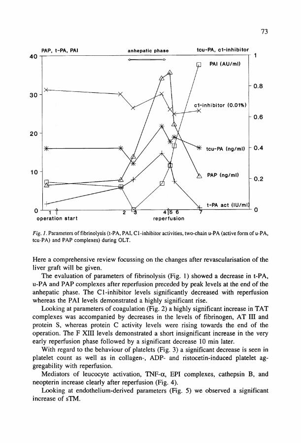
73
PAP, t-PA, PAI anhepatic phase tcu-PA. d- inhibi tor
Operation Start reperfusion
F i g . L Parameters of fibrinolysis (t-PA, PAI, Cl-inhibitor activities, two-chain u-PA (active form of u-PA, tcu-PA) and PAP complexes) during OLT.
Here a comprehensive review focussing on the changes after revascularisation of the liver graft will be given.
The evaluation of parameters of fibrinolysis (Fig. 1) showed a decrease in t-PA, u-PA and PAP complexes after reperfusion preceded by peak levels at the end of the anhepatic phase. The Cl-inhibitor levels significantly decreased with reperfusion whereas the PAI levels demonstrated a highly significant rise.
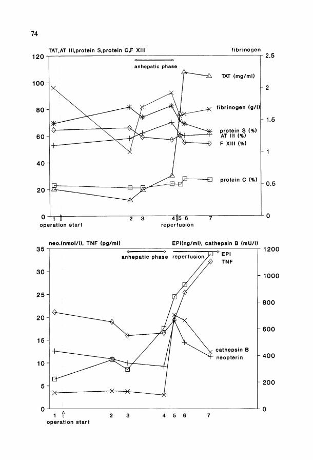
Looking at parameters of coagulation (Fig. 2) a highly significant increase in TAT complexes was accompanied by decreases in the levels of fibrinogen, AT III and protein S, whereas protein C activity levels were rising towards the end of the Operation. The F XIII levels demonstrated a short insignificant increase in the very early reperfusion phase followed by a significant decrease 10 min later.
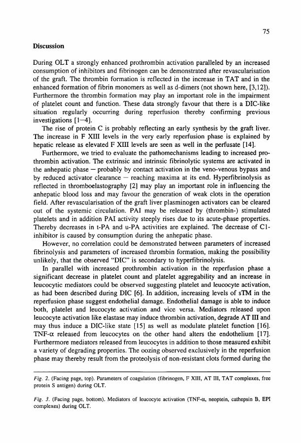
With regard to the behaviour of platelets (Fig. 3) a significant decrease is seen in platelet count as well as in collagen-, ADP- and ristocetin-induced platelet ag-gregability with reperfusion.
Mediators of leucocyte activation, TNF-a, EPI complexes, cathepsin B, and neopterin increase clearly after reperfusion (Fig. 4).
Looking at endothelium-derived parameters (Fig. 5) we observed a significant increase of sTM.
74
TAT,AT lll.protein S.protein C,F XIII fibrinogen
Operation Start reperfusion
neo.(nmol/l), TNF (pg/ml) EPKng/ml), cathepsin B (mU/l)
Operation start
75
Discussion
During OLT a strongly enhanced Prothrombin activation paralleled by an increased consumption of inhibitors and fibrinogen can be demonstrated after revascularisation of the graft. The thrombin formation is reflected in the increase in TAT and in the enhanced formation of fibrin monomers as well as d-dimers (not shown here, [3,12]). Furthermore the thrombin formation may play an important role in the impairment of platelet count and function. These data strongly favour that there is a DIC-like Situation regularly occurring during reperfusion thereby confirming previous investigations [1—4].
The rise of protein C is probably reflecting an early synthesis by the graft liver. The increase in F XIII levels in the very early reperfusion phase is explained by hepatic release as elevated F XIII levels are seen as well in the perfusate [14].
Furthermore, we tried to evaluate the pathomechanisms leading to increased Prothrombin activation. The extrinsic and intrinsic fibrinolytic Systems are activated in the anhepatic phase — probably by contact activation in the veno-venous bypass and by reduced activator clearance — reaching maxima at its end. Hyperfibrinolysis as reflected in thromboelastography [2] may play an important role in influencing the anhepatic blood loss and may favour the generation of weak clots in the Operation field. After revascularisation of the graft liver Plasminogen activators can be cleared out of the systemic circulation. PAI may be released by (thrombin-) stimulated platelets and in addition PAI activity steeply rises due to its acute-phase properties. Thereby decreases in t-PA and u-PA activities are explained. The decrease of C l -inhibitor is caused by consumption during the anhepatic phase.
However, no correlation could be demonstrated between parameters of increased fibrinolysis and parameters of increased thrombin formation, making the possibility unlikely, that the observed "DIC" is secondary to hyperfibrinolysis.
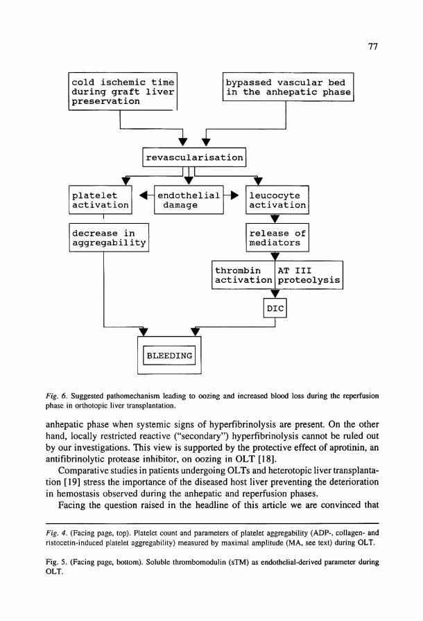
In parallel with increased Prothrombin activation in the reperfusion phase a significant decrease in platelet count and platelet aggregability and an increase in leucocytic mediators could be observed suggesting platelet and leucocyte activation, as had been described during DIC [6]. In addition, increasing levels of sTM in the reperfusion phase suggest endothelial damage. Endothelial damage is able to induce both, platelet and leucocyte activation and vice versa. Mediators released upon leucocyte activation like elastase may induce thrombin activation, degrade AT III and may thus induce a DIC-like State [15] as well as modulate platelet function [16]. TNF-a released from leucocytes on the other hand alters the endothelium [17]. Furthermore mediators released from leucocytes in addition to those measured exhibit a variety of degrading properties. The oozing observed exclusively in the reperfusion phase may thereby result from the proteolysis of non-resistant clots formed during the
F i g . 2. (Facing page, top). Parameters of coagulation (fibrinogen, F XIII, A T III, T A T complexes, free protein S antigen) during OLT.
F i g . 3. (Facing page, bottom). Mediators of leucocyte activation (TNF-a, neoptein, cathepsin B, EPI complexes) during OLT.
76
80 MA (%) anhepatic phase platelets/pl (thousand)
60
40
20
2 3 Operation Start
4f5~6 reperfusion
ristocetin (1.2mg/ml)
platelet count collagen (Vig/ml)
ADP (2|jmol/l)
160
140
120
100
80
60
40
h 20
140
120
100 H
sTM gjg/ml)
1 t Operation start
2 3
140
120
100
4 5
77
cold ischemic time during graft l i v e r preservation
bypassed vascular bed in the anhepatic phase
revascularisat ion
in z plate let activation
endothelial damage
leucocyte act ivat ion
decrease in aggregability
release of mediators
thrombiri act ivat ion
AT III proteolysis
DIC
BLEEDING
F i g . 6. Suggested pathomechanism leading to oozing and increased blood loss during the reperfusion phase in orthotopic liver transplantation.
anhepatic phase when systemic signs of hyperfibrinolysis are present. On the other hand, locally restricted reactive ("secondary") hyperfibrinolysis cannot be ruled out by our investigations. This view is supported by the protective effect of aprotinin, an antifibrinolytic protease inhibitor, on oozing in OLT [18].
Comparative studies in patients undergoing OLTs and heterotopic liver transplantation [19] stress the importance of the diseased host liver preventing the deterioration in hemostasis observed during the anhepatic and reperfusion phases.
Facing the question raised in the headline of this article we are convinced that
F i g . 4. (Facing page, top). Platelet count and parameters of platelet aggregability (ADP-, collagen- and ristocetin-induced platelet aggregability) measured by maximal amplitude (MA, see text) during OLT.
Fig. 5. (Facing page, bottom). Soluble thrombomodulin (sTM) as endothelial-derived parameter during O L T .
78
hemostasis in OLT is a valuable model in humans to study DIC development because the changes described are regularly observed, though differently pronounced, in every patient. There is a well accepted underlying disease, e.g., terminal liver failure with impaired hemostasis. OLT represents an "exogenous" trigger resulting in increased fibrin formation, bleeding and the risk of multi-organ failure. Therefore the main criteria for the diagnosis of DIC are fulfilled. Our investigations provide evidence for a hypothetical pathophysiological pattern (Fig. 6) leading to increased bleeding tendency in the reperfusion phase in OLT and are offering the opportunity to study different therapeutic options to ameliorate the clinical picture.
References
1. Porte RJ, Bontempo FA, Knot EAR, Lewis JH, Kang Y G , Starzl T E . Transplantation 1989;47:978-984.
2. Himmelreich G, Kierzek B, Neuhaus P, Slama K-J, Riess H. Blood Coag Fibrinol 1991;51-59. 3. Harper PL, Luddington RJ, Jennings I, Reardon D, Seaman MJ, Carrell RW, Klinik JR, Smith M ,
Rolles K, Calne R. Transplantation 1989;48:603-607. 4. Dzik WH, Arkin CF, Jenkins RL, Stump DC. Blood 1988;71:1090-1095. 5. Neuhaus P, Blumhardt G, Bechstein WO, Steffen R, Keck H. Transplant Proc 1990;22:1571. 6. Assfalg-Machleidt I, Jochum M , Nast-Kolb D, Siebeck M , Billing A, Joka T, Rothe G , Valet G,
Zauner R, Scheuber HP, Machleidt W. Biol Chem Hoppe-Seyler (1990) 371:211-222. 7. Neumann S, Gunzer G, Hennrich N, Lang H. J Clin Chem Clin Biochem 1984;22:693-697. 8. Himmelreich G, Dooijeward G, Breinl P, Bechstein WO, Neuhaus P, Kluft C, Riess H. Thromb
Haemost 1993;69:56-59. 9. Born GVR, Cross MJ. J Physiol 1963;168:178-195.
10. Himmelreich G, Hundt K, Neuhaus P, Blumhardt G, Riess H. Transplantation 1992;53:582-586. 11. Riess H, Jochum M , Machleidt W, Himmelreich G, Roissant R, Blumhardt G. Transplantation
1991;52:482-490. 12. Himmelreich G, Riewald M , Blumhard G, Neuhaus P, Roissant R, Riess H. Thrombomodulin — a
marker for endothelial damage during orthotopic liver transplantation. Am J Hematol 1993 (in press). 13. Himmelreich G, Jochum M , Machleidt W, Neuhaus P, Riess H. Mediators of leucocyte activation
play a role in DIC during orthotopic liver transplantation. Transplantation 1993 (in press). 14. Himmelreich G, Müller C, Isenberg C, Bechstein WO, Roissant R, Riess H. Sem Thromb Haemostas
1993;19:243-245. 15. Fritz H, Jochum M , Duswald K H , Dittmer H, Kortmann H, Neumann S Lang H. In: Goldberg D M ,
Werner M (eds) Selected Topics in Clinical Enzymology, Vol 2. Berlin-New York, Walter de Gruyter, 1984:306-328.
16. Brower MS, Levin RI, Garry K. J Clin Invest 1985;75:657-666. 17. Westlin WF, Gimbrone MA. Am J Pathol 1993;142:117-128. 18. Himmelreich G, Muser M , Neuhaus P, Bechstein WO, Slama K-J, Jochum M , Riess H. Transplanta
tion 1992;53:132-136. 19. Bakker C M , Metselaar HJ, Gomes MJ, Porte RJ, Groenland TN, Schalm SW, Terpstra OT, Stibbe
J. Thromb Haemost 1993;69:25-28.