pathogenesis of atherosclerosis a review
TRANSCRIPT
DOI 1O.)11 6l /2 41 1-)99X.1OOO}1
Pathogenesis of AtherosclerosisA Review
ln this review, we would discuss the chief pathways involved in the pathophysiology
of atherosclerosis. We would also highlight the end terminal events of thissequel with due consideration to risk factors, clinical features, diagnosis andtreatment. The aim of the review is to get into the fine details of all possible
causes and pathophysiologic mechanisms responsible for atherosclerosis so thatnew treatment modalities can emerge and reduce the morbidity and mortalityassociated with atherosclerosis.
August 05, 2016; August 22, 2OL6; August 29,2016
IntroductionAtherosclerosis has been derived from a Greek word,
Athero meaning gruel [1]. Marchand introduced the term"atherosclerosis" describjng the assosciation of fatty degeneration
and vessel stiffening [2]. lt's the patchy intramural thickening ofthe subintima. The earliest lesion is the fatty streak. Fatty streak
evolve to fibrous plaque and unstable plaque are responsible forclinical events.
Atherosclerosis is marked by atheromas, patchy intimalplaques. Most common location is lumen of medium sized and
large arteries. The plaque has cellular component -namely ofinflammatory cells, smooth muscle cells, a fibrous component of
-connective tissue and a fat component of lipids. Prominent risk
factors of consideration are Hypertension, Diabetes, Dyslipidemia,
obesity, sedentary life style, Family history, smoking. Intraplaque
rupture, bleeding, thrombosis and stenosis cause symptoms.
Diagnosis is clinical and definitive diagnosis is made through
lmaging tests. Management plan includes behavior modifi€ations(Physical activity with low caloric diet, rich in fiber component)
and main class of drugs used in treatment are antiplatelet drugs
and antiatherogenic drugs.
It is the leading cause of morbidity and mortality in the US and
western world. ln the current era cardiovascular disease (CVD)
remains the MCC of death all over the world. In 2008, 17 million
deaths were recorded from CVD. More than 3 million of these
deaths occurred in people below the age of 6o and could have
largely been prevented [3]. There are growing inequalities in theoccurrence and outcome of CVD between countries and social
classes.
o Under License ofCreative Commons Attribution 3-O License I
2016Vol.2No.3:22
Maria Aziz
Director, Pri-Med Care, Lewisville, Texas
75067, U5A.
ref.9723656942
Aziz M, Yadav KS. Pathogenesis ofAtherosclerosis. Med Clin Rev. 2016,2.3.
DiscussionWe would discuss the chief pathways involved in thepathophysiology of atherosclerosis. We would also highlight theend terminal events of this sequel with due consideration to risk
factors, clinical features, diagnosis and treatment.
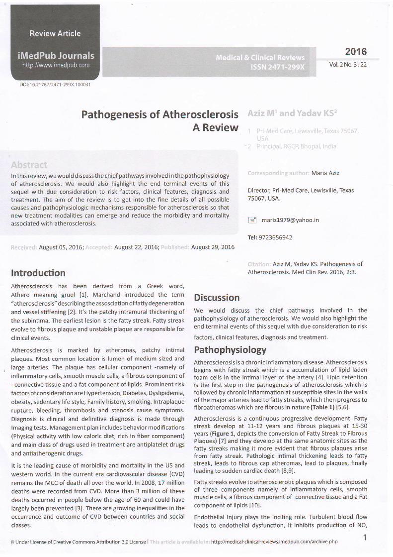
PathophysiologyAtherosclerosis is a chronic inflammatory disease. Atherosclerosisbegins with fatty streak which is a accumulation of lipid ladenfoam cells in the intimal layer of the artery [4]. Lipid retentionis the first step in the pathogenesis of atherosclerosis which is
followed by chronic inflammation at susceptible sites in the wallsof the major arteries lead to fatty streaks, which then progress tofibroatheromas which are fibrous in nature (Table 1) [5,6].
Atherosclerosis is a continuous progressive development. Fattystreak develop at 11-12 years and fibrous plaques at 15 30years (Figure 1, depicts the conversion of Fatty Streak to FibrousPlaques) [7] and they develop at the same anatomic sites as thefatty streaks making it more evident that fibrous plaques arisefrom fatty streak. Pathologic intimal thickening leads to fattystreak, leads to fibrous cap atheromas, lead to plaques, finallyleading to sudden cardiac death [8,9].
Fatty streaks evolve to atherosclerotic plaques which is composedof three components namely of inflammatory cells, smoothmuscle cells, a fibrous component of-connective tissue and a Fat
component of lipids [10].
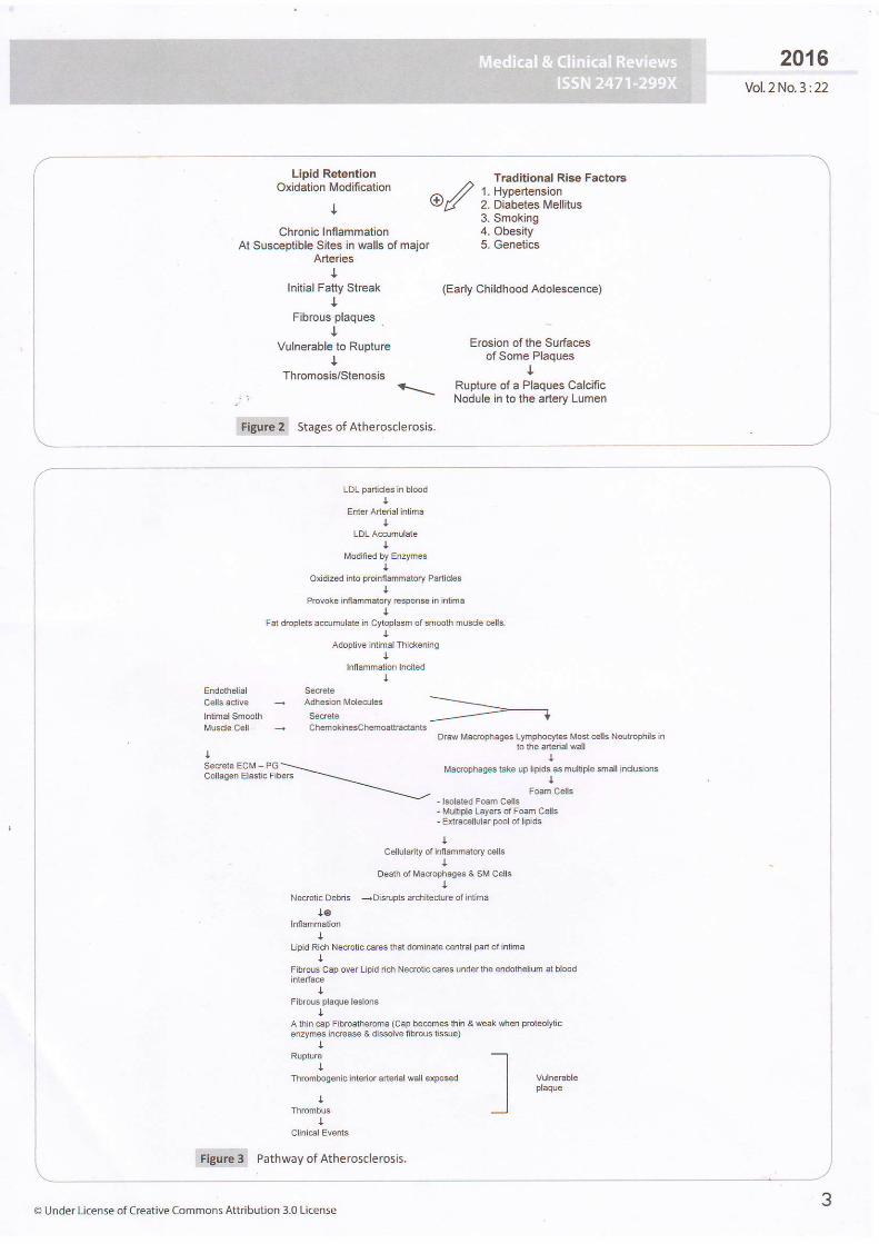
Endothelial Injury plays the inciting role. Turbulent blood flowleads to endothelial dysfunction, it inhibits production of No,
http//medical-clinical-reviews.imedpub.com/archive-php

Table 1 Stages of Atherosclerosis: Modified AHA consensus classification based on morphologic descriptions.
Fatty Streak(11 - 12 year)
Figure 1 Conversion of fatty streak to Fibrous plaques.
a potent vasodilator and stimulates production of adhesion
molecules which attract inflammatorv cells, Other risk factors also
contribute to this step. The net result is Monocytes and T cells
bind to the endothelial cells and migrate to the subendothelialspace. Lipids in the blood, LDL, VLDL bind to endothelial cells and
oxidize in the subendothelial space. Monocytes in subendothelial
space engulf oxidized LDL and transform to foam cells. Thls mark
the first stage i.e-, fatty streak. Macrophages further elaborateproinflammatory cytokines which recruit smooth muscle cells.
There is smooth muscle cell replication and increase in dense
extracellular matrix. End result lesion is a subendothelial fibrousplaque composed of lipid core surrounded by smooth muscle
cells and connective tissue fibers (Figures 2 and 3) [11].
There is sequential involvement of arterial layers, intima, then
media and finally adventitia. Arterial wall lesions have a centralcholesterol rich lipid core surrounded by inflammatory response.
Everv lesion has lipid accumulation and inflammation. Plaque
distorts media/adventitia, increases caliber of arterial lumen
and decreases its size simultaneously. New Vasa Vasorum invade
diseased intima, cause hemorrhage wilhin arterial wall, leading tointramural hemorrhage and increased fibrous tissue. Rupture ofthin fibrous caps leads to thrombosis and healing. cyclic healing
2016Vol.2 No. 3 :22
Absent
Absent
occlusive ornonocclu5rve
Usually nonocclusive
Usually nonocclusive
Absent
of clinically silent ruptures leads to multiple layer of healed tissue
and the end result is sudden cardiac death. calcium depositsas small aggregates convert later to large nodules in the wall.Erosion of endothelium leads to thrombosis. Increase plaque
mass causes stenosis and finally leading to lethal ischemia [12].
There are two types of plaques stable and unstable I131. stableplaques regress or are static or they grow slowly. Unstablesplaques are complicated by erosion, fissure, rupture and cause
stenosis. thrombosis, infarction. Most clinical events result fromcomplications of unstable plaque, hence plaque stabilization can
reduce morbidity and mortality associated with atherosclerosis.
Plaque is ruptured by enzymes secreted by activated macrophagesin the plaque. Once plaque ruptures, plaque contents get exposedto circulating blood and the end result is thrombosis [14]. Theresultant thrombosis may change plaque shape, occlude lumenof blood vessel, may embolise or the plaque contents mayembolise. Low risk plaoues are more fibrous in content andhave low lipids and do not cause 100% blockade while unstableplaques have thick lipid core and thin fibrous cap narrow lumen<50% and tend to rupture unpredictably [15].
The end result of the stenosis caused by the plaques arethe Terminal Events-Acute Coronary Syndrome, MyocardialInfarction, Fatal Arrhythmias, sudden cardiac death (Figures 4and 5 depict the terminal events that arise due to stenosis dueto plaques) [16].
Age, Family history, Male sex, smoking, Diabetes mellitus,hypertension, alcohol, Chlamydia infection, Hyperhomocysteinemia, Obesity, Sedentary lifestyle [17].
Intimalthickening
lntimalxanthoma
Pathologic intimal thickening
Fibrous cap atheroma
Thin cap fibroatheroma
Plaque rupture
Plaque erosion
calcifed nodule
Fibrotic (without calcifi cation)
Fibrocalcific {+/- necrotic core)
Absent
Lesion reference to AHA types V and Vl was discarded, because it failed to account for the 3 different fiorphologies {rupture, erosion, and calcified
nodule)that give rise to acute coronary thrombosis-
Nonatherosclero$c intimal lesions
Normal accumulation of smooth muscle cells (SMCs) in the intima in the absence of lipidor macrophage foam cells.
Superficial accumulation of foam cells without a necrotic core or fibrous cap; based onanimal and human data, such lesions usually regress.
Progressive athercsclerotic lesions
sMC rich plaque with proteoglycan matrix and focal accumulation of extracellular lipid
Early necrosis: focal macrophage infiltration into areas of lipid pools with an overlyingfibrous cap Late necrosis: loss of matrix and extensive cellular debris with an overlying
fibrous cap.
A thin, fibrous cap (< 65 Bm) infiltrat€d by macrophages and lymphocytes with rare orabsence oJ sMcs and a relativelv large underlying necrotic corej intraplaque hemorrhage/- fibrin mav be present.
Lesions with acute thrombiFibroatheroma with fibrous cap disruption; the luminalthrombus communicates with the
underlying necrotic core
Plaque composition, as abovej no communacation ofthe thrombus with the necrotic core;can occur on a plaque substrate of pathologic intimal thickening or fibroatheroma
Eruptive (shedding) of calcified nodules with an underlying fibrocalcific plaque withminimal or absence of necrosis
Lesions with healed thrombiCollagen-rich plaque with 5ignificant luminal stenosis; lesions may contain large areas
of calcification with few inflammatory cells and minimal or absence of necrosis; theselesions may represent healed erosions or ruptures
Fibrous Plaque(15 - 30 yeaf)
hft p://mediaal-clinical-reviews.imedpub.com/archive.php
2016.Vol.2 No.3:22
Lipid RetentionOxidation Modification
+
Chronic lnffammalionAt Suscep ble Sites in walls of major
Arbriest
Initial Fatty StreakI
Fibrous plaques
IVulnerat e to Rupture
IThromosb/Stenosis
^ Traditional Rlso Faciors
/", // |. t+ype{ensionw,B' 2. Diabetes Mellitus3, Smoking4. Obesity5. Genetics
(Early ChiHhood Adole€cance)
1
Erosion otlhe SurfacesofSome Plaques
Rupture of a Plaques CalcificNodule in to lhe artery Lumen
ffi stages of ltherosclerosis.
IDL partclos in bl@d
Enler Arterid inlimaI
LoL A.srmul€tet
Modmed by Enzymes
Ondircd inlo proirilanmaio.y PadtclesI
Povot€ innammato.y .espon$ in h imat
Fat drcplelr admulate in C'ytopl6sm of 3moolh musd€ €i5,I
Adopljve inlimal Thic&ening
Ilniammation Indted
Drd M€oqhag€8 LItrdFqi€s Uct els NdltoPhls in!o t|3 araii vdl
titaftphagB td€ $ littd! ss munide smdl in nr$ms
Fodn Cdts
- MuliCe L€y€rs caFo.m Cdrr-E rEceld.r pool ot [dds
Collsgen Elaslic Fibers
C.llda'ry of ht.mmeto.y cels
Dealh dt Madqhag4 & SM C€{6t
Ne.rolic Debds -Disrupb
archiledre ol lrtm.ta
Upid Rich Ne.rDlic c€rEs that (hnlnals €r ral pad ol i.ntms
Fibd;ap ov€r Upid ddr Nec.otic c€rss und.r ths endoh€liun al dood
!
A rhin cap Frbro8'lerdna (Cap beco|nss hb & wlak $rBn prot€olytcqEyms i@ase & dGs.lve fb.ous lissro)
!+
Thrombog€flic incdd a.tqial Mlt op@ed
a
!
I vurn€,sre
_l p'|aque
p c"thrr"y ot Atherosclerosis.
@ Under License of Creative commohs Attribution 3.0 License

FiSure 4 Steps in terminal events.
Thrombosis due to rupture oiTCFAThrombosis due to erosion of enaotnerum
Thrombosis due to protrusion of a calcifred noduie jnto thearl€riall!men
ITvFibro calcific plaque
I+
Stenosls
I?TerminalEvents
1. Acute Coronary Syndrome
2. MYocardia Intarcnon3. FatalArhythmias
Figure 5 Terminal events that arise due to stenosis due to Plaques.
Elastic and muscular arteries. MC arteries affected are aorta,carotid, coronarv and ileofemoral arteries. MC artery to beinvolved is Aorta. Branch points are the common sites. Proximalcoronary arteries are more susceptible [18].
Atherosclerosis is initially asymptomatic [19]. Symptoms developwhen lesions impede blood flow. When plaques grow, arteriallumen is reduced causing transient ischemic symptoms, stableexertional angina, intermittent claudication, unstable angina,infarction, ischemic stroke, rest pain in the limbs, aneurysm,arterial dissection, sudden death [20].
Patients with signs and symptoms of ischemia should be evaluatedusing history, phvsical examination, fasting lipid profile, plasmaglucose and HbAl-c. Patients with documented disease at onesite should be evaluated at other sites.
CT Angiography, is often used as an initial screening test[21]. Other diagnostic procedures used are catheter basedtests-intravascular ultrasonographV, angioscopy, plaquethermography, elastography, immunoscintigraphy. Certainserum inflammatory markers cRP, LP associated phospholipase42 predict cardiovascular events. other lmaging studies thatdetect plaque are-Angiography, USG, CIMT, MRl.
4
2016Vol.2No.3:22
Behavior Modifications include a diet rich in fruits, vegetables,
fibers with regular physical activity and smoking cessation
could help in getting a favourable lipid profile. Drugs to treat
Dyslipidemia, hypertension and diabetes are often required.
These help in improving endothelial function, reducing
inflammation and give a favourable clinical outcome. Additional
drugs used are statins, antiplatelet drugs (aspirin, clopidogrel),
ACE inhibitors and Beta BIockers. Aspirin is indicated for
prevention of coronary atherosclerosis in high risk patients.
Clopidogrel is used for patients who are intolerant to aspirin and
also for treating ST segment and non ST segment elevation Ml.
Statins, ACE inhibitors, ARB reduce risk of atherosclerosis with
their anti-inflammatory properties. Statins by stabilizing the
plaques, play vital role in management of atherosclerosis.
Statins induce changes in plaque tissues, hence are important
in causing regression in atherosclerosis {Table 2 discusses the
stadns induced changes in plaque tissues) [4,11,18].
Fish oil supplements-omega 3 fatty acids play vital role in
creating favourable lipid profile. Vitamins-Folate, vitamin 86 and
B 12 treat hyper homocysteinemia which is an important cause
of Dvslioidemia.
Table 2i Statins induced changes in Plaque tissues.
T-
Fatty streak
-
http://medical clinical reviews.,medpub-com/archive.php

ConclusionAtherosclerosis is the leading cause of death in the developedcountries. Deep understanding of the causes and underlying
2016Vol.2No.3:22
mechanism of pathogenesis will help to delineate causes andwill help to plan out innovative management. As our knowledgeabout the pathogenesis of atherosclerosis improves moretreatment options will emerge.
56 Under License of Creative Commons Attribution 3.0License

Referencesr Virmani R, Burke AB Farb 4 Xolodgie FD (2006) Pathology of the
vulnerable Dlaque.l Am Coll Cardiol4T: c13-C18,
,' Aschoff L (1933) lntroduction. In: Cowdry EV (ed.) Arteriosclerosis: A
survey ofthe Problem. Macmillan, New York.
-l Beaglehole R, Magnus P (2002) The search for new risk fudors forcoronary heartdisease: occu pationa I the ra py for epidemiologists? IntJ Epidemiol 31: 1117-1122.
' Alexander RW Dzau vl {2000} vascular biology: the past 50years, Circulation 102: lV-112-lV-115.
. Pedrigi RM, de Silva R, Bovens SM, Mehta W, Petretto E, et al. {2014)Thin-cap fibroatheroma rupture is associated with a fine interplay ofsand wall stress. Arterioscler Thromb Vasc Biol34:2224'2237.
Collins R, Armitage l, Parish S, Sleigh P, Peto (2003) R: MRC/BHF
Heart Protedion Study of cholesterol-lowering with simvastatin in
5963 people with diabetes: a randomised placebo-controlled rial.Lancet 351:2005-2016.
I Libby P (2002) lnflammation in atherosclerosis. Nature 420:868-874.
a Meijer wT, Hoes AW Rutgers D, Bots ML, Hofman A, et al. {1998)Peripheral arterial disease in the elderly: the Rotterdam studv.
Arterioscler Thromb Vasc Biol 18: 185-192,
I Simoons L, Saelman M (1994) Effect of simvastatin on coronary
atheroma: the Multicentre Anti-Atheroma Study {MAAS). Lancet
344: 633-638.
1,, Ridker PM, Danielson E, Fonseca F4 Genest l, Gotto AM, etal. {2009) Redudion in C-reactive protein and LDL cholesteroland cardiovascular event rates after initiation of rosuvastatln: a
prospective study of the IUPITER trial. Lancet 373: 1775-7782,
Fowkes FG, Housley E, Riemersma RA, Macintyre cC, cawood
EH, et al. {1992) Smoking, lipids, glucose intolerance, and bloodpressure as risk factors for peripheral atherosclerosis compared
with ischemic heart disease in the Edinburgh Artery Study. Am J
Eoidemiol 135: 331-340.
2016Vol.2 No.3 :22
Scott G, Diane B, Luther C, Richard C, Margo D, et al. (200U
Executive Summary ofThe Third Report of The National CholesterolEducation Program (NCEP) Expert Panel on Detection, Evaluation,and Treatment of High Blood Cholesterol In Adults (AdultTreatmentPanel lll). JAMA 285: 2486-2497.
Ingolfsson lO, Sigurdsson G, Sigvaldason H,Thorgeirsson G, Sigfusson
N, et al. (1994) A marked decline in the prevalence and incidenceof intermittent claudication in lcelandic men 1968-1986: a strongrelationship to smoking and serum cholesterol: the ReykiavikStudy. J
Clin Eoidemiol 47: \237-7243.
Jonason L Bergstrom R (1987) Cessation of smoking in patients withintermittent claudication: effects on the risk of oeripheral vascularcomplications, myocardial infarction and mortality, Ada Med Scand
227:253-260.
Rodriguez-Granillo GA, Agostoni P, Garcia-Garcia HM, Biondi-Zoccai
GG, McFadden E (2007) Meta-analysis of the studies assessing
temporal changes in coronary plaque volume using intravascularultrasound. Am J Cardiol99:5-10.
NaghaviM, Libby B Falk E, casscells 5W Litovsky s, et al. {2003)Fromvulnerable plaque tovulnerable patient a callfor new defnitions andrisk assessment strategies: part l. Ci.culation 108: 1564-1672.
Quick CR, Cotton LT{1982)The measured effect ofstopping smokingon intermittent claudication. BrJ Surg 59: 524-526.
Silva Marques J, Pinto FJ (2014) The vulnerable plaque: Currentconcepts and future perspectives on coronary morphology,composition and wallstress imaging. Rev Po.t Cardiol 33: 101-110.
Law M, Tang JL (19951 An analysis of the effediveness of interventionsintended to helppeople stop smoking. Arch Intern Med 155:1933-1941,
cannon CP, Braunwald E, Mccabe CH, Rader Dl, Rouleau Jl, et al.
{2004) Intensive versus moderdte lipid lowering with statins afteracute coronary syndromes. N Engll Med 350: 1495-1504.
Rosamond w, Flegal K, Furje K, Go A, Greenlund l(, Haase N, et al.(2008) Heart disease and stroke statistics-2oo8 update: a reportfrom the American Heart Association Statistics Cgmmittee and
Stroke Statistics Subcommittee. Circulation 117: e25-e146.
ti
6 '' !t- :. :.t,i:.: .:'., :.- J. , ..i .: : i.,: http://medical-clinical+eviews.imedpub.com/archive.php