pathogenic acanthamoeba spp. secrete a mannose-induced ... · barrier, by inducing apoptosis in a...
TRANSCRIPT

INFECTION AND IMMUNITY, Nov. 2003, p. 6243–6255 Vol. 71, No. 110019-9567/03/$08.00�0 DOI: 10.1128/IAI.71.11.6243–6255.2003Copyright © 2003, American Society for Microbiology. All Rights Reserved.
Pathogenic Acanthamoeba spp. Secrete a Mannose-Induced CytolyticProtein That Correlates with the Ability To Cause Disease
Michael Hurt, Sudha Neelam, Jerry Niederkorn, and Hassan Alizadeh*Department of Ophthalmology, University of Texas Southwestern Medical Center at Dallas, Dallas, Texas 75390
Received 3 April 2003/Returned for modification 20 May 2003/Accepted 8 August 2003
The pathogenesis of Acanthamoeba keratitis begins when Acanthamoeba trophozoites bind specifically tomannosylated glycoproteins upregulated on the surfaces of traumatized corneal epithelial cells. When Acan-thamoeba castellanii trophozoites are grown in methyl-�-D-mannopyranoside, they are induced to secrete a novel133-kDa protein that is cytolytic to corneal epithelial cells. Clinical isolates of Acanthamoeba spp., and not thesoil isolates, were proficient at producing a mannose-induced protein (MIP-133) and generating disease inChinese hamsters. The purified protein was efficient at killing corneal epithelial cells, the first mechanisticbarrier, by inducing apoptosis in a caspase 3-dependent pathway. Subsequent steps in pathogenesis require theamoebae to penetrate and degrade collagen. Only the clinical isolates tested were efficient at migrating througha collagenous matrix in vitro, presumably by MIP-133 degradation of both human type I and human type IVcollagen. A chicken anti-MIP-133 antiserum effectively bound to the protein and blocked collagenolytic activity,migration, and cytopathic effects (CPE) against corneal cells in vitro. Chinese hamsters orally immunized withMIP-133 displayed a >30% reduction in disease. Immunoglobulin A isolated from immunized animals boundMIP-133 and blocked CPE on corneal cells in vitro. Animals induced to generate severe chronic infectionsdisplayed significant reductions in disease symptoms upon oral immunization postinfection. These datasuggest that MIP-133 production might be necessary to initiate corneal disease and that it may play animportant role in the subsequent steps of the pathogenic cascade of Acanthamoeba keratitis. Furthermore, asantibodies produced both prior to and after infection reduced clinical symptoms of disease, the protein mayrepresent an important immunotherapeutic target for Acanthamoeba keratitis.
Acanthamoeba keratitis is a very painful, sight-threateningcorneal infection caused by several species of free-living patho-genic amoebae (22, 40). Acanthamoeba spp. are ubiquitous innature and have been isolated from a wide variety of environ-ments, including swimming pools, hot tubs, lakes, soil, dust,drinking fountains, eyewash stations, and the nasopharyngealmucosae of healthy individuals (3, 4, 6, 12, 19, 27, 30, 39, 41).Despite the wide distribution of amoebae, this disease islargely restricted to contact lens wearers who have experiencedsome sort of trauma to their corneal epithelium (3, 5, 24, 32,33, 40). Moreover, induction of the disease in experimentalanimals requires corneal abrasion prior to exposure to Acan-thamoeba trophozoites (9, 26, 33).
Characteristic symptoms of Acanthamoeba keratitis includea ring-like corneal infiltrate, epithelial destruction, dispropor-tionately severe ocular pain, and resistance to antimicrobialagents (2, 20, 29). Treatment of the disease is very demanding,consisting of hourly applications of brolene, polyhexamethyl-ene biguanide, or chlorhexidine for several weeks. Even withsuch therapies, Acanthamoeba spp. can cause severe damage tothe corneal epithelium and stroma, resulting in the need forcorneal transplantation (2).
Very little is known about how Acanthamoeba can penetratethe ocular defense and enter the stromal layer of the cornea.Upon abrasion of the corneal surface, the corneal epitheliumexpresses elevated concentrations of mannose glycoproteins,
to which the amoebae can adhere with high affinity (11, 25, 42).Subsequently, the amoebae penetrate and destroy the cornealepithelium, perforate Bowman’s membrane, and gain entryinto the underlying stroma. Both Bowman’s membrane and thestroma are primarily comprised of collagen.
Recent studies by our laboratory have shown that whenAcanthamoeba castellanii is stimulated with methyl-�-D-man-nopyranoside, the amoebae secrete a new 133-kDa protein(MIP-133) that is highly cytolytic to both human and hamstercorneal epithelial cells in vitro (10, 16). Secretion of this pro-tein is mannose specific, as neither lactose nor galactose in-duces the production of the protein. Moreover, the cytolyticactivity against corneal epithelial cells is eliminated when theMIP-133 protein is incubated with serine protease inhibitors(10).
In this study, we examined the correlation of this mannose-induced protein with known pathogenic and nonpathogenicstrains of Acanthamoeba, the mechanism behind the cytolyticactivity, and the possible role of the protein in the subsequentsteps in the pathogenic cascade of Acanthamoeba keratitis.Furthermore, we investigated how antibodies may defendagainst the actions of the protein and whether the protein canbe effectively used as an immunogen against disease.
MATERIALS AND METHODS
Animals. Chinese hamsters were purchased from Cytogen Research and De-velopment (West Bury, Mass.). All animals used were from 4 to 6 weeks of age,and all corneas were examined before experimentation to exclude animals withpreexisting corneal defects. Animals were handled in accordance with the Asso-ciation of Research in Vision and Ophthalmology Statement on the Use ofAnimals in Ophthalmic and Vision Research.
* Corresponding author. Mailing address: Department of Ophthal-mology, University of Texas Southwestern Medical Center, 5323 HarryHines Blvd., Dallas, TX 75390. Phone: (214) 648-4732. Fax: (214)648-9061. E-mail: [email protected].
6243
on January 2, 2020 by guesthttp://iai.asm
.org/D
ownloaded from

Amoebae and cell lines. All Acanthamoeba species were originally obtainedfrom the American Type Culture Collection, Manassas, Va. A. castellanii (ATTC30868), Acanthamoeba polyphaga (ATTC 30461), and Acanthamoeba rhysodes(ATTC 50368) were isolated from human corneas. Acanthamoeba culbertsoni(ATTC 30171) was originally isolated from a diseased human kidney. Acan-thamoeba hatchetti (ATTC 30730), Acanthamoeba astronyxis (ATTC 30137), andA. castellanii strain neff (ATTC 30010) were isolated from soil. The chosenspecies represent all three of the different subgroups of Acanthamoeba based onmorphology, isoenzyme analysis, and serology (38). Amoebae were grown asaxenic cultures in peptone-yeast extract-glucose (PYG) at 35°C with constantagitation.
To examine whether the Acanthamoeba spp. produced the MIP-133 protein,trophozoites were grown in 200 ml of PYG either with or without 100 mMmethyl-�-D-mannopyranoside (Sigma Chemical Co., St. Louis, Mo.) on a shakerincubator set at 125 rpm and at 35°C (10). Initial cultures were seeded with atotal of 107 trophozoites. Individual supernatants from cultures at mid-log phasewere collected, centrifuged, filter sterilized, 10-fold concentrated, and immedi-ately examined by sodium dodecyl sulfate-polyacrylamide gel electrophoresis(SDS-PAGE) analysis using 4 to 15% ready gels (Bio-Rad, Hercules, Calif.) orused for detection of the MIP-133 protein by enzyme-linked immunosorbentassay (ELISA).
Human corneal epithelial (HCE) cells were a generous gift from Sherry Wardof the Gillette Company (Gillette Medical Evaluation Laboratories, Gaithers-burg, Md.). Cells were cultured in keratinocyte growth medium with a Bullet kit(catalog number CC-3111; Clonetics, Walkersville, Md.).
Contact lens preparation. Contact lenses were prepared from Spectra/Pordialysis membrane tubing (Spectrum Medical Industries, Los Angeles, Calif.)with a 3-mm-diameter trephine prior to heat sterilization. Lenses were placed insterile 96-well microtiter plates (Costar, Cambridge, Mass.) and incubated with3 � 106 A. castellanii trophozoites at 35°C for 24 h. Attachment of amoebae tothe lenses was verified microscopically before infection.
In vivo corneal infections. Acanthamoeba keratitis was induced as describedpreviously (9, 13, 34). Briefly, the Chinese hamsters were anesthetized withketamine (100 mg/kg of body weight; Fort Dodge Laboratories, Fort Dodge,Iowa), injected peritoneally. Prior to manipulation, the corneas were anesthe-tized by topical application of Alcain (Alcon Laboratories, Fort Worth, Tex.).Approximately 25% of the cornea was abraded with a sterile cotton applicatorbefore the amoeba-laden lens was placed onto the center of the cornea. Theeyelids were then closed by tarsorrhaphy with 6-0 Ethilon sutures (Ethicon,Somerville, N.J.). The contact lenses were removed 3 to 4 days postinfection, andthe corneas were visually inspected by microscopy for severity of disease. Visualinspections were recorded daily as indicated. The infections were scored on ascale of 0 to 5 based on the following parameters: corneal infiltration, cornealneovascularization, and corneal ulceration. The pathology was recorded as fol-lows: 0 indicates no pathology, 1 indicates that �10% of the cornea was involved,2 indicates a 10 to 25% involvement, 3 indicates 25 to 50% involvement, 4indicates 50 to 75% involvement, and 5 indicates 75 to 100% involvement asdescribed previously (17). In Chinese hamsters, Acanthamoeba keratitis resolvesat approximately 3 weeks. At this time, there is a conspicuous absence of cornealopacity, edema, epithelial defects, and stromal necrosis and inflammation.
MIP-133 isolation. The MIP-133 protein was purified as stated previously (10).Briefly, 10-fold-concentrated supernatants from A. castellanii cultures grown with100 mM methyl-�-D-mannopyranoside, were analyzed by SDS–4 to 15% PAGEReady Gels (Bio-Rad) under both reducing and nonreducing conditions. Super-natants were taken from trophozoites at mid-log phase.
For fast protein liquid chromatography, culture supernatants of mannose-stimulated A. castellanii trophozoites were concentrated 10-fold with Ultra-free-15 centrifuge concentrators with a molecular cutoff of 5 kDa (Millipore,Bedford, Mass.). Samples were centrifuged at 3,000 � g for 20 min and passedin 0.5-ml volumes over a Superdex 200 (Amersham Pharmacia Biotech, Piscat-away, N.J.) column with phosphate-buffered saline (PBS) (pH. 7.2). Fractionswere collected every 0.5 ml and examined by SDS–4 to 15% PAGE, fractionscontaining the mannose-induced cytolytic protein were pooled and concentrated10-fold, and the buffer was exchanged three times with 10 mM Tris buffer (pH8.0) with Ultrafree 0.5 concentrators. Two-hundred-microliter samples contain-ing 1 mg of protein were applied to a DEAE ionic-exchange column by using 10mM Tris buffer, pH 8.0 (buffer A). Adsorbed protein was eluted by using agradient of 10 mM Tris buffer, pH 8.0, with 1 M NaCl (buffer B). Fractions wereexamined for the �133-kDa protein by SDS–4 to 15% PAGE. An initial 15%buffer B step removed contaminating proteins, and the �133-kDa protein waseluted between 15 to 30% buffer B run at 0.1 ml/min. Fractions containing the�133-kDa protein were pooled and concentrated 10-fold and washed three timeswith PBS (pH 7.2) to exchange the buffer.
Apoptosis assay. The experiment was carried out by using a TACSAnnexinV-fluorescein isothiocyanate kit (R&D Systems) according to the manufacturer’sinstructions. Briefly, HCE cells were grown to �90% confluence in 24-wellplates. The cells were then incubated with either 1.5 �g of MIP-133, 20 �MZ-DEVD-FMK caspase 3 inhibitor (BD Pharmingen, San Diego, Calif.), 20 �MZ-FA-FMK control inhibitor (BD Pharmingen), or staurosporine (3 �g/ml;Sigma) as a positive control for apoptosis for 18 h. Then the cells weretrypsinized, washed twice with PBS, and stained with Annexin V (1 �l) andpropidium iodide (10 �l) for 15 min, and apoptosis was quantified by flowcytometry at a 488-nm wavelength. All subsequent incubation steps were per-formed on ice, and centrifugation steps were performed at 4°C. For each sample,10,000 ungated events were acquired and the results were analyzed withCellQuest software (BD Biosciences, Franklin Lakes, N.J.). The results areexpressed as percentages of cells that were apoptotic.
Migration assays. The migration assays were performed with 24-well transwellplates (6.5-mm diameter, 3.0-�m pore size; Costar, Corning Inc., Corning, N.Y.).The top chamber membrane was coated with 100 �l of Matrigel (CollaborativeBiomedical Products, Bedford, Mass.) that had been diluted 1:3 in Hanks’ bal-anced salt solution. Excess Matrigel was removed after 10 min, and the mem-branes were allowed to dry at room temperature. Acanthamoeba trophozoites(105) were then placed in the top chamber in 100 �l of PYG. Plates wereincubated at 37°C for 2 h, and acanthamoebae were then counted in the bottomchamber by light microscopy (magnification, �100). Inhibition assays involvedincubating A. castellanii trophozoites with either a 1:75 or a 1:100 dilution ofchicken anti-MIP-133 antiserum or chicken preimmune serum, 1.0 mM phenyl-methylsulfonyl fluoride (PMSF; Sigma), or 10 �M cystatin (Sigma) at 37°C for 30min prior to their addition to the upper chambers. All experiments were per-formed in triplicate.
Collagen digestion assay. Collagen assays were performed in 96-well plates. Inseparate plates, 10-�g samples of human collagen types I (Sigma) and IV (US-Biological, Swampscott, Mass.) were added and incubated at 37°C until theplates were dry. Total volumes were 50 �l in PBS. Plates were then washed threetimes with PBS and then incubated with either 15.6 �g of MIP-133 or PBS or 0.1mg of Clostridium histolyticum collagenase (Sigma) at 37°C for 24 or 72 h. Samplewells were then washed three times with PBS, incubated with a 1:500 dilution ofmouse anti-collagen type IV immunoglobulin G (IgG) or a 1:2,000 dilution ofmouse anti-collagen type I IgG (Sigma) as the primary antibody and goat anti-mouse IgG-horse radish peroxidase (HRP) (Southern Biotechnology Associates,Inc., Birmingham, Ala.) as the secondary antibody. Plates were read at an opticaldensity (OD) at 405 nm in a Molecular Devices (Menlo Park, Calif.) microplatereader. Inhibition studies involved coincubating the MIP-133 protein with thechicken anti-MIP-133 antiserum diluted 1:75. All experiments were performed intriplicate.
Western blot analysis and ELISA. Ninety-six-well assay plates were coatedwith 50 �g of MIP-133 overnight in carbonate buffer. Plates were washed fourtimes with PBS containing 0.05% Tween 20 (wash buffer; Sigma) and thenblocked with 0.5% bovine serum albumin in PBS for 1 h at room temperature.All subsequent antibodies were diluted with blocking buffer and incubated atroom temperature. Chicken anti-MIP-133 antiserum (Aveslabs, Tigard, Oreg.)was added at either a 1:50, 1:75, or 1:100 dilution for 1 h and washed. HRP-conjugated goat anti-chicken IgY (Aveslabs) was added at a 1:10,000 dilutionand incubated for 1 h. Plates were developed by adding 1.0 mM 2,2�-azinobis(3-ethyl-benzthiazoline-6-sulfonic acid) (Sigma) containing 0.003% H2O2 and in-cubated for 30 min at room temperature. After development, 100 �l of 10% SDS(Sigma) was added per well prior to growth being read at an OD at 405 nm witha microplate reader.
Western blot analysis was carried out by conventional techniques. Briefly, 30�g of 10-fold-concentrated crude supernatant taken from A. castellanii tropho-zoites grown in mannose was resolved by using 4 to 25% ready gels. Gels weretransferred to Trans-Immun-Blot PVDF membranes (Bio-Rad) with a Bio-Radminitransfer apparatus. Blots were blocked in 5% dry milk in PBS (blockingbuffer) overnight at 4°C prior to the addition of a 1:200 dilution of chickenanti-MIP-133 antiserum for 1 h at room temperature. After 1 h, membranes werewashed in PBS and incubated in a 1:2,000 dilution of rabbit anti-chicken IgG–alkaline phosphatase (Sigma) in blocking buffer. After 1 h, the membrane waswashed and developed with Nitro Blue Tetrazolium-BCIP (5-bromo-4-chloro-3-indolylphosphate) stock solution (Roche Diagnostics, Mannheim, Germany) asrecommended by the manufacturer.
Assay for CPE. The MIP-133 protein was added at 1.5, 7.8, and 15.6 �g ofprotein (in 25 �l of PBS) to 96-well plates with confluent monolayers of HCEcells and incubated for 18 h at 35°C. Each well contained 200 �l of the respectiverequired growth medium. Inhibition experiments involved incubating the proteinsamples with either a 1:75 dilution of chicken anti-MIP-133 antiserum or of a
6244 HURT ET AL. INFECT. IMMUN.
on January 2, 2020 by guesthttp://iai.asm
.org/D
ownloaded from

chicken preserum control and a 1:50 dilution of pooled enteric washes fromimmunized hamsters or from control (PBS)-immunized animals. Additional con-trol wells consisted of untreated confluent cells. Following incubation, all wellswere washed three times with their respective growth medium and stained withGiemsa stain (Shandon, Inc., Pittsburgh, Pa.). After being stained, the wells werewashed three times with PBS (pH 7.2) and solubilized in 0.1 ml of 5% SDS inPBS and the OD was read at 590 nm in a Molecular Devices microplate reader.Percentages of cytopathic effects (CPE) were calculated according to the follow-ing formula: percent CPE � 100 [(OD of experimental well OD of super-natant alone/OD of control cells alone) � 100]. Assays were performed intriplicate.
Oral immunizations. Animals received 1 ml of 0.1 M sodium carbonate (pH9.6; Sigma) by lavage tube prior to administration of either 100 �g of MIP-133(in 100 �l of PBS) plus 10 �g of cholera toxin (Sigma), 200 �g of MIP-133 plus20 �g of cholera toxin, or 400 �g of MIP-133 plus 40 �g of cholera toxin.
Immunizations were administered once a week for 4 weeks prior to infection withA. castellanii. Control groups included animals immunized with equivalent dosesof cholera toxin alone and untreated animals prior to infection.
Collection of Chinese hamster IgA secretions. Enteric washes were collectedas stated previously (14). Briefly, the enteric washes were collected from Chinesehamsters after the animals were anesthetized with ketamine and euthanized bycervical dislocation. Approximately 20 cm of the small intestine was removed,and 10 ml of PBS was injected through the intestinal section with an 18-gaugeneedle. The enteric washes were collected and centrifuged at 700 � g to removesediments. Protease inhibitor cocktail tablets (Boehringer Mannheim, Indianap-olis, Ind.) were added to the pooled enteric washes at one tablet per 10 ml. Thewash was stored at 80°C until used.
IgA ELISA. Ninety-six-well assay plates were coated with 50 �g of MIP-133overnight in carbonate buffer. Plates were washed four times with PBS contain-ing 0.05% Tween 20 (wash buffer; Sigma) and then blocked with 0.5% bovine
FIG. 1. Correlation between MIP-133 production and the ability of Acanthamoeba soil isolates to cause disease. (A) Acanthamoeba spp. were grownin PYG containing 100 mM methyl-�-D-mannopyranoside. Supernatants were collected at mid-log phase, filter sterilized, concentrated 10-fold, andanalyzed by SDS-PAGE. Lanes: L, molecular weight ladder; 1, A. hatchetti; 2, A. astronyxis; 3, A. castellanii neff; 4, A. castellanii (clinical isolate). The arrowpoints at �133 kDa. (B) Ability of Acanthamoeba spp. to cause keratitis in Chinese hamsters. Animals were infected with Acanthamoeba-laden lensesas described in Materials and Methods. The results are representative of three separate experiments (eight hamsters in each group).
VOL. 71, 2003 ACANTHAMOEBA MANNOSE-INDUCED CYTOLYTIC PROTEIN 6245
on January 2, 2020 by guesthttp://iai.asm
.org/D
ownloaded from

serum albumin in PBS for 1 h at room temperature. All subsequent antibodieswere diluted in blocking buffer and incubated at room temperature. Entericwashes were added at a dilution of 1:2 for 1 h and then washed. Rabbit anti-Chinese hamster IgA hyperimmune serum (15) was then added to a 1:2 dilutionand incubated for 2 h. Plates were washed, and a 1:1,000 dilution of goatanti-rabbit IgG-HRP (Santa Cruz Biotechnology) was added. Plates were devel-oped by adding 1.0 mM 2,2�-azinobis(3-ethyl-benzthiazoline-6-sulfonic acid)(Sigma) containing 0.003% H2O2 and incubated for 30 min at room temperature.After development, 100 �l of 10% SDS (Sigma) was added per well prior togrowth being read on a microplate reader at 405 nm.
Preparation of clodronate liposomes. Multilamellar liposomes were preparedas described earlier (35, 36). Briefly, 8 mg of cholesterol and 86 mg of phos-phatidylcholine (Lipoid GmbH, Ludwigshafen, Germany) were dissolved in 10ml of chloroform in a round-bottomed flask. After low-vacuum rotary evapora-tion at 37°C, a thin film was then dispersed by gentle rotation for 10 min in PBS
for the preparation of PBS containing liposomes. Liposomes were washed twiceby centrifugation in PBS at 100,000 � g for 30 min and resuspended in 4 ml ofPBS that contained 20 mg of clodronate (Roche Diagnostics). Each 100 �l of theclodronate liposome suspension contained 1 mg of clodronate. Clodronate lipo-somes and PBS liposomes were stored at 4°C and used within 7 days of prepa-ration. Clodronate liposomes were tested for in vitro toxicity against macro-phages prior to use.
Liposome-treated animals. Both clodronate and PBS containing liposomeswere administered via subconjunctival injection on days 8, 6, 4, and 2 ofinfection. Fifty microliters of the liposome preparation was injected in fourquadrants of the eye encircling the entire conjunctiva. Acanthamoeba infectionswere performed via contact lens placement as mentioned above.
Animals were orally immunized on days 5, 12, 19, and 26 days postinfectionwith 400 �g of the MIP-133 protein as mentioned above.
FIG. 2. Correlation between MIP-133 production and the ability of clinical isolates of Acanthamoeba to cause disease. (A) Acanthamoeba spp.were grown in PYG containing 100 mM methyl-�-D-mannopyranoside. Supernatants were collected at mid-log phase, filter sterilized, concentrated10-fold, and analyzed by SDS-PAGE. Lanes: L, molecular weight ladder; 1, A. culbertsoni; 2, A. polyphaga; 3, A. rhysodes; 4, A. castellanii. The arrowpoints at �133 kDa. (B) Ability of Acanthamoeba spp. to cause keratitis in Chinese hamsters. Animals were infected with Acanthamoeba-ladenlenses as described in Materials and Methods. The results are representative of three separate experiments (eight hamsters in each group).
6246 HURT ET AL. INFECT. IMMUN.
on January 2, 2020 by guesthttp://iai.asm
.org/D
ownloaded from

Statistics. Statistical analyses of all data except clinical scores were performedby using unpaired Student’s t tests. Clinical-severity scores were analyzed by theMann-Whitney test.
RESULTS
Correlation of MIP-133 production to pathogenic and non-pathogenic strains of Acanthamoeba. A. castellanii has beenshown to produce the MIP-133 protein when it was grown inthe presence of methyl-�-D-mannopyranoside (10). However, itwas important to determine whether there was a correlationbetween production of the protein and the ability of the amoe-bae to generate disease.
When grown in mannose, none of the Acanthamoeba soilisolates tested produced MIP-133 at levels detectable by SDS-PAGE (Fig. 1A). A. castellanii, the positive control, producedsignificant amounts of the MIP-133 protein. Moreover, whentested in vivo, all three of the soil isolates produced either nodisease or significantly less disease than that produced by thestandard ocular isolate of A. castellanii (Fig. 1B). Both A.astronyxis and A. castellanii neff produced symptoms similar tothose of the sterile contact lens control animals, while A. hatch-etti produced a mild infection. The MIP-133 protein was notdetectable in lysates made from the three soil isolates (data notshown).
By contrast, the clinical isolates of Acanthamoeba producedthe MIP-133 protein when they were grown in mannose (Fig.2A). All three clinical isolates tested produced levels similar tothose produced by A. castellanii (Fig. 2B). Additionally, whentested in vivo, the clinical isolates produced severe Acan-thamoeba keratitis.
Mechanism of cell death in vitro. Though the MIP-133 pro-tein is effective at killing both human and hamster cornealepithelial cells in vitro (10), it is unknown how the protein killsthe corneal epithelial cells.
When incubated with fluorescein-encapsulated liposomes,neither 1.5, 7.8, nor 15.6 �g of the MIP-133 protein was able toperforate the liposomes (data not shown). However, whenexamined by Annexin V flow cytometry, the protein was foundto cause cell death by inducing apoptosis (Fig. 3). MIP-133-treated HCE cells displayed a threefold increase in apoptosisover the level in untreated cells. Incubations with a caspase 3inhibitor completely eliminated the apoptosis caused by theMIP-133 protein. By contrast, the control inhibitor did notreduce apoptosis. Staurosporine, known to induce apoptosis,produced a nearly fourfold increase in apoptosis.
Migration assay and collagen degradation. Although theMIP-133 protein correlated with clinical disease and cytotoxicactivity against corneal epithelial cells, it was important todetermine how the protein might play a role in the pathogeniccascade of Acanthamoeba keratitis.
All three of the soil isolates tested were ineffectual in mi-grating through the collagenous Matrigel (number of migratingamoebae, 10 to 18). By contrast, all of the clinical isolates werecapable of migrating through the Matrigel at levels approxi-mately equal to those of the original standard ocular isolate ofA. castellanii (number of migrating amoebae, 60 to 84).
Matrigel is comprised of many components, with collagen(type A) being only one of the components. However, bothBowman’s membrane and the stroma are comprised almostentirely of collagen (types IV and I, respectively).
FIG. 3. Effects of caspase 3 inhibition of MIP-133-mediated apoptosis. HCE cells were treated with 1.7 �g of MIP-133 either alone or with a20 �M concentration of the caspase 3 inhibitor Z-DEVD-FMK (cas-3 Inh), or a 20 �M concentration of the caspase 3 control inhibitor Z-FA-FMK(con Inh). HCE cells were examined by Annexin V flow cytometry as described in Materials and Methods. Additional controls included untreatedcells, and 3 �g of staurosporine (STS) per ml served as a positive control. Bars and error bars represent the means standard errors (SE) of resultsof triplicate experiments. ***, significantly different from the value for the untreated controls (P � 0.001).
VOL. 71, 2003 ACANTHAMOEBA MANNOSE-INDUCED CYTOLYTIC PROTEIN 6247
on January 2, 2020 by guesthttp://iai.asm
.org/D
ownloaded from

The MIP-133 protein was efficient at degrading both humantype I and human type IV collagen (Fig. 4). At 24 h, type IVcollagen was more efficiently degraded than type I. However,by 72 h, both forms of collagen were nearly completely de-graded. Degradation of collagen was similar to that of thecollagenase control at both the 24 and 72 h time points tested.
The PBS control did not show lytic ability against either col-lagen.
Neutralization of MIP-133 and inhibition of trophozoitemigration by anti-MIP-133 antibody. The specificity of thechicken anti-MIP-133 antiserum was confirmed by Westernblotting (Fig. 5A). Preimmune serum did not bind to the man-
FIG. 4. Collagenolytic activity of the MIP-133 protein. Ten micrograms of human collagen types I and IV were incubated and dried onto 96-wellplates. Wells were treated with either 15.6 �g of the MIP-133 protein, PBS, or 0.1 mg of collagenase (Col) for 24 (A) and 72 (B) h. Sample wellswere then washed three times and incubated with mouse anti-collagen type IV IgG or mouse anti-collagen type I IgG as the primary antibody,followed by goat anti-mouse IgG-HRP, as described in Materials and Methods. Plates were developed and read at an OD at 405 nm. Bars anderror bars represent the means SE of triplicate experiments. *, **, and ***, significantly different from values for PBS-treated controls (P � 0.05,P � 0.01, and P � 0.001, respectively).
6248 HURT ET AL. INFECT. IMMUN.
on January 2, 2020 by guesthttp://iai.asm
.org/D
ownloaded from

nose-stimulated protein (data not shown). Moreover, ELISAanalysis indicated specific binding of the anti-MIP-133 anti-serum to the MIP-133 protein at 1:50, 1:75, and 1:100 dilutions(Fig. 5B). As before, there was no ELISA evidence that thepreimmune serum bound to the MIP-133 protein. Addition-ally, neither the preimmune nor the anti-MIP-133 serum
bound to the surfaces of the Acanthamoeba trophozoites (datanot shown).
Accordingly, we tested the ability of anti-MIP-133 antiserumto neutralize the collagenolytic activity of MIP-133 (Fig. 6A).Antiserum treatment reduced the degradation of type I colla-gen by approximately 75% and type IV collagen by 80%. Deg-
FIG. 5. Specific binding of the chicken anti-MIP-133 antiserum. (A) Western blotting. Crude supernatants taken from A. castellanii tropho-zoites grown in mannose were resolved by SDS-PAGE and transferred to PVDF membranes. Membranes were first incubated with chickenanti-MIP-133 antiserum, followed by rabbit anti-chicken IgG-alkaline phosphatase, as described in Materials and Methods. The membranes werethen washed and developed with Nitro Blue Tetrazolium-BCIP stock solution. The arrow indicates the 133-kDa band. (B) ELISA. Ninety-six-wellplates were coated with 50 �g of MIP-133 and allowed to dry. After drying, wells were blocked and incubated with a 1:50, 1:75, or 1:100 dilutionof either the chicken anti-MIP-133 antiserum or preimmune chicken antiserum. Wells were then washed and incubated with goat anti-chickenIgY-HRP. ELISA plates were developed and read at an OD at 405 nm. Bars and error bars represent the means SE of results from triplicateexperiments. ***, significantly different from values obtained with preimmune serum at the same dilutions (P � 0.001).
VOL. 71, 2003 ACANTHAMOEBA MANNOSE-INDUCED CYTOLYTIC PROTEIN 6249
on January 2, 2020 by guesthttp://iai.asm
.org/D
ownloaded from
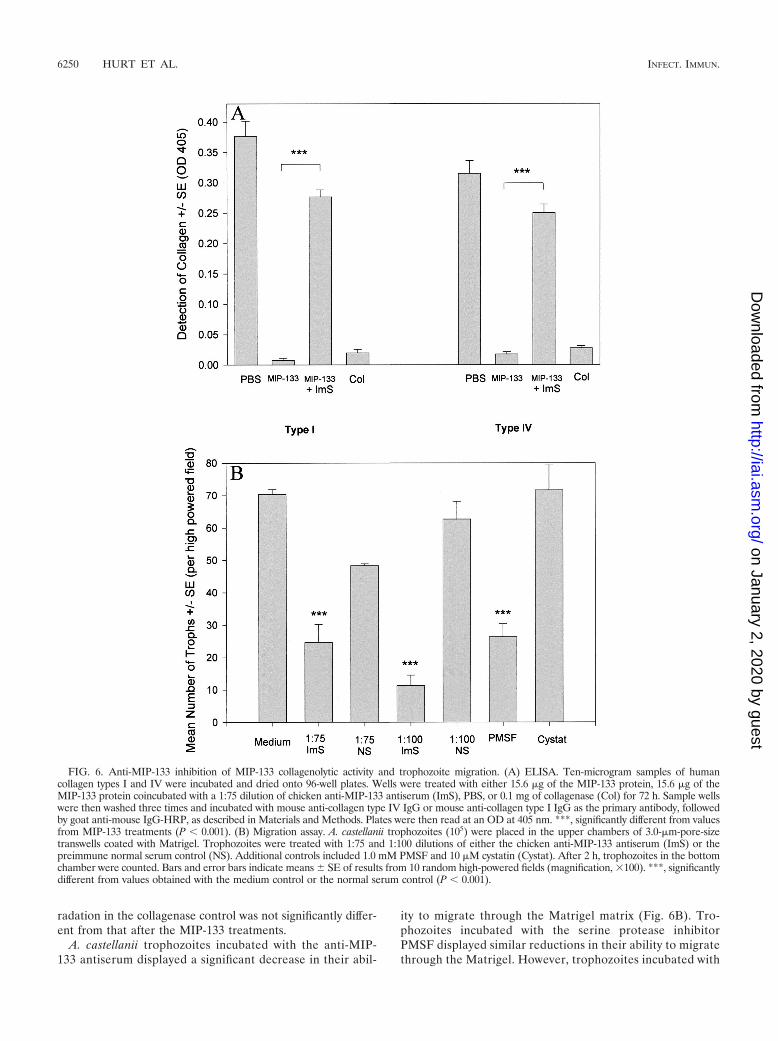
radation in the collagenase control was not significantly differ-ent from that after the MIP-133 treatments.
A. castellanii trophozoites incubated with the anti-MIP-133 antiserum displayed a significant decrease in their abil-
ity to migrate through the Matrigel matrix (Fig. 6B). Tro-phozoites incubated with the serine protease inhibitorPMSF displayed similar reductions in their ability to migratethrough the Matrigel. However, trophozoites incubated with
FIG. 6. Anti-MIP-133 inhibition of MIP-133 collagenolytic activity and trophozoite migration. (A) ELISA. Ten-microgram samples of humancollagen types I and IV were incubated and dried onto 96-well plates. Wells were treated with either 15.6 �g of the MIP-133 protein, 15.6 �g of theMIP-133 protein coincubated with a 1:75 dilution of chicken anti-MIP-133 antiserum (ImS), PBS, or 0.1 mg of collagenase (Col) for 72 h. Sample wellswere then washed three times and incubated with mouse anti-collagen type IV IgG or mouse anti-collagen type I IgG as the primary antibody, followedby goat anti-mouse IgG-HRP, as described in Materials and Methods. Plates were then read at an OD at 405 nm. ***, significantly different from valuesfrom MIP-133 treatments (P � 0.001). (B) Migration assay. A. castellanii trophozoites (105) were placed in the upper chambers of 3.0-�m-pore-sizetranswells coated with Matrigel. Trophozoites were treated with 1:75 and 1:100 dilutions of either the chicken anti-MIP-133 antiserum (ImS) or thepreimmune normal serum control (NS). Additional controls included 1.0 mM PMSF and 10 �M cystatin (Cystat). After 2 h, trophozoites in the bottomchamber were counted. Bars and error bars indicate means SE of results from 10 random high-powered fields (magnification, �100). ***, significantlydifferent from values obtained with the medium control or the normal serum control (P � 0.001).
6250 HURT ET AL. INFECT. IMMUN.
on January 2, 2020 by guesthttp://iai.asm
.org/D
ownloaded from

the cysteine protease inhibitor cystatin did not show reducedmigration.
Inhibition of CPE. The MIP-133 protein has been shown tobe highly cytotoxic for HCE cells in vitro. Being able to blockthis killing with an antibody might be a therapeutic option formitigating disease.
Figure 7 shows that the anti-MIP-133 antiserum was effec-tive at blocking the cytotoxic activity of the MIP-133 protein.The cytotoxic effect of the 1.5-�g dose was completely elimi-nated, while the effect of the 7.8-�g dose was blocked byapproximately 25%. The higher dosage of 15.6 �g was notinhibited by the antiserum. The preimmune serum control didnot significantly reduce the cytolytic ability of the protein atany of the three doses tested.
Clinical disease following MIP-133 immunizations. Wehave previously shown that oral immunizations with antigensconjugated to neutralized cholera toxin is an effective methodof inducing mucosal antibody responses and the appearance ofIgA antibodies in the tears and enteric washes of Chinesehamsters (13, 15). Therefore, Chinese hamsters were immu-nized with either 100, 200, or 400 �g of MIP-133 conjugatedwith neutralized cholera toxin. The results of a typical exper-iment are shown in Fig. 8 and demonstrate that oral immuni-zation with 400 �g of MIP-133 reduced the severity of cornealinfection by 30% and shortened the duration of the disease by5 days (P � 0.01). Immunization with lower doses of MIP-133or with cholera toxin alone did not significantly affect thecourse of disease.
Inhibition of CPE by enteric washes collected from immu-nized hamsters. Enteric washes from orally immunized Chi-nese hamsters were tested by ELISA for the presence of mu-cosal IgA antibodies specific for the MIP-133 protein. Figure9A shows that the pooled enteric washes from the immunizedhamsters bound to the MIP-133 protein. By contrast, enteric
washes taken from the PBS-treated animals did not specificallybind to the MIP-133 protein.
The capacity of the mucosal anti-MIP-133 antibodies to neu-tralize the CPE of MIP-133 was tested in vitro. The results ofa typical experiment are shown in Fig. 9B and demonstrate thatanti-MIP-133 mucosal antibody preparations significantly in-hibited the cytopathic activity of MIP-133 (P � 0.01). Entericwashes from PBS-immunized animals did not inhibit cyto-pathic activity at any of the doses tested.
Oral immunization with MIP-133 mitigates chronic Acan-thamoeba keratitis. Acanthamoeba keratitis is an acute infec-tion in Chinese hamsters that is self-limiting due to the rapidand effective response of the innate system, especially the con-junctival macrophages (7, 35). However, subconjunctival injec-tion of liposomes containing the macrophagicidal drug clodr-onate removes periocular macrophages and results in a chronicinfection that resembles the clinical course of Acanthamoebakeratitis in humans. Accordingly, we wished to determine ifmucosal immunization with MIP-133 would affect a form ofAcanthamoeba keratitis that mimicks the human counterpartin terms of its severity, persistence, and chronicity. Oral im-munization with MIP-133 produced a remarkable mitigation ofcorneal disease in animals pretreated with clodronate-contain-ing liposomes (Fig. 10). By contrast, infected animals treatedwith clodronate-containing liposomes and orally immunizedwith cholera toxin alone displayed clinical disease that was notsignificantly different from that of animals treated with clod-ronate-containing liposomes (data not shown).
DISCUSSION
The purposes of this study were to determine (i) the corre-lation between MIP-133 production and pathogenicity, (ii) thepathogenic mechanisms of MIP-133, (iii) the mechanism of
FIG. 7. Inhibition of MIP-133-mediated CPE against HCE cells. MIP-133 protein samples were adjusted to 1.5, 7.8, and 15.6 �g of protein in25 �l of PBS before addition to HCE cells in 96-well microtiter plates for 18 h. Protein samples were either used alone, or coincubated with a 1:75dilution of chicken anti-MIP-133 antiserum (ImS) or the normal serum control (NS). All final volumes were 200 �l. CPE were assessedspectrophotometrically. Each bar and error bar show the mean SE of triplicate counts. * and ***, significantly different from values for untreatedcontrols (P � 0.05 and P � 0.001, respectively).
VOL. 71, 2003 ACANTHAMOEBA MANNOSE-INDUCED CYTOLYTIC PROTEIN 6251
on January 2, 2020 by guesthttp://iai.asm
.org/D
ownloaded from

MIP-133-induced corneal cell death, and (iv) if neutralizationof MIP-133 affects the clinical course of Acanthamoeba kera-titis.
The results show that production of the MIP-133 proteincorrelates with an amoeba’s ability to cause disease. All clinicalisolates produced MIP-133 and caused significant disease in
Chinese hamsters. In contrast, three soil isolates did not pro-duce the MIP-133 protein and also did not produce severe eyedisease. However, A. hatchetti did consistently generate a milddisease, even though the strain did not produce the MIP-133protein. It is uncertain what factor(s) may be responsible forthis mild disease. A. castellanii neff has been shown by geneticanalysis to be an avirulent strain, and our study further pro-vides evidence that the strain is deficient in both producingdisease and MIP-133 production (8). The correlation of MIP-133 production and the ability to cause disease may provide avaluable tool in further classifying the known pathogenicstrains.
Other pathogenic amoebae, such as Entamoeba histolytica,have been shown to secrete cytolytic peptides in response tobinding to glycoproteins (17, 21, 31, 37). These cytolytic pep-tides, called amoebapores, bind to the cell surface and causecell death by generating pores in the lipid bilayer (18). How-ever, unlike E. histolytica, the MIP-133 protein does not per-forate the lipid bilayers. In contrast, the MIP-133 protein wasfound to be effective at activating a caspase 3-dependent apo-ptosis pathway in HCE cells. How the protein interacts withthe cell surface to cause apoptosis is still unknown.
All of the pathogenic strains of Acanthamoeba tested wereefficient at migrating through a Matrigel. The soil isolates werevery inefficient at penetrating the Matrigel layer. This furthercorrelates pathogenicity with the ability to penetrate an extra-cellular matrix. This ability to penetrate beyond the epithelialcell layer may represent an important hurdle in an organism’sability to generate disease in vivo. Therefore, we examined theability of the MIP-133 protein to degrade an artificial Bow-man’s membrane (type IV collagen) and human stroma (typeI collagen). Our results show that the MIP-133 protein is effi-cient at degrading both forms of collagen, with type IV colla-gen being almost completely degraded by 24 h. As penetrationof Bowman’s membrane and dissolution of the stroma repre-sent the sequential steps in the pathogenic cascade, productionof MIP-133 may correlate with an organism’s ability to kill theouter corneal epithelial cells, penetrate Bowman’s membrane,and gain entry into and dissolve the corneal stroma.
The role of MIP-133 in multiple phases of the pathogeniccascade of Acanthamoeba keratitis suggests that neutralizingthis molecule might prove beneficial. Accordingly, an anti-serum against MIP-133 was raised and was found to block thecytopathic and proteolytic activities of this protein. In addition,the anti-MIP-133 antiserum also inhibited trophozoite inva-sion of Matrigel, which was used as a model of the cornealbasement membrane. Importantly, the antiserum blocked 80%of the MIP-133-induced CPE on corneal epithelial cells.
Based on these in vitro findings, we surmised that the MIP-133 protein might be used as an effective immunogen in vivo.Orally immunized Chinese hamsters have been shown to pro-duce significantly high levels of IgA antibody that can bereadily detected in both tear secretions and enteric washes(14). Since the tears continuously bathe the ocular surface, theanti-MIP-133 IgA antibodies have direct and prolonged con-tact with trophozoites and their products. By contrast, parentalimmunization does not elicit significant accumulation of anti-bodies in the tears (23, 28). Repeated intramuscular immuni-zation with MIP-133 failed to provide any evidence of protec-tion against Acanthamoeba keratitis in Chinese hamsters (data
FIG. 8. Effect of oral immunization with the MIP-133 protein onAcanthamoeba keratitis. Hamsters were orally immunized with either100 (A), 200 (B), or 400 (C) �g of the MIP-133 protein once a weekfor 4 weeks prior to infection with A. castellanii-infected lenses asdescribed in Materials and Methods. Lenses were removed 4 dayspostinfection, and corneas were evaluated for clinical severity at thetimes indicated. At all time points, the animals immunized with 400 �gof MIP-133 (C) displayed infections significantly different (P � 0.01)than those of the cholera toxin and PBS control groups. The resultsshown are representative of three separate experiments for each treat-ment (eight hamsters in each treatment group per experiment).
6252 HURT ET AL. INFECT. IMMUN.
on January 2, 2020 by guesthttp://iai.asm
.org/D
ownloaded from

not shown). Tear IgA in human patients may be vital to pro-tecting against the disease, as patients with Acanthamoebakeratitis have been shown to possess significantly lower levelsof Acanthamoeba-specific IgA than the normal control popu-lation (1). Animals given four oral immunizations prior toAcanthamoeba infection generated protection in a dose-depen-dent fashion. Oral immunizations with 400 �g of MIP-133
provided a �30% reduction in the severity of the disease invivo. Furthermore, enteric washes from the animals treatedwith 400 �g of MIP-133 displayed high levels of IgA specific forbinding to the MIP-133 protein. These IgA washes also effec-tively neutralized nearly 50% of the MIP-133 cytolytic activityagainst HCE cells.
Although oral immunization prior to corneal exposure to
FIG. 9. Inhibition of MIP-133-mediated CPE by mucosal anti-MIP-133 antibodies from immunized Chinese hamsters. (A) ELISA. Ninety-six-well plates were coated with 50 �g of MIP-133 and allowed to dry in carbonate buffer. After drying, wells were blocked and incubated with a1:2 dilution of pooled enteric wash from immunized hamsters. Wells were then washed and incubated with a 1:2 dilution of rabbit anti-Chinesehamster IgA hyperimmune serum, washed, and further incubated with a 1:1,000 dilution of goat anti-rabbit IgG-HRP. ELISA plates weredeveloped and read at an OD at 405 nm. (B) Inhibition of CPE. MIP-133 protein samples were adjusted to 1.5, 7.8, and 15.6 �g of protein in 25�l of PBS before addition to HCE cells in 96-well microtiter plates for 18 h. Protein samples were either used alone or coincubated with a 1:50dilution of pooled IgA enteric wash from immunized hamsters (ImEW) or of control enteric wash from PBS-immunized animals (CEW). All finalvolumes were 200 �l. CPE were assessed spectrophotometrically. Each bar and error bar show the mean SE of triplicate counts. * and **,significantly different from values for untreated controls (P � 0.05 and P � 0.01, respectively).
VOL. 71, 2003 ACANTHAMOEBA MANNOSE-INDUCED CYTOLYTIC PROTEIN 6253
on January 2, 2020 by guesthttp://iai.asm
.org/D
ownloaded from

Acanthamoeba trophozoites was effective in reducing disease,it is not a realistic paradigm for managing patients. Therefore,we employed a more relevant model in which we tested theefficacy of oral immunization with MIP-133 administered afterthe corneal infections had been established. In order to pro-duce a chronic form of Acanthamoeba keratitis in Chinesehamsters, it is necessary to deplete the conjunctival macro-phage population with a macrophagicidal drug, clodronate(35). The clodronate-treated animals developed a chronic formof Acanthamoeba keratitis. However, repeated oral immuniza-tions with MIP-133 conjugated with the mucosal adjuvant,cholera toxin, produced a dramatic amelioration in corneallesions and an eventual resolution of the disease. By contrast,animals treated with cholera toxin alone displayed cornealdisease that was indistinguishable from that of the nonimmu-nized animals.
We are currently investigating the possible role of host cellsurface molecules and their subsequent activation of caspasemolecules upstream from caspase 3. As many caspase path-ways are routed through caspase 3, it is likely that more thanone caspase may be activated following MIP-133 binding to ahost cell receptor. Furthermore, we are currently sequencingthe protein in an effort to clone the protein.
The results offer glimmers of hope for immunotherapy forpersistent, drug-resistant Acanthamoeba keratitis. It will beimportant to enhance the immunogenicity of MIP-133 and tooptimize the oral immunization protocol to obtain higher mu-cosal antibody titers. It may also be possible to directly apply
anti-MIP-133 monoclonal antibodies to the corneas of Acan-thamoeba keratitis patients as a means of neutralizing thepathogenic disease.
ACKNOWLEDGMENTS
This work was supported by Pubic Health Service grant EY09756from the National Institutes of Health, NIAID molecular microbiologytraining grant AI07520, and an unrestricted grant from Research toPrevent Blindness, Inc., New York, N.Y.
We thank Elizabeth Mayhew for assistance with flow cytometryanalysis and Bob Ritter for his assistance with protein purification andscientific discussions. Roche Diagnostics graciously provided clod-ronate for these experiments.
REFERENCES
1. Alizadeh, H., S. Apte, M. S. El-Agha, L. Li, M. Hurt, K. Howard, H. D.Cavanagh, J. P. McCulley, and J. Y. Niederkorn. 2001. Tear IgA and serumIgG antibodies against Acanthamoeba in patients with Acanthamoeba kera-titis. Cornea 20:622–627.
2. Alizadeh, H., J. Y. Niederkorn, and J. P. McCulley. 1996. Acanthamoebakeratitis, p. 1062–1071. In J. S. Pepose, G. N. Holland, and K. R. Wilhelmus(ed.), Ocular infection and immunity. Mosby, St. Louis, Mo.
3. Auran, J. D., M. B. Starr, and F. A. Jacobiec. 1987. Acanthamoeba keratitis.Cornea 6:2–26.
4. Brown, T. J., R. T. M. Cursons, and E. A. Keys. 1982. Amoeba from antarcticsoil and water. Appl. Environ. Microbiol. 44:491–493.
5. Centers for Disease Control. 1987. Acanthamoeba keratitis in soft-contact-lens wearers—United States. Morb. Mortal. Wkly. Rep. 36:397–398, 403–404.
6. Cerva, L., C. Serbus, and V. Skocil. 1973. Isolation of limax amoeba from thenasal mucosa of man. Folia Parasitol. 20:97–103.
7. He, Y. G., J. Y. Niederkorn, J. P. McCulley, G. L. Stewart, D. R. Meyer, R.Silvany, and J. Dougherty. 1990. In vivo and in vitro collagenolytic activity ofAcanthamoeba castellanii. Investig. Ophthalmol. Vis. Sci. 31: 2235–2240.
8. Howe, D. K., M. H. Vodkin, R. J. Novak, G. Visvesvara, and G. L. McLaugh-lin. 1997. Identification of two genetic markers that distinguish pathogenicand nonpathogenic strains of Acanthamoeba spp. Parasitol. Res. 83:345–348.
9. Hurt, M., S. Apte, H. Leher, K. Howard, J. Niederkorn, and H. Alizadeh.2001. Exacerbation of Acanthamoeba keratitis in animals treated with anti-macrophage inflammatory protein 2 or antineutrophil antibodies. Infect.Immun. 69:2988–2995.
10. Hurt, M., J. Niederkorn, and H. Alizadeh. 2003. Effects of mannose onAcanthamoeba castellanii proliferation and cytolytic ability to corneal epi-thelial cells. Investig. Ophthalmol. Vis. Sci. 44:3424–3431.
11. Jaison, P. L., Z. Cao, and N. Panjwani. 1998. Binding of Acanthamoeba to[corrected] mannose-glycoproteins of corneal epithelium: effect of injury.Curr. Eye Res. 17:770–776.
12. Kingston, D., and D. C. Warhurst. 1969. Isolation of amoebae from the air.J. Med. Microbiol. 2:27–36.
13. Leher, H., K. Kinoshita, H. Alizadeh, F. L. Zaragoza, Y. G. He, and J. Y.Niederkorn. 1998. Impact of oral immunization with Acanthamoeba antigenson parasite adhesion and corneal infection. Investig. Ophthalmol. Vis. Sci.39:2337–2343.
14. Leher, H., F. Zaragoza, S. Taherzadeh, H. Alizadeh, and J. Y. Niederkorn.1999. Monoclonal IgA antibodies protect against Acanthamoeba keratitis.Exp. Eye Res. 69:75–84.
15. Leher, H. F., H. Alizadeh, W. M. Taylor, A. S. Shea, R. S. Silvany, F. VanKlink, M. J. Jager, and J. Y. Niederkorn. 1998. Role of mucosal IgA in theresistance to Acanthamoeba keratitis. Investig. Ophthalmol. Vis. Sci. 39:2666–2673.
16. Leher, H. F., R. E. Silvany, H. Alizadeh, J. Huang, and J. Y. Niederkorn.1998. Mannose induces the release of cytopathic factors from Acanthamoebacastellanii. Infect. Immun. 66:5–10.
17. Leippe, M. 1997. Amoebapores. Parasitol. Today 13:178–183.18. Leippe, M., S. Ebel, O. L. Schoenberger, R. D. Horstmann, and H. J.
Muller-Eberhard. 1991. Pore-forming peptide of pathogenic Entamoeba his-tolytica. Proc. Natl. Acad. Sci. USA 88:7659–7663.
19. Lyons, T. B., and R. Kapur. 1977. Limax amoeba in public swimming poolsof Albany, Schenectady, and Ransselear Counties, New York: their concen-trations, correlations, and significance. Appl. Environ. Microbiol. 33:551–555.
20. Mathers, W. D., J. E. Sutphin, R. Folberg, P. A. Meier, R. P. Wenzel, andR. G. Elgin. 1996. Outbreak of keratitis presumed to be caused by Acan-thamoeba. Am. J. Ophthalmol. 121:207–208.
21. McCoy, J. J., B. J. Mann, and W. A. Petri, Jr. 1994. Adherence and cyto-toxicity of Entamoeba histolytica or how lectins let parasites stick around.Infect. Immun. 62:3045–3050.
22. McCulley, J. P., H. Alizadeh, and J. Y. Niederkorn. 1995. Acanthamoebakeratitis. CLAO J. 21:73–76.
FIG. 10. Effects of oral immunization in animals with chronic, pro-gressive Acanthamoeba keratitis. Chinese hamsters were treated withclodronate-encapsulated liposomes administered via subconjunctivalinjection on days 8, 6, 4, and 2 prior to infection with A.castellanii-infected lenses. Hamsters were then either left untreated ororally immunized with 400 �g of the MIP-133 protein on days 5, 12, 19,and 26 postinfection. Lenses were removed 5 days postinfection, andcorneas were evaluated for clinical severity at the times indicated. Alltime points of the immunized clodronate-treated animals displayedinfections that were significantly different (P � 0.001) from those ofthe nonimmunized clodronate group after day 7 postinfection. Theresults shown are representative of two separate experiments for eachtreatment (eight hamsters in each group).
6254 HURT ET AL. INFECT. IMMUN.
on January 2, 2020 by guesthttp://iai.asm
.org/D
ownloaded from

23. Mestecky, J. 1987. The common mucosal immune system and current strat-egies for induction of immune responses in external secretions. J. Clin.Immunol. 7:265–276.
24. Morlet, N., G. Duguid, C. Radford, M. Matheson, and J. Dart. 1997. Inci-dence of Acanthamoeba keratitis associated with contact lens wear. Lancet350:414.
25. Morton, L. D., G. L. McLaughlin, and H. E. Whiteley. 1991. Effects oftemperature, amebic strain, and carbohydrates on Acanthamoeba adherenceto corneal epithelium in vitro. Infect. Immun. 59:3819–3822.
26. Niederkorn, J. Y., H. Alizadeh, H. F. Leher, and J. P. McCulley. 1999. Theimmunobiology of Acanthamoeba keratitis. Springer Semin. Immunopathol.21:147–160.
27. Paszko-Kolva, C., H. Yamamoto, M. Shahamat, T. K. Sawyer, G. Morris,and R. R. Colwell. 1991. Isolation of amoeba and Pseudomonas and Legio-nella spp. from eyewash stations. Appl. Environ. Microbiol. 57:163–167.
28. Peppard, J. V., R. V. Mann, and P. C. Montgomery. 1988. Antibody produc-tion in rats following ocular-topical or gastrointestinal immunization: kinet-ics of local and systemic antibody production. Curr. Eye Res. 7:471–481.
29. Pettit, D. A., J. Williamson, G. A. Cabral, and F. Marciano-Cabral. 1996. Invitro destruction of nerve cell cultures by Acanthamoeba spp.: a transmissionand scanning electron microscopy study. J. Parasitol. 82:769–777.
30. Rivera, F., F. Medina, P. Ramirez, J. Alcocer, G. Vilaclara, and E. Robles.1984. Pathogenic and free-living protozoa cultured from the nasopharyngealand oral regions of dental patients. Environ. Res. 33:428–440.
31. Stanley, S. L. 2001. Pathophysiology of amoebiasis. Trends Parasitol. 17:280–285.
32. Stehr-Green, J. K., T. M. Baily, and G. S. Visvesvara. 1989. The epidemi-ology of Acanthamoeba keratitis in the United States. Am. J. Ophthalmol.107:331–336.
33. Van Klink, F., H. Alizadeh, Y. G. He, J. A. Mellon, R. E. Silvany, J. P.McCulley, and J. Y. Niederkorn. 1993. The role of contact lenses, trauma,and Langerhans cells in the Chinese hamster model of Acanthamoeba ker-atitis. Investig. Ophthalmol. Vis. Sci. 34:1937–1944.
34. Van Klink, F., H. F. Leher, M. J. Jager, H. Alizadeh, W. Taylor, and J. Y.Niederkorn. 1997. Systemic immune response to Acanthamoeba keratitis inthe Chinese hamster. Ocular Immunol. Inflamm. 5:234–244.
35. Van Klink, F., W. M. Taylor, H. Alizadeh, M. J. Jager, N. Van Rooijen, andJ. Y. Niederkorn. 1996. The role of macrophages in Acanthamoeba keratitis.Investig. Ophthalmol. Vis. Sci. 37:1271–1281.
36. Van Rooijen, N. 1989. The liposome-mediated macrophage ‘suicide’ tech-nique. J. Immunol. Methods 124:1–6.
37. Vines, R. R., G. Ramakrishnan, J. B. Rogers, L. A. Lockhart, B. J. Mann,and W. A. Petri, Jr. 1998. Regulation of adherence and virulence by theEntamoeba histolytica lectin cytoplasmic domain, which contains a beta2integrin motif. Mol. Biol. Cell 9:2069–2079.
38. Visvesvara, G. S. 1991. Classification of Acanthamoeba. Rev. Infect. Dis.13(Suppl. 5):S369–S372.
39. Visvesvara, G. S., S. S. Mirra, F. H. Brandt, D. M. Moss, H. M. Mathews,and A. J. Martinez. 1983. Isolation of two strains of Acanthamoeba castellaniifrom human tissue and their pathogenicity and isoenzyme profiles. J. Clin.Microbiol. 6:1405–1412.
40. Visvesvara, G. S., and J. K. Stehr-Green. 1990. Epidemiology of free-livingamoeba infections. J. Protozool. 37:25s–33s.
41. Wang, S. S., and H. A. Feldman. 1967. Isolation of Hartmannella speciesfrom human throats. N. Engl. J. Med. 277:1174–1179.
42. Yang, Z. T., Z. Y. Cao, and N. Panjwani. 1997. Pathogenesis of Acan-thamoeba keratitis: carbohydrate-mediated host-parasite interactions. Infect.Immun. 65:439–445.
Editor: J. M. Mansfield
VOL. 71, 2003 ACANTHAMOEBA MANNOSE-INDUCED CYTOLYTIC PROTEIN 6255
on January 2, 2020 by guesthttp://iai.asm
.org/D
ownloaded from