pathologist's guide
TRANSCRIPT

ESMYA® (ulipristal acetate): Pathologist’s guide
PRM-Associated Endometrial Changes (PAEC)
Important Notice to all pathologists dealing with gynaecological tissue analysis Esmya® (Ulipristal acetate) has been approved for pre-operative and intermittent treatment of moderate to severe symptoms of uterine fibroids in adult women of reproductive age. Ulipristal acetate belongs to the class of Progesterone Receptor Modulators (PRMs), also known as Selective Progesterone Receptor Modulators (SPRMs). Treatment with PRMs results in new endometrial changes described under the name of PRM Associated Endometrial Changes (PAEC). This Pathologist’s guide is intended to describe these changes and to facilitate appropriate histopathologic endometrial assessment in pathology practice. The SmPC is provided in attachment to this Pathologist’s Guide.
This guide has been developed with and validated by Dr. Alistair Williams, University of Edinburgh, UK and Dr. Michael Glant, Medical Director at Orchard Software Corporation, Indianapolis, USA.
CONTACT INFORMATION:
Medical Information
Gedeon Richter UK Ltd
127 Shirland Road, London W9 2EP, UK
Email: [email protected]
Tel: +44 (0) 207 604 8800

Contents 1. Introduction........................................................................................................................................ 3
2. Description of the PAEC observed in Ulipristal Acetate Phase III Clinical Program .............. 5
3. Representative Images of PAEC ..................................................................................................... 7
4. Differences in Histological Features of PAEC, Unopposed Oestrogen Effect, and Endometrial Hyperplasia. .............................................................................................................. 10
5. Conclusion ....................................................................................................................................... 12
September 2015
Confidential 1


1. Introduction
Esmya® 5mg tablet (ulipristal acetate) has been licensed in Europe for pre-operative and intermittent treatment of moderate to severe symptoms of uterine fibroids in adult women of reproductive age. Ulipristal acetate is a member of the class of Progesterone Receptor Modulators (PRMs), also known as Selective Progesterone Receptor Modulators (SPRMs).1
These progesterone receptor ligands exert specific effects on progesterone responsive tissues producing down- stream effect which may be fully or partially agonistic or antagonistic.2
Treatment with PRMs results in class-specific changes which constitute a new kind of endometrial change described under the name of PRM Associated Endometrial Changes (PAEC). The treatment consists of one tablet of 5 mg to be taken orally once daily for treatment courses of up to 3 months each. Treatments should only be initiated when menstruation has occurred: The first treatment course should start during the first week of menstruation. Re-treatment courses should start at the earliest during the first week of the second menstruation following the previous treatment course completion. The changes reverse after treatment is stopped. In some patients, Esmya® also causes endometrial thickening detectable by pelvic ultrasound and this thickening disappears after treatment is stopped and menstruation occurs.
In future practice pathologists may receive hysterectomy specimens from patients pre-treated with Esmya®, and may also receive endometrial biopsies taken by clinicians to investigate possible causes of endometrial thickening. It is important to describe the features of PAEC and how these are different from other changes in the endometrium including the proliferative endometrial response to unopposed oestrogen exposure and forms of endometrial hyperplasia.
This guide is intended:
• to inform you of the PAEC changes and provide a detailed description of them • to compare the specific histological appearances of PAEC to those observed with
unopposed oestrogen effect, and endometrial hyperplasia in order to facilitate appropriate histopathologic endometrial assessment in your clinical practice.
One major target tissue for SPRM action is the endometrium where SPRMs exert specific effects which have not been observed with other agents. These effects may include endometrial thickening and cystic features resembling cystic hyperplasia, yet without the glandular proliferation characteristic of endometrial hyperplasia.3,4
According to Williams et al., the key features distinguishing PAEC from proliferative endometrium or hyperplasia are: (a) low mitotic activity; (b) abortive subnuclear vacuoles; (c) apoptosis; and (d) absence of stromal breakdown and glandular crowding. These changes were reported to reverse when ulipristal acetate treatment is stopped and after menstruation return5. PAEC had been previously reviewed at a US National Institute for Health (NIH) workshop on ‘Progesterone Receptor Modulators and the Endometrium’ in April 20066,3. Since then
Confidential 3

the experience has continued to accumulate as to the nature of the morphological changes in the endometrium. The recent completion of two large, controlled clinical trials of ulipristal acetate with systematic evaluation of endometrial changes (see below) has greatly expanded knowledge on the characteristics of SPRM-induced morphological changes in the endometrium.
This guide describes:
• the specific findings in the morphology of endometrium (PAECs) as reported in two large Phase III randomized double-blind controlled clinical trials (PGL07-021 /PEARL I and PGL07-022/PEARL II) where patients with symptomatic uterine fibroids were treated with 5 mg or 10 mg of ulipristal acetate once daily for 3 months.
• a summary of the differences in histological features of PAEC, unopposed oestrogen effect, and endometrial hyperplasia
• representative images from PAECs, unopposed oestrogen effect, and endometrial hyperplasia (a CD-ROM storing the representatives images with high resolution is also provided as an aid)
1 Chabbert-Buffet N, Mesuri G, Bouchard P, Spitz IM. (2005) Selective progesterone receptor modulators and
progesterone antagonists: mechanisms of action and clinical applications. Human Reproduction Update 11; 293-307. 2 Chwalisz K, Perez MC, DeManno D, Winkel C, Schubert G, Elger W. (2005) Selective progesterone receptor modulator
development and use in the treatment of leiomyomata and endometriosis. Endocrine Reviews 26; 423-438.
3 Mutter GL, Bergeron C, Deligdisch L, et al. The spectrum of endometrial pathology induced by progesterone receptor modulators. Mod Pathol 2008; 21:591-8.
4 Olga B Ioffe, Richard J Zaino and George L Mutter, et al. Endometrial changes from short-term therapy with CDB-4124, a selective progesterone receptor modulator. Modern Pathology (2009) 22, 450–459.
5 Williams AR, Bergeron C, Barlow DH, Ferenczy A. Endometrial Morphology After Treatment of Uterine Fibroids With the Selective Progesterone Receptor Modulator, Ulipristal Acetate. Int J Gynecol Pathol 2012; 31(6):556-69.
6 Horne FM and Blithe DL, (2007) Progesterone receptor modulators and the endometrium: changes and consequences Human Rep Update 13; 1-14.
4 Confidential

2. Description of the PAEC observed in Ulipristal Acetate Phase III Clinical Program
2.1 INTRODUCTION
PGL07-021 (named PEARL I) and PGL07-022 (named PEARL II) were two phase III clinical trials that assessed the efficacy and safety of ulipristal acetate in the treatment of uterine fibroids. Both were randomized, double-blind, parallel group, controlled trials against placebo (PEARL I) or the GnRH agonist, leuproline acetate (PEARL II). Both clinical trials involved pre-menopausal women experiencing excessive uterine bleeding in association with enlarged uterine fibroids.
Endometrial biopsies were performed pre-treatment, at end of treatment (3 months), and after 6 months of follow up with no pharmacological treatment (in patients who did not proceed to hysterectomy or endometrial ablation). Biopsies were assessed by 3 independent blinded expert gynaecopathologists according to a rating scale based on conventional descriptors of endometrial histology, as well as criteria for description of non-physiological changes associated with PAEC. The rating scale is shown in Table 1.
Reversible non-physiological changes in the histology of the endometrium denoted as PAEC were observed in patients treated with ulipristal acetate. In phase III studies, such a pattern has been observed in approximately 60% of patients treated with Esmya® for 3 months. These changes were reversible after treatment cessation. Ulipristal acetate treated endometrium exhibited a variety of changes involving glands, stroma and vessels.
Confidential 5

Table 1 – Rating Scale used to evaluate endometrium biopsies in the pivotal Phase III clinical trials (PEARL I and PEARL II)
Category
Major classes
Subclass
Additional description
Adequacy
Adequacy
No No tissue; endocervix tissue only;
technical issue
Yes
Primary diagnosis
Benign
Benign endometrium
Atrophy; inactive; proliferative; secretory; menstrual; non-physiological; other (describe)
Hyperplasia
EH, simple, non-atypical EH, complex, non-atypical
EH, simple, atypical
EH, complex, atypical
Malignant neoplasm
Endometrial adenocarcinoma Type, grade
Other malignant neoplasm Type
Observations
Polyps
Absent Present
Benign Atrophic
Functional
Hyperplastic Carcinomatous
Other observations
Non-physiological epithelial changes: • Secretion; mitoses; apoptotic changes
Extensive cysts present
Unusual vascular changes present: • Chicken-wire capillaries; thick-walled
vessels; ectatic vessels
Within each Category only one diagnosis of Subclasses should apply for each patient. In case of disagreement between reviewers, con- sensus rules will apply (according to the FDA guideline “Estrogen and Estrogen/Progestin Drug Products to Treat Vasomotor Symptoms and Vulvar and Vaginal Atrophy Symptoms—Recommendations for Clinical Evaluation”), except for Specimen Adequacy: a specimen will be deemed adequate if at least one assessor deems the specimen adequate. Additional Descriptions are recorded without need for consensus
2.2 DETAILS OF THE MORPHOLOGICAL APPEARANCES OF
ENDOMETRIUM OBSERVED WITH ULIPRISTAL ACETATE
2.2.1 Endometrial Glands show architectural irregularity, and there is
often extensive cystic dilatation. The architecture of the glands varied within the endometrium of individuals with many subjects showing scattered cystic glands intermixed with small tubular to more dilated, tortuous or irregular folded glands and a few subjects showing mostly diffusely cystic glands (and rarely a cystic gland is present with a circumferential cellular stromal collar invariably lined by secretory or ciliated metaplastic epithelium). Gland crowding was limited to microscopic foci, typically in areas of variable gland architecture.
6 Confidential
Samples were taken mainly by catheter biopsy, so artefactual gland intussusception was commonly seen. The biopsy disruption often yielded fragments of stroma lined on both sides by epithelium, indicative of fragments from between two large cystic glands or one gland and the surface of the endometrium (stromal pillars).
2.2.2 Glandular epithelium appears inactive with low cuboidal, non-stratified
epithelial cells showing infrequent mitoses. The glandular epithelium usually showed an inactive appearance, the glands being lined by a single layer of cells of cuboidal or low columnar appearance without nuclear stratification. Ciliated metaplasia can be sometimes observed, particularly affecting the epithelium lining of cystically dilated glands.
2.2.3 There is a non-physiological secretory appearance, in which glands are
coiled or tortuous (resembling those of the secretory phase), but with poorly-developed secretory activity.
Mitotic activity was often identifiable but generally seen at a low level, and apoptosis may be observed, but was rarely widespread. There was frequently evidence of non- physiological secretory differentiation, with cytoplasmic vacuolation of glandular epithelial cells. This was often seen focally, with most glands having a non-vacuolated appearance. Surface apocrine-type secretory changes were frequently seen. The lumina of glands including cysts often contained watery secretion.
2.2.4 Glands are irregularly scattered throughout densely cellular stroma
without pre-decidual change. The endometrial stroma was compact without evidence of pre-decidual change, and glands were often widely dispersed in broad columns of cellular stroma. Abnormal vasculature was commonly seen, usually taking the form of aggregates of arteriolar vessels with thickened walls containing smooth muscle cells; “chicken wire capillar- ies” and ectatic thin-walled vessels were occasionally present.
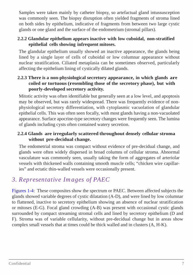
3. Representative Images of PAEC
Figures 1-4: These composites show the spectrum or PAEC. Between affected subjects the glands showed variable degrees of cystic dilatation (A-D), and were lined by low columnar to flattened, inactive to secretory epithelium showing an absence of nuclear stratification or mitoses (E-G). Focal gland crowding (A-B) was present with occasional cystic glands surrounded by compact streaming stromal cells and lined by secretory epithelium (D and F). Stroma was of variable cellularity, without pre-decidual change but in areas show complex small vessels that at times could be thick walled and in clusters (A, H-K).
Confidential 7

Figure 1 A-D (above): Cystic dilatation (A-C), focal gland crowding (A-B), dilated gland with compact streaming stromal cells and lined by secretory epithelium (D), complex small thick walled vessels in clusters (A- arrow) - provided with the courtesy of Dr. M. Glant
Composite Figure 2 E-G (above): Most areas show a low columnar inactive to weakly secretory epithelium. Provided with the courtesy of Dr. M. Glant
8 Confidential

Composite Figure 3 H-K (above): This composite shows stromal vascular changes seen in PAEC. Often small branching clusters of capillaries to small arterioles (H, J, K) to larger thick-wall small arterioles are seen (I). Provided with the courtesy of Dr. M. Glant
Composite Figure 4 L-P (above): This composite shows further changes seen in PAEC. The epithelium is inactive to weakly proliferative (few mitoses and apoptosis, L) and at times shows ciliated metaplasia (N) or unusual secretory changes (M). In areas the glands are folded or stellate and in some cases the disrupted biopsy shows stromal pillars lined on both sides with epithelium, indicative of large cystic glands (O, P). Provided with the courtesy of Dr. M. Glant
Please refer to the CD-ROM for viewing high resolution images of samples of endometrium biopsies.
Confidential 9

4. Differences in Histological Features of PAEC, Unopposed Oestrogen Effect, and Endometrial Hyperplasia.
A pathologist unfamiliar with PAEC may initially consider a diagnosis of unopposed oe- strogen effect or endometrial hyperplasia, as cystic glandular dilatation and architectural irregularity may be seen in these conditions. The process of glandular dilatation differs be- tween PAEC and endometrial hyperplasia.
• In hyperplasia, the dilated glands are lined by epithelium that is stratified and
thicker than normal, with frequent mitotic figures, resembling the appearances of the mid to late proliferative phase.
• In PAEC, the glands are also distended, but lined by an inactive epithelium that is
thinner than that of the normal proliferative phase, and often appears flattened and atrophic.
The key feature seen in all specimens of PAEC is the inactive appearance of the weakly proliferating low cuboidal epithelium, quite different from the mitotically active tall colum- nar epithelial cells seen in unopposed oestrogenic stimulation and endometrial hyperplasia.
Composite Figure 5 Q-U (above): This composite compares changes seen in PAEC with un- opposed oestrogen effect and hyperplasia. The example here of PAEC (Q) has mainly scattered cystic glands with inactive epithelium and the complex hyperplasia has very crowded glands with proliferative epithelium (R,S). The example of disordered proliferative pattern (DPP) (T, U) shows less crowding with dilated venules and proliferative epithelium. In cases of DPP with more cystic glands and less proliferative epithelium, the low power patterns are similar. In such cases the weak secretory changes and vascular changes will aid in correct diagnosis. Provided with the courtesy of Dr. M. Glant
10 Confidential
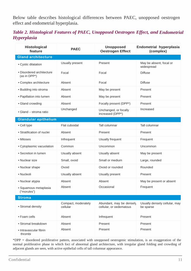
Below table describes histological differences between PAEC, unopposed oestrogen effect and endometrial hyperplasia.
Table 2. Histological Features of PAEC, Unopposed Oestrogen Effect, and Endometrial Hyperplasia
Histological feature
PAEC Unopposed
Oestrogen Effect Endometrial hyperplasia
(complex) Gland architecture
• Cystic dilatation Usually present Present May be absent, focal or
widespread
• Disordered architecture (as in DPP*)
Focal Focal Diffuse
• Complex architecture Absent Focal Diffuse
• Budding into stroma Absent May be present Present
• Papillation into lumen Absent May be present Present
• Gland crowding Absent Focally present (DPP*) Present
• Gland – stroma ratio Unchanged Unchanged, or focally
increased (DPP*) Increased
Glandular epithelium
• Cell type Flat cuboidal Tall columnar Tall columnar
• Stratification of nuclei Absent Present Present
• Mitoses Infrequent Usually frequent Frequent
• Cytoplasmic vacuolation Common Uncommon Uncommon
• Secretion in lumen Usually absent Usually absent May be present
• Nuclear size Small, ovoid Small or medium Large, rounded
• Nuclear shape Ovoid Ovoid or rounded Rounded
• Nucleoli Usually absent Usually present Present
• Nuclear atypia Absent Absent May be present or absent
• Squamous metaplasia (“morules”)
Absent Occasional Frequent
Stroma
• Stromal density
Compact, moderately cellular
Abundant, may be densely cellular, or oedematous
Usually densely cellular, may be sparse
• Foam cells Absent Infrequent Present
• Stromal breakdown Absent Present Present
• Intravascular fibrin thrombi
Absent Present Present
*DPP = disordered proliferative pattern, associated with unopposed oestrogenic stimulation, is an exaggeration of the normal proliferative phase in which foci of abnormal gland architecture, with irregular gland folding and crowding of adjacent glands are seen, with active epithelial cells of tall columnar appearance.
Confidential 11

Please refer to the CD-ROM for viewing high resolution images of samples of endometrium biopsies of PAEC and unopposed oestrogen effect and endometrial hyperplasia (simple and complex).
5. Conclusion
The direct action on the endometrium results in class-specific changes in histology termed, “Progesterone receptor modulator Associated Endometrial Changes” or PAEC. Typically, the histological appearance is an inactive and weakly proliferating epithelium associated with asymmetry of stromal and epithelial growth resulting in prominent cystically dilated glands with admixed oestrogen (mitotic) and progestin (secretory) epithelial effects. Such a pattern has been observed in approximately 60% of patients treated with Esmya for 3 months. These changes are reversible after treatment cessation. These changes should not be confused with unopposed oestrogen effect or endometrial hyperplasia.
12 Confidential

