pathology of hematopoietic systemustavpatologie.upol.cz/_data/section-1/549.pdf · proteinuria...
TRANSCRIPT

Pathology of hematopoietic
system
Leukemias, lymphomas

Bone marrow
Leukemia Neoplastic Proliferations of WBCs in Bone Marrow
Anemia, infection, bleeding
Acute Leukemias
Blast (precursor) cells
Rapidly fatal if not treated
Chronic Leukemias
More mature cells
Longer life expectancy

Leukemia
Acute Chronic
Lymphoid ALL CLL
Myeloid AML CML
Malignant proliferation of
myeloid cells
Myelodysplastic syndrome
Chronic myeloproliferative diseases
(myeloproliferative syndromes)
Acute myelogenous leukemia
Myelodysplastic/myeloproliferative
overlape synndromes

Myeloproliferative syndromes
disorders of the hematopoietic stem cell in
the bone marrow which excess cells
production =effective hematopoiesis
They may evolve into, myelodysplastic
syndrome and acute myeloid leukemia

Chronic myeloproliferative
diseases Philadelphia Chromosome
"positive"
Chronic myelogenous
leukemia (CML)
Philadelphia
Chromosome "negative
Polycythemia vera (PV)
Essential
thrombocytosis (ET)
Primary myelofibrosis
(MF)
Chronic Myelogenous Leukemia
Up to 20% of leukemias
Cause: unknown
radiation
benzen exposure
t(9;22) (bcr-abl) (Philadelphia chromosome)
Proliferation of more mature granulocytes
normal to increased platelet count
anemia

Chronic Myelogenous Leukemia
(CML)
Long chronic phase (matured neutrophils, blasts
less than 10%, basophilia, eosinophiolia)
Accelerated phase (10-20% blatst)
Blast phase - blast crisis
Hydroxyurea, interferons
Bone marrow transplantation

Clinical features
5-6 decades
Slight male predominance
Fatigue, anorexia, weight loss
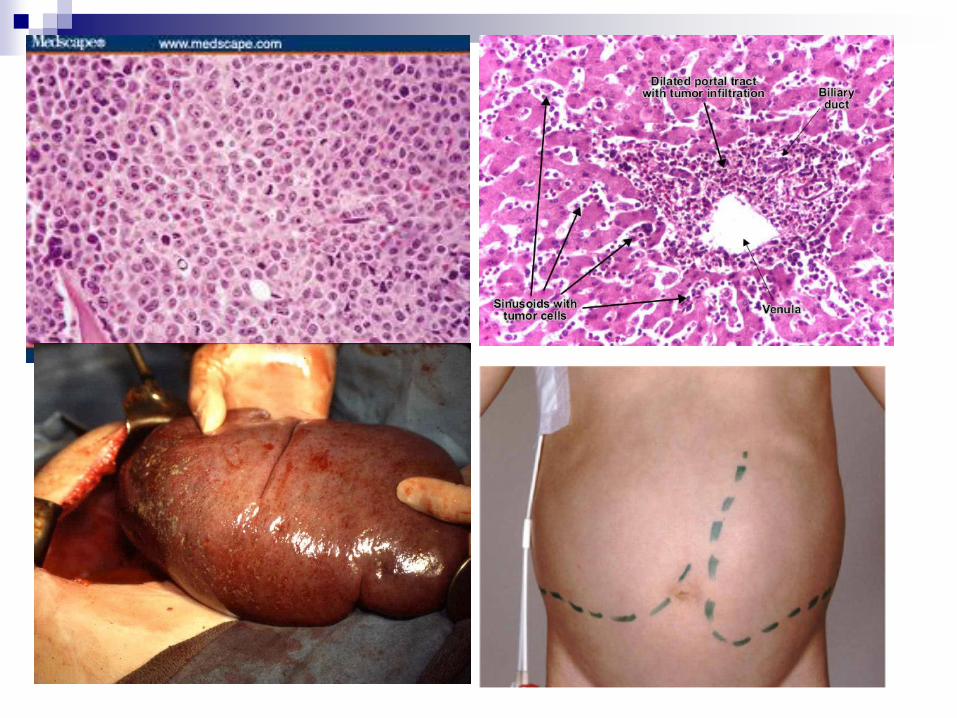
Hepatosplenomegaly- abdominal
dyscomfort
Lymphadenomegaly
Acute leukemia convertion – 80%
Median survival 3-4y

Chronic Myelogenous Leukemia

Polycytemia vera
Clonal hematopoietic stem cell disease
with uncontrolled proliferation of RBCs
JAK 2 mutation
Extramedullary hematopoiesis = myeloid
metaplasia – spleen, liver, lymph nodes
Peak age – 40-60y
Acute leukemic conversion- 10%
Median survival 13y

Polycytemia vera
Insidious onset, nonspecific sy
Plethora
Hematosplenomegaly
Headache, dizzines, visual problems,
angina pectoris, claudication, GIT ulcers
(histamin from basophils),
Thrombosis, infarcts, strokes

Primary (idiopatic) Myelofibrosis
Benzen, radiation, idiopatic
60-70y
Hepatosplenomegaly
Acute leukemic conversion 10%
Median survival 5 y
JAK 2 mutation
Marrow becomes fibrotic
extramedullary hematopoiesis
dry tap

Esential Thrombocytemia
50-70y
Moderate hepatosplenomegaly
Acute leukemic conversion-5%
Median survival – more than 10y
JAK 2 mutation
Thrombosis, hemorrhage

Myelodysplastic syndrome
disorders of the hematopoietic stem cell in
the bone marrow.
In MDS, hematopoiesis (blood production)
is disorderly and ineffective ( The number
and quality of blood-forming cells decline
irreversibly, further impairing blood
production)

New WHO classification 2008
Refractory cytopenia with unilineage dysplasia (Refractory anemia,
Refractory neutropenia, and Refractory thrombocytopenia)
Refractory anemia with ring sideroblasts (RARS)
Refractory cytopenia with multilineage dysplasia (RCMD) includes:
the subset Refractory cytopenia with multilineage dysplasia and ring
sideroblasts (RCMD-RS).
Refractory anemia with excess blasts I and II. RAEB was divided into
*RAEB-I (5-9% blasts) and RAEB-II (10-19%) blasts, which has a poorer
prognosis than RAEB-I.
5q- syndrome (typically seen in older women with normal or high platelet
counts and isolated deletions of the long arm of chromosome 5 in bone
marrow cells, was added to the classification).
Myelodysplasia unclassifiable (seen in those cases of megakaryocyte
dysplasia with fibrosis and others)
Refractory cytopenia of childhood (dysplasia in childhood)
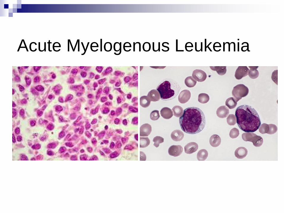
Acute Myelogenous Leukemia


Acute Myelogenous
(myeloid)Leukemia
Proliferation of myeloblasts
anemia, thrombocytopenia, increased WBC
Myeloid, monocytic, RBC, or megakaryocytic
flow cytometry
myeloperoxidase +, TdT-
Auer rod
Over age of 20

Acute Myelogenous Leukemia Radiation, cytotoxic chemotherapy
(alkylationg agents), benzen
In bone marrow –more than 20% of
blasts
Granulocytopenia, thrombocytopenia,
anemia
Opportunistic infection
Bleeding
5y survival less than 30%

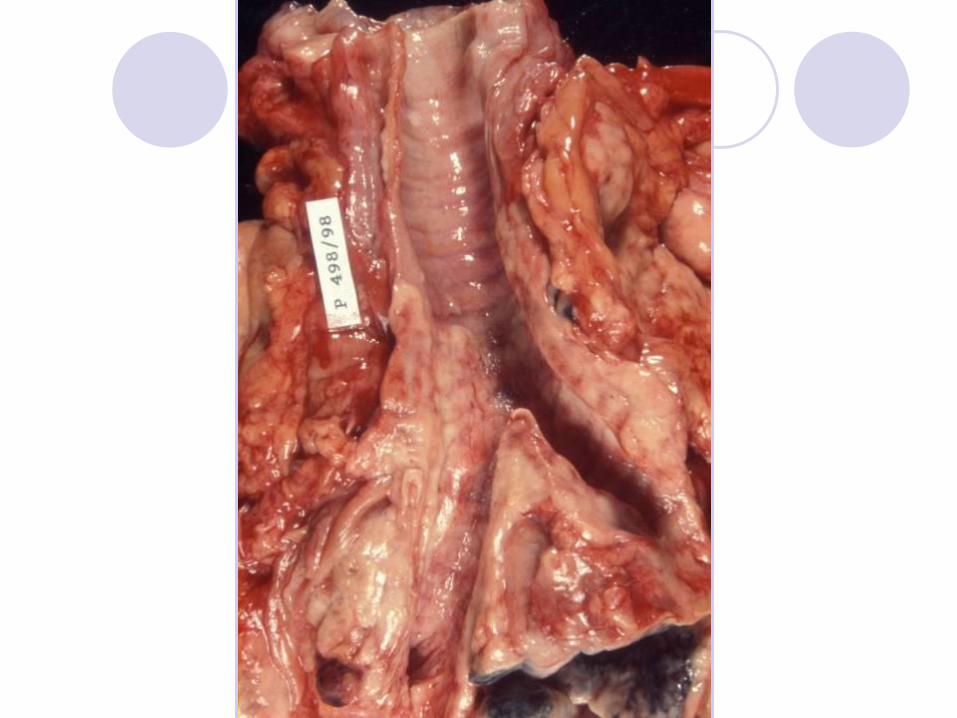
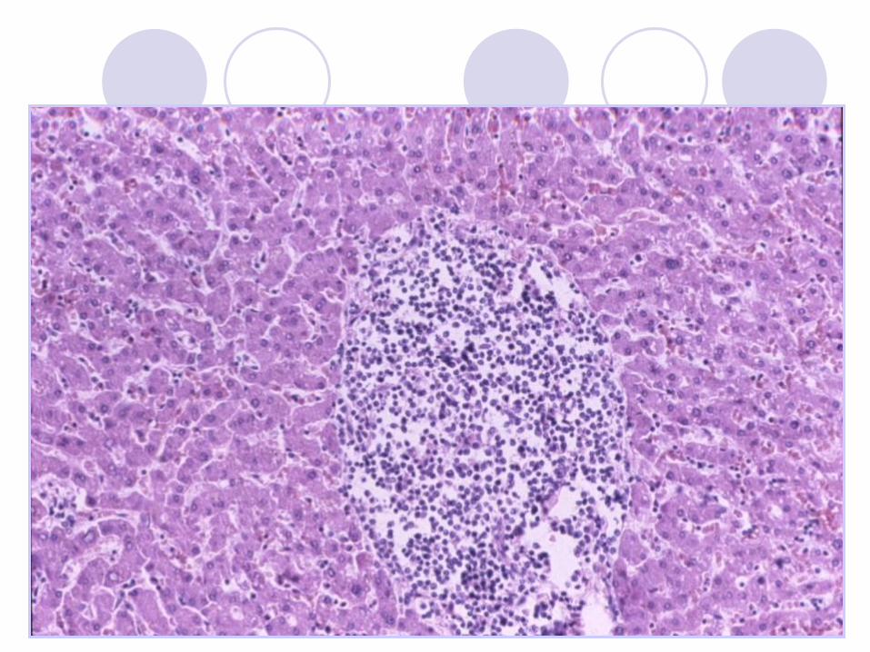
Lymphomas proliferations of lymphoid cells in lymph nodes or extranodal location
(mucoses – BALT, MALT, glands, spleen, skin, brain, bone marrow)
Hodgkin’s lymphoma
Non-Hodgkin’s lymphoma
nodal
extranodal
B cell
T cell/NK
precursor cells
mature cells


Non-hodgkin´s lymphomas (NHL)
WHO klassification B-cell neoplasias
B-cell neoplasias from precursor cells
Lymphoblastic leukemia /lymphoblastic lymphoma
Mature (peripheral ) B-cell neoplasias
B-chronic lymhocytic leukemia/small cell lymphoma (CLL/SLL)
B-prolymhocytic leukemia
Lymphopsmosmocytic lymphoma
SPLENIC MARGINAL ZONE B CELL LYMPHOMA
Hairy cell leukemia
Plasma cell myeloma
Extranodal lymphoma B – cell marginal zone MALT type
Nodal B cell lymphoma – marginal zone
Folicular lymphoma
Mantle cell lymphoma
Difuse large B cell lymphoma
Burkitt´s lymphoma

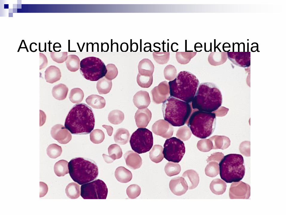
Acute Lymphoblastic Leukemia (ALL)
B- lymphoblastic leukemia/lymphoma NOS
B- lymphoblastic leukemia/lymphoma with repetitive
genetic abnormatilies
t(9;22)(q34;q11) – BCR/ABL
t(v;11q23) – MLL
t(1;19)(q23;p13) – E2A/PBX1
t(12;21)(p12;q22) – ETV/CBF-alfa
hyperdiploidní typ ALL
hypodiploidní typ ALL
T-acute lymhoblastic leukemia from precursor cells
Acute Lymphoblastic Leukemia
(ALL) B- or T-cell precursors (lymphoblasts)
flow cytometry
Most common (80%) leukemia of childhood !!!!!!
anemia, thrombocytopenia, increased WBC
lymphadenopathy/splenomegaly
Good prognosis: Age 4-6y,hyperploidity,
t(12,21)(p13,q22) (TEL/AML1)
Poor outcome:<4, >10y, hypoploid, t(9,22) (BRC/ABL)
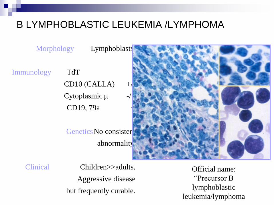
B LYMPHOBLASTIC LEUKEMIA /LYMPHOMA
Morphology Lymphoblasts.
Immunology TdT +
CD10 (CALLA) +/-
Cytoplasmic m -/+
CD19, 79a +
Genetics No consistent
abnormality.
Clinical Children>>adults.
Aggressive disease
but frequently curable.
Official name:
“Precursor B
lymphoblastic
leukemia/lymphoma
Acute Lymphoblastic Leukemia

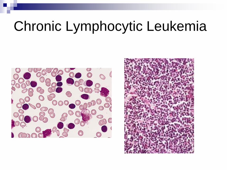
Chronic Lymphocytic Leukemia
(CLL/SLL) Proliferation of small mature B-lymphocytes
flow cytometry (monoclonal Kappa or lambda)
Lymphadenopathy
Lymphocytosis
May be anemia, thrombocytopenia
Hypogamaglobulinemia - infections
May have Ab production and AIHA
50% 6-year survival (but laso 20-30y)
Richter syndrome – transformation to DLBCL“

Age: CLL is most common in older adults, is rare in young adults, and hardly
ever develops in children. About 90% of people diagnosed with CLL are
older than 50.
Gender. Men develop CLL more often than women.
Ethnicity. B-cell CLL is more common in people of Russian and European
descent, and hardly ever develops in people from China, Japan, or
Southeast Asian countries. The reason(s) for this geographic difference is
not known.
Clin sy:
Swelling of lymph nodes in the neck, under the arms, or in the groin. This is
a common symptom that people with CLL usually notice first.
Discomfort or fullness in the upper left part of the abdomen, caused when
the spleen increases in size
Fever and infection
Abnormal bleeding
Shortness of breath
Weight loss
Chronic Lymphocytic Leukemia


IMMUNOCYTOMA /LYMPHOPLASMACYTIC
LYMPHOMA (Waldenstrem macroglobulinemia) Morphology Plasmocytoid lymphocytes,
plasma cells (+/- Dutcher
bodies), lymphocytes.
Immunology Surface IgM +
Cytoplasmatic Ig +
CD5, CD10 -
CD19, 20, 22, 79a +
Genetics No specific abnormalities.
Clinical Adults. Indolent course.
Often associated
with a serum IgM
paraprotein

SPLENIC MARGINAL ZONE B CELL LYMPHOMA Morphology Small centrocyte-like,
cells “monocytoid B cells”,
lymphocytes, plasma cells.
Immunology Surface Ig +
CD5, 10 -
CD19, 20, 22, 79a +
CD23 -
Genetics No specific abnormalities.
Clinical Splenomegaly.
? Always leukemic.
Provisional in REAL scheme.
Corresponds to “splenic
lymphoma with villous
lymhocytes”

Hairy cell leukemia
Rare chronic
Middle aged men
Pancytopenia
Splenomegaly
fried egg appearance

HAIRY CELL LEUKEMIA
Morphology Small lymphoid cells with
bean shaped nuclei and
pale cytoplasm.
Immunology Surface Ig +
CD5, 10, 23 -
CD11c, 25 +
CD19, 20, 22, 79a +
CD103 , DBA44 +
Genetics No specific abnormalities.
Clinical Adults, often with
splenomegaly and pancytopenia.
Indolent course.
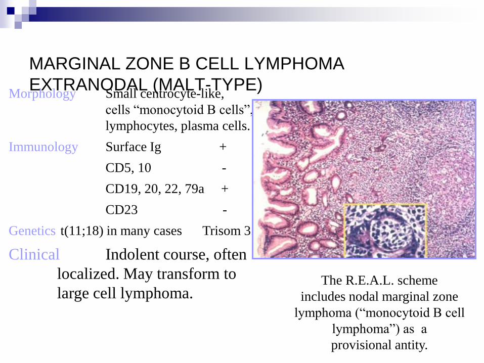
MARGINAL ZONE B CELL LYMPHOMA
EXTRANODAL (MALT-TYPE) Morphology Small centrocyte-like,
cells “monocytoid B cells”,
lymphocytes, plasma cells.
Immunology Surface Ig +
CD5, 10 -
CD19, 20, 22, 79a +
CD23 -
Genetics t(11;18) in many cases Trisom 3.
Clinical Indolent course, often
localized. May transform to
large cell lymphoma.
The R.E.A.L. scheme
includes nodal marginal zone
lymphoma (“monocytoid B cell
lymphoma”) as a
provisional antity.
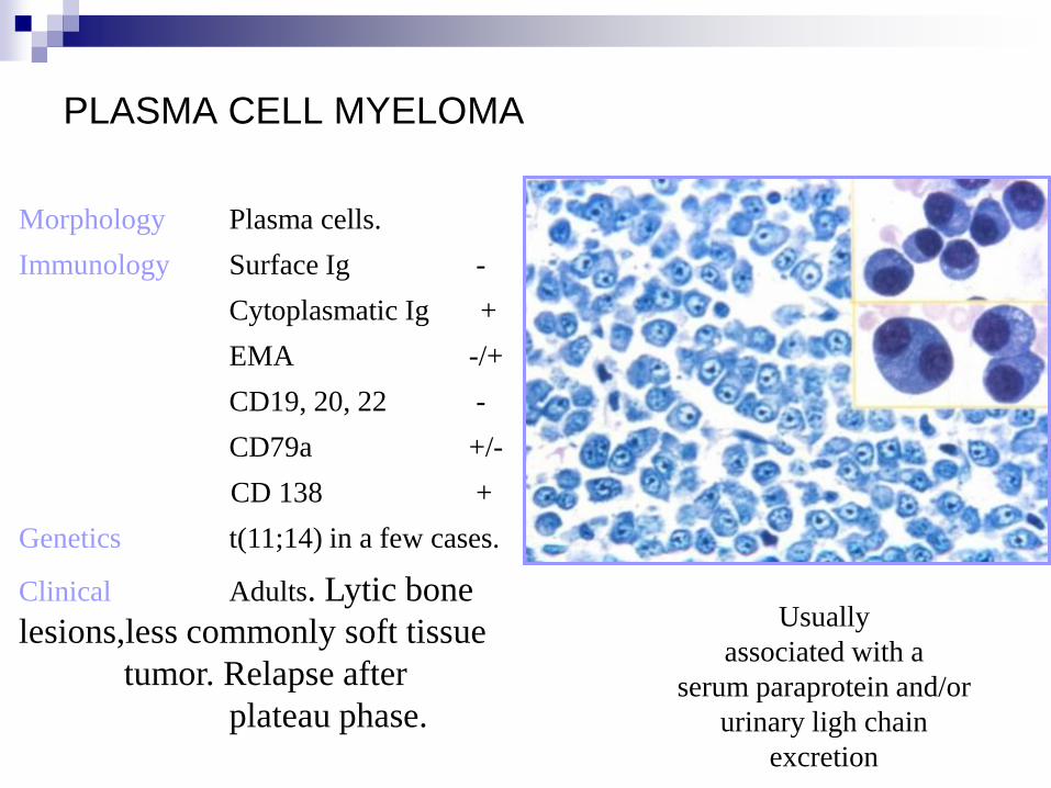
PLASMA CELL MYELOMA
Morphology Plasma cells.
Immunology Surface Ig -
Cytoplasmatic Ig +
EMA -/+
CD19, 20, 22 -
CD79a +/-
CD 138 +
Genetics t(11;14) in a few cases.
Clinical Adults. Lytic bone
lesions,less commonly soft tissue
tumor. Relapse after
plateau phase.
Usually
associated with a
serum paraprotein and/or
urinary ligh chain
excretion
PLASMA CELL MYELOMA
Neoplasm of plasma cells
monoclonal protein in serum (SPEP)
Proteinuria (Bence-Jones) (UPEP)
Lytic lesions in bones
fractures
Anemia, increased globulin
Rouleaux formation
Renal failure/ amyloidosis

PLASMA CELL MYELOMA

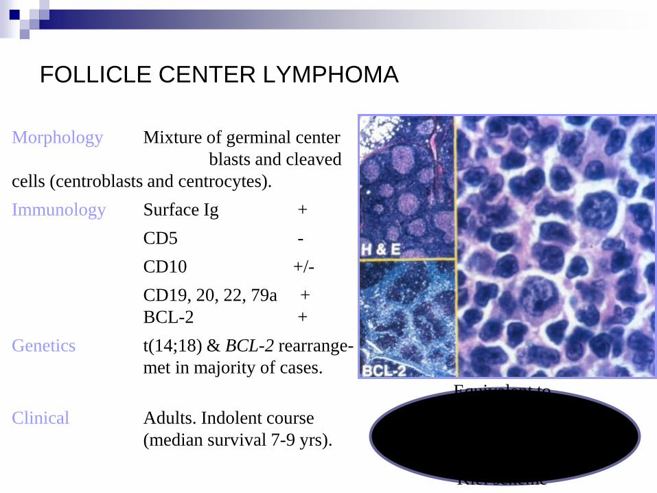
FOLLICLE CENTER LYMPHOMA
Morphology Mixture of germinal center
blasts and cleaved
cells (centroblasts and centrocytes).
Immunology Surface Ig +
CD5 -
CD10 +/-
CD19, 20, 22, 79a +
BCL-2 +
Genetics t(14;18) & BCL-2 rearrange-
met in majority of cases.
Clinical Adults. Indolent course
(median survival 7-9 yrs). +
Equivalent to
“centroblastic/centrocytic”
and “lollicular
centroblastic” lymphomas in
Kiel scheme
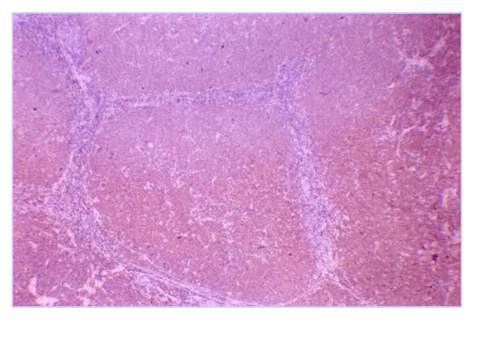
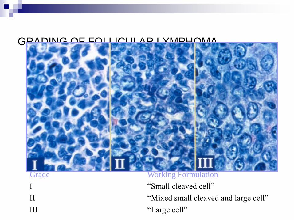
GRADING OF FOLLICULAR LYMPHOMA
Grade Working Formulation
I “Small cleaved cell”
II “Mixed small cleaved and large cell”
III “Large cell”
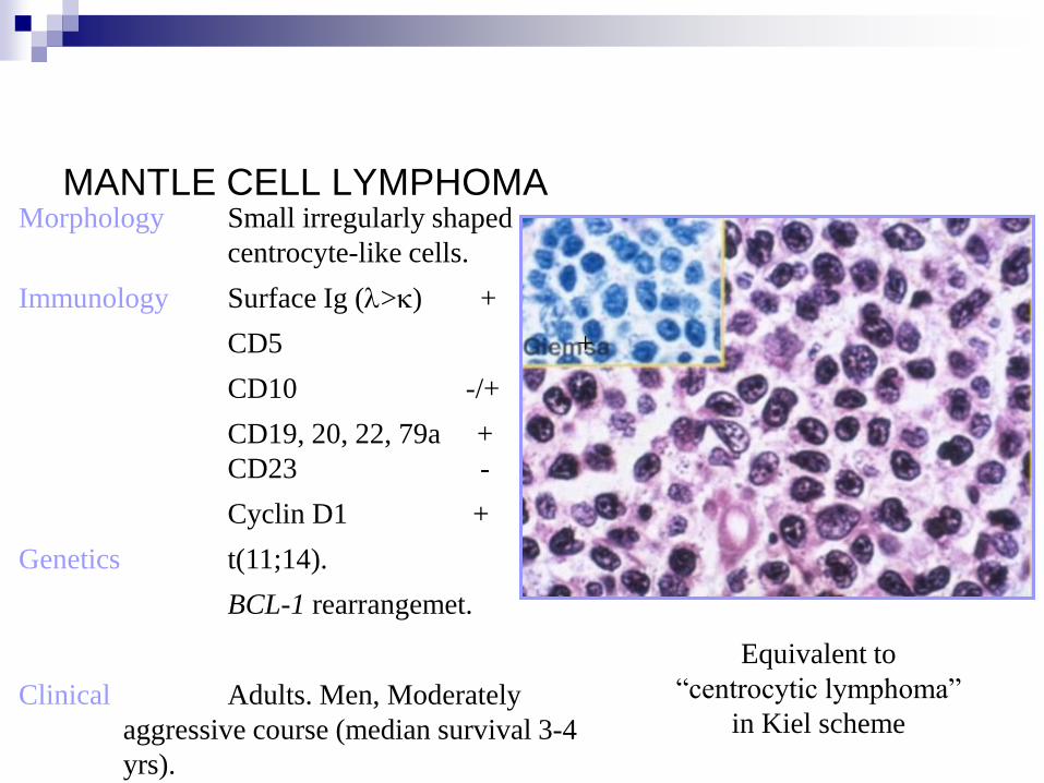
MANTLE CELL LYMPHOMA Morphology Small irregularly shaped
centrocyte-like cells.
Immunology Surface Ig (l>k) +
CD5 +
CD10 -/+
CD19, 20, 22, 79a +
CD23 -
Cyclin D1 +
Genetics t(11;14).
BCL-1 rearrangemet.
Clinical Adults. Men, Moderately
aggressive course (median survival 3-4
yrs).
Equivalent to
“centrocytic lymphoma”
in Kiel scheme
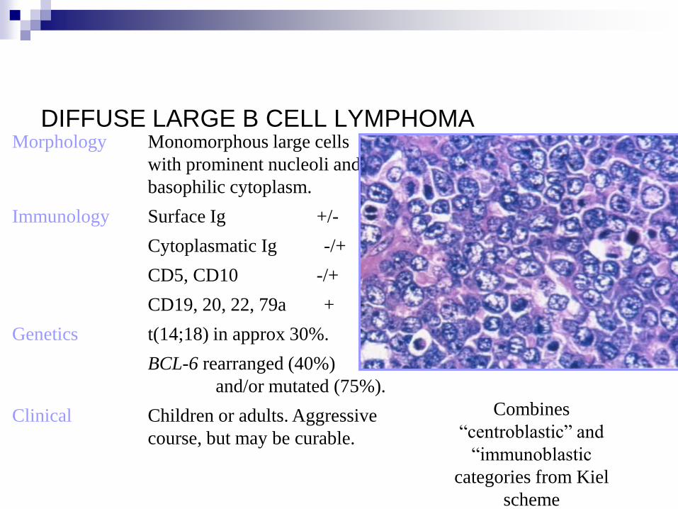
DIFFUSE LARGE B CELL LYMPHOMA Morphology Monomorphous large cells
with prominent nucleoli and
basophilic cytoplasm.
Immunology Surface Ig +/-
Cytoplasmatic Ig -/+
CD5, CD10 -/+
CD19, 20, 22, 79a +
Genetics t(14;18) in approx 30%.
BCL-6 rearranged (40%)
and/or mutated (75%).
Clinical Children or adults. Aggressive
course, but may be curable.
Combines
“centroblastic” and
“immunoblastic
categories from Kiel
scheme


BURKITT’S LYMPHOMA
Morphology Medium sized cells,
basophilic cytoplasm.
“Starry sky” appearance.
High mitotic rate.
Immunology Surface IgM +
CD5, 23 -
CD10 +
CD19, 20, 22, 79a +
Ki 67 >85% of cells
Genetics t(2;8), t(8;14) or t(8;22).
Rearrangement of c-MYC
Clinical Children >> adults.
Aggressive but curable in
children.
“Burkitt-like”
lymphoma is a provisional
entity in the REAL
scheme
WHO classification T- A NK-cell neoplasias
T-cell precursor neoplasis
T lymphoblastic leukemia /lymphoma
MAture T and NK-cell neoplasias
T-prolymphocytic leukemia
T CELL LARGE GRANULAR LYMPHOCYTIC LEUKAEMIA
Aggresive NK-cell neoplasias
T-cell leukemia/lymhoma of adults dospělých (HTLV-1+)
Extranodal NK/T-cell lymphoma- nasal type
ENTEROPATHY- ASSOCIATED T-CELL LYMPHOMA
Hepatosplenic T-cell lymphoma
SUBCUTANEOUS PANNICULITIS-LIKE T-CELL LYMPHOMA
Mycosis fungoides/Sézaryho syndrom
CUTANEOUS ANAPLASTIC LARGE CELL T-CELL LYMPHOMA CD30
POSITIVE
Peripheral T-cell lymphoma NOS
Angioimunoblastic T-cell lymhoma
Anaplastic large cell lymphoma lymfoma ALK pozitive
Anaplastic large cell lymphoma ALK negative

T LYMPHOBLASTIC LEUKEMIA/LYMPHOMA
Morphology Lymphoblasts, identical cytologically
to B lymphoblasts.
Immunology TdT +
CD1a +/-
CD3 +/-
CD7 +
CD4±8 +
Genetics SCL/TAL-1 rearrangement
in approx 25%.
Clinical Frequently involves
mediastinum. Adolescents
and young adults. Highly
aggressive buf potentially
curable.
Official name:
“Precursor T
lymphoblastic
lymphoma/leukemia”

T CELL PROLYMPHOCYTIC LEUKAEMIA
Morphology Small lymphoid cells, with
some nuclear irregularity.
Immunology CD2, 3, 5, 7 +
CD4 +
CD8 -/+
Genetics Inv 14(q11;32) in some
cases. Trisomy 8q
Clinical Adults. Often leukemic.
More aggressive than B cell
chronic lymphocytic
leukemia. Includes T cell
prolymphocytic
leukemia
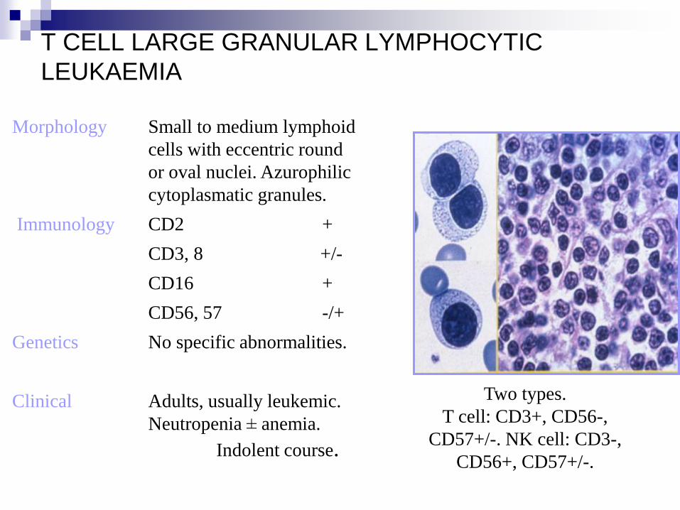
T CELL LARGE GRANULAR LYMPHOCYTIC
LEUKAEMIA
Morphology Small to medium lymphoid
cells with eccentric round
or oval nuclei. Azurophilic
cytoplasmatic granules.
Immunology CD2 +
CD3, 8 +/-
CD16 +
CD56, 57 -/+
Genetics No specific abnormalities.
Clinical Adults, usually leukemic.
Neutropenia ± anemia.
Indolent course.
Two types.
T cell: CD3+, CD56-,
CD57+/-. NK cell: CD3-,
CD56+, CD57+/-.

ENTEROPATHY- ASSOCIATED T-CELL
LYMPHOMA
Morphology Neoplastic cells range
from small lymphocytes to
large bizarre cells.
Immunology CD3, 7 +
CD8 +/-
CD103 +
TIA-1 +
Genetics No specific
abnormalities.
Clinical Adults. Aggressive course,
often with intestinal perforation.
May be associated
with celiac disease
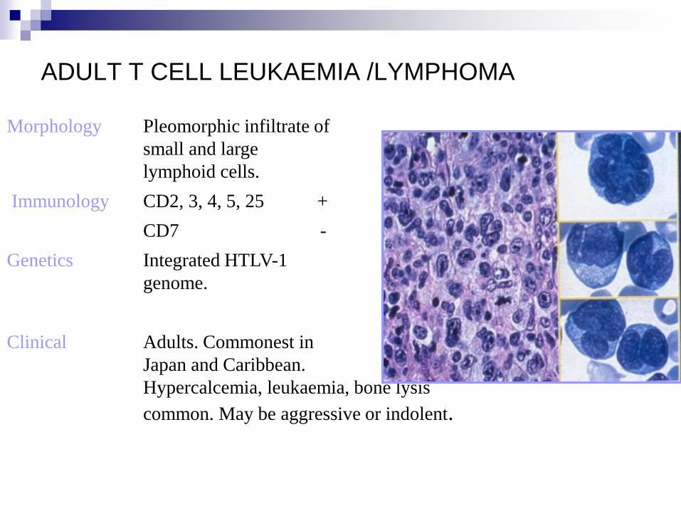
ADULT T CELL LEUKAEMIA /LYMPHOMA
Morphology Pleomorphic infiltrate of
small and large
lymphoid cells.
Immunology CD2, 3, 4, 5, 25 +
CD7 -
Genetics Integrated HTLV-1
genome.
Clinical Adults. Commonest in
Japan and Caribbean.
Hypercalcemia, leukaemia, bone lysis
common. May be aggressive or indolent.
MYCOSIS FUNGOIDES
Clinical Adults. Patches, plaques & tumours.
Preferential location: buttocks, other sun-protected
areas (early phases).
Morphology Small pleomorphic (cerebriform) cells. During the
course of the disease large cell transformation may
occur indicating a worse prognosis (immunoblasts,
large cell anaplastic, large cell pleomorphic)
Immunology CD2, 3, 4, 5 +
CD
Genetics No specific abnormalities. Monoclonal
rearrangement of the TCR may be absent in early
phases.
Treatment guidelines Early phase: PUVA, interferon-2a, retinoids
(alone or in combination); topical
chemotherapy. Advenced disease chemotherapy;
extracorporeal photopheresis; radiotherapy.

EARLY MYCOSIS FUNGOIDES
Epidermotropism of solitary lymphocytes aligned
along basal layer of epidermis
Psoriasiform pattern

MYCOSOS FUNGOIDES PLAQUE STAGE
Plaques of mycosis fungoides arecharacterised by a dense,
band-like infiltrate within the upper dermis.
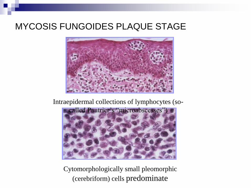
MYCOSIS FUNGOIDES PLAQUE STAGE
Cytomorphologically small pleomorphic
(cerebriform) cells predominate
Intraepidermal collections of lymphocytes (so-
called Pautrier’s “microabscesses”)

Sézary’s Syndrome
Clinical Elderly adults. Pruritic erythroderma, generalised
lymphadenopathy & circulating Sézary cells.
Usually aggressive course.
Morphology Small pleomorphic (cerebriform) cells. During the
course of the disease there may be appearance
of tumours with large cell morphology
immunoblats, large cell anaplastic, large cell
pleomorphic).
Immunology CD2, 3, 4, 5 +
CD8 –
Genetics No specific abnormalities. Monoclonal
rearrangement of the TCR may be absent in early
phases.
Treatment guidelines PUVA, interferon-2a, retinoids (alone or in
combination); extracorporeal photopheresis;
radiotherapy; chlorambucilcombined with
prednisone(Winkelmann scheme); systemic
chemotherapy.

Sézary’s Syndrome
Erythroderma: note enlarged
inguinal lymph nodes Hyperkeratosis of the palms
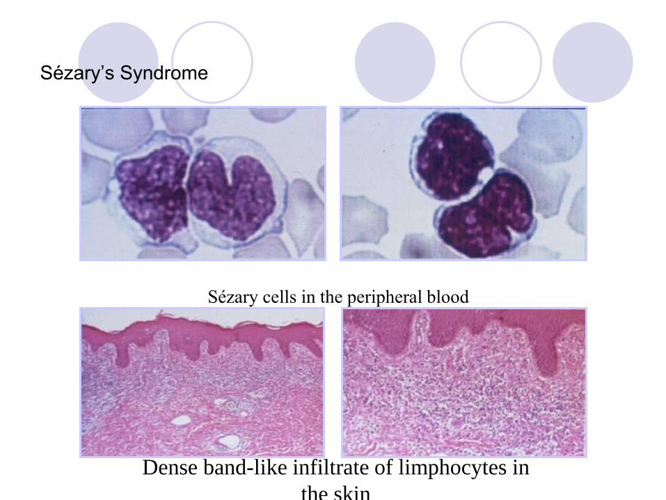
Sézary’s Syndrome
Sézary cells in the peripheral blood
Dense band-like infiltrate of limphocytes in
the skin

CUTANEOUS ANAPLASTIC LARGE CELL
T-CELL LYMPHOMA CD30 POSITIVE
Clinical Adults & younger patients. Solitary or regionally locailsed tumours, often ulcerated. Generally favourable prognosis. Morphology Nodular infiltrates characterised by cohesive sheets of large, CD30 positive cells. Cytomorphology. Usually large anaplastic cells; large pleomorphic cells or immunoblasts. Immunology CD2, 3, 4, 5 + CD30 + CD8 – CD15, EMA – Genetics Usually absence of t(2;5). Monoclonal rearrangement of the TCR detected in the majority of cases. Treatment guidelines Solitary or localised lesions: radiotherapy (with or without previous surgical excision); Generalised lesions: systemic chemotherapy.
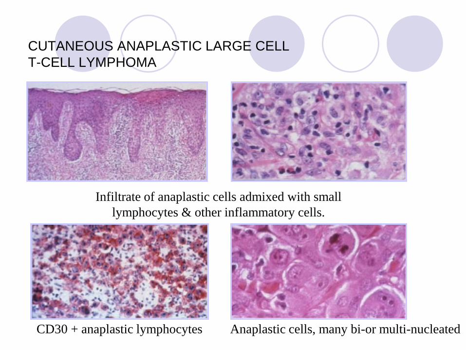
CUTANEOUS ANAPLASTIC LARGE CELL
T-CELL LYMPHOMA
Infiltrate of anaplastic cells admixed with small
lymphocytes & other inflammatory cells.
CD30 + anaplastic lymphocytes Anaplastic cells, many bi-or multi-nucleated

ANAPLASTIC LARGE CELL LYPHOMA
Definition and Cytogenetics
(agrresive, but curable, good prognosis, often extranodal
The (2;5) translocation
Initially thought to be
associated with true
histiocytic malignancy but
then linked to anaplastic
large cell lymphoma.


SUBCUTANEOUS PANNICULITIS-LIKE T-CELL
LYMPHOMA
Occasionally there is
evidence of
haemophagocytosis,
characterised by large
macrophages egulfing
neoplastic lymphocytes
or other blood cells.

PERIPHERAL T CELL LYMPHOMA,
UNSPECIFIED
Morphology Atypical lymphocytes of varying
sizes. Variable reactive
backgroud elements, e.g.
macrophages, vessels, etc.
Immunology CD3 +/-
Variable expression of
other T cell markers.
Genetics No specific abnormalities.
Clinical Adults. Aggressive course,
but potentially curable. “Uncspecified” reflects
suspicion that subtypes
(currently unidentifiable)
exist
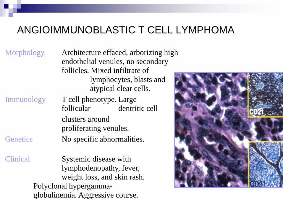
ANGIOIMMUNOBLASTIC T CELL LYMPHOMA
Morphology Architecture effaced, arborizing high
endothelial venules, no secondary
follicles. Mixed infiltrate of
lymphocytes, blasts and
atypical clear cells.
Immunology T cell phenotype. Large
follicular dentritic cell
clusters around
proliferating venules.
Genetics No specific abnormalities.
Clinical Systemic disease with
lymphodenopathy, fever,
weight loss, and skin rash.
Polyclonal hypergamma-
globulinemia. Aggressive course.

5- y survival> 70%:
-Follicular lymphoma
-MALT lymphoma
-Anaplastic large cell lymphoma
5- y survival 50-70%:
-Nodal B cell lymphoma –
marginal zone
-Lymphoplasmocytic lymphoma
-SLL/CLL
5- y survival 30-50%:
-large B cell lymphoma
-Burkitt lymphoma
-Burkitt-like lymfoma
The worst prognosis:
-T lymphoblastic lymphoma
-Peripheral T cell lymphoma
-Mantle cell lymphoma
HODGKIN´s lymphoma/HODGKIN´s disease
EBV infection, genetic changes, immune alteration
2-3 decades, 5 decade
1)Nodular lymphocyte predominant Hodgkin lymphoma
2) Classical Hodgkin lymphoma
Nodular Sclerosis (the most common)
Mixed Cellularity (the highest association with EBV, common
in HIV)
Lymphocyte rich
Lymphocyte Depleted
Nodular lymphocyte
predominant Hodgkin
lymphoma
B cell antigen
Lack CD15, CD30
Popcorn cells
Indolent type
Men under 35y
Cervival, axillary and
inquinal LN
Mediastinum rare
B sings and symptoms in
20%
5y survival in 80%
Classical Hodgkin
lymphoma
LN cervical, anterior
mediastinum
B signs in 40% (low fever,
cyclical, night sweats,
weight loss, pruritus,
alcoholic pain)
HRS cells

HODGKIN’S DISEASE
Lymphocyte Predominance
Low power High power

HODGKIN’S DISEASE
Lymphocyte Predominance B cell
marker on
reactive and
neoplastic
cells
T cell marker
on reactive
cells
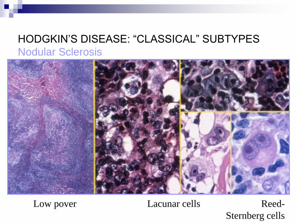
HODGKIN’S DISEASE: “CLASSICAL” SUBTYPES
Nodular Sclerosis
Low pover Lacunar cells Reed-
Sternberg cells


HODGKIN’S DISEASE: “CALSSICAL” SUBTYPES
Mixed Cellularity
High pover “Mummified cells” Reed-Sternberg cells
Low power B cells