pathology of noninfectious vasculitidesbns-hungary.hu/documents/22bns/2015bns_0829_1035.pdf ·...
TRANSCRIPT

Pathology of noninfectious vasculitides
Eva HonsovaInstitute for Clinical and Experimental MedicinePrague, Czech Republic

Systemic noninfectious vasculitidesVasculitis = a general term for inflammation of vessel walls
Any type of vessel in all locations can be affectedin practice – overwhelming number of clinical symptoms andproblems
• Clinical manifestation: very variable, the disease has attacks and spontaneous remissions• the diagnosis is difficult The vasculitides are often serious diseases, prompt recognition and therapy.
Patients suffer from symptoms of systemic inflammation(such as fever, arthralgias, myalgias, weight loss), and can simultaneously have symptoms of local system involvement. They can develop local mass lesion which can lead to the diagnosis of malignant tumor
nodulus imitates metastasis

Historical development of names of vasculitides
1. description of necrotizing arteritis (PAN) Rokitanský (1852)
Periarteritis nodosa (1866 Kussmaul, Maler)1890 Hutchison GCA
1908 Takayasu 1903 Osler SLE
1934 Horton GCA
1936 Wegener
1942 Rich, hypersen. angiitis 1948-52 Zeek, hypersens. angiitis
1978 Fauci, hypersen.a. small vessel vasculitis
mainly skin involvement, 1951 Churg- Strauss
1954 Godman and Churg (MPA x PAN)
1982 Davis, ANCA antibodies
GCA PAN ANCA associated (GPA, MPO, EGPA)
Takayasu Kawasaki Immune complex small vessel v.
For decades PAN was the term used for virtually any patient with necrotizing arteritis

First description of necrotizing arteritis with aneurysmata (PAN, 1852)Karel Rokitanský
• Karel Rokitanský was born in Hradec Králové, started his medicine study in Prague under Purkyně.
• When Purkyně left in r. 1823 for WroclawRokitanský went to Vienna.
• Aneurysmal lesions in numerous arteries in a 23-year-old shoemaker (without histology)
• Eppinger: histological confirmation
• 1866; Kussmaul and Maier: 27-year-old tailor; periarteritis nodosa with histology (with the involvement of gli)
Death of colleague Dr. Koletscko, who succumbed the sepsis after injury at autopsy room, has inspired Dr. Ignaz Semmelweis (a pioneer in antiseptic medical practice).


2012 revised international CHCC nomenclature of vasculitides
ANCA-associated small vessel v.GPA (Wegener g.)MPA (microscopic polyangitis)EGPA (Churg-Strauss sy)
Large vessel vasculitisTakayasu a.Giant cell a.
Medium vessel v.PAN
Kawasaki disease.
Immune complex small vessel v.Anti-GBMIgA v. (HSP)Cryoglobulinemic v..
Hypocomplementic urticarial v.
According to the article: Jennette et al. Arthritis Rheum. 2013; 65:1-11.

Nomenclature noninfectious vasculitides1. Large vessel vasculitis: GCA a Takayasu
2. Medium vessel vasculitis: PAN a Kawasaki
3. Small vessel vasculitis (arteries, arterioles, venules, veins):
a) ANCA-associated vasculitis (most frequent vasculitis of adults!!!)
b) immune complex:
IgA v.(Henoch-Schönlein), anti-GBM (ANCA : anti-GBM = 100 : 1), cryoglobulins, hypocomplementic urticarial (anti-C1q) v., serum sickness 4.
Variable vessel vasculitis (Cogan’s, Behcet’s, etc.)
Single organ vasculitis (cutaneous SVV, primary CNS vasculitis, etc.)
Vasculitis associated with systemic diseases (e.g. Rheumatoid, Lupus, Sarcoid, etc.)
Vasculitis associated with probable etiologies (e.g. HBV, HCV, drug, cancer, etc.)

Large Vessel Vasculitis, 2012 CHCC definition
Vasculitis affecting large arteries more often than other vasculitides. Large
arteries are the aorta and its major branches. Any size artery may be affected.
1. Takayasu arteritisPatients: women under 50, rare in the Czech Rep.
• Morphology: shares histological features of GCA
(granulomatous arteritis), no fibrinoid necrosis
2. Giant cell (temporal) arteritis (GCA)• Patients: over 50, common (with polymyalgia rheumatica)
The distinction between the two entities is made on the basis of
a patient´s age!!!

PAN, 2012 CHCC definition
• Necrotizing arteritis of medium or small arteries without glomerulonephritis or vasculitis in arterioles, capillaries, or venules; and not associated with ANCA.
• Very rare
No GN !!!

Kawasaki disease; 2012 CHCC definition Arteritis associated with the mucocutaneus lymph node syndrome, predominantly affecting medium and small arteries. Coronary arteries are often involved. Aorta and large arteries maybe involved. Usually occurs in infants and young children.

Small vessel vasculitides
1. ANCA-associated smallvessel v.
• GPA (Wegener g.)
• MPA (microscopicpolyangitis)
• EGPA (Churg-Strauss sy)
2. Immune complex smallvessel v.
• Anti-GBM
• IgA v. (HSP)
• Cryoglobulinemic v.
• Hypocomplementicurticarial v.
IgAIgG

ANCA antibodies (Anti-Neutrophil Cytoplasmatic Antibody)
• ABs specific to antigens of neutrophil granules and to lysosoms of monocytes;
• c-ANCA (cytoplasmatic) p-ANCA(perinuclear)
• Many different antigens in the group c- or p- ( proteinasa 3, myeloperoxidasa-MPO, lactoferin, cathepsin G, elastasa, lysosym, azurocidin atd.)
• ANCA positive vasculitides: c-ANCA: PR3 (proteinasa 3),
p-ANCA: MPO (myeloperoxidasa)
• IF determine the group c or p
• ELISA distinguishes the precise antigens
• (PR3 or MPO) which are associated with AAV
• New methods for identification of specific epitopes (MALDI-TOF/TOF-MALDI-MS)
Epitope specificity defines pathogenicity
(Roth A. at al. Epitope specificity determines
pathogenicity and detectability
in ANCA-associated vasculitis. J Clin Invest. 2013;
123:1773-1783.)
ANCA associated vasculitis
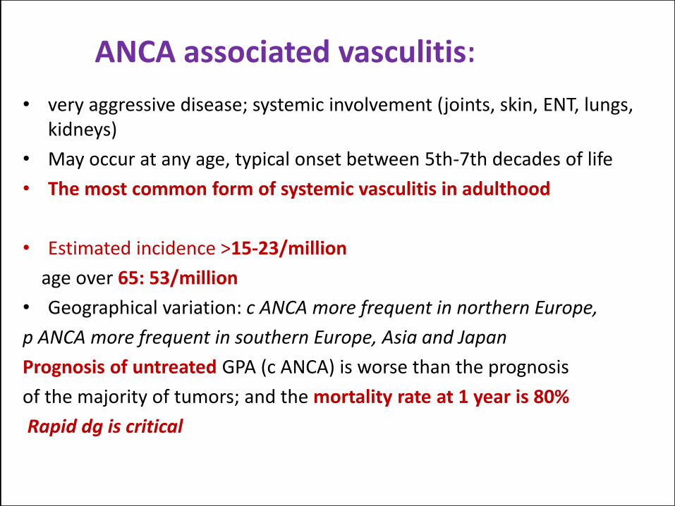
• very aggressive disease; systemic involvement (joints, skin, ENT, lungs, kidneys)
• May occur at any age, typical onset between 5th-7th decades of life
• The most common form of systemic vasculitis in adulthood
• Estimated incidence >15-23/million
age over 65: 53/million
• Geographical variation: c ANCA more frequent in northern Europe,
p ANCA more frequent in southern Europe, Asia and Japan
Prognosis of untreated GPA (c ANCA) is worse than the prognosis
of the majority of tumors; and the mortality rate at 1 year is 80%
Rapid dg is critical

location HSP cryoglob MPA GPA EGPA
skin 90% 90% 60% 50 - 70% 60%
kidney 50% 55% 90% 80% 45%
lung <5% <5% 50% 90% 70%
ENT <5% <5% 35% 90% 50%
joint 75% 70% 60% 60% 50%
neurol. 10% 40% 30% 50% 70%
GIT 60% 30% 30% 50% 50%
Occurrence of systemic involvement(Jennette J. N. Eng J Med: 1997; 337:1512-1523).
The concept that MPO- and PR3-AAV are genetically distinct diseases with phenotypic overlap and that studies and clinical practice may benefit from clustering according to serotype.
Lyons P. A. et al. Genetically distinct subset within ANCA-associated vasculitis. N.Engl. J. Med. 2012; 367: 214-223.

ANCA-associated vasculitides
• Any organ may be afflicted (kidney, lungs, ENT...)
• Kidney involvement:
pauciimmune necrotizing crescentic
(rapidly progressive) glomerulonephritis
75% of patients with GN have systemic
vasculitis
• Renal biopsy:
Gold standard to confirm the diagnosis
To assess the prognosis
Not absolutely required,
but recommended whenever possible

ANCA-associated GN: morphology: gli pauciimmune necrotizing GN with crescents
Acute lesions: necrosis + crescentsTypically scattered with normal gli or their parts, later with combination of acute and chronic lesions

ANCA-associated GN: morphology: gli Crescents: epithelial/cellular, fibro-epithelial, fibrotic

ANCA-associated GN:morphologyvessels & intersticium
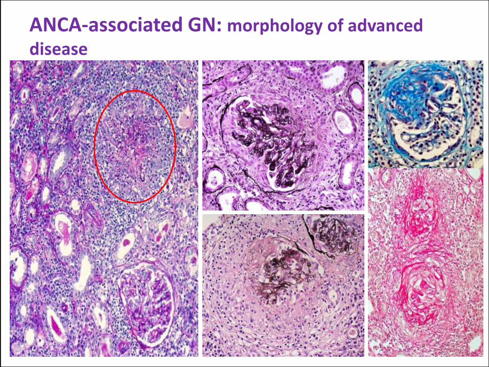
ANCA-associated GN: morphology of advanced disease

Time period during the course of vasculitis in morphology

When it is difficult to diagnosed AAV
In a kidney biopsy:
• Early acute lesion: normal morphology; sampling error
• Advanced lesion (destruction and high number of crescents)
• Chronic scaring lesions
• Combinations: with IgA and or other diseases (modify the morphological features); anti-GBM, DM, SLE: mainly MPA with slow progression with segmental sclerotic lesions
In other locations/organs:
• More difficult to diagnose vasculitis (often second opinion can help)

Case no. 1.: 63-year-old woman
• Autumn 2002 worse hearing, ENT in normal range, CT showed inflammation in the middle ear
• She visited internist in January next year• She requested lungs examination, normal• February: artralgias of small joints, susp. RA • May: S-Cr 197 umol/l, microhematurie• June: S-Cr 273 umol/l;• only then she is sent to nephrologist• Weight loss: 6 kg
AAV advanced morphological features (MPA, p-ANCA)
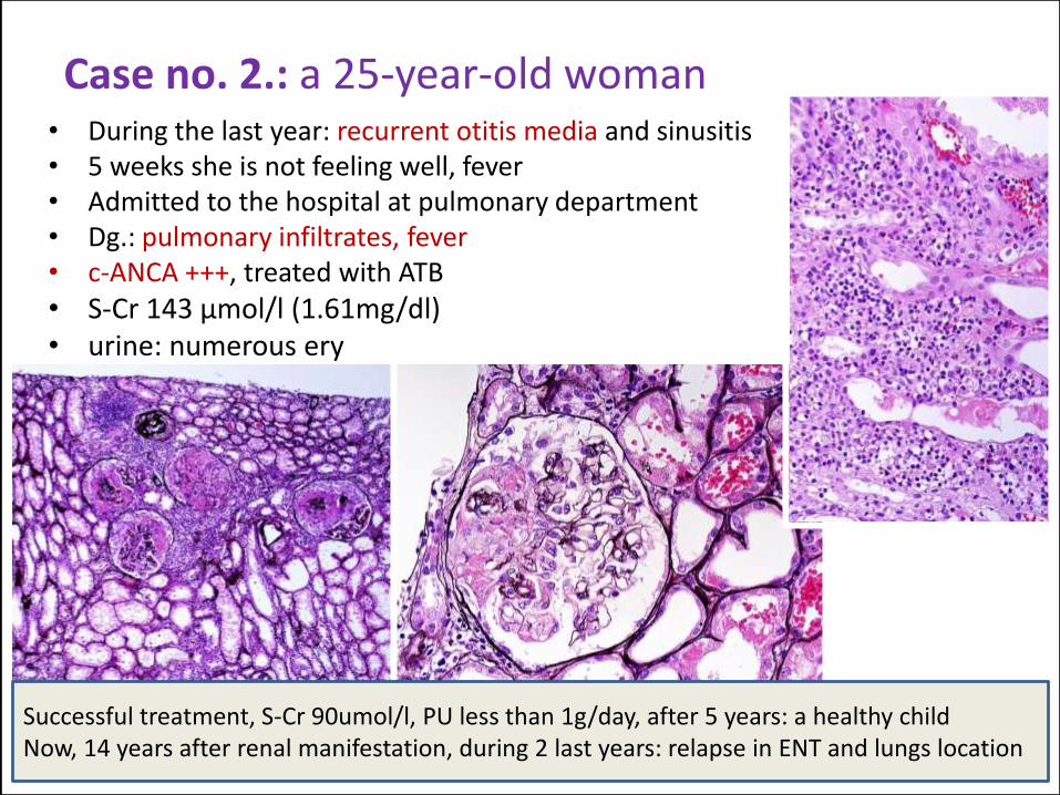
Case no. 2.: a 25-year-old woman• During the last year: recurrent otitis media and sinusitis• 5 weeks she is not feeling well, fever• Admitted to the hospital at pulmonary department• Dg.: pulmonary infiltrates, fever• c-ANCA +++, treated with ATB• S-Cr 143 μmol/l (1.61mg/dl) • urine: numerous ery
Successful treatment, S-Cr 90umol/l, PU less than 1g/day, after 5 years: a healthy child Now, 14 years after renal manifestation, during 2 last years: relapse in ENT and lungs location

Case no. 3.: 28-year-old man
• 5 mths of non-specific symptoms: myalgia, artralgia, unintended weight loss (10 kg)
• chronic otitis media with hypacusis l. sin.
acute polyneuropathy: hypoesthesia and paresthesia
• mild hematuria and proterinuria (1 g/day), sterile pyuria
• CRP 45 mg/dl, sCr 120μmol/l; 1.36 mg/dl, Hb 99
• US and CT: mass of left kidney – exploratory surgery
• firm and whitish appearance;
• perioperative biopsy – no malignant cells, granulomatous inflammation
• mass seemed malignant – nephrectomy completed

Case no. 3.: nephrectomy specimen

Case no. 3.: nephrectomy specimen

Case no. 3.: nephrectomy specimen
colonic vasculitis, necrotizing colitis
pelvis

Case no. 3.: nephrectomy specimen

Case no. 3.
• diagnosis of granulomatosis with polyangiitisconfirmed by c-ANCA positive (IF and ELISA)
• treated with corticoids and cyclophosphamide until complete recovery
• remission in the follow-up (14 years) – normal renal function (S-Cr 76μmol/l; 0.86 mg/dl), ANCA negative, only mild proteinuria (0.6 g/day)
• maintainence therapy – low-dose azathioprine

nodulus imitates metastasis
AAV, systemic involvementstomach
skin

Czech Registry of ANCA-associatedvasculitides (AAV)
Local database of AAV in Prague since 1993
National level & online data collection since 2009
Main aims:
To obtain consistent epidemiologic and clinical data on patients with AAV in the Czech Republic
To support modern therapeutic strategies in (young and/or refractory) AAV patients
16 centres in 7 cities:
9x Nephrology
4x Rheumatology
2x Immunology
1x Pediatrics
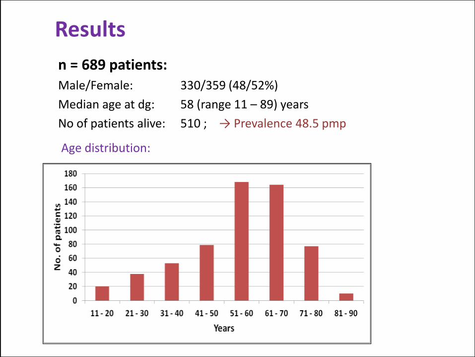
Results
n = 689 patients:Male/Female: 330/359 (48/52%)
Median age at dg: 58 (range 11 – 89) years
No of patients alive: 510 ; → Prevalence 48.5 pmp
Age distribution:

ANCA type (ever) ELISA and/or IIF
p/anti-MPO, 38%
c/anti-PR357%
Atypical, N/A2% 0%
Negativní3%

Cumulative organ involvement (n: 689)
0,0%10,0%20,0%30,0%40,0%50,0%60,0%70,0%80,0%90,0%

Causes of death
2.9x increased risk of death due to cardiovascular cause, in patients aged 15-64 years: 10x increased risk of mortality (all causes)
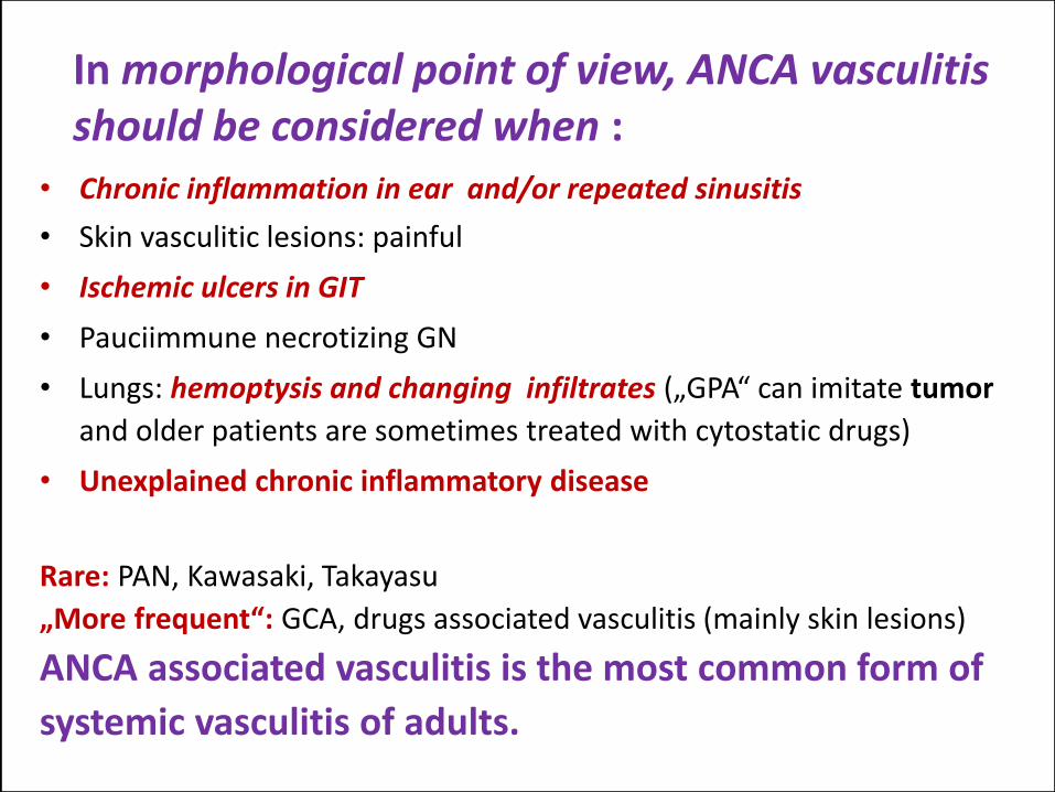
In morphological point of view, ANCA vasculitis should be considered when :
• Chronic inflammation in ear and/or repeated sinusitis
• Skin vasculitic lesions: painful
• Ischemic ulcers in GIT
• Pauciimmune necrotizing GN
• Lungs: hemoptysis and changing infiltrates („GPA“ can imitate tumor
and older patients are sometimes treated with cytostatic drugs)
• Unexplained chronic inflammatory disease
Rare: PAN, Kawasaki, Takayasu
„More frequent“: GCA, drugs associated vasculitis (mainly skin lesions)
ANCA associated vasculitis is the most common form of
systemic vasculitis of adults.

16th Prague Postgraduate Training Course in Nephrology and ERA-EDTA CME Course (22. – 23. 1. 2016)Organized by CNS, ERA- EDTA immunonephrology WG, RPS, and Nephropath®
Nephropathology for the nephrologists
Introduction to renal pathology and approach to diagnosis (clinic-pathology correlations)
When to add molecular pathology to the diagnosis of kidney diseases (Helen Liapis, US)
Clinical up-data of membranous nephropathy: Is it time to change the guidelines? (Pierre Ronco)
Should the patients with a clear diagnosis of AAV be biopsied? (V. Tesař)
How I treat a patient with AAV (3 case reports)
Interpretation of renal allograft biopsies: an algorithmic approach with up-grade of Banff classification system (A.Perkowska-Ptasińská)