patient-specific human induced pluripotent stem cell model...
TRANSCRIPT

Title
Patient-specific human induced pluripotent stem cell modelassessed with electrical pacing validates S107 as a potentialtherapeutic agent for catecholaminergic polymorphicventricular tachycardia
Author(s)
Sasaki, Kenichi; Makiyama, Takeru; Yoshida, Yoshinori;Wuriyanghai, Yimin; Kamakura, Tsukasa; Nishiuchi, Suguru;Hayano, Mamoru; Harita, Takeshi; Yamamoto, Yuta;Kohjitani, Hirohiko; Hirose, Sayako; Chen, Jiarong;Kawamura, Mihoko; Ohno, Seiko; Itoh, Hideki; Takeuchi,Ayako; Matsuoka, Satoshi; Miura, Masaru; Sumitomo,Naokata; Horie, Minoru; Yamanaka, Shinya; Kimura, Takeshi
Citation PLOS ONE (2016), 11(10)
Issue Date 2016-10-20
URL http://hdl.handle.net/2433/217229
Right
© 2016 Sasaki et al. This is an open access article distributedunder the terms of the Creative Commons Attribution License,which permits unrestricted use, distribution, and reproductionin any medium, provided the original author and source arecredited.
Type Journal Article
Textversion publisher
Kyoto University

RESEARCH ARTICLE
Patient-Specific Human Induced Pluripotent
Stem Cell Model Assessed with Electrical
Pacing Validates S107 as a Potential
Therapeutic Agent for Catecholaminergic
Polymorphic Ventricular Tachycardia
Kenichi Sasaki1, Takeru Makiyama1*, Yoshinori Yoshida2*, Yimin Wuriyanghai1,3,
Tsukasa Kamakura1, Suguru Nishiuchi1, Mamoru Hayano1, Takeshi Harita1,
Yuta Yamamoto1, Hirohiko Kohjitani1, Sayako Hirose1, Jiarong Chen1,
Mihoko Kawamura3, Seiko Ohno3, Hideki Itoh3, Ayako Takeuchi4, Satoshi Matsuoka4,
Masaru Miura5, Naokata Sumitomo6, Minoru Horie3, Shinya Yamanaka2, Takeshi Kimura1
1 Department of Cardiovascular Medicine, Kyoto University Graduate School of Medicine, Kyoto, Japan,
2 Kyoto University iPS Cell Research and Application, Kyoto, Japan, 3 Department of Cardiovascular and
Respiratory Medicine, Shiga University of Medical Science, Otsu, Japan, 4 Department of Integrative and
Systems Physiology, Faculty of Medical Sciences, University of Fukui, Fukui, Japan, 5 Division of
Cardiology, Tokyo Metropolitan Children’s Medical Center, Tokyo, Japan, 6 Department of Pediatric
Cardiology, Saitama Medical University International Medical Center, Saitama, Japan
* [email protected] (TM); [email protected] (Y. Yoshida)
Abstract
Introduction
Human induced pluripotent stem cells (hiPSCs) offer a unique opportunity for disease
modeling. However, it is not invariably successful to recapitulate the disease phenotype
because of the immaturity of hiPSC-derived cardiomyocytes (hiPSC-CMs). The purpose of
this study was to establish and analyze iPSC-based model of catecholaminergic polymor-
phic ventricular tachycardia (CPVT), which is characterized by adrenergically mediated
lethal arrhythmias, more precisely using electrical pacing that could promote the develop-
ment of new pharmacotherapies.
Method and Results
We generated hiPSCs from a 37-year-old CPVT patient and differentiated them into cardio-
myocytes. Under spontaneous beating conditions, no significant difference was found in
the timing irregularity of spontaneous Ca2+ transients between control- and CPVT-hiPSC-
CMs. Using Ca2+ imaging at 1 Hz electrical field stimulation, isoproterenol induced an
abnormal diastolic Ca2+ increase more frequently in CPVT- than in control-hiPSC-CMs
(control 12% vs. CPVT 43%, p<0.05). Action potential recordings of spontaneous beating
hiPSC-CMs revealed no significant difference in the frequency of delayed afterdepolariza-
tions (DADs) between control and CPVT cells. After isoproterenol application with pacing at
PLOS ONE | DOI:10.1371/journal.pone.0164795 October 20, 2016 1 / 17
a11111
OPENACCESS
Citation: Sasaki K, Makiyama T, Yoshida Y,
Wuriyanghai Y, Kamakura T, Nishiuchi S, et al.
(2016) Patient-Specific Human Induced Pluripotent
Stem Cell Model Assessed with Electrical Pacing
Validates S107 as a Potential Therapeutic Agent for
Catecholaminergic Polymorphic Ventricular
Tachycardia. PLoS ONE 11(10): e0164795.
doi:10.1371/journal.pone.0164795
Editor: Tomohiko Ai, Indiana University, UNITED
STATES
Received: June 4, 2016
Accepted: September 30, 2016
Published: October 20, 2016
Copyright: © 2016 Sasaki et al. This is an open
access article distributed under the terms of the
Creative Commons Attribution License, which
permits unrestricted use, distribution, and
reproduction in any medium, provided the original
author and source are credited.
Data Availability Statement: All relevant data are
within the paper and its Supporting Information
files.
Funding: This work was supported by Japan
Society for the Promotion of Science KAKENHI
Grant Number 25461054 and Suzuken Memorial
Foundation Grant Number 10-097. The funders had
no role in study design, data collection and
analysis, decision to publish, or preparation of the
manuscript.

1 Hz, 87.5% of CPVT-hiPSC-CMs developed DADs, compared to 30% of control-hiPSC-
CMs (p<0.05). Pre-incubation with 10 μM S107, which stabilizes the closed state of the rya-
nodine receptor 2, significantly decreased the percentage of CPVT-hiPSC-CMs presenting
DADs to 25% (p<0.05).
Conclusions
We recapitulated the electrophysiological features of CPVT-derived hiPSC-CMs using
electrical pacing. The development of DADs in the presence of isoproterenol was signifi-
cantly suppressed by S107. Our model provides a promising platform to study disease
mechanisms and screen drugs.
Introduction
Catecholaminergic polymorphic ventricular tachycardia (CPVT) is a hereditary arrhythmicdisorder characterized by bidirectional ventricular tachycardia (VT) that is triggered by emo-tional stress or physical exercise and leads to syncope or sudden cardiac death without struc-tural heart disease. CPVT is caused by autosomal dominant mutations in the cardiacryanodine receptor gene (RyR2) [1], the calmodulin gene (CALM1, CALM2) [2, 3] and theinward rectifying potassium channel gene (KCNJ2) [4] and autosomal recessive mutations inthe cardiac calsequestrin gene (CASQ2) [5] and the triadin gene (TRDN) [6]. Approximately50–55% of CPVT cases are associated with RyR2 mutations [7] and 1–2% are due to CASQ2mutations [8]. Beta-blockers are the first-line therapy for CPVT, but they often fail to preventfatal arrhythmias [9]. Recently, flecainide, a class Ic Na+ channel blocker, has been reported tobe effective for treating CPVT patients [10].
The advent of human induced pluripotent stem cell (hiPSC) technology has enabled us touse human cardiomyocytes that have the same genetic background as the patients. There havebeen several reports of CPVT iPSC-basedmodel [11–15], however, it is becoming clearer thathiPSC-derived cardiomyocytes (hiPSC-CMs) have immature electrophysiological and struc-tural properties compared to adult human cardiomyocytes [16, 17], which hamper us to ana-lyze their phenotype precisely. In the present study, we demonstrated that hiPSC-CMs beat inan irregular disorganized pattern even in those of control, and suggested the usefulness of elec-trical pacing during Ca2+ transient and action potential (AP) recordings in hiPSC-CMs. Inaddition, we investigated the efficacy of S107, a 1,4-benzothiazepinederivative that is a promis-ing candidate drug for treating CPVT. S107 was discovered by Marks et al and reported to cor-rect leaky RyR1, 2 by stabilizing interactions between RyR 1, 2 and calstabin 1, 2 [18]. Thepotency of S107 for preventing the development of DADs in CPVT-hiPSC-CMs indicates thatour model could be useful for investigating new pharmacotherapies.
Materials and Methods
hiPSC generation and cardiomyocyte differentiation
Human dermal fibroblasts were obtained from a patient after written informed consent wasobtained. The fibroblasts were retrovirally transduced with a combination of 4 transcriptionfactors (Oct3/4, Sox2, Klf-4, c-Myc) to generate hiPSCs. This study was approved by KyotoUniversity ethics review board (G259) and conformed to the Declaration of Helsinki. Allpatients provided written informed consent. The control hiPSC line, 201B7, was generated
S107 Suppresses DADs in Human Induced Pluripotent Stem Cell-Based Model of CPVT
PLOS ONE | DOI:10.1371/journal.pone.0164795 October 20, 2016 2 / 17
Competing Interests: The authors have declared
that no competing interests exist.

from a healthy individual using the same transcription factors [19]. The hiPSCs were differenti-ated into cardiomyocytes using an embryoid body (EB) differentiating system describedprevi-ously [20]. For Ca2+ imaging, EBs were treated with collagenase B (Roche, Indianapolis, IN,USA) and trypsin EDTA (Nacalai Tesque, Kyoto, Japan) at day 21 of differentiation and dis-persed into single cells or small clusters which were plated onto gelatin-coated dishes. For geneexpression analyses, EBs were plated onto fibronectin-coateddishes without dissociation atday 21. After being plated on dishes, hiPSC-CMs were maintained in culture medium consist-ing of DMEM/F12 supplemented with 2% fetal bovine serum, 2 mmol/L L-glutamine, 0.1mmol/L non-essential amino acids, 0.1 mmol/L β-mercaptoethanol, 50 U/ml penicillin, and50 μg/ml streptomycin [21]. The medium was renewed every 2–3 days.
Genomic sequencing and karyotyping
Genomic DNA was isolated from control and CPVT-hiPSC lines by GenElute MammalianGenomic DNA Miniprep kit (Sigma-Aldrich, St Louis, MO, USA). PurifiedDNA was ampli-fied with specific primers and analyzed by 3100 Genetic Analyzer and Big Dye Terminator v1.1(Applied Biosystems, Foster City, CA, USA). Chromosomal G-banding analysis was performedusing a standard procedure (Nihon Gene Research Laboratories, Sendai, Japan). Primers aredetailed in S1 Table.
Immunocytochemistry
The hiPSC colonies were fixed in 4% paraformaldehyde (PFA) for 20 min. The cells were per-meabilized in 0.2% Triton X-100 (Nacalai Tesque). The samples were stained with the follow-ing primary antibodies:mouse monoclonal anti-OCT3/4 (1:50; Santa Cruz Biotechnology,Delaware, CA, USA), mouse monoclonal anti-SSEA4 (1:200; Santa Cruz Biotechnology), andmouse monoclonal anti-TRA 1–60 (1:200; Santa Cruz Biotechnology). The secondary antibodywas donkey anti-mouse Alexafluor 488 (1:1000, Invitrogen, Carlsbad, CA, USA). The nucleiwere stained with DAPI (1:2000, Wako Pure Chemical Industries, Osaka, Japan). The speci-mens were observedunder a fluorescencemicroscope (Biozero BZ-9000; KEYENCE, Osaka,Japan).
Teratoma formation
In order to determine the pluripotency of hiPSCs, we performed teratoma formation inimmune-compromised mice. Two NOD/SCID male mice were maintained under a 12 hourslight / 12 hours dark cycle and fed ad libitium. Mice were inspected daily by the veterinarystaff. Following anesthesia with pentobarbital (50 mg/kg), the hiPSCs were injected as cellclumps into NOD/SCID mice under the testis capsule. The injection site was monitored fortumor growth weekly. Tumor samples were collected at 8 weeks, fixed in 10% formalin andstained with hematoxylin and eosin. Mice were euthanized by cervical dislocation. All animalexperiments were performed in accordance with the ‘Guide for the Care and Use of LaboratoryAnimals’ (2011) of the National Institutes of Health and the Regulation on Animal Experimen-tation at Kyoto University, and approved by Ethics Committee of Kyoto University (PermitNumber: kei 31–18).
Analysis of mRNA expression by real-time quantitative polymerase
chain reaction (qPCR)
Total RNA was isolated using TRIzol Reagent (Invitrogen, Carlsbad, CA, USA) from 20 to 30spontaneously beating EBs microdissectedat day 30 and day 90, and treated with TURBO
S107 Suppresses DADs in Human Induced Pluripotent Stem Cell-Based Model of CPVT
PLOS ONE | DOI:10.1371/journal.pone.0164795 October 20, 2016 3 / 17

DNA-free Kit (Applied Biosystems, Foster City, CA, USA). Total RNA from human wholeheart tissue (BioChain Institute, Newark, CA, USA) was also reverse transcribed into comple-mentary DNA (cDNA) for comparison. The cDNA was synthesized from 1 μg of total RNA, ina total volume of 20 μl, using oligo (dT)18 primer with Transcriptor First Strand cDNA Synthe-sis Kit (Roche). TaqMan real-time PCR assay was performed using FastStart Universal ProbeMaster (Rox) and an appropriate probe from Universal ProbeLibrary Set (Roche). The expres-sion of genes of interest was normalized to that of GAPDH. Relative quantification was calcu-lated according to the ΔΔCT method. The changes in gene expression levels were comparedwith those of adult human heart. The fold change is expressed as mean ± SEM. A list of theprimers used in these experiments is provided in S1 Table.
Ca2+ imaging
The hiPSC-CMs were dispersedwith collagenase B and Trypsin EDTA and plated onto glasscoverslips coated with fibronectin (BD Biosciences, San Jose, CA, USA). After 5–7 days, disso-ciated hiPSC-CMs on a coverslip were loaded with 2 μmol/L Fluo-8 (AAT Bioquest, Sunnyvale,CA, USA) in the culture medium described above. After incubation for 30 min at 37°C in 5%CO2, the medium was replaced with normal tyrode solution containing (in mmol/L): 140NaCl, 0.33 NaH2PO4, 5.4 KCl, 1.8 CaCl2, 0.5 MgCl2, 5.0 HEPES, and 5.5 D-Glucose. Spontane-ously contracting or electrically stimulated single cells were analyzed at 36.0 ± 1.0°C. The imag-ing of fluo-8 was analyzed for average pixel intensities of regions of interest drawn to includewhole cell, following background correction, using an Aquacosmos image-processing system(Hamamatsu Photonics, Hamamatsu, Japan). The hiPSC-CMs were stimulated at 0.5 and 1.0Hz with 4 ms depolarizing pulses at 20–30 V using platinum electrodes,with an interelectrodedistance of 12 mm (Intermedical, Osaka, Japan). The rhythmicity of the spontaneous Ca2+
transient was assessed by calculating the cycle length variability index, defined as the standarddeviation of the cycle length/mean cycle length. A solution containing 100 nM isoproterenol(LKT Laboratories, Saint Paul, MN, USA) was prepared fresh before the experiment andapplied 5–10 min before data collection.A 10 μM ryanodine solution (Wako Pure ChemicalIndustries) was added 2–3 min before isoproterenol administration.
Electrophysiological recordings
The hiPSC-CMs were enzymatically dissociated with collagenase B and Trypsin EDTA andplated onto glass coverslips coated with fibronectin (BD Biosciences). APs were recorded at36.0 ± 1.0°C in a current clamp mode using a perforated patch-clamp technique. The pipettesolution contained 300 μg/ml amphotericin B (Sigma-Aldrich, St Louis, MO, USA) and the fol-lowing (in mM): 150 KCl, 5 EGTA, 5 MgATP, 10 HEPES, 5 NaCl, 2 CaCl2, pH was adjusted to7.2 by KOH. The experiments were performed under continuous perfusion of the extracellularsolution containing (in mM): 150 NaCl, 5.4 KCl, 1.8 CaCl2, 1 MgCl2, 15 glucose, 15 HEPES, 1Na-pyruvate (pH adjusted to 7.40 with NaOH) [22]. Patch-clamp pipettes, formed from boro-silicate glass with PP-830 (Narishige, Tokyo, Japan) and had a resistance of 4–7 MΩ. APs wererecorded from spontaneously contracting and quiescent hiPSC-CMs. All signals were acquiredat 10 kHz, digitizedwith a Digidata 1332A (Axon instruments, CA, USA) and analyzed with apCLAMP 10.4 software (Axon instruments). Current clamp recordings were performed usinga MultiClamp 700B amplifier. Solutions containing 100 nM isoproterenol were applied using agravitational flow system 5–10 min prior to data collection.We added 1 and 10 μM freshly pre-pared S107 (Cayman Chemical, Ann Arbor, MI, USA) to the culture medium 2–3 hours priorto the experiments.
S107 Suppresses DADs in Human Induced Pluripotent Stem Cell-Based Model of CPVT
PLOS ONE | DOI:10.1371/journal.pone.0164795 October 20, 2016 4 / 17

Statistical analysis
Continuous variables are presented as the mean ± SEM. Categorical variables are expressed asfrequencies. Differences in the means between two groups were compared using Student’s t-tests. Categorical differences between two groups were evaluated using chi-squared tests. Avalue of p< 0.05 was considered statistically significant.
Results
Generation of patient-specific CPVT-hiPSCs
We generated hiPSCs from a 37-year-old female patient with CPVT, whose clinical featureswere previously reported [23]. She first experienced syncope during running or swimming at15 years of age. Her next episodes of syncope were while riding a bicycle and when under emo-tional stress caused by a traffic accident at 30 years of age. She then remained free of syncopeuntil the age of 36 years when she experienced an episode upon awakening from a nightmare,at which time she was admitted to the hospital. Polymorphic VT was recorded during an exer-cise test and an epinephrine provocation test. She was diagnosedwith CPVT and began takingcarvedilol.However, she had an episode of syncope and polymorphic VT was recorded againduring an exercise test despite having received an oral beta-blocker. Subsequently, she under-went implantation of an implantable cardioverter-defibrillator (ICD). At the age of 37, anappropriate ICD discharge occurred and she began taking flecainide, which was effective inpreventing polymorphic VT. Genetic analyses identified a missense mutation, c.13759 A>G, p.I4587V, in the RyR2 gene. Her son also had a history of syncope and was a carrier of an identi-cal mutation.
Dermal fibroblasts were obtained from the proband and retrovirally transduced with 4genes (Oct3/4, Sox2, Klf-4, and c-Myc). As a control, we used an hiPSC line, 201B7, which wassimilarly generated from a healthy individual [19]. The CPVT-hiPSCs exhibited characteristichuman embryonic stem cell morphology and stained positively for pluripotency markers(OCT3/4, SSEA4, TRA1-60) (Fig 1A). The CPVT-hiPSC lines displayed a normal karyotype(Fig 1B). We confirmed the RyR2-I4587V mutation in the CPVT-hiPSCs, but not in the con-trol-hiPSCs (Fig 1C). In order to confirm the pluripotency of generated hiPSCs, we injectedhiPSCs into severe combined immunodeficiency (SCID) mice, which led to the formation ofteratomas, containing tissue derivatives of three germ layers: pigmented epithelium (ecto-derm), gut-like structures (endoderm), and cartilage tissue (mesoderm) (Fig 1D).
Gene expression of hiPSC-CMs
We differentiated control- and CPVT-hiPSCs into spontaneously contracting cardiomyocytesusing an EB differentiation system. Spontaneously beating EBs started to appear at day 7 of dif-ferentiation. EBs were plated on fibronectin-coateddishes at day 21 and maintained in the cul-ture medium described above for an additional 2 months. We conducted quantitative real-timePCR in control- and CPVT-hiPSC-CMs at days 30 and 90, and the expression levels of thegenes involved in Ca2+ handling are shown in Fig 2. Consistent with previously reported tran-scriptional profile data on human iPSC/ESC-derived cardiomyocytes [24], CASQ2 expressionlevels were extremely low compared to those of adult human cardiomyocytes. However, RyR2and SERCA2 were already expressed at 1 month similar to adult human myocytes in the con-trol- and CPVT-hiPSC-CMs. Innositol-1,4,5-trisphosphate receptor 2 (IP3R2) and calreticulinwere highly expressed in the control- and CPVT-hiPSC-CMs compared to adult human cardi-omyocyts (Fig 2). In this study, there was no significant difference in Ca2+ handling geneexpression between control- and CPVT-hiPSC-CMs.
S107 Suppresses DADs in Human Induced Pluripotent Stem Cell-Based Model of CPVT
PLOS ONE | DOI:10.1371/journal.pone.0164795 October 20, 2016 5 / 17

CPVT-hiPSC-CMs presented diastolic Ca2+ waves with adrenergic
stimulation upon Ca2+ imaging
We performedCa2+ imaging of spontaneously contracting control- and CPVT-hiPSC-CMs. Aconsiderable number of both control- and CPVT-hiPSC-CMs showed irregular beatingrhythms at baseline and after isoproterenol administration (Fig 3A). To evaluate the irregular-ity parameter, we calculated the cycle length variability index, also known as the coefficient ofvariation, defined as the cycle length standard deviation divided by the mean of the cyclelength. The cycle length variability indices were higher in hiPSC-CMs compared to those in
Fig 1. Characterization of CPVT-hiPSCs. (A) CPVT-hiPSC colonies derived from the dermal fibroblasts of a patient with CPVT
expressed pluripotency markers, shown by immunostaining. Scale bars = 200 μm. (B) CPVT-hiPSCs maintained the normal
karyotype. (C) Sequencing analysis of the RYR2 gene identified the I4587V heterozygous point mutation in the CPVT-hiPSCs. (D)
Hematoxylin-eosin staining of teratomas formed from CPVT-hiPSCs showed differentiation of the cells into various tissues derived
from all three germ layers: pigmented epithelium (ectoderm), gut-like structures (endoderm), and cartilage tissue (mesoderm).
Scale bar = 100 μm.
doi:10.1371/journal.pone.0164795.g001
S107 Suppresses DADs in Human Induced Pluripotent Stem Cell-Based Model of CPVT
PLOS ONE | DOI:10.1371/journal.pone.0164795 October 20, 2016 6 / 17

isolated rabbit nodal cells [25], but there was no significant difference between control andCPVT cells (Fig 3B). In spontaneous beating hiPSC-CMs, we found no significant difference inthe frequency of diastolic Ca2+ waves (increased intracellular Ca2+ concentration during thediastolic phase) between control and CPVT cells (Fig 3C).
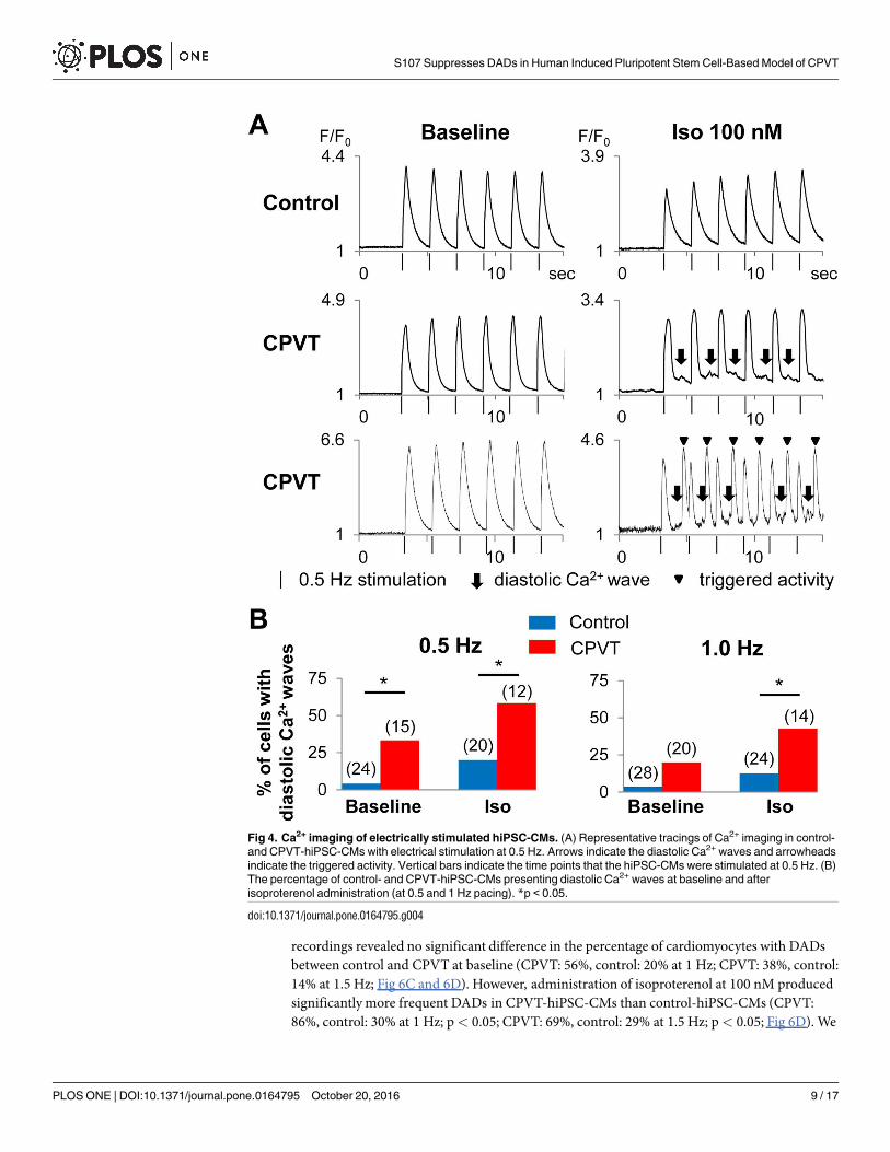
Therefore, to minimize the effect of beating intervals, we used electrical field stimulationwhen recording Ca2+ transients to assess the arrhythmic phenotype in hiPSC-CMs (Fig 4A). Atbaseline, CPVT-hiPSC-CMs showed diastolic Ca2+ waves significantly more frequently thancontrol-hiPSC-CMs at 0.5 Hz pacing (CPVT: 33%, control: 4%; p< 0.05; Fig 4B). After admin-istration of 100 nM isoproterenol, significantly more frequent diastolic Ca2+ waves wererecorded in CPVT-hiPSC-CMs than control-hiPSC-CMs (CPVT: 58%, control: 20% at 0.5 Hz;p< 0.05; CPVT: 43%, control: 12% at 1 Hz; p< 0.05; Fig 4B).
We treated the CPVT-hiPSC-CMs with ryanodine, a strong ligand of the ryanodine recep-tor (Fig 5A). With 10 μM ryanodine, the percentage of CPVT-hiPSC-CMs that presented dia-stolic Ca2+ waves after isoproterenol administration significantly decreased to 9% at 0.5 Hz and7% at 1 Hz pacing (Fig 5B).
Fig 2. Gene expression of calcium handling proteins in hiPSC-CMs. Quantitative real-time PCR of spontaneously
contracting embryoid bodies differentiated from control- and CPVT-hiPSCs showed comparable expression levels of the
studied calcium handling proteins. All values are relative to the adult human heart and were normalized to
glyceraldehyde 3-phosphate dehydrogenase (GAPDH). PCR = polymerase chain reaction
doi:10.1371/journal.pone.0164795.g002
S107 Suppresses DADs in Human Induced Pluripotent Stem Cell-Based Model of CPVT
PLOS ONE | DOI:10.1371/journal.pone.0164795 October 20, 2016 7 / 17

β-adrenergic stimulation-induced DADs in CPVT-hiPSC-CMs
The percentage of CPVT-hiPSC-CMs that developedDADs during AP recordings did not dif-fer from that of control-hiPSC-CMs under the spontaneously beating condition at baseline andafter isoproterenol administration (Fig 6A and 6B). Under regular electrical stimulation, AP
Fig 3. Ca2+ imaging of spontaneously beating hiPSC-CMs. (A) Representative tracings of Ca2+ imaging in spontaneously
contracting control- and CPVT-hiPSC-CMs. (B) Cycle length variability indices, defined as the standard deviation of the cycle
length/mean cycle length of spontaneously beating hiPSC-CMs showed no significant difference between control and CPVT. (C)
The percentage of spontaneously contracting control- and CPVT-hiPSC-CMs presenting diastolic Ca2+ waves at baseline and
after isoproterenol administration.
doi:10.1371/journal.pone.0164795.g003
S107 Suppresses DADs in Human Induced Pluripotent Stem Cell-Based Model of CPVT
PLOS ONE | DOI:10.1371/journal.pone.0164795 October 20, 2016 8 / 17
recordings revealed no significant difference in the percentage of cardiomyocytes with DADsbetween control and CPVT at baseline (CPVT: 56%, control: 20% at 1 Hz; CPVT: 38%, control:14% at 1.5 Hz; Fig 6C and 6D). However, administration of isoproterenol at 100 nM producedsignificantly more frequent DADs in CPVT-hiPSC-CMs than control-hiPSC-CMs (CPVT:86%, control: 30% at 1 Hz; p< 0.05; CPVT: 69%, control: 29% at 1.5 Hz; p< 0.05; Fig 6D). We
Fig 4. Ca2+ imaging of electrically stimulated hiPSC-CMs. (A) Representative tracings of Ca2+ imaging in control-
and CPVT-hiPSC-CMs with electrical stimulation at 0.5 Hz. Arrows indicate the diastolic Ca2+ waves and arrowheads
indicate the triggered activity. Vertical bars indicate the time points that the hiPSC-CMs were stimulated at 0.5 Hz. (B)
The percentage of control- and CPVT-hiPSC-CMs presenting diastolic Ca2+ waves at baseline and after
isoproterenol administration (at 0.5 and 1 Hz pacing). *p < 0.05.
doi:10.1371/journal.pone.0164795.g004
S107 Suppresses DADs in Human Induced Pluripotent Stem Cell-Based Model of CPVT
PLOS ONE | DOI:10.1371/journal.pone.0164795 October 20, 2016 9 / 17

found no early afterdepolarization in the AP recordings of control- and CPVT-hiPSC-CMswhen paced at 1 or 1.5 Hz.
S107 exerted an antiarrhythmic effect on CPVT-hiPSC-CMs
We treated CPVT-hiPSC-CMs with the 1,4-benzothiazepinederivative S107. Pre-incubationwith S107 for 2–3 hours suppressed the frequency of DADs in CPVT-hiPSC-CMs in a concen-tration-dependentmanner (Fig 7A). Incubation with 1 μM S107 significantly decreased thepercentage of CPVT-hiPSC-CMs presenting DADs to 33% at 1 Hz pacing after isoproterenoladministration. Incubation with 10 μM S107 further decreased the percentage of CPVT-hiPSC-CMs with DADs compared to those lacking S107 (25% at 1 Hz; p< 0.05, 10% at 1.5 Hz;p< 0.05; Fig 7B).
Fig 5. Ryanodine suppressed diastolic Ca2+ waves in CPVT-hiPSC-CMs. (A) Representative tracings of Ca2+
imaging in CPVT-hiPSC-CMs with (lower) and without (upper) ryanodine. Note diastolic Ca2+ waves (arrows)
without ryanodine (upper, right), however, no diastolic Ca2+ waves with ryanodine (lower, right). Vertical bars
indicate the time points that the CPVT-hiPSC-CMs were stimulated at 0.5 Hz. (B) Fraction (in %) of CPVT-
hiPSC-CMs that showed diastolic Ca2+ waves with and without ryanodine. *p < 0.05.
doi:10.1371/journal.pone.0164795.g005
S107 Suppresses DADs in Human Induced Pluripotent Stem Cell-Based Model of CPVT
PLOS ONE | DOI:10.1371/journal.pone.0164795 October 20, 2016 10 / 17

Discussion
Since the recognition of RyR2 as a gene responsible for CPVT [1], functional analyses have sug-gested that DADs following Ca2+ leakage from the sarcoplasmic reticulum (SR) are associatedwith ventricular arrhythmia in CPVT; however, the mechanisms underlying SR Ca2+ leak havenot been elucidated. Even now, some patients with CPVT experience syncope or sudden car-diac death. Although we must develop novel therapeutic approaches, it is uncertainwhetherthe results of drug testing in mouse models are replicated in humans. Patient-specific hiPSC-based models of CPVT offer new opportunities for studying drug effects and pathogeneticmechanisms in human cardiomyocytes, however, it is not invariably successful to recapitulatethe phenotypes due to the electrophysiological immaturity of hiPSC-CMs. In this study, weprecisely evaluated the electrophysiological properties using electrical pacing, and confirmedabnormal diastolic Ca2+ waves during Ca2+ imaging and DADs during AP recordings inCPVT-hiPSC-CMs, which are two major phenotypes shown in CPVT knock-inmouse models[26, 27]. In addition, S107 prevented the development of DADs in CPVT-hiPSC-CMs.
Fig 6. AP recordings of hiPSC-CMs. (A) Representative tracings of AP recordings from spontaneously beating control- and
CPVT-hiPSC-CMs. Both control- and CPVT-hiPSC-CMs showed DADs. (B) No significant difference was found in the
percentage of spontaneously contracting hiPSC-CMs presenting DADs between control and CPVT. (C) Representative
tracings of AP recordings during 1 Hz pacing from control- and CPVT-hiPSC-CMs. CPVT-hiPSC-CMs showed DADs,
especially after isoproterenol administration. (D) The percentage of control- and CPVT-hiPSC-CMs that developed DADs at
1 and 1.5 Hz pacing. *p < 0.05.
doi:10.1371/journal.pone.0164795.g006
S107 Suppresses DADs in Human Induced Pluripotent Stem Cell-Based Model of CPVT
PLOS ONE | DOI:10.1371/journal.pone.0164795 October 20, 2016 11 / 17

Cardiomyocytes derived from human embryonic stem cells (hESC-CMs) have functionalIP3-dependent Ca2+ release and the gene expression level of IP3R2 progressively declines withthe maturation of the hESC-CMs [28]. For the first time, we showed that IP3R2 was highlyexpressed in hiPSC-CMs compared to adult human cardiomyocytes. IP3-dependent Ca2+ sig-naling has been shown to play an important role during the process of cardiac development.Calreticulin is also an important Ca2+ buffer and a regulator of Ca2+ homeostasis during fetallife. High expression levels of IP3R2 and calreticulin, together with extremely low expressionlevels of CASQ2, indicate immature Ca2+ handling properties in hiPSC-CMs.
The hiPSC-CMs have irregular beating rhythms resulting from immature electrophysiologi-cal properties, which hamper the precise electrophysiological analyses. The cycle length vari-ability indices of spontaneously beating control- and CPVT-hiPSC-CMs were remarkablyhigher compared to those of rabbit pacemaker cells [25], indicating highly irregular contractionof hiPSC-CMs. Under spontaneously beating conditions, there were no significant differencesin the frequency of diastolic Ca2+ waves or DADs between control- and CPVT-hiPSC-CMs.Ca2+ transients, ion currents (Ica(L), Ito, INaK and INaL), and intracellular ion concentrations(Na+ and K+) in myocytes were reported to vary depending on pacing frequency using in-silicoanalyses [29, 30]. Therefore, we used electrical stimulation to perform the experiments at equalbeating rates and successfully demonstrated significantly more frequent diastolic Ca2+ wavesand DADs in CPVT-hiPSC-CMs compared to control cells. Kujala et al reported the presenceof early afterdepolarizations in CPVT-hiPSC-CMs [14]; however, in our experiments, we did
Fig 7. S107 prevented DADs in CPVT-hiPSC-CMs. (A) Representative tracings of AP recordings from CPVT-hiPSC-CMs
following a 2–3 h pre-incubation with 10 μM S107. DADs were not found after isoproterenol application. (B) Fraction (in %) of
CPVT-hiPSC-CMs that showed DADs after isoproterenol administration with and without S107 pre-incubation. S107
suppressed DADs in a concentration-dependent manner. *p < 0.05 versus the absence of S107.
doi:10.1371/journal.pone.0164795.g007
S107 Suppresses DADs in Human Induced Pluripotent Stem Cell-Based Model of CPVT
PLOS ONE | DOI:10.1371/journal.pone.0164795 October 20, 2016 12 / 17

not identify any early afterdepolarizations during 1 or 1.5 Hz pacing. The use of 10 μM ryano-dine significantly suppressed the diastolic Ca2+ waves of CPVT-hiPSC-CMs. This ligand fullycloses ryanodine receptors at micromolar concentrations. Therefore, our results indicate thatryanodine receptors play an important role in the mechanisms of diastolic Ca2+ waves inCPVT-hiPSC-CMs.
Human RyR2 is a large intracellular Ca2+-permeable channel and consists of the N-termi-nal, central, and C-terminal domains [31]. The C-terminal domain is composed of the I-domain and the transmembrane domain which forms the channel pore. K201 (JTV519), a1,4-benzothiazepinederivative, was formerly a promising candidate drug for CPVT and wasthought to stabilize RyR2 by correcting defective domain-domain interactions in the N-termi-nal and central domains. K201 prevented the SR Ca2+ leak in failing cardiomyocytes byimproving the defective inter-domain interaction between the N-terminal and central domains[32]. However, it failed to present cardioprotective effects on catecholamine-inducedDADsand arrhythmia generation in the R4496C+/- mouse model of CPVT with a mutation in theRyR2 transmembrane domain [26]. In addition, K201 was reported to be a multichannelblocker that suppressed ICa(L) and INCX (33 ± 5% reduction in the peak Ica(L) with 0.3 μM K201,the concentration generally used in in vitro assays) [33]. In this context, another 1,4-benzothia-zepine derivative, S107, was developed. S107 is one of the Rycals1, small molecules that pre-vent Ca2+ leakage from the SR by promoting the binding of calstabin or calstabin2 (a channelstabilizing protein) to ryanodine receptor isoform 1 or RyR2. S107 has high specificity forRyR2 and no off-target activity up to 10 μM [18]. In mouse model studies, S107 was effectivefor preventing ventricular arrhythmias, seizures and atrial fibrillation in CPVT [18]. In thepresent study, we first confirmed the efficacyof S107 in CPVT-hiPSC-CMs. Three mechanismshave been proposed to underlie leaky RyR2 channels: dissociation of a channel-stabilizing pro-tein (calstabin2) from RyR2 [34], defective inter-domain folding of RyR2 [35], and changes inthe sensitivity of RyR2 to cytosolic and luminal Ca2+ [36]. The augmented binding of calsta-bin2 to RyR2 is the only mechanism demonstrated to underlie the effect of S107. Regarding thebinding site of calstabin2 on RyR2, the central domain of RyR2 has been reported to interactwith calstabin2 [37]. However, Zissimopoulos and colleagues demonstrated that the C-termi-nal domain (containing the transmembrane domain) of RyR2 interacts with calstabin2 [38]. Inour CPVT model, S107 might exert an antiarrhythmic effect by improving binding betweencalstabin2 and the transmembrane domain of RyR2. S107 can also prevent stress-induced cog-nitive dysfunction [39], the progression of heart failure [40], and muscle weakness in aging[41] or Duchenne muscular dystrophy [42]. The lead Rycal program is in phase 2 clinical stud-ies for the treatment of heart failure and arrhythmias. These compounds may have great prom-ise for treating patients with CPVT.
There are several limitations in this study. We analyzed CPVT-hiPSC-CMs derived fromone RyR2 mutation. It is therefore unclear whether the findings in our model would be con-firmed in another CPVT hiPSC-model harboring different mutations in RyR2 or other candi-date genes. Other limitations are immature electrophysiological and structural properties ofhiPSC-CMs compared to adult CMs, and a fundamental issue that we have developed a cell-based model which did not recapitulate the complex environment of a mammalian heart.
Conclusions
We generated a stem cell-basedCPVT model harboring an RyR2 mutation in the transmem-brane domain, and it successfully recapitulated the disease phenotype. Electrical pacing washighly useful for analyzing arrhythmogenic features in CPVT-hiPSC-CMs, and the antiar-rhythmic effect of S107 was confirmed in this model. This CPVT hiPSC-basedmodel assessed
S107 Suppresses DADs in Human Induced Pluripotent Stem Cell-Based Model of CPVT
PLOS ONE | DOI:10.1371/journal.pone.0164795 October 20, 2016 13 / 17

with electrical pacing is a powerful tool that will provide us with a better understanding of theunderlying mechanisms and new approaches for screening drugs to establish personalizedmedicine.
Supporting Information
S1 Table. Primers used for sequencing of the human RYR2 gene (exon 94) and qRT-PCR.(XLSX)
Acknowledgments
We thank Masako Tanaka, Kyoko Yoshida and Aya Umehara for their technical assistance.
Author Contributions
Conceptualization:TM Y. Yoshida KS.
Data curation:YW T. Kamakura SN M. Hayano TH Y. Yamamoto HK SH JC.
Formal analysis:KS.
Funding acquisition: TM T. Kimura.
Investigation: KS.
Methodology:TM KS SM AT.
Project administration:TM Y. Yoshida M. Horie.
Resources:NS MM MK HI.
Software: SM AT.
Supervision:TM Y. Yoshida SO SY M. Horie T. Kimura.
Validation: TM Y. Yoshida.
Visualization: TM KS.
Writing – original draft:KS.
Writing – review& editing: TM Y. Yoshida M. Horie.
References1. Priori SG, Napolitano C, Tiso N, Memmi M, Vignati G, Bloise R, et al. Mutations in the cardiac ryano-
dine receptor gene (hRyR2) underlie catecholaminergic polymorphic ventricular tachycardia. Circula-
tion. 2001; 103(2):196–200. PMID: 11208676.
2. Hwang HS, Nitu FR, Yang Y, Walweel K, Pereira L, Johnson CN, et al. Divergent regulation of ryano-
dine receptor 2 calcium release channels by arrhythmogenic human calmodulin missense mutants.
Circ Res. 2014; 114(7):1114–24. doi: 10.1161/CIRCRESAHA.114.303391 PMID: 24563457; PubMed
Central PMCID: PMCPMC3990285.
3. Makita N, Yagihara N, Crotti L, Johnson CN, Beckmann BM, Roh MS, et al. Novel calmodulin muta-
tions associated with congenital arrhythmia susceptibility. Circ Cardiovasc Genet. 2014; 7(4):466–74.
doi: 10.1161/CIRCGENETICS.113.000459 PMID: 24917665; PubMed Central PMCID:
PMCPMC4140998.
4. Vega AL, Tester DJ, Ackerman MJ, Makielski JC. Protein kinase A-dependent biophysical phenotype
for V227F-KCNJ2 mutation in catecholaminergic polymorphic ventricular tachycardia. Circ Arrhythm
Electrophysiol. 2009; 2(5):540–7. doi: 10.1161/CIRCEP.109.872309 PMID: 19843922; PubMed Cen-
tral PMCID: PMCPMC2766080.
S107 Suppresses DADs in Human Induced Pluripotent Stem Cell-Based Model of CPVT
PLOS ONE | DOI:10.1371/journal.pone.0164795 October 20, 2016 14 / 17

5. Lahat H, Eldar M, Levy-Nissenbaum E, Bahan T, Friedman E, Khoury A, et al. Autosomal recessive
catecholamine- or exercise-induced polymorphic ventricular tachycardia: clinical features and assign-
ment of the disease gene to chromosome 1p13-21. Circulation. 2001; 103(23):2822–7. PMID:
11401939.
6. Roux-Buisson N, Cacheux M, Fourest-Lieuvin A, Fauconnier J, Brocard J, Denjoy I, et al. Absence of
triadin, a protein of the calcium release complex, is responsible for cardiac arrhythmia with sudden
death in human. Hum Mol Genet. 2012; 21(12):2759–67. doi: 10.1093/hmg/dds104 PMID: 22422768;
PubMed Central PMCID: PMCPMC3363337.
7. Priori SG, Napolitano C, Memmi M, Colombi B, Drago F, Gasparini M, et al. Clinical and molecular
characterization of patients with catecholaminergic polymorphic ventricular tachycardia. Circulation.
2002; 106(1):69–74. PMID: 12093772.
8. di Barletta MR, Viatchenko-Karpinski S, Nori A, Memmi M, Terentyev D, Turcato F, et al. Clinical phe-
notype and functional characterization of CASQ2 mutations associated with catecholaminergic poly-
morphic ventricular tachycardia. Circulation. 2006; 114(10):1012–9. doi: 10.1161/CIRCULATIONAHA.
106.623793 PMID: 16908766.
9. Hayashi M, Denjoy I, Extramiana F, Maltret A, Buisson NR, Lupoglazoff JM, et al. Incidence and risk
factors of arrhythmic events in catecholaminergic polymorphic ventricular tachycardia. Circulation.
2009; 119(18):2426–34. doi: 10.1161/CIRCULATIONAHA.108.829267 PMID: 19398665.
10. van der Werf C, Kannankeril PJ, Sacher F, Krahn AD, Viskin S, Leenhardt A, et al. Flecainide therapy
reduces exercise-induced ventricular arrhythmias in patients with catecholaminergic polymorphic ven-
tricular tachycardia. J Am Coll Cardiol. 2011; 57(22):2244–54. doi: 10.1016/j.jacc.2011.01.026 PMID:
21616285; PubMed Central PMCID: PMCPMC3495585.
11. Fatima A, Xu G, Shao K, Papadopoulos S, Lehmann M, Arnaiz-Cot JJ, et al. In vitro modeling of ryano-
dine receptor 2 dysfunction using human induced pluripotent stem cells. Cell Physiol Biochem. 2011;
28(4):579–92. doi: 10.1159/000335753 PMID: 22178870; PubMed Central PMCID:
PMCPMC3709175.
12. Jung CB, Moretti A, Mederos y Schnitzler M, Iop L, Storch U, Bellin M, et al. Dantrolene rescues
arrhythmogenic RYR2 defect in a patient-specific stem cell model of catecholaminergic polymorphic
ventricular tachycardia. EMBO Mol Med. 2012; 4(3):180–91. doi: 10.1002/emmm.201100194 PMID:
22174035; PubMed Central PMCID: PMCPMC3376852.
13. Itzhaki I, Maizels L, Huber I, Gepstein A, Arbel G, Caspi O, et al. Modeling of catecholaminergic poly-
morphic ventricular tachycardia with patient-specific human-induced pluripotent stem cells. J Am Coll
Cardiol. 2012; 60(11):990–1000. doi: 10.1016/j.jacc.2012.02.066. PMID: 22749309.
14. Kujala K, Paavola J, Lahti A, Larsson K, Pekkanen-Mattila M, Viitasalo M, et al. Cell model of catechol-
aminergic polymorphic ventricular tachycardia reveals early and delayed afterdepolarizations. PLoS
One. 2012; 7(9):e44660. doi: 10.1371/journal.pone.0044660 PMID: 22962621; PubMed Central
PMCID: PMCPMC3433449.
15. Di Pasquale E, Lodola F, Miragoli M, Denegri M, Avelino-Cruz JE, Buonocore M, et al. CaMKII inhibi-
tion rectifies arrhythmic phenotype in a patient-specific model of catecholaminergic polymorphic ven-
tricular tachycardia. Cell Death Dis. 2013; 4:e843. doi: 10.1038/cddis.2013.369 PMID: 24113177;
PubMed Central PMCID: PMCPMC3824678.
16. Doss MX, Di Diego JM, Goodrow RJ, Wu Y, Cordeiro JM, Nesterenko VV, et al. Maximum diastolic
potential of human induced pluripotent stem cell-derived cardiomyocytes depends critically on I(Kr).
PLoS One. 2012; 7(7):e40288. doi: 10.1371/journal.pone.0040288 PMID: 22815737; PubMed Central
PMCID: PMCPMC3396384.
17. Kamakura T, Makiyama T, Sasaki K, Yoshida Y, Wuriyanghai Y, Chen J, et al. Ultrastructural matura-
tion of human-induced pluripotent stem cell-derived cardiomyocytes in a long-term culture. Circ J.
2013; 77(5):1307–14. PMID: 23400258.
18. Lehnart SE, Mongillo M, Bellinger A, Lindegger N, Chen BX, Hsueh W, et al. Leaky Ca2+ release chan-
nel/ryanodine receptor 2 causes seizures and sudden cardiac death in mice. J Clin Invest. 2008; 118
(6):2230–45. doi: 10.1172/JCI35346 PMID: 18483626; PubMed Central PMCID: PMCPMC2381750.
19. Takahashi K, Tanabe K, Ohnuki M, Narita M, Ichisaka T, Tomoda K, et al. Induction of pluripotent stem
cells from adult human fibroblasts by defined factors. Cell. 2007; 131(5):861–72. doi: 10.1016/j.cell.
2007.11.019 PMID: 18035408.
20. Yang L, Soonpaa MH, Adler ED, Roepke TK, Kattman SJ, Kennedy M, et al. Human cardiovascular
progenitor cells develop from a KDR+ embryonic-stem-cell-derived population. Nature. 2008; 453
(7194):524–8. doi: 10.1038/nature06894 PMID: 18432194.
21. Moretti A, Bellin M, Welling A, Jung CB, Lam JT, Bott-Flugel L, et al. Patient-specific induced pluripo-
tent stem-cell models for long-QT syndrome. N Engl J Med. 2010; 363(15):1397–409. doi: 10.1056/
NEJMoa0908679 PMID: 20660394.
S107 Suppresses DADs in Human Induced Pluripotent Stem Cell-Based Model of CPVT
PLOS ONE | DOI:10.1371/journal.pone.0164795 October 20, 2016 15 / 17

22. Ma J, Guo L, Fiene SJ, Anson BD, Thomson JA, Kamp TJ, et al. High purity human-induced pluripotent
stem cell-derived cardiomyocytes: electrophysiological properties of action potentials and ionic cur-
rents. Am J Physiol Heart Circ Physiol. 2011; 301(5):H2006–17. doi: 10.1152/ajpheart.00694.2011
PMID: 21890694; PubMed Central PMCID: PMCPMC4116414.
23. Dochi K, Matsumoto Y, Nagaoka I, Ito M, Ashihara T, Ito H, et al. A novel missense mutation in the
human cardiac ryanodine receptor gene (I4587V) in a patient with catecholaminergic polymorphic ven-
tricular tachycardia. JPN J Electrocardiology. 2007; 27(3):7.
24. Liu J, Fu JD, Siu CW, Li RA. Functional sarcoplasmic reticulum for calcium handling of human embry-
onic stem cell-derived cardiomyocytes: insights for driven maturation. Stem Cells. 2007; 25(12):3038–
44. doi: 10.1634/stemcells.2007-0549 PMID: 17872499.
25. Wilders R, Jongsma HJ. Beating irregularity of single pacemaker cells isolated from the rabbit sino-
atrial node. Biophys J. 1993; 65(6):2601–13. doi: 10.1016/S0006-3495(93)81289-X PMID: 8312495;
PubMed Central PMCID: PMCPMC1226001.
26. Liu N, Colombi B, Memmi M, Zissimopoulos S, Rizzi N, Negri S, et al. Arrhythmogenesis in catechol-
aminergic polymorphic ventricular tachycardia: insights from a RyR2 R4496C knock-in mouse model.
Circ Res. 2006; 99(3):292–8. doi: 10.1161/01.RES.0000235869.50747.e1 PMID: 16825580.
27. Kashimura T, Briston SJ, Trafford AW, Napolitano C, Priori SG, Eisner DA, et al. In the RyR2(R4496C)
mouse model of CPVT, β-adrenergic stimulation induces Ca waves by increasing SR Ca content and
not by decreasing the threshold for Ca waves. Circ Res. 2010; 107(12):1483–9. doi: 10.1161/
CIRCRESAHA.110.227744 PMID: 20966392.
28. Satin J, Itzhaki I, Rapoport S, Schroder EA, Izu L, Arbel G, et al. Calcium handling in human embryonic
stem cell-derived cardiomyocytes. Stem Cells. 2008; 26(8):1961–72. doi: 10.1634/stemcells.2007-
0591 PMID: 18483424.
29. Hund TJ, Rudy Y. Rate dependence and regulation of action potential and calcium transient in a canine
cardiac ventricular cell model. Circulation. 2004; 110(20):3168–74. doi: 10.1161/01.CIR.0000147231.
69595.D3 PMID: 15505083; PubMed Central PMCID: PMCPMC1851913.
30. Maleckar MM, Greenstein JL, Giles WR, Trayanova NA. K+ current changes account for the rate
dependence of the action potential in the human atrial myocyte. Am J Physiol Heart Circ Physiol. 2009;
297(4):H1398–410. doi: 10.1152/ajpheart.00411.2009 PMID: 19633207; PubMed Central PMCID:
PMCPMC2770776.
31. Tunwell RE, Wickenden C, Bertrand BM, Shevchenko VI, Walsh MB, Allen PD, et al. The human car-
diac muscle ryanodine receptor-calcium release channel: identification, primary structure and topologi-
cal analysis. Biochem J. 1996; 318 (Pt 2):477–87. PMID: 8809036; PubMed Central PMCID:
PMCPMC1217646.
32. Tateishi H, Yano M, Mochizuki M, Suetomi T, Ono M, Xu X, et al. Defective domain-domain interac-
tions within the ryanodine receptor as a critical cause of diastolic Ca2+ leak in failing hearts. Cardio-
vasc Res. 2009; 81(3):536–45. doi: 10.1093/cvr/cvn303 PMID: 18996969; PubMed Central PMCID:
PMCPMC2721653.
33. Chen YJ, Chen YC, Wongcharoen W, Lin CI, Chen SA. Effect of K201, a novel antiarrhythmic drug on
calcium handling and arrhythmogenic activity of pulmonary vein cardiomyocytes. Br J Pharmacol.
2008; 153(5):915–25. doi: 10.1038/sj.bjp.0707564 PMID: 17994112; PubMed Central PMCID:
PMCPMC2267278.
34. Wehrens XH, Lehnart SE, Huang F, Vest JA, Reiken SR, Mohler PJ, et al. FKBP12.6 deficiency and
defective calcium release channel (ryanodine receptor) function linked to exercise-induced sudden
cardiac death. Cell. 2003; 113(7):829–40. PMID: 12837242.
35. Ikemoto N, Yamamoto T. Postulated role of inter-domain interaction within the ryanodine receptor in
Ca(2+) channel regulation. Trends Cardiovasc Med. 2000; 10(7):310–6. PMID: 11343972.
36. Fernandez-Velasco M, Rueda A, Rizzi N, Benitah JP, Colombi B, Napolitano C, et al. Increased Ca2+
sensitivity of the ryanodine receptor mutant RyR2R4496C underlies catecholaminergic polymorphic
ventricular tachycardia. Circ Res. 2009; 104(2):201–9, 12p following 9. doi: 10.1161/CIRCRESAHA.
108.177493 PMID: 19096022; PubMed Central PMCID: PMCPMC2796688.
37. Marx SO, Reiken S, Hisamatsu Y, Jayaraman T, Burkhoff D, Rosemblit N, et al. PKA phosphorylation
dissociates FKBP12.6 from the calcium release channel (ryanodine receptor): defective regulation in
failing hearts. Cell. 2000; 101(4):365–76. PMID: 10830164.
38. Zissimopoulos S, Lai FA. Interaction of FKBP12.6 with the cardiac ryanodine receptor C-terminal
domain. J Biol Chem. 2005; 280(7):5475–85. doi: 10.1074/jbc.M412954200 PMID: 15591045.
39. Liu X, Betzenhauser MJ, Reiken S, Meli AC, Xie W, Chen BX, et al. Role of leaky neuronal ryanodine
receptors in stress-induced cognitive dysfunction. Cell. 2012; 150(5):1055–67. doi: 10.1016/j.cell.
2012.06.052 PMID: 22939628; PubMed Central PMCID: PMCPMC3690518.
S107 Suppresses DADs in Human Induced Pluripotent Stem Cell-Based Model of CPVT
PLOS ONE | DOI:10.1371/journal.pone.0164795 October 20, 2016 16 / 17

40. Shan J, Betzenhauser MJ, Kushnir A, Reiken S, Meli AC, Wronska A, et al. Role of chronic ryanodine
receptor phosphorylation in heart failure and β-adrenergic receptor blockade in mice. J Clin Invest.
2010; 120(12):4375–87. doi: 10.1172/JCI37649 PMID: 21099115; PubMed Central PMCID:
PMCPMC2993577.
41. Andersson DC, Betzenhauser MJ, Reiken S, Meli AC, Umanskaya A, Xie W, et al. Ryanodine receptor
oxidation causes intracellular calcium leak and muscle weakness in aging. Cell Metab. 2011; 14
(2):196–207. doi: 10.1016/j.cmet.2011.05.014 PMID: 21803290; PubMed Central PMCID:
PMCPMC3690519.
42. Bellinger AM, Reiken S, Carlson C, Mongillo M, Liu X, Rothman L, et al. Hypernitrosylated ryanodine
receptor calcium release channels are leaky in dystrophic muscle. Nat Med. 2009; 15(3):325–30. doi:
10.1038/nm.1916 PMID: 19198614; PubMed Central PMCID: PMCPMC2910579.
S107 Suppresses DADs in Human Induced Pluripotent Stem Cell-Based Model of CPVT
PLOS ONE | DOI:10.1371/journal.pone.0164795 October 20, 2016 17 / 17