patterns and processes of molecular evolution in rickettsia - diva
TRANSCRIPT


3
To my parents

4
Main referencesThe thesis is based on the following papers, which will be referred to in the text by
roman numerals I-VII.
I. Syvänen, A.-C., Amiri, H., Jamal, A., Andersson, S. G. E. & C. G. Kurland. 1996. A
chimeric disposition of the elongation factor genes in Rickettsia prowazekii. J.
Bacteriol. 178:6192-6199.
II. Amiri, H., Alsmark, U. C. M. & S. G. E. Andersson. 2002. Proliferation and
deterioration of Rickettsia palindromic elements. Mol. Biol. Evol. In press.
III. Amiri, H., Karlberg, O. & S. G. E. Andersson. 2002. In the phylogenetic footsteps of
ATP- ADP translocases. Manuscript.
IV. Amiri, H., Davids, W. & S. G. E. Andersson. 2002. Evolution of intergenic DNA in
Rickettsia. Manuscript.
V. Davids, W., Amiri, H., & S. G.E. Andersson. 2002. Loss of gene function: Clues from
expressed gene fragments in Rickettsia. Trends Genetics In press.
VI. Frank, C., Amiri, H. & S. G. E. Andersson. 2002. Genome deterioration: Loss of
repeated sequences and accumulation of junk DNA. Genetica. In press
VII. Amiri, H. & S. G. E. Andersson. 2001. Rickettsia: The highly rearranged cousin of
mitochondria. Recent Res. Dev. Microbiol. 5:321-329.
Previously published papers are reprinted with permission from the publishers.

5
Table of contents
MAIN REFERENCES…………………………………………………………………………………………….3
TABLE OF CONTANTS…………………………………………………………………………..4
INTRODUCTION………………………………………………………………………………..5
Molecular phylogeny and clinical features of Rickettsia………………………………………………..5
The size and G+C composition of Rickettsia genomes………………………………………………….8EVOLUTION OF NOVEL TRANSPORT SYSTEMS RICKETTSIA………………………………………...9
Duplication of ATP/ADP translocase in Rickettsia……………………………………………………………………9
The ancient origin of ATP/ADP transporters in Rickettsia…………………………………………………………...11
ATP/ADP translocases provide insights into the evolution of mitochondria…………………………………… 12
GENOME REARRANGMENTS IN RICKETTSIA…………………………………………………………..14
Ancient origin of gene rearrangments in Rickettsia……………………………………………………………………...18
GENOMIC STREAMLINING IN RICKETTSIA……………………………………………………………..19
Rapid and extensive gene loss ……………………………………………………………………………………19
Stepwise degradation and loss of genetic information in Rickettsia……………………………………………...20
Deletional profile in Rickettsia……………………………………………………………………………………22
THE EVOLUTION OF SELFISH DNA in Rickettsia……………………………………………23Rickettsia palindromic elements (RPEs) - long time residents …………………………………………………..23
RPEs - victoms of eductive genome evolution…………………………………………………………………...24
Are RPEs long-term hichhikers?……………………………………………………………...25THE INFLUENCE OF DELETIONAL BIAS ON GENOME SIZE IN BACTERIA……………………...27
Deletional frequencies may depend on the density of repetitive elements………………….…………….28CONCLUDING REMARKS……………………………………………………………………………………31
Conclutions……………………………………………………………………………………………………….31
Future prospects…………………………………………………………………………………………………..32
REFERENCES…………………………………………………………………………………34ACKNOWLEDGEMENTS…………………………………………………………………………………….39

6
Introduction
Rickettsia prowazekii, the etiologic agent of body-louse typhus is a gram negative, rod-
shaped bacterium that is incapable of growing outside of host cells (1). Epidemic or louse-
borne typhus caused the death of millions people during the first and second world wars (2).
As recently as 1995, there was a large outbreak of louse-borne epidemic typhus in Burundi.
Due to the failure of public health programs and the appalling sanitation the disease
subsequently swept across the higher and colder regions of the country (3).
Although there is a long history of human suffering due to microbes such as
Yersinia pestis and R. prowazekii, the evolution of pathogenicity is a new challenge to
scientists. Knowledge about the molecular mechanisms of pathogenicity reveals that the
relationship between humans and bacteria is very dynamic. While scientists try to come up
with new strategies to counter the challenges posed by microorganisms, bacterial genomes are
exposed to intensive rearrangements, providing fast adaptations to environmental changes.
In addition to its medical interest, Rickettsia is an interesting model organism for
evolutionary studies. The small genome size of Rickettsia is a product of several modes of
reductive evolution and its close phylogenetic relationship to mitochondria makes this
organism even more interesting. The transition to the obligate intracellular lifestyle has
caused Rickettsia to undergo evolutionary processes such as genomic rearrangements,
duplications and deletions.
In this thesis, I have focused on the origin and duplication of genes encoding
ATP/ADP translocases as an example of the evolution of novel transport systems in
Rickettsia. I have also studied genomic rearrangements and reductive evolution in Rickettsia,
including both rapid and stepwise gene loss, evolution of palindromic repeat sequences, and
finally, the influence of deletional bias on the genome size of bacteria.
Molecular phylogeny and clinical features of Rickettsia
The genus Rickettsia belongs to the α−subclass of the proteobacterial phylum (4) (figure 1).
Based on the characteristics of vectors, hosts and antigenic cross-reactivity, the genus is
further divided into three biotypes, namely the spotted fever group (SFG), the typhus group
(TG) and the scrub typhus group (STG) (1). However, the tick-borne species Rickettsia bellii
dose not fit into these three groups; these bacteria have characteristics common to both the

7
spotted fever group and the typhus group biotypes and also exhibit some unique features (1).
In addition, phylogenetic analyses reconstructed from the 16S and 23S rRNA gene sequences
indicate that R. bellii is not a member of either the SFG or the TG. Instead, this species
appears to have diverged prior to separation of the TG and SFG, but subsequent to their
divergent from the STG. In cell culture, R. bellii is capable of growing both inside of the
cytoplasm and within the nucleus (5, 6).
Fig. 1. Phylogenetic tree based on rRNA genes shows the position of Rickettsia prowazekii within the
α−proteobacteria and its close phylogenetic relationship to mitochondria. The tree is modified and taken from
Ref. 4.
More than 20 different members of SFG are known so far and new species continue to be
recognized in different geographical regions. The SFG whose representatives are globally
distributed consists of both pathogenic and non-pathogenic species. The non-pathogenic
group includes for example R. montana, R. parkeri, R. rhipicephali, and R. massiliae (7,8).
The most common diseases caused by the pathogenic members of SFG are typhus-like
rickettsial diseases such as Rocky Mountain spotted fever (RMSF), African tick typhus and
rickettsial pox.
8
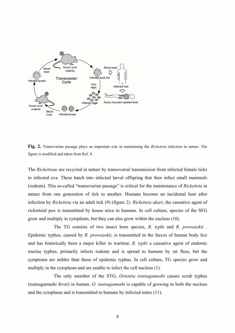
Fig. 2. Transovarian passage plays an important role in maintaining the Rickettsia infection in nature. The
figure is modified and taken from Ref. 9.
The Rickettsiae are recycled in nature by transovarial transmission from infected female ticks
to infected ova. These hatch into infected larval offspring that then infect small mammals
(rodents). This so-called “transovarian passage” is critical for the maintenance of Rickettsia in
nature from one generation of tick to another. Humans become an incidental host after
infection by Rickettsia via an adult tick (9) (figure 2). Rickettsia akari, the causative agent of
rickettsial pox is transmitted by house mice to humans. In cell culture, species of the SFG
grow and multiply in cytoplasm, but they can also grow within the nucleus (10).
The TG consists of two insect born species, R. typhi and R. prowazekii .
Epidemic typhus, caused by R. prowazekii, is transmitted in the faeces of human body lice
and has historically been a major killer in wartime. R. typhi a causative agent of endemic
murine typhus, primarily infects rodents and is spread to humans by rat flees, but the
symptoms are milder than those of epidemic typhus. In cell culture, TG species grow and
multiply in the cytoplasm and are unable to infect the cell nucleus (1).
The only member of the STG, Orientia tsutsugamushi causes scrub typhus
(tsutsugamushi fever) in human. O. tsutsugamushi is capable of growing in both the nucleus
and the cytoplasm and is transmitted to humans by infected mites (11).
9
The size and G + C composition of Rickettsia genomes
The G+C content of bacterial genomes vary from approximately 25% to 75% (15), with
tRNA and rRNA genes being highly conserved in base composition features (4). In protein
coding genes the G+C content values vary for different sites of the codon. The variation in
G+C content at the first codon position is about 50% in known bacterial genomes. This value
is decreased to 10% at the second codon position. However, nucleotide composition at the
third codon position can vary over a 10-fold range from 10% in Mycoplasma to 92 % in
Streptomyces (12,13). This is explained by the redundancy of genetic code, i.e. most of the
changes in base composition at the third codon position result in silent substitutions (14).
In general, obligate intracellular bacteria have small genome sizes. The genomes of
known Rickettsia species vary in size from 1.1 Mb in R. prowazekii to 1.66 Mb in R. bellii. In
the TG Rickettsia, G+C composition is about 29%. However, the SFG Rickettsia has a
slightly higher G+C content value of 32-33% (16-19). The G+C content of the intergenic
regions is estimated to be lower than the total genomic G+C content value in Rickettsia. As an
example, in eight investigated intergenic regions, the GC content is about 21-23 % and 30-
31% for the TG and the SFG respectively (paper IV).

10
Evolution of novel transport systems in Rickettsia
During the course of evolution towards an obligate intracellular life style, the bacteria appear
to have abandoned much of the genetic material that was previously essential for their
survival, such as a large number of biosynthetic genes. This has forced obligate intracellular
bacteria to establish efficient transport systems in order to obtain essential metabolites and
coenzymes from the host cell cytoplasm. Novel genes, such as those involved in protein
transport, can be obtained by horizontal gene transfer or by gene duplication (20). Through
horizontal gene transfer, genetic information is transferred from a donor organism to a
distantly related recipient cell. This process is more common in free-living bacteria than in
obligate intracellular bacteria (21). Another source of evolution of novel gene functions is
gene duplication followed by sequence divergence. If we assume that the probability of these
events are very low in obligate intracellular bacteria, we would expect that both of these
evolutionary events have taken place early in the evolutionary history of Rickettsia, i.e. prior
or at the time of its entrance to the intracellular habitat.
Comparative analyses of the distantly related obligate intracellular parasites may
provide valuable information about the characteristics of these bacteria that have evolved as a
consequence of their life style. Indeed, the comparative analyses of the two genomes of R.
prowazekii and Chlamydia trachomatis revealed a few striking examples of reductive
convergent evolution (22). In both genomes the genes coding for enzymes involved in the
biosynthesis of purines or pyrmidines are absent (23,26). However, intracellular specialists
such as Rickettsia and Chlamydia have found a way to cover this loss by gaining efficient
membrane transport systems. For example, the genome sequence of Chlamydia trachomatis
revealed the existence of 13 ABC transporters primarily associated with amino acid and
oligopeptide transport. Furthermore, the identification of permeases in the Chlamydia genome
makes this intracellular parasite able to transfer magnesium, phosphate, nitrate, and sulphate
from the host cell (23).
In the next section, I will discuss the evolution of ATP/ADP translocases as an
example of a novel and specific transport system in Rickettsia.
Duplication of ATP/ADP translocases in Rickettsia
The identification of genes coding for ATP/ADP translocases in the genomes of Rickettsia

11
and Chlamydia indicates the presence of a unique transport system in these bacteria (23-27).
This transport system is also found in organelles and in the eukaryotic parasite,
Encephalitozoon cuniculi but not in any other bacterial genomes known to date (28-41). In
mitochondria, ATP is generated by oxidative phosphorylation and secreted into the cytoplasm
by the aid of ATP/ADP translocases. Unlike the mitochondrial ATP/ADP translocases, these
proteins are responsible for uptake of ATP from the host cell cytoplasm in the intracellular
parasites Rickettsia and Chlamydia. ATP/ADP translocase proteins are 500 amino acid long
monomers and have similar topology in Rickettsia, Chlamydia, and plastids (25,27).
However, mitochondrial ATP/ADP translocase has six transmembrane domains and exhibits
an internal sequence triplication of 100 amino acids containing two membrane-spanning
α−helices (30-34).
There are five ATP/ADP translocases in the typhus group as well as in the two
investigated members of the spotted fever group, R. rickettsii and R. montana. It has been
shown experimentally that one of the translocases (tlc1) in R. prowazekii catalyze the
transport of ADP and ATP (24). In addition, studies of transcription regulation in R.
prowazekii reveals that the amount of the transcript is correlated with the concentration of
ATP in the host cells cytoplasm. That is, the level of expression decreases in cells that are
heavily infected with the R. prowazekii. At the same time, expression of genes encoding
proteins involved in ATP-generating metabolic pathways are upregulated when the
concentration of ATP is low in the host cell cytoplasm. This means that Rickettsia exploit the
host cells ATP during an early phase of infection but are capable of producing their own ATP
during the late phase of infection (42 ).
It has been previously shown that the five paralogous tlc genes in R. prowazekii
are expressed in cell culture systems (42). In addition, sequence data based on these genes
from the R. typhi, R. montana and R. ricketsii shows that they do not contain any stop codons
or frame shifts, i.e. they are most likely also expressed in these species (paper III). So far it’s
not known whether all five tlc genes in Rickettsia encode proteins with identical functions or
whether they code for proteins with somewhat different functions.
The discovery of five duplicated, conserved genes in Rickettsia seems not to be
a characteristic feature of resident genomes. Since obligate intracellular parasites such as
Rickettsia tend to lose genetic information during the course of evolution gene loss rather than
gene duplication is expected. However, a relaxed degree of reductive evolution would be
obtained if there were a strong selection for the function of the duplicated gene. Thus, the
duplication of the tlc genes in Rickettsia is most likely explained by their important role in
maintaining an efficient uptake and transport system of host cytoplasmic ATP.

12
The ancient origin of ATP/ADP transporters in Rickettsia
Sequence homology and phylogenetic analyses indicate an independent origin of the
rickettsial ATP/ADP translocases and the mitochondrial ATP/ADP translocase (paper III). It
has been suggested that mitochondrial ATP/ADP translocases have evolved by vertical
transmission from an ancestral gene with a broader transport function (43 ).
In contrast, the sporadic distribution of ADP/ATP translocases in
phylogenetically distant genomes may be explained by recent acquisition of these genes by
horizontal gene transfer events. Indeed, Wolf, Aravind and Koonin suggest that two
horizontal gene transfer events explain the acqusition of these genes in Chlamydia and
Rickettsia (44). According to this suggestion, the tlc genes were first transferred to Chlamydia
from plants and then by a second horizontal gene transfer event they were transferred from
Chlamydia to Rickettsia (44).
However, an amino acid sequence comparison between the chlamydial and
rickettsial ATP/ADP transporters did not reveal any strong sequence homology. There are
only 19 amino acids that are uniquely shared between Rickettsia and Chlamydia. This is much
less than the number of shared amino acids between Chlamydia and plastids translocases (148
amino acids). In addition, a phylogenetic reconstruction based on tlc amino acid sequences
indicates that the rickettsial ATP/ADP translocases do not cluster with the plastid and the
Chlamydia ATP/ADP translocases. Thus, they seem to have diverged prior to the divergence
of ATP/ADP translocases in plastids and Chlamydia. Furthermore, superimposition of the two
phylogenetic trees belonging to mitochondria and plastid/parasite translocases reveals that the
origin of plastid/parasite translocases is as deep as for the mitochondrial translocases (paper
III).
We speculate that, similar to the mitochondrial ATP/ADP translocases,
duplication and divergence of membrane proteins with other functions may have resulted in
the plastid/parasite type of ATP/ADP translocases. Indeed, we have found a possibly similar
hypotetical protein in the ψ-proteobacterium Xylella fastidiosa. This protein is 441 amino
acids long and is predicted to have 9 transmembrane helices. Amino acid comparisons of the
known ADP/ATP transporters with the hypotetical protein from Xylella fastidiosa reveal
identical amino acids at three conserved positions.
Taken together, our sequence and phylogenetic analyses are not consistent with
a recent acquisition of the ATP/ADP translocases in Rickettsia by horizontal gene transfer.
These transporter proteins seem to be of ancient origin, and may have originated by
duplication and divergence of other membrane transport proteins.

13
ATP/ADP translocases provide insights into the evolution of mitochondria
Sequence comparisons of mitochondrial genomes with the relevant bacterial genome
sequences together with phylogenetic reconstruction of related proteins have provided
overwhelming support for an endosymbiotic, eubacterial origin of mitochondria (45-50). The
question no longer is whether it occurred, but why, by what process (es), and what was the
characteristics feature of the endosymbiont?
All standard models of the endosymbiotic theory rely on the original role of the
mitochondria as the ATP-generating organelles of eukaryotes. According to these models, the
most likely reason for the symbiosis leading to the mitochondrial lineages was the ability of
the symbiont (free-living α−proteobacteria) to provide the anaerobic host with aerobically
generated ATP. The discovery of a non-bacterial origin of mitochondrial ATP/ADP
translocases has challenged this view (51-53). We now know that ATP/ADP translocaes are
only found in the two obligate intracellular parasites Chlamydia and Rickettsia, since free-
living bacteria are not capable of obtaining ATP from the environment (52-54). These data
strongly suggest that, unlike the modern mitochondria, the free-living ancestor of
mitochondria was unable to export ATP or import proteins. The hypothesis that the ATP
generation was not the driving force for the origin of the mitochondria may sound a little
astonishing to us since we are used to see the mitochondria as the powerhouse of the
eukaryotic cells.
However, another functional role of the mitochondria is to reduce the
intracellular oxygen to water through oxidative phosphorylation, a process accomplished of
five membrane bound protein complexes including cytochrome oxidase and cytochrom b.
These proteins form a monophyletic group with the corresponding proteins in R. prowazekii
(55). This suggests that the α−proteobacterial ancestor of mitochondria had already acquired
an aerobic respiratory chain at the time of symbioses.
According to the so-called “Ox-Tox” hypothesis, the driving force for
endosymbiosis was to reduce the concentration of ancient oxygen and thereby protect the
anaerobic host from its toxic effects (53,56). Roughly two billion years ago, when the
concentration of atmospheric oxygen began to rise dramatically, the need for an
endosymbiotic relationship for the anaerobic host with an aerobic symbiont was essential
(57). Even today, modern organisms have enzymes such as peroxidases, catalyses, and
superoxide dismutases that protect the cell from the toxic effect of oxygen respiration.
In addition, based on the Ox-Tox model, the evolution of the mitochondrion

14
from the endosymbiont required a system for mitochondrial import and export that has been
shown to be derived from nuclear, eukaryotic genes (58-60). The recruitment of nuclear
derived ATP/ADP translocases by mitochondria is one example of such a system. These
proteins are found in all mitochondria and evolved prior to divergence of the major branches
of eukaryotes. As a result of the novel acquisition of ATP/ADP translocases in the
mitochondrial proteome, the host cell had access to an efficient supply of ATP. For this
reason, ATP/ADP translocases may serve as a hallmark for the transformation of the
endosymbiont into an organelle (59).

15
Genome rearrangements in Rickettsia
Genome comparison between closely related species have revealed that gene rearrangements
and genome downsizing appear to have resulted from extensive recombination events
between dispersed repetitive sequences. Depending on the orientation of the repetitive
sequences, the outcome of homologous intrachromosomal recombination can be either a
deletion or an inversion of one or two repetitive sequences and the corresponding intervening
sequence (60-62). Comparison of the two genome sequences of Mycobacterium tuberculosis
(4.41 Mb) and Mycobacterium leprae (3.27 Mb) has shown that the smaller genome size of
M. leprae is the result of large-scale rearrangements and deletions arising from homologous
recombination events between related repetitive sequences. These repeptitive sequences
include transfer RNA genes and copies of the three major repeats, RLEP, REPLEP and
LEPREP (63-64).
Numerous research investigations of Rickettsia, including sequence analyses of
the R. prowazekii genome, indicate that the genomes of these intracellular parasites are highly
derived and rearranged (paper I&VIII, 65,66 ). The small genome size of Rickettsia is a result
of reductive evolution. This reductive mode of evolution has had an effect not only on the
genome size of these organisms but has also changed their genomic architecture. We have
observed several examples of a disrupted gene orders in Rickettsia (66-68). It is noteworthy
that the organization of these genes is otherwise highly conserved in a broad range of
microbial genomes (69-76). The major mechanism responsible for genomic rearrangements in
Rickettsia is homologous intra-chromosomal recombination. The outcome of intra-
chromomsomal recombination between inversely oriented repetitive sequences is an inversion
of the intervening sequences (60,61). Such an inversion has been observed in Rickettsia in the
so-called super-ribosomal protein gene operon (77-79). This operon is highly conserved in a
broad range of bacteria and archaea and consists of about 40 genes locating in seven operons
with the conserved order secE-nusG, L11, L10, str, S10, spc and α (figure 3).

16
Fig. 3. Schematic representation of the organisation of super ribosomal protein gene cluster in a variety ofbacteria. a) The expected organization of the ribosomal protein gene cluster in the common ancestor of Bacteriaand Archaea. These operons are conservatively arranged in the genome of b) Bacillus subtilis and c) Escherichiacoli, but have dispersed organisation in the genome of d) Haemophilus influenzae, e) Mycoplasma genitaliumand f) Rickettsia prowazekii.
The conserved organization of 40 genes in these seven operons is thought to
reflect the ancient origin of the super ribosomal protein gene operon (60). Despite the
conserved orientation of the genes in the ribosomal protein gene operon in many bacteria such
as E. coli and Bacillus subtilis, these genes are scattered around the genomes of Haemophilus
influenzae, Mycoplasma genitalium and R. prowazekii (73,60). A more detail study of
rickettsial genes encoding for elongation factor Tu (tuf) and G (fus) suggest that homologous
intra-chromosomal recombination is the main mechanism responsible for the unique order of
genes in these organisms (paper I). The tuf gene is found in two or multiple copies in a broad
spectrum of microbial genomes (74-76, 78). In general, one copy is positioned in the so-called
streptomycin operon (str) that comprises the two ribosomal protein genes, rpsL and rpsG,
followed by the genes encoding for elongation factors G (fus) and Tu (tufA) respectively. The
second copy of the tuf gene is located in tufB operon upstream of the two-membrane proteins
secE and nusG (70-71).
The genome of R. prowazekii is atypical in that it contains only one tuf gene.
Furthermore, the gene organization deviates from the conventional gene cluster. That is, the
single tuf gene is not a member of the str operon and the flanking genes at the 3` end of the
fus gene in the str operon are secE, nusG and rpsJKL that are normally located downstream of
the tuf genes in other species. The present disposition of elongation factor genes and the

17
corresponding flanking regions in R. prowazekii can be explained by an inversion of the
intervening sequences as a consequence of homologous intra-chromosomal recombination at
the two ancestral tuf genes and the subsequent deletion of one tuf gene (paper I).

18
Fig. 4. Schematic representation of the postulated homologous intra-chromosomal recombination eventbetween the two ancestral tuf genes that gave rise to the present gene organization and the subsequent deletion ofone tuf gene. The figure is modified and taken from Ref.

19
Ancient origin of gene rearrangements in RickettsiaTwo major genomic rearrangements in Rickettsia occurred at the elongation factor genes and
the rRNA genes. We suggest that the rearrangements at the tuf genes may have lead to a
major inversion of the Rickettsia genome that occurred early in the evolution of Rickettsia.
Indeed, our phylogenetic analyses based on the tuf, fus and the rRNA gene sequences indicate
that the rearrangement of the duplicated genes and the subsequent deletions of the redundant
tuf and rRNA genes occurred prior to speciation in Rickettsia. The finding of multiple tuf
copies with the conventional organization in two closely related α−proteobacterial species,
Rhodobacter capsulatus and Agrobacter tumefaciens, shows that this rearrangement event
occurred subsequent to divergence of Rickettsia from R. capsulatus and A. tumefaciens (paper
II).

20
Genomic streamlining in Rickettsia
Genome downsizing in bacteria is generally thought to be a consequence of a bias for
deletions. The extent to which there is a bias for deletions in bacterial genomes has been
examined for a broad range of bacteria (80). It has been proposed that this phenomenon plays
a significant role in balancing genome size expansions in free-living bacteria and also in
reducing the genome size of obligate intracellular bacteria. In other words, the compactness of
bacterial genomes is a consequence of an ongoing deletional bias. This bias may result in the
elimination of large segments of DNA by homologous recombination (rapid gene loss) or
stepwise degradation and subsequent elimination of small segments of DNA (slow gene loss).
The bias for deletions has been studied extensively in a variety of Rickettsia species and is the
major point of the following sections in this thesis.
Rapid and extensive gene loss
It has been suggested that small obligate intracellular bacteria such as Rickettsia (1.1 Mb),
Mycoplasma (0.58 Mb), Coxiella (1 Mb) and Chlamydia (1.0 Mb) have evolved from a free-
living ancestor that had much larger genome size (1). During the course of evolution these
obligate intracellular bacteria have lost a significant fraction of their genomes. As stated
previously, large-scale deletions of genes and DNA sequences (rapid gene loss) can arise
from intra-chromosomal recombination at directly oriented repetitive sequences.
The first victims of rapid gene extinction are those genes that are present in
more than one copy in bacterial genomes, since the deletion of one or more copies will most
likely not be lethal for the bacterial organism. Examples of such genes are those encoding
rRNA genes and elongation factor proteins. In bacteria with an obligate intracellular lifestyle,
such as Rickettsia, Chlamydia and Mycoplasma, Buchnera, these genes are found as single
copy (52,53,54). Since duplicated genes such as tuf- and rRNA genes can serve as repetitive
sequences, the deletion of one or multiple copies of these genes from the genome of
intracellular bacteria may occur by intra-chromosomal recombination (61). Due to the
consumption of repetitive sequences with each recombination event we expect that the rate of
homologous recombination will decline during the course of evolution of obligate
intracellular bacteria. Accordingly, we predict that the majority of homologous
recombination-mediated deletion events in obligate intracellular bacteria occurred in the early

21
phase of the intracellular lifestyle.
Sequence comparison of the identified open reading frames (ORFs) located in
the intergenic regions of different Rickettsia species has provided several examples of
homologous recombination as the responsible mechanism for gene deterioration. For
example, we found two pairs of short internal repeat elements in the putative ORF locating in
the intergenic region of the queA gene, encoding S-adenosylmethionine tRNA and the gene
encoding an abc transporter protein (abcT3). This ORF is absent from the TG Rickettsia and
is inactivated and degraded within the SFG. We found that the size of this inactivated ORF
varies between different members of the SFG. We suggest that size differences in the SFG are
a result of independent homologous recombination between the internal repeat elements in
this region (paper IV). The degree and positions of deletions caused by intra-chromosomal
recombination in Rickettsia is different among the species, which suggests that the
homologous recombination is an ongoing process that may result in an ongoing loss of genes
under weak or no selection pressure.
Stepwise degradation and loss of genetic information in Rickettsia
The patterns and rates of mutations in resident genomes such as obligate intracellular
parasites, endosymbionts and cellular organelles, is different from that of free-living bacteria.
Small population size, lack of recombination, and bottlenecks during the transmission to the
next host are characteristic features of resident genomes such as Rickettsia (81). As a result,
the fixation rate of the slightly deleterious mutations is higher in obligate intracellular
parasites compared to that of free-living bacteria (82). The tendency to accumulate deleterious
mutations in asexual populations living on the inside of a host cell is thought to be an effect of
Mullers ratchet. Mullers ratchet may result in the gradual accumulation of harmful mutations,
which in the absence of compensatory back mutations may lead to the extinction of the
obligate intracellular parasites (83). It has been shown that Mullers ratchet is acting on the
resident genomes such as those of Buchnera and Rickettsia, and it has been suggested that this
phenomenon will generate smaller and smaller genomes (84-88).
We have already discussed that genomic streamlining may be caused by intra-
chromosomal recombination at duplicated sequences resulting in the deletion of one or
multiple copies of the duplicated sequence. Alternatively, the loss of genetic information may
be accomplished by a gradual accumulation of small deleterious mutations. Due to the higher
fixation rate of slightly harmful mutations in obligate intracellular bacteria, such as Buchnera

22
and Rickettsia, compared to free-living bacteria (89), we would expect an overall increase in
the number of inactivated genes in these obligate intracellular bacteria. Detailed sequence
comparison analyses of the pseudogenes and long intergenic regions belonging to the TG and
the SFG provide numerous examples of gene shrinkage and loss in these intracellular
parasites (paper IV&61). The genomic shrinkage can be triggered by accumulation of random
mutations in DNA sequences that may not be any longer under purifying selection..
Indeed, it has been shown that the gene encoding for AdoMet synthetase is not
active in the Madrid E strain of R. prowazekii since it has an internal termination codon (90).
However, this gene is highly conserved among a wide range of bacteria as well as eukaryotes
(65). The AdoMet synthetase is responsible for synthesis of AdoMet from methionine and
ATP. The AdoMet is a source of methyl-groups that are essential for methylation of DNA,
RNA, protein side-chain modifications, biosynthesis of amines, creatine, and carnitine (65).
For these reasons, it is thought that the enzyme AdoMet synthetase is essential for all living
organisms. Comparative analyses of several Rickettsia species have revealed that the AdoMet
synthetase gene is intact in R. typhi and the Madrid B strain of R. prowazekii but contain stop
codons and frame shift mutations (deletions/insertions) in Rickettsia species belonging to the
SFG. The inactivation and degradation of essential genes such as AdoMet synthetase in the
obligate intracellular parasites Rickettsia and Chlamydia may be compensated by
establishment of a novel transport system by which the parasite can get access to essential
compounds (90,91). Preliminary data suggests that such a transport system is present in
Rickettsia (H. Winkler, personal communication).
Comparative analysis of long intergenic regions from several different species
of Rickettsia provides additional support for an ongoing degradation and elimination of genes
in Rickettsia. Examples of such genes are gabD, ampG4 and fic. The gabD gene encodes
succinate semialdehyde dehydrogenase oxidoreductase (SSDH) (paper IV). This gene
catalyzes the formation of succinate from succinate semialdehyde by reducing
NAD+/NAD(P)+ to NADH/NADPH. Succinate is one of the compounds participating in
citric acid cycle. The enzyme SSDH is necessary for glutamate, tyrosin and butanoate
metabolism (92). While this gene is intact in the SFG it is highly degraded in the TG
Rickettsia (only traces of the gene at 3´ end are recognizable.) (paperIV).
The genes coding for AmpG are found in four copies in R.conorii and in three
copies in R. prowazekii (93). These genes may make their carriers more resistant to some
antibiotics such as β-lactam (93). The ampG4 gene has been closely studied in five Rickettsia
species, belonging to the TG and the SFG. The ampG4 gene was found to be intact in three

23
members of the SFG, namely in R. conorii, R. rickettsii and R. montana while they are
reduced in length in the TG. Furthermore, the gene contains many frameshifts and termination
codons in members of the TG. The degradation of the ampG gene in the TG Rickettsia is
consistent with the higher resistance to antibiotics observed in the SFG as compared to the
TG. Finally, the gene encoding philamentation protein (fic) is intact in the investigated SFG
Rickettsia but present as pseudogene in the TG Rickettsia. This gene contains deletions of 279
and 573 basepairs as well as 13 and 2 termination codons in R. prowazekii and R. typhi,
respectively (paper IV). Apparently, these genes, gabD, ampG4 and fic are still functional in
the SFG, however, but accumulate mutations in a neutral manner in the TG Rickettsia.
Deletion profile in Rickettsia
Comparative analyses of long intergenic regions belonging to different Rickettsia species
confirms that the unexpectedly high fraction of non-coding DNA (24%) in R. prowazekii
represent remnants of inactivated and degraded genes (paper IV, 94). As was mentioned
earlier, the degradation and subsequent deterioration of genes is a result of a bias for deletion
mutations in Rickettsia, as observed by comparative analysis of the deletional profile of
pseudogenes and highly fragmented genes. Reconstruction of the putative ancestral sequences
for a large number of pseudogenes belonging to the TG and SFG Rickettsia, indicates that the
number of deletions are considerably higher than the number of insertion mutations (94, paper
IV). The size heterogeneity of pseudogenes between different species is a consequence of a
mutation bias for deletions in these regions (paper IV).

24
The evolution of selfish DNA in Rickettsia
The term selfish DNA refers to repeat sequences that are capable of self-replication. Selfish
genetic elements comprise a large diverge group, and due to their self-propagating feature
they are found in most organisms (95). For example, a large fraction of eukaryotic genomes
(up to 50%) consists of selfish DNA, such as retrotransible elements and transposons (96).
The functional role of these self-replicating elements is still a matter of debate. There is now
overwhelming evidence for a role in the regulation of gene expression. They are also
responsible for gene or exon shuffling and duplications (97-99). Selfish DNA can also play a
significant role in increasing the fitness of their host. For instance, the insertion of
transposable element (P-element) in the third intron of the methuselah (mth) gene resulted in
an extension (35%) of the life span of mutant flies (containing the inserted P-elements)
compared to non-mutant flies (without no P-element insertion). The mutant line also showed
increased resistance to starvation, high temperature and other stress factors. The accumulation
of selfish DNA in the host genome may also have deleterious effects on the fitness of the host
by inducing mutations in the genes (genetic diseases) (100). In this case, selection favours the
survival of the propagating selfish DNA rather than the host’s biology. How these selfish
genetic elements originated and what was their role in the evolution of their host organisms
has not been resolved.
Recently, a novel type of palindromic repetitive elements in the genome of
Rickettsia has been identified (101). Due to the sporadic distribution of these repetitive
sequences in Rickettsia genome, including insertion into the protein-coding genes, they have
been classified as selfish DNA. However the selfish feature of these repeat elements has not
been determined experimentally. In the following section, I am going to bring up questions
concerning how early they have been acquired in Rickettsia, and what can be their possible
destiny in the obligate parasite genome of Rickettsia.
Rickettsia palindromic elements (RPEs) - Long time residentsRPEs varies in length from 106-150 bp in Rickettsia and have palindromic structure (101).
RPE-like repetitive elements with palindromic structure are also found in enterobacteria
(102-104). However, unlike enterobacterial selfish DNA that is found in the non-coding
regions, these elements are present in both genes and non-coding sequences. The abundance
of palindromic selfish elements in protein coding genes has raised questions concerning their

25
origin and possible function in Rickettsia. There are 44 RPEs in R. conorii. Nineteen of these
selfish elements are located in genes coding for a variety of proteins. Comparative analyses of
the two Rickettsia genomes, R. conorii and R. prowazekii, has shown that RPEs are targeted
to different genes in different species of Rickettsia (101).
The different distribution of RPEs in various Rickettsia species can be explained
in two possible ways. First, the variation in the distribution pattern may be explained by
recent proliferation of RPEs in the Rickettsia genomes. If RPEs indeed represents new
residents of the Rickettsia genomes, we would expect to find a considerable homology
between their sequences within the same species. In other words, RPEs from the same species
would cluster together with high bootstrap support and be separated by short branches in the
phylogentic tree. However, phylogenetic reconstructions based on the nineteen RPEs in R.
conorii indicate that these repeat elements are very diverged and do not cluster together
(paper II). An alternative interpretation is that the RPEs were introduced into the Rickettsia
genomes early in evolution and then deteriorated during the course of evolution. If so we
would expect that RPE orthologs (RPE from different species) are more closely related than
RPE paralogs (RPE within the same species). Indeed, sequence and phylogenetic analyses of
RPEs located in the non-coding regions downstream of the genes encoding the elongation
factors Tu and G (RPE-tuf and RPE-fus respectively) clearly indicates that these RPEs are
long time residents of the Rickettsia genomes. In addition, the finding that the insertion sites
of RPE-tuf and RPE-fus are identical in the different species suggest that their entrance into
the Rickettsia genomes occurred some times prior to speciation of Rickettsia (paper II).
RPEs – Victims of reductive genome evolution
The independent loss of RPEs in some Rickettsia species is suggested to be responsible for
species-specific differences in the host proteins targeted by RPEs (paper II). Indeed, it seems
not unrealistic to believe that these selfish elements are temporary residents of the Rickettsia
genome and that their elimination is caused by reductive genome evolution. The decay of
RPEs in Rickettsia may be accomplished by homologous recombination between inversely
oriented repeated sequences. One example of such a process has been identified in several
Rickettsia species in which the RPE is located between two short repeated sequences.
Alternatively, the RPEs may be degraded from the genome gradually by accumulating small
deletions. This type of degradation is expected to happen more frequently than rapid loss,
especially during the later stages of reductive evolution.

26
Indeed, the degradation of RPE-fus is an example of an RPE in different stages
of degradation in different species of Rickettsia. This RPE is intact in some species of the
SFG, it has accumulated some deletions mutations and termination codons in the TG and is
hardly recognizable in R. bellii. Furthermore, we have found that deletions dominate over
insertions in RPE-fus. This is consistent with the indel spectrum previously obseved in
pseudogenes and other neutrally evolving genes in Rickettsia. In general there is a tendency
for a higher deterioration rate in the TG compared to the SFG (paper II). This tendency has
been seen in many sequences that are under weak selection pressure (paper IV,V). For this
reason there is a considerably lower frequency of RPEs present in the R. prowazekii genome
compared to that of R. conorii. Almost all of the few RPEs still present in the R. prowazekii
genome are located inside ORFs. The RPEs that are incorporated inside the three genes, kdtA,
polA and RP545, have been shown to have higher substitution rates than their flanking coding
sequences (73). This provides additional support for the view that there is an ongoing
degradation of RPEs in the Rickettsia genomes. Thus, it seems unlikely that the RPEs have
proliferated recently in obligate intracellular parasites such as R. conorii that tend to eliminate
the large fractions of their genomes during the course of evolution.
Are RPEs long-term hitchhikers?
The abundance of RPEs in different protein coding genes together with the conserved
palindromic nature of these selfish elements is consistent with a possible functional role for
the proteins in which they reside. It has been found that most of the RPEs presented in R.
conorii are able to form stable hairpin-like mRNA secondary structures. However, the
palindromic feature of RPEs belonging to R. prowazekii is less clear and only two out of ten
are predicted to form hairpin structures (74). Indeed, the finding of unstable hairpin-like
mRNA secondary structure for the majority of RPEs located in protein-coding genes suggest
that this selfish DNA may have lost its function in most of the RPEs in R. prowazekii. This is
also supported by a high evolutionary degradation rate in R. prowazekii. This means that the
lack of purifying selection on RPEs has resulted in the extinction of most RPEs from the
genome of R. prowazekii. However, they may still be under weak selection in the R. conorii
genome, especially if inserted into protein coding genes. However, based on hydropathy
analyses, they are predicted to be located at the surface of the various proteins (74). Due to
their position on the surface of these proteins, the loss of these elements may not be critical
for their hosts and may not affect the function of their carrier protein. Thus, these selfish

27
elements may possibly have had some functional role early in the history of Rickettsia but
their role is currently thought to be insignificant and they seem to be in a process of rapid
deterioration.

28
The influence of deletions on genome size in bacteria
In eukaryotes, genome size can vary from about 10 7 to 10 11 basepairs (80). This broad
variation in genome size is not related to the eukaryotic organisms complexity (the C-value
paradox) (105). For instance, Arabidopsis thaliana, with the smallest genome in the plant
kingdom known todate, has a 3000 fold smaller genome than the far simpler plant Psilotum
nudum (sometimes called the "whisk fern"). It is also worth noting that the large fraction
(80%) of the P. nudums genome consists of repetitive DNA. Other examples are the genomes
of amphibians that contain 30 times as much DNA as human genome (106). The question is,
if the enormous variation in genome size is not linear with organismal complexity, what is the
explanation for this paradox? Dmitri A. Petrov and his coworkers have suggested that the
answer to this long-standing mystery is that the rate at which junk DNA is eliminated from a
genome is different among different organisms. The genome size of the Hawaiian crickets
(Laupala) is about the 11-fold larger than that of Drosophila. It has been shown that the
spontaneous deletions rate of the non-essential DNA is more than 40 times slower in Laupala
than in Drosophila. The high deletion rate of unconstrained sequences such as dead-on-arrival
(DOA) copies of non-LTR retrotransposable elements in Drosophila is suggested to be
congruent with the general feature of mutation in this organism (105).
The C-value paradox may not solely be determined by differences in the
insertion and deletion mutational spectra. For example, C. elegans has a very compact
genome, smaller than that of D. melanogaster, although it has a lower rate of DNA loss
(105). It is clear that the huge difference in genome size among organisms is not related to
their complexity but the size of the non-coding region may be correlated to the organisms
complexity. The most striking example is the human genome with a non-coding fraction that
comprises ca 98% of the genome (106).
However, in bacteria, genome sizes do not vary as much as in eukaryotes.
Known bacterial genome sizes vary over a 20-fold range from 580 kb in Mycoplasma
genitalium to more than 10 Mb in several cynobacterial and myxobacterial species (108). In
contrast to eukaryotic genomes, a large fraction of the bacterial genome consists of genic
DNA and non-coding DNA comprises only 10% of the bacterial genomes. For this reason the
size differences in bacterial genomes is often consistent with organismal complexity. The
balance of two major forces, horizontal gene transfer and deletional bias can explain most of
the genome size diversity among bacteria. The widespread impact of lateral gene transfer on
29
bacterial evolution should not be underestimated. Horizontal gene transfer occurs more
frequently in free-living bacteria compared to bacteria in intracellular habitats. Lateral gene
transfer results in extremely dynamic genomes and allow bacteria to adapt rapidly to changing
environments by providing recipient organisms with new metabolic traits (for instance the
acquisition of the lac-operon in E.coli), antibiotic resistance genes and genes that contribute to
virulence such as pathogenicity islands (108). In addition to its significant role for creation of
dynamic genomes, lateral gene transfer may contribute to expansion of genome size in
bacteria. However, if lateral gene transfer were the only process affecting bacterial genomes,
we would expect an ongoing expansion of genome size.
On the contrary, bacterial genomes are compact and the influence of horizontal
gene transfer seems not to contribute to the expansion of genome size in bacteria. The
compactness of bacterial genomes may be explained by reductive evolutionary forces that
induce gene loss, which counteracts the influx of foreign DNA by horizontal gene transfer.
The molecular details of reductive evolution are best understood by comparative analysis of
known pseudogenes with their functional counterparts within the same taxa or closely related
taxa. Since pseudogenes are neutral sequences, the profiles of deleterious mutations reflect the
spontanous spectrum of mutations. As I discussed earlier there is a bias for deletions over
insertions in obligate intracellular bacteria such as Rickettsia and Buchnera. Deletional bias
also exists in a broad range of other bacteria such as Neissera meningitidis, Helicobacter
pylori, Sulfolobus solfataricus, Rhodobacter spharoides (80). The effect of the deletional bias
on genome size may be more visible in bacteria with obligate intracellular lifestyle where
lateral gene transfer does not occur frequently and since the mutational degradation of genes
as a consequence of this bias is normally counterbalanced by selection on gene function.
Taken together, deletional bias contributes to maintaining constant genome size in free-living
bacteria, but result in small genome size in obligate intracellular bacteria.
Deletion frequencies may depend on the density of repetitive elements
Studies on the processes of reductive genome evolution in obligate intracellular parasites
have revealed the important role of repetitive sequences in affecting genome size and
structure. The highly rearranged genome of R. prowazekii indicates that these organisms have
undergone a large number of genomic rearrangements as well as extensive gene loss. Genome
shrinkage in Rickettsia has been shown to be an ongoing process that results in loss of genetic
material over time. As was mentioned earlier, the loss of large segments of genetic
30
information can occur rapidly by homologous recombination at directly oriented repetitive
sequences. Since repetitive sequences are major substrates for homologous recombination, the
variation in frequency of repetitive sequences in bacterial genomes and also in closely related
species may contribute to different sizes and structures. Thus, we expect that genomes with a
high number of repetitive sequences undergo homologous recombination more frequently,
which in turn may result in a rapid deletion of DNA and a fast reduction of genome size over
time.
A global survey of microbial genomes has shown that free-living bacteria with
large genomes have a high content of repeated sequences and self-propagating DNA such as
transposons and bacteriophages. In contrast, obligate intracellular bacteria tend to have small
genomes with a low content of repeated sequences and no or few genetic parasites (paper VI).
The frequency of the repeated sequences and genome size in microbial genomes may be
determined by the lifestyle of the organism. With each homologous recombination (between
directly oriented repeats) one or two repeat sequences will be lost. In free-living bacteria, the
loss of repeated sequences by homologous recombination can be compensated by the import
of novel repeat sequences from other bacteria by horizontal gene transfer or intra-specific
recombination. Therefore, repeated sequences may have small effects on genome size in free-
living bacteria. In contrast, Mullers ratchet will cause an irreversible loss of DNA including
repeated sequences in obligate intracellular bacteria. Thus, the loss of repetitive sequences in
isolated bacteria can not be compensated by a corresponding influx from other species and
will therefore result in a small genome size.
The consumption of repeated sequences during the course of evolution may
affect the rate of homologous recombination in resident genomes. We predict that the
frequency of homologous recombination has declined during the evolution in obligate
intracellular bacteria (paper VI). This in turn may affect the rate of genomic deletion. The
latter will decrease over time under the assumption that the rate of fixation of slightly
deleterious mutations is constant or less than zero. Genome size variation and differences in
repeat content between free-living bacteria and obligate intracellular bacteria are the
consequence of the lifestyle of these organisms. However, the difference in genome size and
repeat content in obligate intracellular bacteria with identical lifestyles may be explained by a
variety of factors, such as the number of hosts they infect, the extent of bottlenecks, the time
since species divergence and the rate of evolution. Indeed, a comparative analysis of R.
prowazekii and R. conorii as well as a separate study of eight non-coding regions in 5
different Rickettsia species (paperIV) suggests that reductive evolution has been influenced
the TG Rickettsia to the larger extent than the SFG.

31
Concluding remarks
I have discussed the evolution of Rickettsia and the influence of obligate intracellular
lifestyle on shaping their genomic architectures. Different evolutionary processes such as
genome rearrangements, loss of genetic information and gene duplications have the been
major focus of this thesis:
• Genome rearrangements
Rickettsia genomes are highly rearranged. Large genomic rearrangement is suggested to be
the resulted of intra-chromosomal recombination at duplicated sequences. For example, we
suggest that the unique organisation of the super ribosomal protein gene operon in Rickettsia
is a consequence of intra-chromosomal recombination at the inversely oriented ancestral tuf
genes followed by deletion of one tuf gene. Phylogenetic reconstructions based on tuf and fus
genes from different species of Rickettsia and their closely related α-proteobacteria suggest
that the rearrangement occurred at an early stage of the transition to an obligate intracellular
life style, but prior to speciation within the genus Rickettsia.
• Reductive genome evolution
Reductive evolution is the major evolution process that has influenced the genomes of the
Rickettsia. Reduction in genome size will primarily occur through by homologous
recombination between directly oriented repetitive sequences. This will result in the deletion
of the intervening DNA sequences. We suggest that a major loss of genetic information
occurred via homologous recombination at repeated sequences (tuf genes, rRNA genes, and
repetitive elements). The rate of homologous recombination has declined during the evolution
due to consumption of repeated sequences during the recombination events. However, the
elimination of genetic material will continue even after the consumption of repeated
sequences, albeit with a slower rate by the accumulation of short deletion mutations. This in
turn will give rise to the formation and temporary accumulation of inactive genes and junk
DNA. The produced pseudogenes will serve as new victims of sequence degradation.
Therefore, like any other neutrally evolving genes that are not under selective constraints,
they are expected to slowly deteriorate from the genome. Gene loss in bacteria seems to be a
consequence of a general bias for deletions. This process plays an important role in free-living

32
bacteria by balancing the influx of genetic information via horizontal gene transfer. In
addition, the bias for deletions has resulted in small genomes in obligate intracellular bacteria
such as Rickettsia. Comparative analyses of long intergenic regions that belong to the TG and
SFG Rickettsia indicate that many genes that contain termination codons or frameshifts or that
are completely deleted from one species may remain active in another species. Sequence and
phylogenetic analyses suggest that deletion mutations occur more frequently than the
insertion mutations in Rickettsia.
• The invention of novel transport system
The loss of essential biosynthetic genes during the transition to obligate intracellular life
style has triggered the intracellular specialists to establish or develop novel transport system.
One example of such a transport system is the ATP/ADP translocases that are only found in
obligate intracellular parasites and organelles. We have found no support for the hypothesis
that these transport systems have been transmitted among obligate intracellular parasites by
recent horizontal gene transfer. On the contrary, these transport proteins seem to have an
ancient origin and was acquired in Rickettsia long before speciation, possibly already during
the establishment of the endosymbiont that later gave rise to the mitochondrion. We have
suggested that the ATP/ADP transporters in Rickettsia have evolved by duplication and
subsequent divergence from other transmembrane proteins long before the divergence of TG
from SFG Rickettsia.
Future prospects
Comparative sequence analysis of a variety of Rickettsia species has provided a general
overview of the different evolutionary processes that shape the genome of this obligate
intracellular parasite. We have observed that reductive evolution is the major evolutionary
force operating on these genomes, which has resulted in the inactivation and gradual
elimination of genes and repetitive elements such as RPEs from Rickettsia. Comparison of the
genomes of R. prowazekii (1.1 Mb) and R. conorii (1.3 Mb) indicate that the R. conorii
genome contain a total number of 552 genes that are not present in R. prowazekii. A detailed
comparative analysis indicates that as many as 200 of the unique genes in R. conorii
correspond to inactivated gene fragments in R. prowazekii and that another 200 genes have
33
been completely eliminated from the R. prowazekii genome.
A comparison of active genes with their slightly degraded orthologs in R.
prowazekii and R. conorii indicates that some of these degraded genes, also called split genes,
are expressed (92). However, the substitution frequencies at non-synonymous and
synonymous sites show no significant difference between the non-expressed ORFs and the
expressed ORFs from the same set of split genes (paper V). This finding suggests that neither
the expressed, nor the non-expressed split genes may be functional. In order to understand the
mechanism of gene inactivation and degradation, we need to determine whether the expressed
degraded genes are translated and if so whether the gene products are able to perform their
original function and if so, how their expression is regulated. A genome-wide analysis of the
Rickettsia genomes at the protein level will provide a clear picture of the functional roles of
the encoded gene products. It will also highlight the role of genes involved in different
metabolic pathways and tells us whether multiple functions may be encoded by some genes
and define the role of the transport machinery in gaining essential compounds that can not be
synthesized by the obligate intracellular parasite. In addition, functional genomics studies
may shed light on the extent to which Muller ratchet have altered enzyme functions and
kinetics in obligate intracellular parasites such as Rickettsia, and thereby their fait in the
eukaryotic host cell.

34
References
1. Weiss, E., and J. W. Moulder. 1984. Order I. Rickettsiales Gieszczkiewicz 1939, 25 AL .In J. H. Holt (ed.), Bergeys manual of systematic bacteriology vol. 1. The Williams andWilkins Co., Baltimore, pp. 687-704.2.Gross, L. 1996. How Charles Nicolle of the Pasteur Institute discovered that epidemictyphus is transmitted by lice: reminiscences from my years at the Pasteur Institute in Paris.Proc. Natl Acad. Sci USA 93:10539-105403. Raoult, D., Ndihokubwayo, J. B., Tissot-Dupont, H., Roux, V., Faugere, B., AbegbinniR. and Birtle, R. J. 1998. Outbreak of epidemic typhus associated with trench fever inBurundi. The Lancet. 352:353-358.4.Olsen, G. J., C. R. Woese, and R. Overbeek. 1994. The winds of (evolutionary) change:breathing new life into microbiology. J. Bacteriol. 176:1-6.5. Stothard, D. R., J. B. Clark, and P. A. Fuerst. 1994. The phylogenetic relationship ofRickettsia bellii to the spotted fever and typhus group of Rickettsia, and the antiquity of thegenus. Int. J. Syst. Bacteriol. 44:798-804.6. Stothard, D. R., and P. A. Fuerst. 1995. Evolutionary analysis of the spotted fever andtyphus groups of Rickettsia using 16S rRNA gene sequences. Syst. Appl. Bacteriol. 18:52-61.7. Raoult D, Roux V. 1997. Rickettsiases as paradigms of new or emerging infectiousdiseases. Clin Microbiol Rev. 10:694-719.8. Azad, AF., Beard CB. 1998. Rickettsial pathogens and their arthropod vectors. EmergInfect Dis 4:179-86.9. Thorner, A. R., Walker, D. H., Petri W. A. Jr. 1998 Rocky. Mountain Spotted Fever.Clin Infect Dis 6:1353-9.10. Wisseman, C. L. JR., E. A. Edlinger, A. D. Waddell, and M. R. Jones. 1976. Infectioncycle of Rickettsia rickettsii in chick embryo and L-929 cells in culture. Infect Immun.14:1052-1064.11. Tamura, A., N. Ohashi, H. Urakami, and S. Miyamura. 1995. Classification ofRickettsia tsutsugamushi in a new genus, Orientia gen. Nov., as orientia tsutsugamushi comb.Nov. int. J. Syst. Bacteriol. 45:589-591.12. Ohkubo, S., Muto, A., Kawauchi, Y., Yamao, F., Osawa, S. 1987. The ribosomalprotein gene cluster in Mycoplasma capricolum. Mol Gen Genet 210:314-322.13. Wright, F., Bibb, M. J. 1992. Codon usage in the G+C- rich streptomyces genome. Gene113:55-65.14. Kimura, M. 1983. The neutral theory of molecular evolution. Cambridge UniversityPress, Cambridge, UK.15. Muto, A., and Osawa, S. 1987. The guanine and cytosine content of genomic DNA andbacterial evolution. Proc. Natl. Acad. Sci. U.S.A. 84:166-169.16. Eremeeva, M. E., Roux, V., Raoult, D. 1993. Determination of genome size andrestriction pattern polymorphism of Rickettsia prowazekii and Rickettsia typhi by pulsed fieldgel electrophoresis. FEMS Microbiol Lett 112:105-12.17. Roux, V., Raoult, D. 1993. Genotypic identification and phylogenetic analyses of thespotted fever group rickettsiae by pulsed-field gel electrophoresis. J Bacteriol. 175:4895-904.18. Tyeryar, Jr. F. J., Weiss, E, Millar, D. B., Bozeman, F. M., Ormsbee, R. A. 1973.DNA base composition of rickettsiae. Science 180:415-7.19. Schramek, S. 1974. Deoxyribonucleic acid base composition of Rickettsiae belonging tothe Rocky Mountain spotted fever group isolated in Czechoslovakia. Acta Virol (Praha)18:173-4.

35
20. Li, W. H. and Graur, D. 1991. Fundamentals of Molecular Evolution. Sinauerassociates, INC. publishers. U.S.a.21. Andersson, S. G. E. and Kurland, C. G. 1998. Reductive evolution of resident genomes.Trends in Micro. Biol. 6:263-268.22. Zomorodipour, A. & Andersson, S. G. E. 1999. Obligate intracellular parasites:Rickettsia prowazekii and Chlamydia trachomatis. FEBS Lett. 452:11-15.23. Stephens, R. S., Kalman, S., Lammel, C., Fan, J., Marathe, R., Aravind, L., Mitchell,W., Olinger, L., Tatusov, R. L., Zhao, Q., Koonin, E. V. & Davis, R. W. 1998. Genomesequence of an obligate intracellular pathogen of humans: Chlamydia trachomatis. Science282:7854-759.24. Winkler, H. H. 1976. Rickettsial permeability: An ADP-ATP transport system. J. biolChem 251:389-396.25. Williamson, L. R., Plano, G. V., Winkler, H. H., Krause, D. C. & Wood, D. O. 1989.Nucleotide sequence of the Rickettsia prowazekii ADP/ATP translocase encoding gene. Gene80:269-278.26. Andersson, S. G. E., Zomorodipour, A., Andersson, J. O., Sicheritz-Ponten, T.,Alsmark, U. C. M., 1998 Podowski, R. M., Näslund A. K., Winkler, H. H. &Kurland, C. G. 1998. The genomesequence of Rickettsia prowazekii and the origin of mitochondria. Nature 96:133-14027. Hatch, T. P., Al-Houssaine, E. & Silvermann, J. A. 1982. Adenine nucleotide andlysine transport in Chlamydia psittaci. J Bacteriol 150:662-670.28. Katinka, M. D., Duprat, S., Cornillot, E., Metenier, G., Thomara, F. et al., 2001.Genome sequence and gene compaction of the eukaryotic parasite Encephalitozoon cuniculi.Nature 414:450-453.29. Vignais, P. V. 1976. Molecular and physiological aspects of adenine nucleotide transportin mitochondria. Biochim Biophys Acta 456:1-38.30. Klingenberg, M. 1989. Molecular aspects of adenine nucleotide carrier frommitochondria. Arch Biochem Biophys 270:1-14.31. Heldt, H. W. 1969. Adenine nucleotide translocation in spinach chloroplasts. FEBS Lett5:11-14.32. Pozueta-Romero, J., Frehner, M., Viale, A. M. & Akazawa, T. 1991. Direct transportof ADP glucose by an adenylate translocator is linked to starch biosynthesis in amyloplasts.Proc Natl Acad Sci USA 88:5769-5773.33. Neuhaus, H. E., Henrichs, G. & Scheibe, R. 1993. Characterization of glucose-6-phosphate incorporation into strach by isolated intact cauliflower-bud plastids. Plant Physiol(Rock) 101:573-578.34. Schunemann, D., Borchert, S., Flugge, U. I. & Heldt, H. W. 1993. ADP/ATPtranslocator from pea root plastids. Comparison with translocators from spinach chloroplastsand pea leaf mitochondria. Plant Physiol (Rock) 103:131-137.35. Kampfenkel, K., Möhlmann, T., Batz, O., Van Montagu, M., Inze, D. & Neuhause,H. E. 1995. Molecular characterisation of an Arabidopsis thaliana cDNA encoding a novelputative adenylate translocator of higher plants. FEBS Letters 374:351-355.36. Möhlmann, T., Tjaden, J., Schwöppe, C., Winkler, H. H., Kampfenkel, K. &Neuhaus, H. E. 1998. Occurrence of two plastidic ADP/ATP transporter in Arabidopsisthaliana. Eur J Biochem 252:353-359.37. Runswick, M. J., Powell, S. J., Nyren, P. & Walker, J.E. 1987. Sequence of the bovinemitochondrial carrier protein: structural relationship to ADP/ATP translocase and the brownfat mitochondria uncoupling protein. EMBO J 6:1367-1373.38. Saraste, M. & Walker, J. E. 1982. Internal sequence repeats and the path of polypeptidein mitochondrial ADP/ATP translocase. FEBS Lett 144:250-254.

36
39. Klingenberg, M. 1993a. Dialectics in carrier research: the ADP/ATP carrier and theuncoupling protein. J Bioenerg Biomembr. 25:447-57.40. Aquila H, Link TA, Klingenberg M. 1987. Solute carriers involved in energy transfer ofmitochondria from a homologous protein family. FEBS Lett. 212:1-9.41. Hackenberg H, Klingenberg M. 1980. Molecular weight and hydrodynamic parametersof the adenosine 5´-diphosphate-adenosine 5´-triphosphate carrier in Triton X-100.Biochemistry. 19:548-55.42. Cai, J. & Winkler, H. H. 1996. Transcriptional regulation in the obligateintracytoplasmic bacterium Rickettsia prowazekii. J Bacteriol 178: 5543-5545.43. Kuan, J. & Saier, M. H. 1993. The mitochondrial carrier family of transport proteins:structural, functional, and evolutionary relationships. Crit Rev Biochem Mol Biol 28:209-233.44. Wolf, Y. I., Aravind, L. & Koonin, E. V. 1999. Rickettsiae and Chlamydiae: evidence ofhorizontal transfer and gene exchange. Trends Genet 15:173.45. Yang, D., Oyaizu, Y., Olsen G. J., Woese, C. R. 1985. Mitochondrial origins. Proc NatlAcad Sci USA 82:4443-4447.46. Gray, M. W., Cedergren, R., Abel, Y, Sankoff, D. 1989. On the evolutionary origin ofthe plant mitochondrion and its genome. Proc Natl Acad Sci USA. 86:2267-2271.47. Gray, M. W. 1998. Rickettsia, typhus and the mitochondrial connection. Nature. 396:109-110.48. Doolittle, W. F. 1998. A paradigm gets shifty. Nature. 392:15-16.49. Gray, M. W. 1992. The endosymbiont hypothesis revisited. Int. Rev. Cytol. 141:233-357.50. Gray, M. W., and B. F. Doolittle. 1982. Has the endosymbiont hypothesis been proven?Microbiol. Rev. 46:1-42.51. Kurland, C. G., and Andersson, S. G. E., 2000, Origin and Evolution of theMitochondrial proteome. Microbiol. Molecularbiol Rev. 64:786-820.52. Andersson, S. G. E. 1998. Bioenergetics of the obligate intracellular parasite Rickettsiaprowazekii. Biochim. Biophys. Acta 1365:105-111.53. Andersson, S. G. E., and C. G. Kurland. 1999. Origin of mitochondria andhydrogenosomes. Curr. Opin. Microbiol. 2:535-541.54. Martin, W., and M. Mueller. 1998. The hydrogen hypothesis for the first eukaryote.Nature. 392:37-41.55. Vellai, T., K. Takacs, and G. Vida. 1998. A new aspect to the origin and evolution ofeukaryotes. J. Mol. Evol. 46:499-507.56. Sichertz-ponten, T., C. G. Kurland and S. G. E. Andersson. 1998. A phylogeneticanalyses of the cytochrome b and cytochrome c oxidase I genes supports an origin ofmitochondria from within the Rickettsiaceae. Biochim. Biophys. Acta 1365:545-551.57. Holland, H. D. 1994. Early proterozoic atmospheric change, p. 237-244. In S. Bengtson(ed.), Early life on earth. Columbia University Press, New York, N. Y.58. Margulis, L. 1970. Origin of eukaryotic cells. Yale University Press, New Haven, Conn.59. Karlberg, O., B. Canabäck, C. G. Kurland, and S. G. E. Andersson. 2000. The dualorigin of the yeast mitochondrial proteome. Yeast Comp. Functional Genomics. 17:170-187.60. Andersson, S. G. E. and Kurland, C. G. 1998. Reductive evolution of resident genomes.Trends in Microbiol. 6:263-268.61. Krawiec, S. and Riley, M. 1990. Microbiol. Rev. 54:502-598.62. Segall, A., Mahan, M. and Roth, J. R. 1988. Science. 241: 1314-1318.63. Cole, S. T., Eiglmeier, K. et al. 2001. Massive gene decay in the leprosy bacillus. Nature.409:1007-1011.64. Cole, S. T. et al. 1998. Deciphering the biology of Mycobacterium tuberculosis from thecomplete genome sequence. Nature 393: 537-544.
37
65. Andersson, J. O. & Andersson, S. G. E. 1997. Genomic rearrangement during evolutionof the obligate intracellular parasite Rickettsia prowazekii as inferred from an analyses of52015 bp nucleotide sequence. Microbiology. 143:2783-2795.66. Andersson, S. G. E. et al. 1995. J. Bacteriol. 177:4171-4175.67. Keeling, P. J., Charlebois, R. L., and Doolittle, W. F. 1994. Curr. Opin. Genet. Dev.4:816-822.68. Sicheritz-Ponten, T. and Andersson, S. G. E. 1997. Microb. Comp. Genomics 2:123-139.69. Munson, M .A. et al. 1993. Gene. 137:171-178.70. Friesen, An. G., and Friesen J. D. 1980. The nucleotide sequence of tufB and fournearby tRNA structural genes of Escherichia coli. Gene 12:33-39.71. Auer, J., Lechner, K., and Böck, A. 1989. Gene organization and structure of twotranscriptional units from Methanococcus coding for ribosomal proteins and elongationfactors. Can. J. Microbiol. 35:200-204.72. Buttarelli, F. R., Caloger, R. A., Tiboni, O., Gualersi, C. O., and Pon, C. L. 1989.Characterization of the str operon from Spirulina platensis and their evolutionary relationshipto those of other prokaryotes. Mol. Gen. Genet. 217:97-104.73. Fraser, C. M., Gocayne, J. D., White, O., Adams, M., Clayton, R. et al. 1995. Theminimal gene complement of Mycoplasma genitalium. Science 270:397-403.74. Furano, A. V. 1978. Direct demonstration of duplicate tuf genes in enteric bacteria. Proc.Natl. Acad. Sci. USA 75:3104-3108.75. Jaskunas, S. R., Lindhal, L., Nomura, M., and Burgess, R. R. 1975. Identification oftwo copies of the gene for elongation factor EF-Tu in E. coli. Nature. 257:458-462.76. Meng, B. Y., Shinozaki, K., and Masahiro, S. 1989. Genes for the ribosomal proteinsS12 and S7 and elongation factors EF-G and EF-Tu of the cyanobacterium Anacystisnidulans: Structural homology between 16S rRNA and S7 mRNA. Mol. Gen. Genet. 216:25-30.77. Pang, H., Winkler, H. H. 1993. Copy number of the 16S rRNA gene in Rickettsiaprowazekii. J. Bacteriol. 175:3893-3896.78. Sela, S., Yogev, D., Razin, S., and Bercovier, H. 1989. Duplication of the tuf gene: anew insight into the phylogeny of eubacteria. J. Bacteriol. 171:581-584.79. Zurawski, G., and Zurawski, S. M. 1985. Structure of the Escherichia coli S10ribosomal protein operon. Nucleic Acid Res. 13:4521-4526.80. Mira, A., Ochman, H., and Moran, N. A. 2001. Deletional bias and evolution ofbacterial genomes. Trends in Genetic. 17:589-59681. Brynnel, E. U., Kurland, C. G., Moran, N. A., Andersson., S. G. E. 1998. EvolutionaryRates for tuf genes in endosymbionts of Aphids. Mol. Biol. Evol. 15:574-582.82. Shigenobu, S., et al. 2000. Genome sequence of the endocellular bacterial symbiont ofaphids Buchnera sp. Nature. 407:81.83. Muller, H. J. 1964. Mutat. Res. 1: 2-9.84. Felsenstein, J. 1974. Genetics 52:737-756.85. Chao, L. 1990. Fitness of RNA virus decreased by Muller ratchet. Nature. 348:454-455.86. Chao, L. et al. 1992. Muller ratchet and the advantage of sex in the RNA virus PHI-6Evolution. 46:289-299.87. Chao, L. 1997. Evolution of sex and the molecular clock in RNA viruses.Gene. 205: 301-308.88. Andersson, D. I. & Hughes, D. 1995. Proc. Natl. Acad. Aci. U. S. A. 93:906-907.89. Moran, N. A. 1996. Accelerated evolution and Mullers ratchet in endosymbiotic bacteria.Proc. Natl. Acad. Sci. USA. 93:2873-2878.
38
90. Andersson, J. O., & Andersson, S. G. E. 1999a. Genome degradation is an ongoingprocess in Rickettsia. Mol. Biol. Evol. 16:1178-1191.91. Andersson, J. O. & Andersson, S. G. E. 1999b. Insights into the evolutionary process ofgenome degradation. Curr. Opin. Genet. Dev. 9:664-671.92. KEG Database, http://www.genome.ad.jp/kegg/kegg2.html.93. Ogata, H., et al. 2001. Mechanisms of evolution in Rickettsia conorii and R. prowazekii.Science. 293:2093-2098.94. Andersson, J. O. & Andersson, S. G. E. 2001. Pseudogenes, Junk DNA, and theDynamics of Rickettsia Genomes. Mol. Biol. Evol. 18:829-839.95. Makalowski, W. 1995. The impact of short interspersed elements (SINEs) on the HostGenome edited by Maraia, R. J., R. G. Landes Company, Austin.96. Moran, J. V., Deberardinis, R. J., Kazazian, Ir., 1999. Science 283:1530.97. Kato, S., et al. 1998. Curr. Genet. 34:303.98. Britten, R. J., 1996. Proc. Natl. Acad. Sci. U. S. A. 93:937499. Kidwell, M. G., Lisch, D. R., 2000, Perspective: Transposable elements, Parasitic DNA,and Genome Evolution. Evolution. 55:1-24100. Lin, Y. J., et al. 1998. Extended life-span and stress resistance in the Drosophila mutantmethuselah. Science. 282:943-946.101. Ogata, H., et al. 2000. Selfish DNA in protein-coding genes of Rickettsia. Science.290:347-350.102. Rudd, K. E. 2000. Novel intergenic repeats of Eschericjia coli K-12. Research inMicrobiology. 150:653-664.103. Belkum, A. V., et al. 1998. Short-sequence DNA repeats in prokaryotic genomes.MMBR. 62:275-293.104. Herdman, M. 1985. The evolution of bacterial genomes. In: T. Cavalier-Smith, Editor,The evolution of genome size, John Wiley & Sons pp. 37-68.105. Petrov, D. A. et al. 1996. High intrinsic mutation rate of DNA loss in Drosophila.Nature. 384:346-9. 106.106. Mattick, J. S. 2001. Non-coding RNAs: the architects of eukaryotic complexity. EMBOreports. 2:986-991.107. Rocha, E. P. C. et al. 1999. Analyses of long repeats in bacterial genomes revealsalternative evolutionary mechanisms in Bacillus subtilis and other cometent prokaryotes. Mol.Biol. Evol. 16:1219-1230.108. Ochman, H., et al. 2000. Lateral gene transfer and the nature of bacterial innovation.Nature. 405:299-304
39
Acknowledgements
I would like to thank everybody who have encouraged and helped me during this work at the
former department of Molecular Biology and present department of Molecular Evolution. I
am especially grateful to:
The former head of our department, Professor Charles G. Kurland, for giving me theopportunity to join this group. I greatly appreciate his outstanding scientific knowledge,encouragement and support. I also would like to express my gratitute to my supervisor andcurrent head of the department, Professor Siv Andersson. You have been alwaysunderstanding and helped me during the very tough and difficult period. I appreciate verymuch for all help that I got from you, especially during the last period of my research.I am very grateful to all my colleges and friends. I have had great time with all of you and Iam going to remember it as a very enjoyable time of my life.
I would thank more specifically:Vildan, my friend, for the nice conversations and great time at the former department ofMolecular Biology.Ivica or Ivan or Tamas, my college, friend and former room maid, for always being helpfuland kind.Boris, you are wonderful.Wagied, for all computer help and nice talking moments.Carolin and Dave for critical reading of this thesis, and I am especially thankful to youCarolin for your efforts for arranging music for the disputation party.Mats, you are and have been real gentleman to me. Thanks for all discussions and sharingknowledge.Ann-Sofie and Kristina, thank you for all help and for nice and pleasant moments that youhave shared with me during this time in this department.Björn (known as gulligull), Hans henrik, Ola, Ulla and Olof for entertaining discussionsduring lunch, and Hans Henrik for your future help to fix the music, Ola for nice figures andOlof for great help with the manuscript.Karin Olsson, for being helpful and kind whenever I need your help.Robert, for being always nice and expanding my Swedish vocabulary by teaching me theancient Swedish words.A big thank to Cessie, Micke, Lisa (lilla gumman), Cissi, Sean, Otto, Håkan, Karin, Rolf,Magnus, Linus …. It has been wonderful to work with you.Laleh, for your help, for the all PCR and sequencing reactions you did for me.I also thank Ann-Christin Syvenän for her excellent supervising when I began my researchat the former Molecular Biology department.My precious family, My parents, for their great support during my whole life and specially inthe last period of my scientific work.My sisters, Shahnaz, Ladi, Maryam, My brother Sharid for their unconditional supportand love.Finally, I will thank you Farshid for your care, love, encouragements and understandingsespecially during the last weeks of my work.My children Arian and Lilian for giving me happiness, and lots of fun which I needed to getthrough this.Thank you every -body including those that I have forgotten to mention.