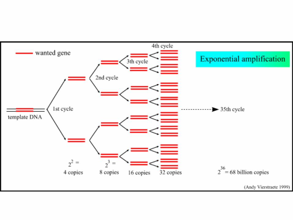
pcr - polymerase chain reaction
DESCRIPTION
PCR - Polymerase Chain Reaction. PCR is an in vitro technique for the amplification of a region of DNA which lies between two regions of known sequence. PCR amplification is achieved by using oligonucleotide primers. - PowerPoint PPT PresentationTRANSCRIPT

PCR - Polymerase Chain Reaction• PCR is an in vitro technique for the amplification of a region of DNA
which lies between two regions of known sequence. • PCR amplification is achieved by using oligonucleotide primers.
– These are typically short, single stranded oligonucleotides which are complementary to the outer regions of known sequence.
• The oligonucleotides serve as primers for DNA polymerase and the denatured strands of the large DNA fragment serves as the template. – This results in the synthesis of new DNA strands which are
complementary to the parent template strands. – These new strands have defined 5' ends (the 5' ends of the
oligonucleotide primers), whereas the 3' ends are potentially ambiguous in length.


• http://ocw.mit.edu/NR/rdonlyres/Civil-and-Environmental-Engineering/1-89Fall-2004/321BF8FF-75BE-4377-8D74-8EEE753A328C/0/11_02_04.pdf

Primer selection• Primer is an oligonucleotide sequence – will target
a specific sequence of opposite base pairing (A-T, G-C only) of single-stranded nucleic acids
• For example, there is a– ¼ chance (4-1) of finding an A, G, C or T in any given DNA
sequence; there is a – 1/16 chance (4-2) of finding any dinucleotide sequence (eg.
AG); a – 1/256 chance of finding a given 4-base sequence.
• Thus, a sixteen base sequence will statistically be present only once in every 416 bases (=4 294 967 296, or 4 billion): this is about the size of the human or maize genome, and 1000x greater than the genome size of E. coli.

Primer Specificity
• Universal – amplifies ALL bacterial DNA for instance
• Group Specific – amplify all denitrifiers for instance
• Specific – amplify just a given sequence

Forward and reverse primers
• If you know the sequence targeted for amplification, you know the size which the primers should be anealing across
• If you don’t know the sequence… What do you get?

DNA Polymerase• DNA Polymerase is the enzyme responsible for
copying the sequence starting at the primer from the single DNA strand
• Commonly use Taq, an enzyme from the hyperthermophilic organisms Thermus aquaticus, isolated first at a thermal spring in Yellowstone National Park
• This enzyme is heat-tolerant useful both because it is thermally tolerant (survives the melting T of DNA denaturation) which also means the process is more specific, higher temps result in less mismatch – more specific replication

RFLP• Restriction Fragment Length Polymorphism
• Cutting a DNA sequence using restriction enzymes into pieces specific enzymes cut specific places
Starting DNA sequence:5’-TAATTTCCGTTAGTTCAAGCGTTAGGACC3’-ATTAAAGGCAATCAAGTTCGCAATAATGG
Enzyme X5’-TTC-3”-AAG-
Enzyme X5’-TTC-3”-AAG-
5’-TAATTT3’-ATTAAA
5’-CCGTTAGTT3’-GGCAATCAA
5’-CAAGCGTTAGGACC3’-GTTCGCAATAATGG

RFLP• DNA can be processed by RFLP either directly (if
you can get enough DNA from an environment) or from PCR product
• T-RFLP (terminal-RFLP) is in most respects identical except for a marker on the end of the enzyme
• Works as fingerprinting technique because different organisms with different DNA sequences will have different lengths of DNA between identical units targeted by the restriction enzymes– specificity can again be manipulated with PCR primers
Liu et al. (1997) Appl Environ Microbiol 63:4516-4522

Electrophoresis
• Fragmentation products of differing length are separated – often on an agarose gel bed by electrophoresis, or using a capilarry electrophoretic separation


DGGE• Denaturing gradient gel electrophoresis
– The hydrogen bonds formed between complimentary base pairs, GC rich regions ‘melt’ (melting=strand separation or denaturation) at higher temperatures than regions that are AT rich.
• When DNA separated by electrophoresis through a gradient of increasing chemical denaturant (usually formamide and urea), the mobility of the molecule is retarded at the concentration at which the DNA strands of low melt domain dissociate. – The branched structure of the single stranded moiety of the
molecule becomes entangled in the gel matrix and no further movement occurs.
– Complete strand separation is prevented by the presence of a high melting domain, which is usually artificially created at one end of the molecule by incorporation of a GC clamp. This is accomplished during PCR amplification using a PCR primer with a 5' tail consisting of a sequence of 40 GC.
Run DGGE animation here – from http://www.charite.de/bioinf/tgge/

RFLP vs. DGGEDGGE
• Advantages– Very sensitive to variations in
DNA sequence– Can excise and sequence
DNA in bands• Limitations
– Somewhat difficult– ”One band-one species” isn’t
always true– Cannot compare bands
between gels– Only works well with short
fragments (<500 bp), thus limiting phylogenetic characterization
RFLP• Advantages
– Relatively easy to do– Results can be banked for
future comparisons• Limitations
– Less sensitive phylogenetic resolution than sequencing
– Each fragment length can potentially represent a diversity of microorganisms
– Cannot directly sequence restriction fragments,making identification indirect

FISH• Fluorescent in-situ hybridization
– Design a probe consisting of an oligonucleotide sequence and a tag
– Degree of specificity is variable!– Hybridize that oligonucleotide sequence to the
rRNA of an organism – this is temperature and salt content sensitive
– Image using epiflourescence, laser excitation confocal microscopy
• Technique DIRECTLY images active organisms in a sample

16S gene
16S rRNA
CellCellmembranemembrane
DNA
16S gene**
*
*
* *
Fluorescent in situ hybridisation(FISH) using DNA probes
TAGCTGGCAGTAUCGACCGUCACGU
Fluorescein
AU
ProbeProbe(( 20 bases) 20 bases)
Fluorescent in site hybridization

10 µm
DAPI FER656
B Drift Slime Streamer


Oligunucleotide design

FISH variations
• FISH-CARD – instead of a fluorescent probe on oligo sequence, but another molecule that can then bond to many fluorescent probes – better signal-to-noise ratio
• FISH-RING – design of oligo sequence to specific genes – image all organisms with DSR gene or nifH for example

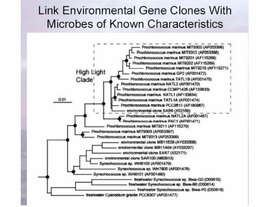
Clone Library
• http://ocw.mit.edu/NR/rdonlyres/Civil-and-Environmental-Engineering/1-89Fall-2004/321BF8FF-75BE-4377-8D74-8EEE753A328C/0/11_02_04.pdf

http://www.ifa.hawaii.edu/UHNAI/NAIweb/presentations/astrobiol6.pdf