pdf (979 kb) - iopscience
TRANSCRIPT

Advances in Natural Sciences:Nanoscience and Nanotechnology
PAPER • OPEN ACCESS
Catalytic activity of allamanda mediatedphytosynthesized anisotropic gold nanoparticlesTo cite this article: Rajesh K Gangwar et al 2013 Adv. Nat. Sci: Nanosci. Nanotechnol. 4 045005
View the article online for updates and enhancements.
You may also likeGold Nanoparticles; Synthesis,Characterization and Comparative Studiesof Their Antimicrobial ActivitiesIrshad Ahmed Wani and Ajit Khosla
-
Spherical aggregates composed ofgold nanoparticlesChi-Chang Chen, Ping-Lin Kuo and Yu-Chen Cheng
-
Fabrication of Nano-Patterns Composed ofMetal Nanoparticles with Photo-NanoimprintYutaka Kuwahara, Minoru Morita, KeishiroYoshimori et al.
-
This content was downloaded from IP address 46.70.187.60 on 14/02/2022 at 11:11

IOP PUBLISHING ADVANCES IN NATURAL SCIENCES: NANOSCIENCE AND NANOTECHNOLOGY
Adv. Nat. Sci.: Nanosci. Nanotechnol. 4 (2013) 045005 (5pp) doi:10.1088/2043-6262/4/4/045005
Catalytic activity of allamanda mediatedphytosynthesized anisotropic goldnanoparticlesRajesh K Gangwar1, Vinayak A Dhumale1, S W Gosavi2,Rishi B Sharma3 and Suwarna S Datar1
1 Department of Applied Physics, Defence Institute of Advanced Technology (DU), Girinagar,Pune 411025, India2 Department of Physics, University of Pune, Pune 411007, India3 Directorate of Personnel, Defence Research and Development Organization Bhawan, Rajaji Marg,New Delhi 110011, India
E-mail: [email protected] and [email protected]
Received 8 June 2013Accepted for publication 17 July 2013Published 14 August 2013Online at stacks.iop.org/ANSN/4/045005
AbstractA simple and eco-friendly method has been developed for the synthesis of gold nanoparticlesusing allamanda flower extract. In this green synthesis method, chloroauric acid (HAuCl4)solution was reduced with the help of allamanda flower extract. The synthesized goldnanoparticles were characterized by atomic force microscopy (AFM), transmission electronmicroscopy (TEM) and x-ray diffraction technique for their morphological and structuralanalysis. The size of the spherical and triangular gold nanoparticles was found to be in therange of 5–40 and 20–70 nm, respectively. The x-ray diffraction analysis revealed that thecrystallite size of face-centered cubic (FCC) gold nanoparticles was ∼11 nm. Thesesynthesized gold nanoparticles exhibit good catalytic activity towards the reduction of H2O2.The fabricated sensor exhibits good sensitivity of 21.33 µA mM−1 cm−2 with linearrelationship (R2
= 0.996) in the range from 2 to 10 mM of H2O2 concentration. This work canbe extended further for potential applications such as antimicrobial studies, bio-imaging anddrug-delivery owing to the known properties of the allamanda flower extract.
Keywords: gold nanoparticles, allamanda flower, biosynthesis, catalytic application
Classification numbers: 2.01, 4.00, 5.00, 5.06
1. Introduction
In recent years, metal nanoparticles have been an intensetopic of research due to their size, shape, surrounding mediaand inter-particle distance dependent physical, chemical,optical, electronic and electrical properties [1–6]. Inparticular, gold nanoparticles have attracted the attentionof researchers because of their unique optical propertiesoriginating from surface plasmon resonance (SPR) [7].
Content from this work may be used under the terms ofthe Creative Commons Attribution 3.0 licence. Any further
distribution of this work must maintain attribution to the author(s) and thetitle of the work, journal citation and DOI.
There are various methods for the synthesis of goldnanoparticles such as wet chemical [8], biological [9],laser ablation [10], electrochemical [11], etc. In chemicalmethods, various precursors, reducing agents and cappingagents are used which could be hazardous for humanbeings and the environment. To overcome these problems,researchers are using bacteria [12], yeast [13] and plant/flowerextracts [14, 15] for the synthesis of nanoparticles, whichis called green synthesis/biosynthesis. The green synthesishas drawn attention from researchers because of its rapid,clean, nontoxic, economical, eco-friendly procedure [16,17].Nanoparticles synthesized by this technique show the sameproperties as those synthesized by any other technique suchas wet chemical, laser ablation and electrochemical.
2043-6262/13/045005+05$33.00 1 © 2013 Vietnam Academy of Science & Technology

Adv. Nat. Sci.: Nanosci. Nanotechnol. 4 (2013) 045005 R K Gangwar et al
For the biosynthesis of gold nanoparticles, researchershave reported various plants, leaves, fruits and flower extractssuch as lemongrass [18], coriander [19], neem [20], aloe vera[21], rosa rugosa [22], cypress leaves [23], barbated skullcupherb [24], tansy fruit [25], magnolia kobus and diopyroskaki [26]. Recently, Maity et al [27] have synthesized goldnanoparticles using gum polysaccharide of cochlospermumreligiosum (katira gum), and studied their role as a catalystin the reduction of 4-nitrophenol (4-NP) to 4-aminophenol(4-AP). Ghoreishi et al [28] have also reported green synthesisof silver, gold nanoparticles using rosa damascena and theirprimary applications in electrochemistry.
The present paper delineates biosynthesis of goldnanoparticles by allamanda flower extract. To the best ofour knowledge, this is the first report that describes thebiosynthesis of gold nanoparticles by allamanda flowerextract. Allamanda also known as golden trumpet exhibitsvarious medicinal properties. The plant is used againstjaundice, malaria and enlarged spleen. The allamanda floweracts as a laxative and also shows antibiotic properties [29].Synthesized gold nanoparticles were found to be anisotropicwith the size of the spherical and triangular gold nanoparticlesto be in the range of 5–40 and 20–70 nm, respectively.The x-ray diffraction (XRD) analysis revealed that thecrystallite size of FCC gold nanoparticles was ∼11 nm. Thenanoparticles further showed good catalytic activity towardsthe reduction of H2O2.
2. Experimental
Gold chloride (HAuCl4 · 3H2O), potassium dihydrogen orthophosphate (KH2PO4), sodium hydroxide (NaOH) andhydrogen peroxide (H2O2 30%) were purchased fromThomas Baker, India. All chemicals were used as receivedwithout further purification. The allamanda cathartica (family,apocynaceae) flowers were collected from the garden at DIATcampus. Deionized water (DI water, ≈18 M�) was used in theexperiment.
Twenty-five allamanda flowers (∼20 g) were washed andthen boiled in 100 ml of DI water for 2 h. Flower extract wasfiltered thrice and stored at 4 ◦C for further experiment. Thecolor of the filtered flower extract (60 ml) was found to be afaint yellow. For the synthesis of gold nanoparticles, 20 ml of1 mM gold chloride was stirred at 50 ◦C for 10 min in threedifferent flasks simultaneously. A quantity of 5, 7.5 and 10 mlof flower extract was added rapidly into the three differentprecursor solutions. All these samples were labeled as R1, R2
and R3, respectively. Within 10 min, the color of the solutionof sample R1 and R2 changed from pale yellow to purplish redindicating the formation of gold nanoparticles. The color ofsample R3 changed from pale yellow to blue indicating somekind of agglomeration. In order to ensure the completion ofthe reaction, the solution was continuously stirred for another30 min but no color change was observed. The synthesizedgold nanoparticles were stable for several weeks.
Synthesized gold nanoparticles were examined underUV–Vis spectroscopy (Ocean Optics, HR 4000), Fouriertransform infrared (FTIR) spectroscopy (Perkin Elmer),atomic force microscopy (AFM, Asylum Research) andtransmission electron microscopy (TEM, FEI-Tecnai G2 20).
For TEM characterization, highly diluted samples wereprepared and directly deposited on carbon coated coppergrid. These samples were dried under an IR lamp. The XRDsamples were prepared by adding several drops of the goldnanoparticles solution on cleaned glass substrate followed bydrying.
For the electrochemical studies, glassy carbon electrodes(GCEs) were polished with 1.0, 0.3 and 0.05 µm aluminapowder and rinsed with ethanol and DI water successively.These electrodes were dried in natural environment andmodified by casting the 10 µl drop of synthesized Aunanoparticles. Further, a 1.8 µl drop of polyaniline (PANI)0.8% was also casted onto the GCE and kept in an ovenfor 12 h at 60 ◦C. Similarly for reference, gold nanoparticlesand PANI modified electrodes were also prepared.All electrochemical experiments were performed withelectrochemical workstation (CHI 1100 B), three-electrodecell, in 0.1 M phosphate buffer solution (PBS) at 7.4 pH.Ag/AgCl and platinum wire electrodes were used as referenceand counter electrodes, respectively, to measure the potentials.
3. Results and discussion
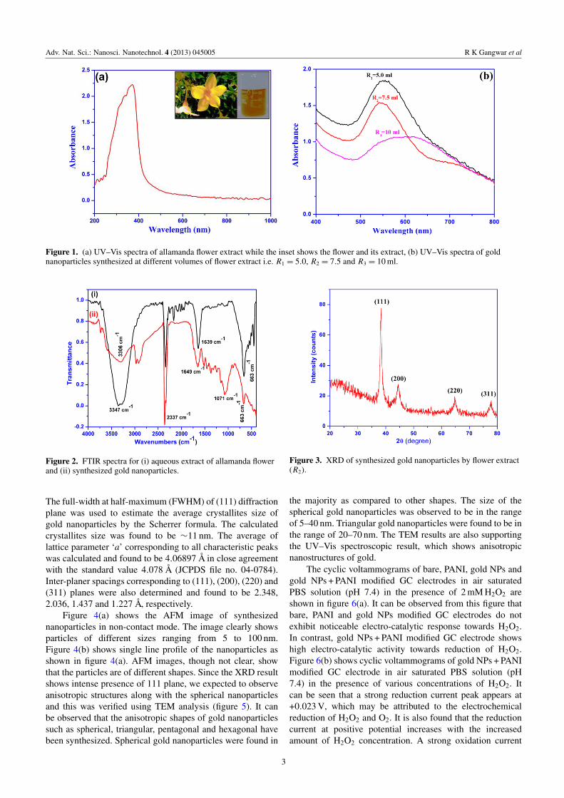
Figure 1(a) shows UV–Vis spectra of allamanda flower extractwhile the inset shows the yellow flower and its extract. TheUV–Vis spectra show a strong absorption peak at ∼ 370 nm.Figure 1(b) shows UV–Vis spectra of gold nanoparticlessynthesized at different volumes of flower extract i.e. R1 =
5.0 ml, R2 = 7.5 ml and R3 = 10 ml. It can be observed fromthe spectra that at low volume (R1) of flower extract anabsorption band appears at ∼552 nm which is a characteristicpeak of gold nanoparticles due to surface plasmon resonance.When the volume of flower extract was increased to R2,a small absorption band appears at ∼715 nm along withthe characteristic band of Au nanoparticles at ∼545 nm.These two distinct absorption bands indicate asymmetry inthe structure of gold nanoparticles and may be associatedwith the transverse and longitudinal plasmon absorption. Athigher volume of flower extract (R3), a broad absorptionband appeared at ∼590 nm indicating aggregation of goldnanoparticles within the solution. Samples R1 and R2 wereused for further characterization and catalytic application.
Figure 2 shows the FTIR spectra for (i) aqueous extractof allamanda flower and (ii) synthesized gold nanopar-ticles. Here spectra (i) show intense peaks at 3347 cm−1
(O H stretch), 2337 cm−1 (C N stretch), 1639 cm−1 (C Cstretch), 1071 cm−1 (C N stretch) and 663 cm−1 (C Sstretch). All these peaks in FTIR spectra confirm variouscomponents of allamanda floral extract [30]. Spectra (ii)also shows all these intense peaks with shift in O Hstretching from 3347 to 3306 cm−1 and C C stretching from1639 to 1649 cm−1. These shifts may be attributed to thereduction of Au3+ to Au0 and capping of synthesized goldnanoparticles.
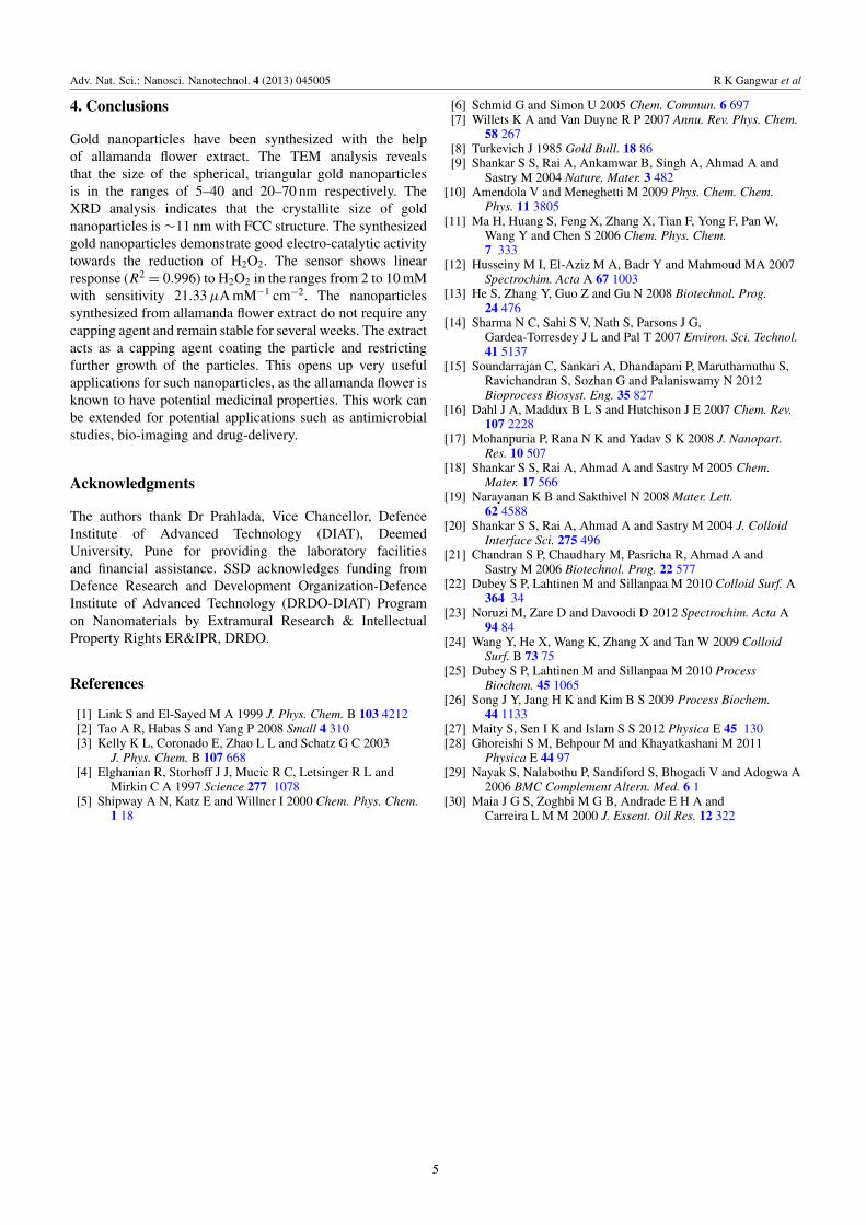
Figure 3 shows the XRD spectra of sample R2. The XRDpattern exhibits sharp reflections which are characteristicsof face-centered cubic (FCC) gold. The spectra shows fourcharacteristic peaks of gold at 38.28◦, 44.44◦, 64.77◦ and77.72◦ in the 2θ range of 20–80◦ which are indexed as (111),(200), (220) and (311) planes (JCPDS file no. 04-0784).
2
Adv. Nat. Sci.: Nanosci. Nanotechnol. 4 (2013) 045005 R K Gangwar et al
Figure 1. (a) UV–Vis spectra of allamanda flower extract while the inset shows the flower and its extract, (b) UV–Vis spectra of goldnanoparticles synthesized at different volumes of flower extract i.e. R1 = 5.0, R2 = 7.5 and R3 = 10 ml.
Figure 2. FTIR spectra for (i) aqueous extract of allamanda flowerand (ii) synthesized gold nanoparticles.
The full-width at half-maximum (FWHM) of (111) diffractionplane was used to estimate the average crystallites size ofgold nanoparticles by the Scherrer formula. The calculatedcrystallites size was found to be ∼11 nm. The average oflattice parameter ‘a’ corresponding to all characteristic peakswas calculated and found to be 4.06897 Å in close agreementwith the standard value 4.078 Å (JCPDS file no. 04-0784).Inter-planer spacings corresponding to (111), (200), (220) and(311) planes were also determined and found to be 2.348,2.036, 1.437 and 1.227 Å, respectively.
Figure 4(a) shows the AFM image of synthesizednanoparticles in non-contact mode. The image clearly showsparticles of different sizes ranging from 5 to 100 nm.Figure 4(b) shows single line profile of the nanoparticles asshown in figure 4(a). AFM images, though not clear, showthat the particles are of different shapes. Since the XRD resultshows intense presence of 111 plane, we expected to observeanisotropic structures along with the spherical nanoparticlesand this was verified using TEM analysis (figure 5). It canbe observed that the anisotropic shapes of gold nanoparticlessuch as spherical, triangular, pentagonal and hexagonal havebeen synthesized. Spherical gold nanoparticles were found in
Figure 3. XRD of synthesized gold nanoparticles by flower extract(R2).
the majority as compared to other shapes. The size of thespherical gold nanoparticles was observed to be in the rangeof 5–40 nm. Triangular gold nanoparticles were found to be inthe range of 20–70 nm. The TEM results are also supportingthe UV–Vis spectroscopic result, which shows anisotropicnanostructures of gold.
The cyclic voltammograms of bare, PANI, gold NPs andgold NPs + PANI modified GC electrodes in air saturatedPBS solution (pH 7.4) in the presence of 2 mM H2O2 areshown in figure 6(a). It can be observed from this figure thatbare, PANI and gold NPs modified GC electrodes do notexhibit noticeable electro-catalytic response towards H2O2.In contrast, gold NPs + PANI modified GC electrode showshigh electro-catalytic activity towards reduction of H2O2.Figure 6(b) shows cyclic voltammograms of gold NPs + PANImodified GC electrode in air saturated PBS solution (pH7.4) in the presence of various concentrations of H2O2. Itcan be seen that a strong reduction current peak appears at+0.023 V, which may be attributed to the electrochemicalreduction of H2O2 and O2. It is also found that the reductioncurrent at positive potential increases with the increasedamount of H2O2 concentration. A strong oxidation current
3

Adv. Nat. Sci.: Nanosci. Nanotechnol. 4 (2013) 045005 R K Gangwar et al
Figure 4. (a) AFM image of synthesized gold nanoparticles by flower extract (R2), (b) single line profile of synthesized nanoparticles asshown in (a).
Figure 5. TEM images of synthesized gold nanoparticles by flower extract (R2).
Figure 6. (a) Cyclic voltammograms of bare, PANI, gold NPs and gold NPs + PANI modified GC electrodes in air saturated PBS solution(pH 7.4) in the presence of 2 mM H2O2, (b) cyclic voltammograms of gold NPs + PANI modified GC electrode in air saturated PBS solution(pH 7.4) in the presence of various concentrations of H2O2. Inset shows the corresponding calibration curve (R2
= 0.996) between theanodic peak current and H2O2 concentrations.
peak is also observed at −0.26 V and may be attributed tothe oxidation of H2O2 in presence of O2. The inset showsthe calibration curve between the anodic peak currents and
H2O2 concentrations. A good linear relationship (R2= 0.996)
with sensitivity 21.33 µA mM−1 cm−2 has been observed inthe ranges from 2 to 10 mM.
4
Adv. Nat. Sci.: Nanosci. Nanotechnol. 4 (2013) 045005 R K Gangwar et al
4. Conclusions
Gold nanoparticles have been synthesized with the helpof allamanda flower extract. The TEM analysis revealsthat the size of the spherical, triangular gold nanoparticlesis in the ranges of 5–40 and 20–70 nm respectively. TheXRD analysis indicates that the crystallite size of goldnanoparticles is ∼11 nm with FCC structure. The synthesizedgold nanoparticles demonstrate good electro-catalytic activitytowards the reduction of H2O2. The sensor shows linearresponse (R2
= 0.996) to H2O2 in the ranges from 2 to 10 mMwith sensitivity 21.33 µA mM−1 cm−2. The nanoparticlessynthesized from allamanda flower extract do not require anycapping agent and remain stable for several weeks. The extractacts as a capping agent coating the particle and restrictingfurther growth of the particles. This opens up very usefulapplications for such nanoparticles, as the allamanda flower isknown to have potential medicinal properties. This work canbe extended for potential applications such as antimicrobialstudies, bio-imaging and drug-delivery.
Acknowledgments
The authors thank Dr Prahlada, Vice Chancellor, DefenceInstitute of Advanced Technology (DIAT), DeemedUniversity, Pune for providing the laboratory facilitiesand financial assistance. SSD acknowledges funding fromDefence Research and Development Organization-DefenceInstitute of Advanced Technology (DRDO-DIAT) Programon Nanomaterials by Extramural Research & IntellectualProperty Rights ER&IPR, DRDO.
References
[1] Link S and El-Sayed M A 1999 J. Phys. Chem. B 103 4212[2] Tao A R, Habas S and Yang P 2008 Small 4 310[3] Kelly K L, Coronado E, Zhao L L and Schatz G C 2003
J. Phys. Chem. B 107 668[4] Elghanian R, Storhoff J J, Mucic R C, Letsinger R L and
Mirkin C A 1997 Science 277 1078[5] Shipway A N, Katz E and Willner I 2000 Chem. Phys. Chem.
1 18
[6] Schmid G and Simon U 2005 Chem. Commun. 6 697[7] Willets K A and Van Duyne R P 2007 Annu. Rev. Phys. Chem.
58 267[8] Turkevich J 1985 Gold Bull. 18 86[9] Shankar S S, Rai A, Ankamwar B, Singh A, Ahmad A and
Sastry M 2004 Nature. Mater. 3 482[10] Amendola V and Meneghetti M 2009 Phys. Chem. Chem.
Phys. 11 3805[11] Ma H, Huang S, Feng X, Zhang X, Tian F, Yong F, Pan W,
Wang Y and Chen S 2006 Chem. Phys. Chem.7 333
[12] Husseiny M I, El-Aziz M A, Badr Y and Mahmoud MA 2007Spectrochim. Acta A 67 1003
[13] He S, Zhang Y, Guo Z and Gu N 2008 Biotechnol. Prog.24 476
[14] Sharma N C, Sahi S V, Nath S, Parsons J G,Gardea-Torresdey J L and Pal T 2007 Environ. Sci. Technol.41 5137
[15] Soundarrajan C, Sankari A, Dhandapani P, Maruthamuthu S,Ravichandran S, Sozhan G and Palaniswamy N 2012Bioprocess Biosyst. Eng. 35 827
[16] Dahl J A, Maddux B L S and Hutchison J E 2007 Chem. Rev.107 2228
[17] Mohanpuria P, Rana N K and Yadav S K 2008 J. Nanopart.Res. 10 507
[18] Shankar S S, Rai A, Ahmad A and Sastry M 2005 Chem.Mater. 17 566
[19] Narayanan K B and Sakthivel N 2008 Mater. Lett.62 4588
[20] Shankar S S, Rai A, Ahmad A and Sastry M 2004 J. ColloidInterface Sci. 275 496
[21] Chandran S P, Chaudhary M, Pasricha R, Ahmad A andSastry M 2006 Biotechnol. Prog. 22 577
[22] Dubey S P, Lahtinen M and Sillanpaa M 2010 Colloid Surf. A364 34
[23] Noruzi M, Zare D and Davoodi D 2012 Spectrochim. Acta A94 84
[24] Wang Y, He X, Wang K, Zhang X and Tan W 2009 ColloidSurf. B 73 75
[25] Dubey S P, Lahtinen M and Sillanpaa M 2010 ProcessBiochem. 45 1065
[26] Song J Y, Jang H K and Kim B S 2009 Process Biochem.44 1133
[27] Maity S, Sen I K and Islam S S 2012 Physica E 45 130[28] Ghoreishi S M, Behpour M and Khayatkashani M 2011
Physica E 44 97[29] Nayak S, Nalabothu P, Sandiford S, Bhogadi V and Adogwa A
2006 BMC Complement Altern. Med. 6 1[30] Maia J G S, Zoghbi M G B, Andrade E H A and
Carreira L M M 2000 J. Essent. Oil Res. 12 322
5