california cabg outcomes reporting program data ... · california cabg outcomes reporting...
TRANSCRIPT

Updated 1/13/2017
California CABG Outcomes Reporting Program
Data Abstractor Training Manual
Version 6.3

Updated 1/13/2017
Document Revision History
Date Version Description
1/22/2016 6.2 Myomectomy was corrected to myectomy on p. 54, Added revised Definitions for Isolated CABG at the end
2/17/2016 6.3 Added STS clarifications from their training manual updated 1/2016: http://www.sts.org/sites/default/files/documents/ACSD_January2016.pdf
Change data element Isolated CABG to Type of CABG eff with 1.1.2016 discharges.
3/23/2016 6.3 Added clarification for Type of CABG-Other Non-isolated.
1/13/2017 6.3 Added STS clarifications from their various training manual updates July 2016-January 2017.
http://www.sts.org/sites/default/files/documents/ACSD_JULY2016.pdf
http://www.sts.org/sites/default/files/documents/ACSD_AUGUST2016.pdf
http://www.sts.org/sites/default/files/documents/ACSD_SEPTEMBER2016.pdf
http://www.sts.org/sites/default/files/documents/ACSD_JANUARY-2017.pdf
Changes are in green.
http://www.sts.org/sites/default/files/documents/ACSD_February-2017_0.pdf
7/5/2017 Corrected typo on page 57 Isolated CABG -> Type of CABG. Corrections are in red.

Updated 1/13/2017
CCORP
Office of Statewide Health Planning and Development
CORC Hotline: (916) 326-3865
Confidential Fax: (916) 445-7534

Data Elements and Definitions
California CABG Outcomes Reporting Program
Updated 1/13/2017
4

Data Elements and Definitions
California CABG Outcomes Reporting Program
Updated 1/13/2017
5
Data Elements in Export Order
Effective with July 1, 2014 Discharges
Type of CABG Data Element Change is effective with January 1, 2016 Discharges

Data Elements and Definitions
California CABG Outcomes Reporting Program
Updated 1/13/2017
6
Overview: DATA ELEMENT EXPORT ORDER
Data Element Classification Origin
1. Medical Record Number Demographics STS
2. Type of CABG (eff. 1/1/2016 discharges) Isolated CABG (eff up to 12/31/2015 discharges)
Operative Non-STS
3. Date of Surgery Hospitalization STS
4. Date of Birth Demographics STS
5. Patient Age Demographics STS
6. Sex Demographics STS
7. Race Documented Demographics STS
8. Race – White Demographics STS
9. Race – Black/African American Demographics STS
10. Race – Asian Demographics STS
11. Race – American Indian/ Alaskan Native Demographics STS
12. Race – Native Hawaiian/ Pacific Islander Demographics STS
13. Race – Other Demographics STS
14. Hispanic or Latino or Spanish Ethnicity Demographics STS
15. Date of Discharge Hospitalization STS
16. Discharge Status Mortality STS
17. Date of Death Mortality STS
18. Responsible Surgeon Name ( 3 separate fields)
Operative Non-STS
18a. Surgeon Last Name 18b. Surgeon First Name 18c. Surgeon Middle Initial
Operative Operative Operative
Non-STS Non-STS Non-STS
19. Responsible Surgeon CA License Number Operative Non-STS
20. Height (cm) Risk Factors STS
21. Weight (kg) Risk Factors STS
22. Diabetes Risk Factors STS
23. Diabetes Control Risk Factors STS
24. Dialysis Risk Factors STS
25. Hypertension Risk Factors STS
26. Endocarditis Risk Factors STS
27. Infectious Endocarditis Type Risk Factors STS
28. Chronic Lung Disease Risk Factors STS

Data Elements and Definitions
California CABG Outcomes Reporting Program
Updated 1/13/2017
7
29. Liver Disease Risk Factors STS
30. Immunocompromise Risk Factors STS
31. Peripheral Arterial Disease (PVD) Risk Factors STS
32. CVD Risk Factors STS
33. Prior CVA Risk Factors STS
34. Prior CVA When Risk Factors STS
35. CVD TIA Risk Factors STS
36. CVD – Carotid Stenosis Risk Factors STS
37. CVD Carotid Stenosis – Right Risk Factors STS
38. CVD Carotid Stenosis – Left Risk Factors STS
39. CVD Prior Carotid Surgery Risk Factors STS
40. Last Creatinine Level Risk Factors STS
41. Total Albumin Risk Factors STS
42. Total Bilirubin Risk Factors STS
43. INR Risk Factors STS
44. Previous CABG Previous Cardiac Interventions STS
45. Previous Valve Previous Cardiac Interventions STS
46. Previous PCI Previous Cardiac Interventions STS
47. Previous PCI – Interval Previous Cardiac Interventions STS
48. Prior MI Preoperative Cardiac Status STS
49. MI When Preoperative Cardiac Status STS
50. Heart Failure within 2 weeks Preoperative Cardiac Status STS
51. Classification – NYHA Preoperative Cardiac Status STS
52. Cardiogenic Shock Preoperative Cardiac Status STS
53. Resuscitation Preoperative Cardiac Status STS
54. Cardiac Arrhythmia Preoperative Cardiac Status STS
55. Cardiac Arrhythmia – Vtach/VFib Preoperative Cardiac Status STS
56. Cardiac Arrhythmia - AFlutter Preoperative Cardiac Status STS
57. Cardiac Arrhythmia – Third Degree Heart Block
Preoperative Cardiac Status STS
58. Cardiac Arrhythmia – Atrial Fibrillation Preoperative Cardiac Status STS
59. Meds – Coumadin Preoperative Medications STS
60. Warfarin Use (within 5 days) Preoperative Medications Non-STS
61. Coronary Anatomy/Disease Known Hemodynamics / Cath / Echo STS
62. Number of Diseased Vessels Hemodynamics / Cath / Echo STS
Data Elements and Definitions
California CABG Outcomes Reporting Program
Updated 1/13/2017
8
63. Percent Native Artery Stenosis Known Hemodynamics / Cath / Echo STS
64. Percent Stenosis Left Main Hemodynamics / Cath / Echo STS
65. Ejection Fraction Done Hemodynamics / Cath / Echo STS
66. Ejection Fraction (%) Hemodynamics / Cath / Echo STS
67. PA Systolic Pressure Measured Hemodynamics / Cath / Echo STS
68. PA Systolic Pressure Hemodynamics / Cath / Echo STS
69. Insufficiency – Mitral Hemodynamics / Cath / Echo STS
70. Incidence Operative STS
71. Status Operative STS
72. Urgent of Emergent Reason Operative STS
73. CPB Utilization Operative STS
74. CPB Utilization – Combination Plan Operative STS
75. IMA Used Coronary Bypass STS
76. Reason for No IMA Coronary Bypass STS
77. Valve Operative STS
78. Aortic Valve Valve Surgery STS
79. Aortic Valve Procedure Valve Surgery STS
80. Mitral Valve Valve Surgery STS
81. Mitral Valve Procedure Valve Surgery STS
82. Tricuspid Valve Valve Surgery STS
83. Tricuspid Valve Procedure Valve Surgery STS
84. Pulmonic Valve Valve Surgery STS
85. Pulmonic Valve Procedure Valve Surgery STS
86. Reoperation for Bleed Postoperative Events STS
87. Reintervention – Graft Occlusion Postoperative Events STS
88. Deep Sternal Infection/ Mediastinitis Postoperative Events STS
89. Neuro – Stroke Permanent Postoperative Events STS
90. Pulm – Ventilation Prolonged Postoperative Events STS
91. Renal – Renal Failure Postoperative Events STS
92. Renal – Dialysis Requirement Postoperative Events STS
93. Other – A Fib Postoperative Events STS
94. Facility Identification Number Hospitalization Non-STS

Data Elements and Definitions
California CABG Outcomes Reporting Program Updated 7/5/2017
9
Data Element Valid Values Definition
1. Medical Record Number STS Sequence #: 85
Alphanumeric Indicate the patient's medical record number at the hospital where surgery occurred. This field should be collected in compliance with state/local privacy laws.
2. Type of CABG (eff.
1/1/2016 discharges) __________________ Isolated CABG (eff. Up
to 12/31/2016 discharges) CCORP-specific variable
1 = Isolated 3 = CABG + Valve 4= Other Non-isolated CABG ______________________ 1 = Yes 2 = No
Indicate whether the surgery was considered an isolated CABG, CABG + Valve, or all other CABG. Other Non-isolated must include a CABG (not isolated valve). _________________________________________________________________ Indicate whether the surgery was considered an isolated CABG. CCORP Clarification/Comments: See reference on pages 55 – 59. Q: We have a patient that had aortic aneurysm repair. A Cabrol procedure was done to perfuse around the aortic root using two venous conduits. Do we answer yes to CAB and fill out all the information in the CAB procedure section including the CAB worksheet? A: Do not code the Cabrol procedure as a CAB.
3. Date of Surgery STS Sequence #: 310
Numeric: mmddyyyy Indicate the date of index cardiac surgical procedure. Index cardiac surgical procedure is defined as the initial major cardiac surgical procedure of the hospitalization. CCORP Clarification/Comments: The date the patient enters the operating room for surgery.
4. Date of Birth STS Sequence #: 65
Numeric: mmddyyyy Indicate the patient's date of birth using 4-digit format for year. This field should be collected in compliance with state/local privacy laws.
5. Patient Age STS Sequence #: 70
Numeric Indicate the patient's age in years, at time of surgery. This should be calculated from the date of birth and the date of surgery, according to the convention used in the USA (the number of birthdate anniversaries reached by the date of surgery).
6. Sex STS Sequence #: 75
1 = Male 2 = Female
Indicate the patient’s sex at birth as either male or female. CCORP Clarification/Comments: Patients who have undergone gender reassignment surgery maintain the risk associated with their chromosomal gender. Code gender at birth.

Data Elements and Definitions
California CABG Outcomes Reporting Program Updated 7/5/2017
10
7. Race Documented STS Sequence #: 150
1 = Yes 2 = No
Indicate whether race is documented. STS Clarification: Race should be self-reported by the patient/family. Do not assign race or make assumptions if race is not documented.
8. Race – White STS Sequence #: 155
1 = Yes 2 = No 3 = Patient declined to disclose
Indicate whether the patient's race, as determined by the patient or family, includes White. "White" refers to a person having origins in any of the original peoples of Europe, the Middle East, or North Africa. It includes people who indicated their race(s) as "White" or reported entries such as Irish, German, Italian, Lebanese, Arab, Moroccan, or Caucasian. STS Clarification: You may choose more than one race category. The Census Bureau collects race data in accordance with guidelines provided by the U.S. Office of Management and Budget and these data are based on self-identification. The racial categories included in the census form generally reflect a social definition of race recognized in this country, and are not an attempt to define race biologically, anthropologically or genetically. In addition, it is recognized that the categories of the race item include racial and national origin or socio-cultural groups. People may choose to report more than one race to indicate their racial mixture, such as “American Indian and White.” People who identify their origin (ETHNICITY) as Hispanic, Latino or Spanish may be of any race. In addition, it is recognized that the categories of the race item include both racial and national origin and socio-cultural groups.
9. Race – Black/African American STS Sequence #: 160
1 = Yes 2 = No
Indicate whether the patient's race, as determined by the patient or family, includes Black/African- American. "Black or African-American" refers to a person having origins in any of the black racial groups of Africa. It includes people who indicated their race(s) as “Black, African Am., or Negro” or reported entries such as African American, Kenyan, Nigerian, or Haitian. STS Clarification: This includes a person having origins in any of the black racial groups of Africa. Terms such as "Haitian" or "Negro" can be used in addition to "Black or African American."
10. Race – Asian STS Sequence #: 165
1 = Yes 2 = No
Indicate whether the patient's race, as determined by the patient or family, includes Asian. "Asian" refers to a person having origins in any of the original peoples of the Far East, Southeast Asia, or the Indian subcontinent, including, for example, Cambodia, China, India, Japan, Korea, Malaysia, Pakistan, the Philippine Islands, Thailand, and Vietnam. It includes people who indicated their race(s) as "Asian"
Data Elements and Definitions
California CABG Outcomes Reporting Program Updated 7/5/2017
11
or reported entries such as "Asian Indian", "Chinese", "Filipino", "Korean", "Japanese", "Vietnamese", and "Other Asian" or provided other detailed Asian responses.
11. Race – American Indian/ Alaskan Native STS Sequence #:170
1 = Yes 2 = No
Indicate whether the patient's race, as determined by the patient or family, includes American Indian/Alaskan Native. "American Indian or Alaska Native" refers to a person having origins in any of the original peoples of North and South America (including Central America) and who maintains tribal affiliation or community attachment. This category includes people who indicated their race(s) as "American Indian or Alaska Native" or reported their enrolled or principal tribe, such as Navajo, Blackfeet, Inupiat, Yup’ik, or Central American Indian groups or South American Indian groups.
12. Race – Native Hawaiian/ Pacific Islander STS Sequence #: 175
1 = Yes 2 = No
Indicate whether the patient's race, as determined by the patient or family, includes Native Hawaiian / Pacific Islander. "Native Hawaiian or Other Pacific Islander" refers to a person having origins in any of the original peoples of Hawaii, Guam, Samoa, or other Pacific Islands. It includes people who indicated their race(s) as "Pacific Islander" or reported entries such as "Native Hawaiian", "Guamanian or Chamorro", "Samoan", and "Other Pacific Islander" or provided other detailed Pacific Islander responses.
13. Race – Other STS Sequence #: 180
1 = Yes 2 = No
Indicate whether the patient's race, as determined by the patient or family, includes any other race. "Some Other Race" includes all other responses not included in the White, Black or African American, American Indian or Alaska Native, Asian, and Native Hawaiian or Other Pacific Islander race categories described above.
14. Hispanic or Latino or Spanish Ethnicity STS Sequence #: 185
1 = Yes 2 = No 3 = Not Documented
Indicate if the patient is of Hispanic, Latino or Spanish ethnicity as reported by the patient/family. "Hispanic, Latino or Spanish" refers to a person of Cuban, Mexican, Puerto Rican, South or Central American, or other Spanish culture or origin regardless of race. CCORP Clarification/Comments: People who identify their origin as Hispanic, Latino or Spanish may be of any race. STS FAQ: My facility does not document ethnicity. If there is no mention of ethnicity in the medical record how should this this be coded? Answer: You cannot make the assumption that the patient is not Hispanic, Latino or Spanish without clear documentation in the medical record. Code not documented.

Data Elements and Definitions
California CABG Outcomes Reporting Program Updated 7/5/2017
12
15. Date of Discharge STS Sequence #: 315
Numeric: mmddyyyy Indicate the date the patient was discharged from the hospital (acute care) even if the patient is going to a rehab or hospice or similar extended care unit within the same physical facility. If the patient died in the hospital, the discharge date is the date of death. CCORP Clarification/Comments: Do not include transfers to other services, such as renal care unit. If the patient is discharged (given a new account number) to hospice care but remains in the same bed/unit, the discharge date is that date. If the patient is discharged (given a new account number) to a psychiatric or rehab unit, even if located in the same building, the discharge date is that date.
16. Discharge Status STS Sequence #: 5010
1 = Alive 2 = Dead
Indicate whether the patient was alive or dead at discharge from the hospitalization in which surgery occurred. Include patients who died after transfer to another acute care hospital. CCORP Clarifications/Comments: It is not necessary to report operative mortalities. CCORP uses the death file from the state’s Vital Statistics program to verify deaths after discharge.
17. Date of Death STS Sequence #: 5030
Numeric: mmddyyyy Indicate the date the patient was declared dead.
18. Responsible Surgeon Name CCORP-specific variable
18a. Surgeon Last Name 18b. Surgeon First Name 18c. Surgeon Middle Initial
Indicate the Surgeon’s name. This field must have controlled data entry where a user selects the surgeon name from a user list. This will remove variation in spelling, abbreviations and punctuation within the field. Note: Surgeon name is encrypted in the analysis database. Punctuation, abbreviations and spacing differences cannot be corrected at the warehouse. CCORP Clarification/Comments: Hospitals are encouraged to look up their surgeon names and licensing information DIRECTLY from the California Medical Board. http://www.mbc.ca.gov/Breeze/License_Verification.aspx **See reference on page 56.
19. Responsible Surgeon CA License Number CCORP-specific variable
California physician license number of responsible surgeon assigned by the Medical Board of California of the Department of Consumer affairs. See page 56 of this training manual for more information criteria. CCORP Clarification/Comments: Hospitals are encouraged to look up their surgeon names and licensing information DIRECTLY from the California Medical Board. http://www.mbc.ca.gov/Breeze/License_Verification.aspx

Data Elements and Definitions
California CABG Outcomes Reporting Program Updated 7/5/2017
13
20. Height (cm) STS Sequence #: 330
Usual Range: 122.0 – 213.0 Low/High: 20.0 – 251.0
Indicate the height of the patient in centimeters nearest to the date of surgery. CCORP Clarification/Comments: Used to calculate BSA (body surface area), a field for risk calculation. To convert Inches to centimeters, multiply # of inches by 2.54. 1 inch = 2.54 centimeters. STS FAQ: If a pt is a bilateral leg amputee due to PVD, should we use current height or height prior to amputation? Cath PCI wants original height but I thought STS wanted current Height prior to surgery, after amputation. Answer: Code the patient’s height prior to amputation.
21. Weight (kg) STS Sequence #: 335
Usual Range: 40.0 – 170.0 Low/High: 10.0 – 250.0
Indicate the weight of the patient in kilograms closest to the date of surgery. CCORP Clarification/Comments: Used to calculate BSA (body surface area), a field for risk calculation. To convert pounds to kilograms, divide # of lbs by 2.2 1 kg = 2.2 pounds.
22. Diabetes STS Sequence #: 360
1 = Yes 2 = No 3 = Unknown
History of diabetes diagnosed and/or treated by a healthcare provider. The American Diabetes Association criteria include documentation of the following: i. Hemoglobin A1c >=6.5%; or ii. Fasting plasma glucose >=126 mg/dL (7.0 mmol/l); or iii. 2-hour Plasma glucose >=200 mg/dL (11.1 mmol/l) during an oral glucose tolerance test; or iv. In a patient with classic symptoms of hyperglycemia or hyperglycemic crisis, a random plasma glucose >=200 mg/dL (11.1 mmol/l) This does not include gestational diabetes. 2013 ACCF/AHA Data Standards Cannon et al. JACC Vol. 61, No. 9, 2013 CCORP Clarification/Comments: Diabetes = yes only if the diagnosis is documented and/or treated by a physician in the medical record. ADA criteria are informational only and data managers should not diagnose diabetes themselves. In particular, glucose may be elevated transiently in the absence of diabetes. The STS and CCORP make an exception for Hgb A1C >=6.5% which is sufficient to codes diabetes = yes because it reflects chronic elevation of glucose over 2-3 months.

Data Elements and Definitions
California CABG Outcomes Reporting Program Updated 7/5/2017
14
STS Clarification: Indicate if the patient has a history of diabetes mellitus regardless of duration of disease or need for anti-diabetic agents. Exclusions are steroid induced hyperglycemia and gestational (transient), without elevated HbA1c and/or treatment, code “No”. Not all patients receiving diabetic medications are considered diabetic. It is important to remember, some medications used to treat diabetes may be used to treat other conditions. A hemoglobin A1C value of >= 6.5%, collected within 3 months prior to surgery, is acceptable to use for documentation of diabetes = "Yes". STS FAQ: The surgeon documented "history of non-insulin-dependent diabetes, which by report has resolved, secondary to weight loss”. Should this still be coded yes to diabetes? Answer: Yes, code diabetes.
23. Diabetes Control STS Sequence #: 365
1 = None 2 = Diet only 3 = Oral 4 = Insulin 5 = Other 6 = Other subcutaneous medication 7 = Unknown
Indicate the patient’s diabetes control method as presented on admission. Patients placed on a pre-procedure diabetic pathway of insulin drip at admission but whose diabetes was controlled by diet or oral methods are not coded as being treated with insulin. STS Clarification: "Control type is the long term management therapy used." Other subcutaneous medications may include: exenatide (Byetta, Bydureon), liraglutide, (Victoza), Pramlintide (Symlin). Oral treatments may include: Sulfonylureas - Diabinese, glipizide (Glucotrol, Glucotrol XL), glyburide (Micronase, DiaBeta, Glynase), and glimepiride (Amaryl). Meglitinides - Repaglinide (Prandin) and nateglinide (Starlix). Biguanides - metformin (Glucophage). Thiazolidinediones - rosiglitazone (Avandia) and pioglitazone (Actos). Alpha-glucosidase inhibitors - acarbose (Precose) and meglitol (Glyset). DPP-4 inhibitor - sitagliptin (Januvia). Q: How should diabetes control be coded for the patient who has had a pancreatic transplant? A: Code “other”, since the insulin from the new pancreas is not exogenous. STS FAQ: The surgeon documented "history of non-insulin-dependent diabetes, which by report has resolved, secondary to weight loss”. I have coded Seq. 360 as yes since the patient does have a history, but how would I code Seq. 365? Should I code as "diet only" or "none"? Answer: Code diet only.

Data Elements and Definitions
California CABG Outcomes Reporting Program Updated 7/5/2017
15
24. Dialysis STS Sequence #: 375
1 = Yes 2 = No 3 = Unknown
Indicate whether the patient is currently (prior to surgery) undergoing dialysis. Refers to whether the patient is currently on dialysis, not distant past history. STS Clarification/Comments: Includes any form of dialysis including peritoneal or hemodialysis, which the patient is receiving at the time of admission. Also, may include Continuous Veno-Venous Hemofiltration (CVVH, CVVH-D), and Continuous Renal Replacement Therapy (CRRT) as dialysis. Code “No” for renal dialysis if ultra-filtration is the only documentation found in the record since this is for volume management.
25. Hypertension STS Sequence #: 380
1 = Yes 2 = No 3 = Unknown
Indicate if the patient has a current diagnosis of hypertension defined by any 1 of the following: i. History of hypertension diagnosed and treated with medication, diet, and/or exercise; ii. Prior documentation of blood pressure >140 mmHg systolic and/or 90 mmHg diastolic for patients without diabetes or chronic kidney disease, or prior documentation of blood pressure >130 mmHg systolic or 80 mmHg diastolic on at least 2 occasions for patients with diabetes or chronic kidney disease; iii. Currently undergoing pharmacological therapy for treatment of hypertension. CCORP Clarification/Comments: A clinician has to state in the medical record that the patient has hypertension. Hypertensive medications are used for other symptoms besides hypertension. Do not code “Yes” based on medications alone. Code “Yes” for hypertension if patient has normal blood pressure readings but has a documented history of hypertension.
26. Endocarditis STS Sequence #: 385
1 = Yes 2 = No
Indicate whether the patient has a history of endocarditis. STS Clarification: This applies to any history of endocarditis; even remote history can result in valve damage. According to the CDC: Endocarditis of a natural or prosthetic heart valve must meet at least 1 of the following criteria: i. Patient has organisms cultured from valve or vegetation. ii. Patient has 2 or more of the following signs or symptoms with no other recognized cause: fever (>38°C), new or changing murmur*, embolic phenomena*, skin manifestations* (i.e., petechiae, splinter hemorrhages, painful subcutaneous nodules), congestive heart failure*, or cardiac conduction abnormality* (*With no other recognized cause) AND at least 1 of the following: a). Organisms cultured from 2 or more blood cultures b). Organisms seen on Gram’s stain of valve when culture is negative or not done
Data Elements and Definitions
California CABG Outcomes Reporting Program Updated 7/5/2017
16
c). Valvular vegetation seen during a surgical operation or autopsy d). Positive antigen test on blood or urine (e.g., H influenzae, S pneumoniae, Nmeningitides, or Group B Streptococcus) e). Evidence of new vegetation seen on echocardiogram and if diagnosis is made antemortem, physician institutes appropriate antimicrobial therapy Choose "Yes" for patients with pre-operative endocarditis who begin antibiotics post-op. Code yes for patients who are diagnosed intraoperatively. This is a case where operative or autopsy findings can change a pre-operative risk factor. Marantic Endocarditis (Nonbacterial Thrombotic Endocarditis) (Lupus) should not be coded as infectious endocarditis.
27. Infectious Endocarditis Type STS Sequence #: 390
1 = Treated 2 = Active
Indicate the type of endocarditis the patient has. If the patient is currently being treated for endocarditis, the disease is considered active. If no antibiotic medication (other than prophylactic medication) is being given at the time of surgery, then the infection is considered treated. CCORP Clarification/Comments: If the patient is currently being treated with antimicrobials for endocarditis, the disease is considered active. STS Clarification: Active - currently being treated; also include patients who were diagnosed in the OR but began treatment postop. Treated - no antibiotic medication at time of surgery (other than prophylactic medication).
28. Chronic Lung Disease STS Sequence #: 405
1 = No 2 = Mild 3 = Moderate 4 = Severe 5 = Lung disease documented, severity unknown 6 = Unknown
Indicate whether the patient has chronic lung disease, and the severity level according to the following classification: 1. No; 2. Mild: FEV1 60% to 75% of predicted, and/or on chronic inhaled or oral bronchodilator therapy. 3. Moderate: FEV1 50% to 59% of predicted, and/or on chronic steroid therapy aimed at lung disease. 4. Severe: FEV1 < 50% and/or Room Air pO <60 or pCO2 > 50. 5. Chronic Lung Disease present, severity not documented 6. Unknown A history of chronic inhalation reactive disease (asbestosis, mesothelioma, black lung disease or pneumoconiosis) may qualify as chronic lung disease. Radiation induced pneumonitis or radiation fibrosis also qualifies as chronic lung disease (if above criteria is met). A history of atelectasis is a transient condition and does not qualify. Chronic lung disease can include patients with chronic obstructive pulmonary disease, chronic bronchitis, or emphysema. It can also include a patient who is

Data Elements and Definitions
California CABG Outcomes Reporting Program Updated 7/5/2017
17
currently being chronically treated with inhaled or oral pharmacological therapy (e.g., beta-adrenergic agonist, anti-inflammatory agent, leukotriene receptor antagonist, or steroid). Patients with asthma or seasonal allergies are not considered to have chronic lung disease. CCORP Clarification/Comments: The diagnosis of chronic lung disease is not based solely on the fact that a person has or currently is smoking, or is on home oxygen. Diagnostic testing and or pharmacological criteria must be met. Chest x-ray findings alone are not included in the data specs for inclusion as chronic lung disease and should not be coded as “Yes”. STS Clarification: DLCO values should not be used for determining chronic lung disease Time Frame: Do not use values obtained more than 12 months prior to the date of surgery Patients on home oxygen without documentation of COPD or PFT testing are coded as Unknown [note: this supersedes earlier CCORP clarification to code home oxygen as severe lung disease.] Asthma is not considered chronic lung disease; therefore, do not code chronic lung disease for those patients who are treated with steroids for their asthma. ONLY systemic steroids qualify for chronic lung disease (not inhaled steroids). Q: Nothing in the history indicates COPD, the surgeon documents that the patient’s lungs are covered in blebs. Can this be coded chronic lung disease? A: No, there is no way to quantify lung disease in this scenario. Q: Can PFTs alone be used for chronic lung disease? A: Yes, you can use the values from PFTs to code chronic lung disease, unless the patient’s condition is asthma which is not obstructive lung disease. This Q&A means you can meet PFT criteria but not have chronic lung disease (eg, asthma). Similarly, in hospital PFTs done immediately prior to CABG may be abnormal due to heart failure and should not be coded as CLD unless there is reason to believe the PFT abnormality is due to lung disease (eg, patient is not in heart failure and has a significant smoking history). STS Clarification: Pts on home oxygen without documentation of COPD or PFT/ABG testing are coded as “Unknown.” STS Clarification: If a patient is on NO medication, has no O2 need, no PFT/ABG, no notation of prior history of COPD, yet the H&P states the pt has "severe COPD", do we code as "severe"? Answer: Lung disease documented, severity unknown.

Data Elements and Definitions
California CABG Outcomes Reporting Program Updated 7/5/2017
18
STS FAQ: When the physician documents "severe COPD" (or other lung disease), but the FEV1 and home meds do not support "severe", do we still code it as "severe"? Answer: Code chronic lung disease based on the severity indicated in diagnostic testing and/or medications. STS FAQ: As it reads right now, it is causing some confusion if inhaled or oral bronchodilator therapy alone qualifies for chronic lung disease without a diagnosis of any disease. We have a patient who was on Albuterol but did not have a diagnosis for asthma or any chronic lung disease. Are we safe to assume that any chronic inhaled or oral bronchodilator therapy drug, without a corresponding diagnosis to include or exclude it, would qualify as Mild Chronic Lung Disease? Answer: To code “yes”, you MUST have a diagnosis of some type of chronic lung disease, (other than asthma). Steroid inhalers qualify for moderate CLD. ONLY systemic steroids qualify for moderate CLD. Steroid inhalers do not count for preoperative steroids, or immunosuppression. STS FAQ: If the physician documents that the patient has "reactive COPD" or any type of chronic lung disease and a PFT is not performed and the patient is not on a chronic inhaled or oral bronchodilator, or chronic steroid therapy aimed at lung disease, does the abstractor mark the lowest form of chronic lung disease, such as "Mild," and then select reactive as noted above? (Does the patient need to be on chronic lung meds and/or have PFT results to answer this element?) Answer: Code lung disease documented, severity unknown. STS FAQ: Question: PFT - FEV1=82%; Chest CT shows emphysema and MDs note states "moderate COPD." Pt has a 1.2 - 2 PPD current smoker history. What is the best way to code this element? Answer: You can code Lung Disease Documented, severity unknown. The FEV1 does not meet criteria. STS FAQ: If there are pulmonary function studies that meet the criteria to quantify chronic lung disease, can they be used to code CLD? Answer: Yes, you can code CLD from the pulmonary function studies.

Data Elements and Definitions
California CABG Outcomes Reporting Program Updated 7/5/2017
19
29. Liver Disease STS Sequence #: 485
1 = Yes 2 = No 3 = Unknown
Indicate whether the patient has a history of hepatitis B, hepatitis C, cirrhosis, portal hypertension, esophageal varices, chronic alcohol abuse or congestive hepatopathy. Exclude NASH in the absence of cirrhosis. STS Clarification: Liver diseases such as hepatitis B, hepatitis C, cirrhosis, portal hypertension, esophageal varices, chronic alcohol abuse and congestive hepatopathy affect the cells, tissues, structures, or functions of the liver. Severity can range from mild to severe and will be quantified by the MELD score. Hepatitis A is a transient condition- do not code as liver disease. Liver fibrosis with recurrent ascites should be coded as "yes" if documented appropriately and is supported by the MELD score. Do not code liver disease for the liver transplant patient, if the patient has no residual anatomic or systemic issue OR if the MELD score does not quantify liver disease. LFTs or a MELD score alone cannot be used to code "yes" to liver disease since other conditions impact these lab values. Do not code liver cancer as liver disease, code as cancer. STS FAQ: Because there are medications to treat Hepatitis-C that will give a negative test result, should liver disease be coded no for these patients? Answer: No, these patients should be coded as yes to liver disease.
30. Immunocompromise STS Sequence #: 490
1 = Yes 2 = No 3= Unknown
Indicate whether immunocompromise is present due to immunosuppressive medication therapy within 30 days preceding the operative procedure or existing medical condition. This includes, but is not limited to systemic steroid therapy, anti-rejection medications and chemotherapy. This does not include topical steroid applications, one time systemic therapy or preprocedure steroid protocol. CCORP Clarification/Comments: DO NOT include topical creams or inhalers that are steroidal in form. DO NOT include patients who receive a one or two time dose of systemic treatment, or a pre-operative/pre-cath protocol. Patients post organ transplant or with rheumatologic conditions may be on immunosuppressive therapy other than corticosteroids such as: Cyclosporine (Gengraf, Neoral, Sandimmune), Azathioprine (Imuran), Cyclophosamide (Cytoxan), Methotrexate, Tacrolimus (Prograf), Sirolimus (Rapamune, Mycophenolate-Mofetil-MMF (Cellcept).
STS Clarification: There are multiple classes of drugs considered to be immunosuppressive. Corticosteroids (only if taken systemically). Cytotoxix drugs, Antimetabolites, Cyclosporine, and Biologics (biologic response modifiers ex: (Actemra, Cimzia, Enbrel, Humira, Kineret, Orencia, Remicade, Rituxan, Simponi). Biologics are genetically engineered proteins derived from human genes. They are designed to inhibit specific components of the immune system that play pivotal

Data Elements and Definitions
California CABG Outcomes Reporting Program Updated 7/5/2017
20
roles in fueling inflammation. Immunosuppression can result from radiation therapy, malnutrition, or removal of the spleen. Immunodeficiency can be inherited or acquired. Examples of conditions causing immunocompromise include Hypogammaglobulinemia and HIV infection. If patient has had a previous splenectomy code “Yes” to immunocompromised. Patients with a history of receiving chemotherapeutic medications greater than 30 days prior to surgery should be coded as “No”. Positive Coombs test alone is not indicative of immunocompromised. If a patient has receive a short treatment of prednisone (5 days) for respiratory problems within 30 days of CABG, do I code yes or no in this category? Answer: No
STS FAQ: FAQ: Can immunosuppression be coded for an anorexic patient with acute weight loss of more than 20 pounds in the last month?
Answer: No, there is not sufficient evidence to code immunosuppression.
STS FAQ: Is a patient who is being treated with IVIG for chronic lymphocytic leukemia (CLL) considered immunocompromised?
Answer: Yes, IVIG is treatment for immunosuppression in patients with CLL.
31. Peripheral Arterial Disease (PVD) STS Sequence #: 505
1 = Yes 2 = No 3 = Unknown
Indicate whether the patient has a history of peripheral arterial disease (includes upper and lower extremity, renal, mesenteric, and abdominal aortic systems). This can include: i. Claudication , either with exertion or at rest, ii. Amputation for arterial vascular insufficiency, iii. Vascular reconstruction, bypass surgery, or percutaneous intervention to the extremities (excluding dialysis fistulas and vein stripping), iv. Documented abdominal aortic aneurysm with or without repair, v. Positive noninvasive test (e.g., ankle brachial index =< 0.9, ultrasound, magnetic resonance or computed tomography imaging of > 50% diameter stenosis in any peripheral artery, i.e., renal, subclavian, femoral, iliac) or angiographic imaging CCORP Clarification/Comments: Peripheral arterial disease excludes disease in the carotid or cerebrovascular arteries. STS Clarification: Capture subclavian artery stenosis. PAD sometimes called PVD.

Data Elements and Definitions
California CABG Outcomes Reporting Program Updated 7/5/2017
21
32. Cerebrovascular Disease (CVD) STS Sequence #: 525
1 = Yes 2 = No 3 = Unknown
Indicate whether the patient has a current or previous history of any of the following: i. Stroke: is an acute episode of focal or global neurological dysfunction caused by brain, spinal cord, or retinal vascular injury as a result of hemorrhage or infarction, where the neurological dysfunction lasts for greater than 24 hours. ii. TIA: is defined as a transient episode of focal neurological dysfunction caused by brain, spinal cord, or retinal ischemia, without acute infarction, where the neurological dysfunction resolves within 24 hours. iii. Noninvasive or invasive arterial imaging test demonstrating >=50% stenosis of any of the major extracranial or intracranial vessels to the brain iv. Previous cervical or cerebral artery revascularization surgery or percutaneous intervention. This does not include chronic (nonvascular) neurological diseases or other acute neurological insults such as metabolic and anoxic ischemic encephalopathy. CCORP Clarification/Comments: DO NOT include any of the peripheral arterial disease processes. STS Clarification: Subdural hematoma is not cerebrovascular disease.
33. Prior CVA STS Sequence #: 530
1 = Yes 2 = No 3 = Unknown
Indicate whether the patient has a history of stroke. Stroke is an acute episode of focal or global neurological dysfunction caused by brain, spinal cord, or retinal vascular injury as a result of hemorrhage or infarction, where the neurological dysfunction lasts for greater than 24 hours STS Clarification/Comments: Include any confirmed neurological deficit of abrupt onset caused by a disturbance in cerebral blood supply that did not resolve within 24 hours of the event. The physical deficit can be in the form of extremity weakness, facial asymmetry, language (speech and/or cognitive thinking) impairment. Code “Yes” if a patient may have had a permanent stroke with residual when over time and/or with therapy regained all deficit function. The intent is to differentiate between neurological events that resolve and those that don’t. STS FAQ 07/2016: If the patient has no history of stroke and no symptoms but the CT reports an infarct do you code yes to prior CVA? Answer: Yes, code prior CVA. STS FAQ 08/2016: The patient is admitted with active endocarditis and the MRI demonstrates small punctate foci that favor small ischemic infarcts. How are endocarditis emboli collected for these events preoperatively? Answer: Code these events as prior CVAs.
Data Elements and Definitions
California CABG Outcomes Reporting Program Updated 7/5/2017
22
34. Prior CVA When STS Sequence #: 535
3 = Recent <= 30 days 4 = Remote > 30 days
Indicate when the CVA events occurred. Those events occurring within 30 days prior to the surgical procedure are considered recent, while all others are considered remote.
35. CVD TIA STS Sequence #: 540
1 = Yes 2 = No 3 = Unknown
Indicate whether the patient has a history of a Transient Ischemic Attack (TIA). Transient ischemic attack (TIA) is defined as a transient episode of focal neurological dysfunction caused by brain, spinal cord, or retinal ischemia, without acute infarction, where the neurological dysfunction resolves within 24 hours. STS Clarification: Unknown should be selected if some neurologic dysfunction occurred or was suspected, was resolved in 24 hours, and could not be confirmed or if patient/family unable to provide history.
36. CVD – Carotid Stenosis STS Sequence #: 545
1 = None 2 = Right 3 = Left 4 = Both
Indicate which carotid artery was determined from any diagnostic test to be >= 50% stenotic. CCORP Clarification/Comments: Diagnostic studies may include ultrasound, Doppler, angiography, CT, MRI or MRA. If more than one test was performed with different results, choose the highest level of stenosis reported. STS Clarification: Code what is found at the time of surgery (even if prior stent is in place) Question: A pt had both an ultrasound and CT to assess carotid stenosis. The procedures were done two days apart. Should I record the worst % stenosis or the results from the procedure closest to surgery? Answer: CT angiogram would be more definitive.
37. CVD Carotid Stenosis – Right STS Sequence #: 550
1 = 80-99% 2 = 100% 3 = 50-79% 4 = Not Documented
Indicate the severity of stenosis reported on the right carotid artery. STS Clarification: -Choose 100% for stenosis labeled as "total". -Choose 80-99% for stenosis labeled as “critical” or “severe” or “subtotal”. -Choose 50 - 79% for stenosis labeled as "moderate".
38. CVD Carotid Stenosis – Left STS Sequence #: 555
1 = 80-99% 2 = 100% 3 = 50-79% 4 = Not Documented
Indicate the severity of stenosis reported on the left carotid artery. STS Clarification: -Choose 100% for stenosis labeled as "total". -Choose 80-99% for stenosis labeled as “critical” or “severe” or “subtotal”. -Choose 50 - 79% for stenosis labeled as "moderate".

Data Elements and Definitions
California CABG Outcomes Reporting Program Updated 7/5/2017
23
39. CVD Prior Carotid Surgery STS Sequence #: 560
1 = Yes 2 = No
Indicate whether the patient has a history of previous carotid artery surgery and/or stenting. STS Clarification: Carotid endarterectomy is a surgical procedure during which a surgeon removes atherosclerotic plaque or other material obstructing the flow of blood from the artery. This procedure eliminates a substance called plaque from the artery and can restore blood flow. Carotid artery stenting is a procedure in which a slender, metal-mesh tube, called a stent, is inserted and expands inside the carotid artery to increase blood flow in areas blocked by plaque. STS FAQ: The patient does not have a history of stroke but has a history of internal carotid aneurysm which was coiled. This does not seem to meet the definition of carotid surgery or stent, but it seems significant. Should this be coded as yes? Answer: Yes, code prior carotid surgery and/or stenting .
40. Last Creatinine Level STS Sequence #: 585
Usual Range: 0.10 – 9.00 Low/ High: 0.10 – 30.00
Indicate the creatinine level closest to the date and time prior to surgery but prior to anesthetic management (induction area or operating room). STS Clarification/Comments: Anesthetic management begins when a member of the anesthesiology team initiates care. The administration of IV fluids in the holding area can cause dilution of blood. Do not capture labs drawn after the patient receives fluids in the holding area or O.R.
41. Total Albumin STS Sequence #: 590
Usual range: 3.50 - 5.00 Low/High: 1.00 - 10.00 (mg/dL)
Indicate the total albumin closest to the date and time prior to surgery but prior to anesthetic management (induction area or operating room). STS Clarification/Comments: You can capture results up to 6 weeks prior to surgery provided there is no known acute liver disease process. Anesthetic management begins when a member of the anesthesiology team initiates care. The administration of IV fluids in the holding area can cause dilution of blood. Do not capture labs drawn after the patient receives fluids in the holding area or O.R.
42. Total Bilirubin STS Sequence #: 595
Usual range: 0.20 - 1.30 Low/High: 0.10 - 50.00 (mg/dL)
Indicate the total Bilirubin closest to the date and time prior to surgery but prior to anesthetic management (induction area or operating room).
Data Elements and Definitions
California CABG Outcomes Reporting Program Updated 7/5/2017
24
STS Clarification/Comments: You can capture results up to 6 weeks prior to surgery provided there is no known acute liver disease process. Anesthetic management begins when a member of the anesthesiology team initiates care. The administration of IV fluids in the holding area can cause dilution of blood. Do not capture labs drawn after the patient receives fluids in the holding area or O.R.
43. INR STS Sequence #: 610
Usual range 0.90 - 1.30 Low/High: 0.50 - 30.00
Indicate the International Normalized Ratio (INR) closest to the date and time prior to surgery but prior to anesthetic management (induction area or operating room). STS Clarification/Comments: Anesthetic management begins when a member of the anesthesiology team initiates care. The administration of IV fluids in the holding area can cause dilution of blood. Do not capture labs drawn after the patient receives fluids in the holding area or O.R.
44. Previous CABG STS Sequence #: 670
1 = Yes 2 = No
Indicate whether the patient had a previous Coronary Bypass Graft prior to the current admission. STS Clarification/Comments: This applies only to surgical approach to revascularization. Angioplasty or other catheter based coronary artery occlusion treatment does not apply.
45. Previous Valve STS Sequence #: 675
1 = Yes 2 = No
Indicate whether the patient had a previous surgical replacement and/or surgical repair of a cardiac valve. This may also include percutaneous valve procedures. STS Clarification/Comments: This may include percutaneous valve procedures such as percutaneous valvotomy or valvuloplasty, as well as surgical or transcatheter valve repair or replacement. These do not have to be in order of chronology.
46. Previous PCI STS Sequence #: 775
1 = Yes 2 = No
Indicate whether a previous Percutaneous Coronary Intervention (PCI) was performed any time prior to this surgical procedure. Percutaneous coronary intervention (PCI) is the placement of an angioplasty guide wire, balloon, or other device (e.g. stent, atherectomy, brachytherapy, or thrombectomy catheter) into a native coronary artery or coronary artery bypass graft for the purpose of mechanical coronary revascularization. CCORP Clarification/Comments: There is no time limit on its historical occurrence. PCI refers to those non-surgical methods that unblock narrowed coronary arteries. A PCI may have been performed during this same admission, BUT prior to the surgical procedure.

Data Elements and Definitions
California CABG Outcomes Reporting Program Updated 7/5/2017
25
STS Clarification: An attempted, even if unsuccessful, PCI should be coded as a Previous CV intervention-PCI. This is in an effort to harmonize with ACC-NCDR. PCIs may include coronary angioplasties, stents and/or atherectomies done by interventional cardiologists. Include patients who had PCI prior to surgery as part of a planned, staged hybrid procedure.
47. Previous PCI – Interval STS Sequence #: 800
1 = ≤ 6 Hours 2 = > 6 Hours
Indicate the interval of time between the PCI procedure and the current surgical procedure. CCORP Clarification/Comments: Intervals are calculated from the time of the conclusion of the PCI procedure (removal of the coronary dilation catheter) and surgical skin incision cut time. This field is intended to capture PCIs done during the same episode of care prior to the surgical procedure. Include patients who were transferred for surgery from another facility following PCI. Include patients who had PCI prior to surgery as part of a planned, staged hybrid procedure. Do not code PCIs done after the surgical procedure. STS Clarification: The choices are ≤ 6 hours or > 6 hours prior to OR entry. The timing of surgery after PCI may influence outcomes such as renal failure due to contrast given during PCI. STS FAQ 07/2016: Prior to admission, pt is in NSR. In the operative report section, “Findings at Operation: Paroxysmal afib. The pt developed afib intraoperatively." This is noted after CPB was commenced and the pt was observed to have a large LAA. Consent is for AVR only, but a modified MAZE (LAA and PVI) is also performed unexpectedly. Is it best to capture the afib post op or pre op?
Answer: Intraop Afib wouldn’t be in either the preop or postop section. The patient was in NSR preop. STS FAQ: A pt had TOF with a VSD repair in 1969. The VSD was repaired with a Teflon patch. Does this count as a "closure device, ventricular septal defect"? Answer: No, this is not a closure device should be coded as congenital cardiac repair, surgical.
48. Prior MI STS Sequence #: 885
1 = Yes 2 = No 3 = Unknown
Indicate if the patient has had at least one documented previous myocardial infarction at any time prior to this surgery.

Data Elements and Definitions
California CABG Outcomes Reporting Program Updated 7/5/2017
26
CCORP Clarification/Comments: Medical record documentation of prior myocardial infarction is sufficient. ECG or enzyme documentation in the current chart is not required. Data abstractors should not diagnose MI; the medical record should document that a clinician made the diagnosis. STS Clarification: A myocardial infarction is evidenced by any of the following: a). rise and fall of cardiac biomarkers (preferably troponin) with at least one of the values in the abnormal range for that laboratory [typically above the 99th percentile of the upper reference limit (URL) for normal subjects] together with at least one of the following manifestations of myocardial ischemia; b). Ischemic symptoms; c). ECG changes indicative of new ischemia (new ST-T changes, new left bundle branch block, or loss of R- wave voltage); d). Development of pathological Q- waves in 2 or more contiguous leads in the ECG (or equivalent findings for true posterior MI); e). Imaging evidence of new loss of viable myocardium or new regional wall motion abnormality; f). Documentation in the medical record of the diagnosis of acute myocardial infarction based on the cardiac biomarker pattern in the absence of any items enumerated in a-d due to conditions that may mask their appearance (e.g., peri-operative infarct when the patient cannot report ischemic symptoms; baseline left bundle branch block or ventricular pacing) ECG changes associated with prior myocardial infarction can include the following (with or without prior symptoms): a). Any Q-wave in leads V2-V3 >=0.02 seconds or QS complex in leads V2 and V3. b). Q-wave >=0.03 seconds and >=0.1 mV deep or QS complex in leads I, II, aVL, aVF, or V4-V6 in any two leads of a contiguous lead grouping (I, aVL, V6; V4-V6; II, III, and aVF). c). R-wave >=0.04 seconds in V1-V2 and R/S >=1 with a concordant positive T-wave in the absence of a conduction defect. Imaging evidence of a region with new loss of viable myocardium at rest in the absence of a non-ischemic cause. This can be manifested as: a). Echocardiographic, CT, MR, ventriculographic or nuclear imaging evidence of left ventricular thinning or scarring and failure to contract appropriately (i.e., hypokinesis, akinesis, or dyskinesis) b). Fixed (non-reversible) perfusion defects on nuclear radioisotope imaging (e.g., MIBI, thallium) Medical records documentation of prior myocardial infarction. Do not use phrases such as “cannot rule out”, “suggestive”, “probable”, “cannot exclude”, etc. to code. STS FAQ: I had a patient who had the Impella(right) inserted on 3/3 for VSD and shock. She had surgery on 3/11. The Impella was removed. Does this count as a previous intervention?

Data Elements and Definitions
California CABG Outcomes Reporting Program Updated 7/5/2017
27
Answer: Impella is a catheter based assist device only. Do not code as previous CV intervention.
49. MI When STS Sequence #: 890
1 = ≤ 6 Hrs 2 = > 6 Hrs but < 24 Hrs 3 = 1 to 7 Days 4 = 8 to 21 Days 5 = > 21 Days
Indicate the time period between the last documented myocardial infarction and surgery. STS Clarification/Comments: Time of surgery is documented as the hour the patient entered the operating room. Select the time-interval category based on information available on when the MI occurred. MI occurrence is the time of diagnosis and/or when confirmation of the last MI is documented prior to surgery. Note: If the EKG indicates a prior MI of undetermined age Code as >21 days if the patient has no recently reported or documented symptoms. More recent infarctions would likely be described as “evolving” on the EKG.
50. Heart Failure within 2 weeks STS Sequence #: 910
1 = Yes 2 = No 3 = Unknown
Indicate if there is physician documentation or report that the patient has been in a state of heart failure within the past 2 weeks. Heart failure is defined as physician documentation or report of any of the following clinical symptoms of heart failure described as unusual dyspnea on light exertion, recurrent dyspnea occurring in the supine position, fluid retention; or the description of rales, jugular venous distension, pulmonary edema on physical exam, or pulmonary edema on chest x-ray presumed to be cardiac dysfunction. A low ejection fraction alone, without clinical evidence of heart failure does not qualify as heart failure. An elevated BNP without other supporting documentation should not be coded as CHF.
CCORP Clarification/Comments: Since evidence of recent HF symptoms is not always available in current medical record, CCORP accepts chart documentation that the patient was diagnosed with a HF episode within the two weeks prior to surgery (if presented at outside hospital within 2 weeks). STS Clarification: The intent is to capture the patient's actual status in the weeks before surgery, the new diagnosis or exacerbation of an existing heart failure condition. DO NOT code stable or asymptomatic compensated failure or patients whose symptoms improved after medical therapy. A low ejection fraction (EF) without clinical presentation does not qualify for history of heart failure. See examples under NYHA class.
Data Elements and Definitions
California CABG Outcomes Reporting Program Updated 7/5/2017
28
51. Classification – NYHA STS Sequence #: 915
1 = Class I 2 = Class II 3 = Class III 4 = Class IV
Indicate the patient's worst dyspnea or functional class, coded as the New York Heart Association (NYHA) classification within the past 2 weeks. This is to be used for heart failure only, is not intended to classify angina. CCORP Clarification/Comments: Select the highest level of heart failure within the two weeks leading up to episode of hospitalization or at the time of the procedure. STS Clarification: NYHA classification is used for congestive heart failure (CHF). Select the highest level of heart failure within the two weeks leading up to episode of hospitalization or at the time of the procedure. The intent is to capture the highest level of failure. If the NYHA class is not documented, use the guidelines below to assign a class based on documented symptoms. Class I: Patient has cardiac disease but without resulting limitations of ordinary physical activity. Ordinary physical activity (e.g., walking several blocks or climbing stairs) does not cause undue fatigue, palpitation, dyspnea, or anginal pain. Limiting symptoms may occur with marked exertion. Class II: Patient has cardiac disease resulting in slight limitation of ordinary physical activity. Patient is comfortable at rest. Ordinary physical activity such as walking more than two blocks or climbing more than one flight of stairs results in limiting symptoms (e.g., fatigue, palpitation, dyspnea, or anginal pain). Class III: Patient has cardiac disease resulting in marked limitation of physical activity. Patient is comfortable at rest. Less than ordinary physical activity (e.g., walking one to two level blocks or climbing one flight of stairs) causes fatigue, palpitation, dyspnea, or anginal pain. Class IV: Patient has dyspnea at rest that increases with any physical activity. Patient has cardiac disease resulting in inability to perform any physical activity without discomfort. Symptoms may be present even at rest. If any physical activity is undertaken, discomfort is increased. Q: The physician documents new onset CHF with an EF of 25% and SOB. There is no indication of what level of activity causes the SOB. How do I code NYHA classification? A: You cannot code the NYHA classification if there is no supportive documentation in the record. Code yes to Heart Failure and leave NYHA classification blank. Q: Pt. is diagnosed with STEMI and has a cardiac arrest. Prior to surgery he has documented respiratory failure and pulmonary edema. Should NYHA Class be IV based on the fact he was intubated and remained intubated for days? A: This should be coded as heart failure, NYHA class IV because of pulmonary edema secondary to cardiac failure, whether or not he remained intubated.
Data Elements and Definitions
California CABG Outcomes Reporting Program Updated 7/5/2017
29
52. Cardiogenic Shock STS Sequence #: 930
2 = No 3 = Yes, at the time of procedure 4 = Yes, not at the time of procedure, but within prior 24 hours
Indicate if the patient developed cardiogenic shock. Cardiogenic shock is defined as a sustained (>30 min) episode of hypoperfusion evidenced by systolic blood pressure <90 mm Hg and/or, if available, cardiac index <2.2 L/min per square meter determined to be secondary to cardiac dysfunction and/or the requirement for parenteral inotropic or vasopressor agents or mechanical support (e.g., IABP, extracorporeal circulation, VADs) to maintain blood pressure and cardiac index above those specified levels. CCORP Clarification/Comments: “Shock” = Yes if the patient: 1) currently SBP <90 mmHg or cardiac index <2.2 or 2) previously had a SBP < 90 or CI <2.2 but now are on inotropes/ IABP to maintain higher #s. NOTE: sustained (>30 min) episode STS: “or requirement for … vasopressor agents ….to maintain blood pressure and cardiac index” Patients left on inotropes/pressors/IABP whose BP/CI has improved so that it is probable BP/CI would be above criteria off therapy should be coded “No.” This is more often the case the longer the patient has received these therapies prior to surgery.
1) CI < 2.2 or unassisted/unaugmented SBP < 90 shock 2) CI>=2.8 or unassisted/unaugmented SBP >=130 not shock 3) CI 2.2-2.39, unassisted/unaugmented SBP 90-99 on any active inotrope/vasopressor/IABP
or impella shock 4) CI 2.4-2.79, unassisted/unaugmented SBP 100-129 on high dose inotrope/ vasopressor/
impella shock 5) CI 2.4-2.79, unassisted/unaugmented SBP 100-129 on low dose inotrope/ vasopressor/
IABP not shock a. Dopamine < 5 mcg/kg/min b. Dobutamine < 5 mcg/kg/min c. Milrinone < 0.375 mcg/kg/min d. Norepinephrine (Levophed) < 0.3 mcg/kg/min e. Epinephrine < 0.3 mcg/kg/min f. Phenylephrine < 0.5 mcg/kg/min g. Vasopressin < 0.03 units per min
6) VAD, ECMO shock 7) Chart label “shock,” inotrope/pressor/IABP, but no CI/BP criteria not shock
IABPs are often used to treat coronary ischemia in absence of shock and their use alone does not meet shock criteria (eg, IABP put in for severe left main disease and ACS to stabilize ischemia while waiting for surgery). Some patients have mild cardiogenic shock which does not meet STS criteria

Data Elements and Definitions
California CABG Outcomes Reporting Program Updated 7/5/2017
30
even if treated with IABP, inotropes, or pressors. Inotropes may be used or continued to augment diuresis in patients not meeting shock criteria. Note IABPs usually lower systolic BP (assisted SBP < unassisted SBP) therefore assisted SBP should not be used as evidence for shock. The additional time options were added to harmonize with NCDR. Note: “At the time of the procedure” is defined as incision time. This includes patients with CS who have been stabilized on IABP/inotropes at the time of surgery Do not code yes to shock for patients with a low cardiac index who are asymptomatic and do not require mechanical or inotropic support. STS Clarification: Note: Transient episodes of hypotension reversed with IV fluid or atropine do not constitute cardiogenic shock. The hemodynamic compromise (with or without extraordinary supportive therapy) must persist for at least 30 min. ACCF/AHA 2013. Q: Going into the procedure the patient was on IABP, CI: 2.3-2.5 pre-op and SBP>80. The IABP was in place 24 hrs prior to the procedure due to hypotension during cardiac catheterization, does this count for cardiogenic shock? A: NO STS Clarification: A patient with bacterial endocarditis, respiratory failure, septic shock and multisystem failure prior to arrival. How do I capture the degree of her illness? I did count IV inotropes. The only shock question available to us is cardiogenic shock. Answer: If patient meets the criteria: sustained (>30 min) episode of hypoperfusion evidenced by systolic blood pressure <90 mm Hg and/or, if available, cardiac index <2.2 L/min per square meter determined to be secondary to cardiac dysfunction and/or the requirement for parenteral inotropic or vasopressor agents or mechanical support (e.g., IABP, extracorporeal circulation, VADs) to maintain blood pressure and cardiac index above those specified levels then cardiogenic shock can be coded.

Data Elements and Definitions
California CABG Outcomes Reporting Program Updated 7/5/2017
31
If the physician documents the patient is in cardiogenic shock, yet the patient does NOT MEET ANY elements in the definition, do we still code it? Answer: No, it is important to have supportive physician documentation but the patient must meet the clinical definition; please read the data specification. STS FAQ 09/2016: The patient was admitted for elective CAB and developed cardiogenic shock after induction but prior to incision. Can cardiogenic shock be coded as a risk factor after the patient enters the operating room? Answer: No, hemodynamic issues that could be contributed to anesthesia induction problems should not count in the preoperative status of the patient. Also, elective procedures should not be coded as cardiogenic shock. See following diagram.
Data Elements and Definitions
California CABG Outcomes Reporting Program Updated 7/5/2017
32
53. Resuscitation STS Sequence #: 935
2 = No 3 = Yes, within 1 hour of start of the procedure 4 = Yes, more than 1 hour before, but less than 24 hours of the start of the procedure
Indicate whether the patient required cardiopulmonary resuscitation before the start of the operative procedure which includes the institution of anesthetic management. Capture resuscitation timeframe: within 1 hour or 1-24 hours pre-op. CCORP Clarification/Comments: Impella is NOT complete circulatory support and does not qualify as ongoing resuscitation (trumps STS). STS Clarification: This may include complete circulatory support such as ECMO/other mechanical assist devices (ex. LVAD) initiated emergently prior to surgery. Do not code yes for resuscitation started after induction of anesthesia, the goal is to capture patients who required CPR prior to entering the OR.
54. Cardiac Arrhythmia STS Sequence #: 945
1 = Yes 2 = No 3 = Unknown
Indicate whether the patient has a history of a cardiac rhythm disturbance before the start of the operative procedure which includes the institution of anesthetic management. STS Clarification: In the fields that follow, for each arrhythmia, choose: none, remote (more than 30 days pre op), or recent (within 30 days of procedure). The arrhythmia must have been treated with one or more of the definitional list of therapies and/or clinically documented. These do not include arrhythmias such as 1st degree heart block, occasional premature ventricular contractions (PVC’s), or supraventricular tachycardia (SVT) that did not require treatment. To define “treated for an arrhythmia”: a patient is considered to be treated for arrhythmia if they are on a medication specifically to treat an arrhythmia. Today, most arrhythmias are treated with antiarrhythmic medications. Coumadin is considered a treatment for A-fib, but a patient on Coumadin with documented a-fib rhythm would be coded yes. Patients may take Digoxin to treat arrhythmias. In the past Digoxin was used to treat A-fib, but patients can also be on Digoxin to increase contractility, etc. Therefore, do not assume that all patients that are on Digoxin are being treated for A-fib. If the patient had a history of an arrhythmia (i.e. a-fib or V-tach) and is currently on medication to control rate and rhythm, and presents in sinus rhythm, code the patient as having the arrhythmia. Treatment may include: -Ablation therapy - surgical and/or catheter based -Implantable cardioverter/defibrillator (ICD) -Pacemaker -Pharmacological treatment -Electrocardioversion

Data Elements and Definitions
California CABG Outcomes Reporting Program Updated 7/5/2017
33
55. Cardiac Arrhythmia – Vtach/VFib STS Sequence #: 950
1 = None 2 = Remote (> 30 days) 3 = Recent (≤ 30 days)
Indicate whether arrhythmia was VTach or VFib. CCORP Clarification/Comments: CCORP suggests the rhythm be sustained for 30 seconds or longer, or require cardioversion. Treatment not a criteria. STS Clarification: V-tach rhythm must be sustained/persistent or paroxysmal sufficient to require some type of intervention (pharmacological and/or electrical) to interrupt and cease the arrhythmia.
56. Cardiac Arrhythmia – Aflutter STS Sequence #: 960
1 = None 2 = Remote (> 30 days) 3 = Recent (≤ 30 days)
Indicate whether arrhythmia was atrial flutter. STS Clarification: Atrial flutter is an abnormal heart rhythm that occurs in the atria of the heart. When it first occurs, it is usually associated with a fast heart rate or tachycardia (beats over 100 per minute), and falls into the category of supraventricular tachycardias. While this rhythm occurs most often in individuals with cardiovascular disease (e.g. hypertension, coronary artery disease, and cardiomyopathy) and diabetes, it may occur spontaneously in people with otherwise normal hearts. It is typically not a stable rhythm, and frequently degenerates into atrial fibrillation. However, it does rarely persist for months to years. If rhythm is described as fib/flutter, code fibrillation.
57. Cardiac Arrhythmia – Third Degree Heart Block STS Sequence #: 970
1 = None 2 = Remote (> 30 days) 3 = Recent (≤ 30 days)
Indicate whether arrhythmia was third degree heart block.
58. Cardiac Arrhythmia – Atrial Fibrillation STS Sequence #: 980
1 = None 2 = Paroxysmal 3 = Continuous/persistent- 3 includes “Long-Standing Persistent” and “Permanent”
Indicate whether arrhythmia was atrial fibrillation and if so, which type. STS Clarification: In atrial fibrillation, the electrical signals that coordinate the muscle of the upper chambers (atria) of the heart become rapid and disorganized; resulting in an irregular heartbeat (arrhythmia) often greater than 300 beats per minute. The likelihood of developing these arrhythmias increases with age. After age 65, between 3 percent and 5 percent of people have AF.
Paroxysmal
Recurrent AF (>2 episodes). Terminates spontaneously within 7 days
Continuous/ Persistent Sustained episode > 7 days, or lasting < 7 days, but necessitating pharmacologic or electrical

Data Elements and Definitions
California CABG Outcomes Reporting Program Updated 7/5/2017
34
cardio-conversion
Long-Standing Persistent
Continuous episode of > 1 year duration
Permanent AF at a point in which no further treatment of any kind is considered
Many patients with paroxysmal AF eventually develop permanent AF. The signs and symptoms of AF vary, and may include a sudden flutter of the heart, anxiety, shortness of breath, weakness and difficulty exercising, chest pain, sweating, dizziness or fainting. AF may have no known cause, or it may be related to coronary artery disease, thyroid disease, high blood pressure, structural defects of the heart and its valves, lung disease or other disorders. AF is diagnosed by electrocardiogram (ECG), or with devices that are worn by the patient to monitor the heart over time (Holter monitors and/or event recorders). AF may increase the risk of blood clots and stroke. Medications can be prescribed to prevent blood clots from forming. AF sometimes requires treatment with medications, controlled electric shocks to the heart or procedures that destroy the heart tissue that gives rise to the irregular heart rhythm. Less often, a pacemaker or other device is implanted to monitor and control the heart's rhythm. STS FAQ: Prior to admission, pt is in NSR. In the operative report section, “Findings at Operation: Paroxysmal afib. The pt developed afib intraoperatively." This is noted after CPB was commenced and the pt was observed to have a large LAA. Consent is for AVR only, but a modified MAZE (LAA and PVI) is also performed unexpectedly. Answer: Is it best to capture the afib post op or pre op? Intraop Afib wouldn’t be in either the preop or postop section. The patient was in NSR preop.
59. Meds – Coumadin STS Sequence #: 1075
1 = Yes 2 = No 4 = Unknown
Indicate whether the patient received Coumadin within 24 hours preceding surgery. STS Clarification/Comments: While Anisindione is taken orally, it is not Coumadin and should not be captured here. Received Coumadin within 24 hours preceding surgery where surgery = entry into the O.R. Yes - Capture those who are prescribed to take medications on a regular schedule and are presumed to be at a therapeutic level within 24 hours preceding surgery (entry into the OR) No – Patient did not receive a Coumadin within 24 hours preceding surgery Unknown – conflicting information in the medical record and/or with the patient/family or no information is available

Data Elements and Definitions
California CABG Outcomes Reporting Program Updated 7/5/2017
35
60. Warfarin Use (within 5 days) CCORP-specific variable
1 = Yes 2 = No 4 = Unknown
Indicate whether the patient received Warfarin (Coumadin) within 5 days preceding surgery. CCORP Clarification/Comments: The purpose of this data element is to determine whether the reported INR value was influenced by the patient taking Warfarin within 5 days of surgery, which may raise the INR independently and lead to false indications of liver disease. Note: patients on chronic Warfarin therapy who have stopped or been switched to an alternative anticoagulant 5-7 days prior to surgery should be coded as “No”. Notes in the admission H&P or Nurse's assessment (e.g., “stopped 1 week ago", "switched to Lovenox", "held x 1 week") may help in making this determination.
61. Coronary Anatomy/Disease Known STS Sequence #: 1155
1 = Yes 2 = No
Indicate whether coronary artery anatomy and/or disease is documented and available prior to surgery. STS Clarification: “Documented prior to surgery”: Sometimes the results are known and verbally communicated to the surgeon, but the Cath Lab Report is not documented in the medical record until after surgery has started. This is particularly true for Emergent cases. This can be captured even if dictation was not completed until after the surgery.
62. Number of Diseased Vessels STS Sequence #: 1170
1 = None 2 = One 3 = Two 4 = Three
Indicate the number of diseased major native coronary vessel systems: LAD system, Circumflex system, and/or Right system with >= 50% narrowing of any vessel preoperatively. CCORP Clarification/Comments: The number of diseased vessels may not necessarily match the number of bypass grafts performed. The number of vessels refers to the number of major coronary arteries which are diseased. Consider a major coronary artery as diseased if it or one of its first order branches has a greater than or equal to 50% stenosis. The three major coronary arteries and their first order branches are: a) the left anterior descending (LAD) with its branches the diagonals; b) the circumflex (Cx) with its branches the obtuse marginals (OM’s) or circumflex marginals; and c) the right coronary artery (RCA) with its branch the posterior descending artery (PDA). STS Clarification: There are three (3) major coronary systems; Left Anterior Descending (LAD), Circumflex, and Right Coronary System (RCA). Each system has “branches” that are considered part of their corresponding system. Vessel stenosis or narrowing is measured in percentages (%), most often expressed as a range of “stenosis”. The Ramus Intermedius is a vessel that can function as part of the LAD system or as part of the Circumflex system depending on its course. If the Ramus is part of the LAD system and functions much like a diagonal, code 1 vessel disease. If the Ramus is part of the Circumflex system and functions much like an obtuse marginal AND the patient has LAD disease, code 2 vessel disease. If there is any
Data Elements and Definitions
California CABG Outcomes Reporting Program Updated 7/5/2017
36
confusion about the distribution of the Ramus as it relates to the LAD or Circumflex coronary artery, consult with your surgeon. A patient may never have more than three vessel disease. Once a coronary artery is found to be diseased, for the purposes of the STS, the vessel is considered diseased for the remainder of the patient’s life and all subsequent reoperations. Notes: Left main disease (≥ 50%) is counted as TWO vessels (LAD and Circumflex). For example, left main and RCA would count as a total of three. If bypass is performed for an anomalous, kinked or damaged vessel, this vessel is counted as one diseased or abnormal vessel. Code the number of vessels diseased only for those vessels that have a stenosis greater than or equal to 50%.
63. Percent Native Artery Stenosis Known STS Sequence #: 1175
1 = Yes 2 = No
Indicate whether the percent stenosis of native coronary stenosis is known. STS Clarification: Yes – coronary artery % stenosis is documented in the medical record. No – coronary artery % stenosis is not documented in the medical record. Notes: If multiple sources are available, select surgeon’s documentation degree of stenosis. This is the degree of stenosis (s)he used to develop the operative plan unless there is a documented decrease. How do you code the percent stenosis for intracoronary thrombus? Answer: Fresh clot usually means 100% occlusion
64. Percent Stenosis Left Main STS Sequence #: 1195
Usual Range: 0 – 100 Low/ High: 0 – 100
Indicate the highest percent stenosis in this vessel at the time of this surgery. CCORP Clarification/Comments: “Subtotal” = 99%, “Critical” = 90%, “Severe” = 80%, “Tight” = 80%, “Significant” = 70%, “Borderline” = 50%, “Moderate” = 35%, “Mild” = 20% Terms such as ‘plaquing’ or ‘luminal irregularity’ should be considered mild.
Data Elements and Definitions
California CABG Outcomes Reporting Program Updated 7/5/2017
37
STS Clarification: The intent is to capture % stenosis for vessels with documented stenosis >=50% If ‘Native Artery % Stenosis Known’ (field 1175) is marked yes, at least one vessel must have a percent stenosis marked to avoid a missing data flag in the DQR. If there is no stenosis or no documentation or mention of a vessel, leave the selection blank in instances where multiple lesions are present, enter the single highest percent stenosis noted in that vessel when ranges are reported, such as 45- 50% for stenosis, report as the highest percent in range, in this case 50%. Stenosis at the ostia of the LAD and circumflex is not considered left main disease for the purpose of Society of Thoracic Surgeons (STS). Stenosis needs to be in the left main artery. If the cath report states 40% disease, but the Intravascular Ultrasound (IVUS) shows 70%, code 70%. Note: If multiple sources are available, select surgeon’s documentation degree of stenosis. This is the degree of stenosis (s)he used to develop the operative plan.
65. Ejection Fraction Done STS Sequence #: 1540
1 = Yes 2 = No
Indicate whether the Ejection Fraction was measured prior to the induction of anesthesia. CCORP Clarification/Comments: Collect data from the most recent source before surgery, even it is several months (less than 6). STS Clarification: Some patients may not have had an LV Gram performed during cardiac catheterization due to existing clinical conditions. Ejection fraction (EF) and hemodynamic pressures may be obtained from other sources other than coronary angiogram, such as echo, or MUGA. Note: Because anesthesia can alter the values to be collected, do not collect data from intra-operative transesophageal echo (TEE) after the induction of anesthesia, unless you have no other source to collect the information. Do not use results more than 6 months prior to this operation.
66. Ejection Fraction (%) STS Sequence #: 1545
Usual Range: 5.0 – 90.0 Low/ High: 1.0 – 99.0
Indicate the percentage of the blood emptied from the left ventricle at the end of the contraction. Use the most recent determination prior to the surgical intervention documented on a diagnostic report. CCORP Clarification/Comments: Ejection fraction (EF) is an important predictor of risk. Make every effort to obtain it when available. The official number on a report (documented source) outweighs a surgeon’s estimate! If a range of EF’s are given, enter the mean value (e.g. for “30 to 35%”, enter “33” - the system has no space for 32.5).

Data Elements and Definitions
California CABG Outcomes Reporting Program Updated 7/5/2017
38
STS Clarification: Time Frame: Collect the last value closest to incision, not greater than 6 months. Use the most recent determination prior to the induction of anesthesia documented on a diagnostic report, regardless of the diagnostic procedure to obtain it. Notes: If no diagnostic procedural report specifying an EF is in the medical record, a value documented in the progress record is acceptable. Only if there is no documentation of a pre-op EF, then it is acceptable to code the EF from the intra-op TEE prior to induction of anesthesia. Q: You have two EF % values, one from the cardiologist and one from the surgeon. Which do you take? A: Take the surgeons value, this is the estimation (s)he used to plan operative care [Per CCORP: official report trumps either]. If two studies report differing values, then take the one closest to surgery.
67. PA Systolic Pressure Measured STS Sequence #: 1570
1 = Yes 2 = No
Indicate whether the PA systolic pressure was measured prior to induction of anesthesia. CCORP Clarification/Comments: PA systolic pressure, measured pre-op is preferable but values obtained in O.R. (awake or after induction) prior to incision can be reported if no other results are available. If more than one preoperative measurement is available, choose the HIGHEST PA systolic pressure recorded before the incision. STS Clarification: Elevated pulmonary artery pressures are indicative of pulmonary hypertension, mitral valve disease and other pulmonary/cardiac diseases. Normal mean pulmonary artery pressure readings are between 9-17mm of pressure. If there are no PA pressures recorded or available from heart cath –one may use PA pressure values from Swan Ganz Catheter inserted for surgery prior to induction of anesthesia.
68. PA Systolic Pressure STS Sequence #: 1575
Usual Range: 15.0 – 30.0 Low/High: 10.0 – 150.0
Capture highest PA systolic pressure recorded prior to induction of anesthesia. STS Clarification: Elevated pulmonary artery pressures are indicative of pulmonary hypertension, mitral valve disease and other pulmonary/cardiac diseases. Normal mean pulmonary artery pressure readings are between 9-17mm of pressure. If there are no PA pressures recorded or available from heart cath –one may use PA pressure values from Swan Ganz Catheter inserted for surgery prior to induction of anesthesia. -If more than one preoperative measurement is available, choose the HIGHEST PA systolic pressure recorded before the induction. (revised 1/2016) -If PA systolic pressure is not available it is acceptable to code the peak RV systolic pressure (RSVP). RVSP and PA systolic pressures will be the same as long as there is no pulmonary valve disease or outflow obstruction.

Data Elements and Definitions
California CABG Outcomes Reporting Program Updated 7/5/2017
39
-If more than one preoperative measurement is available, choose the HIGHEST PA systolic prior to induction of anesthesia. Clarify coding of valve disease from echocardiograms. If there is a preoperative echo, use those values UNLESS the diagnostic information from the TEE changes the procedure performed. If there is no preop information, you may use the pre-incision intraoperative TEE. EF: Code the ejection fraction from the TEE prior to the induction of anesthesia if you do not have a preoperative value.
69. Insufficiency – Mitral STS Sequence #: 1680
0 = None 1 = Trivial/Trace 2 = Mild 3 = Moderate 4 = Severe 5 = Not Documented
Indicate whether there is evidence of Mitral valve insufficiency/regurgitation. Enter level of valve function associated with highest risk (i.e., worst performance). CCORP Clarification/Comments: Enter the highest level recorded in the chart, i.e., worst performance level. “Moderately severe” should be coded as “severe”. If a range of mitral valve regurgitation is given, enter the higher value (e.g. for “2 (mild) to 3 (moderate)” enter “3” or moderate). Since operative conditions may artifactually alter ejection fraction and mitral regurgitation, readings from preoperative trans-thoracic echocardiograms are generally more accurate than those from trans-esophageal echocardiograms (TEE’s) done during surgery. Mitral prolapse and rheumatic fever are the most common cause of mitral valve regurgitation. Capture even if patient is not scheduled for valve repair and/or replacement when available. STS Clarification: Mitral regurgitation/insufficiency may be an acute or chronic condition manifesting itself as increased left heart filling pressures which increase the left ventricular stroke volume (amount of blood ejected from the Left Vent. with each heart beat). Over time and depending upon the severity, MR can result in pulmonary edema and systemic volume overload. In chronic MR, Left Ventricular Hypertrophy may result. Mitral prolapse and rheumatic fever are the most common cause of MR. Time Frame: Collect the last value closest to incision, not greater than 6 months. Choose the highest level of valve dysfunction when there are differences in interpretation of the most recent study. Capture even if patient is not scheduled for valve repair and/or replacement when available. STS FAQ 01/2017: Can valve data be obtained from MRI reports?

Data Elements and Definitions
California CABG Outcomes Reporting Program Updated 7/5/2017
40
Answer: Yes, if the information is included in the MRI dictation it can be used to document valve disease. STS FAQ 01/2017: Can data that is 7 months old be used for patients being worked up for LVAD/Transplant? Answer: No, information should be from studies done within 6 months of the procedure.
70. Incidence STS Sequence #: 1970
1 = First cardiovascular surgery 2 = First re-op cardiovascular surgery 3 = Second re-op cardiovascular surgery 4 = Third re-op cardiovascular surgery 5 = Fourth or more re-op cardiovascular surgery
Indicate if this is the patient's: -First surgery -First re-op surgery -Second re-op surgery -Third re-op surgery -Fourth or more re-op surgery CCORP Clarification/Comments: -CV surgeries INCLUDE: CABG, valve replacement/repair, intracardiac repairs (ASD, VSD), ventricular aneurysmectomy, or surgery on the aortic arch. Use of CPB is not required. -CV surgeries DO NOT INCLUDE: PCI’s and non-cardiac vascular surgeries such as abdominal aortic aneurism repairs or fem-pop bypasses, percutaneous aortic stent grafts, percutaneous valves or pacemaker/ICD implantations. The intent of this field is to capture the incidence of the procedure that the patient is about to go through during the current hospitalization, as compared to those procedures prior to this hospitalization. First operative means the patient has never had any procedure on the heart and/or great vessels. STS Clarification: The patient had a coarctation of the aorta corrected at age 33. He returns 15 years later (now) for an AVR. Is the AVR the first CV surgery or the first Re-do? Answer: First Reoperation. If a patient has a Convergent A. Fib ablation procedure in the cath lab by a cardiothoracic surgeon and followed by and EPS and Cryo ablation by a cardiologist (the patient has never had a heart procedure done before), would I capture this as First Cardiovascular Surgery or do I leave it blank?
Data Elements and Definitions
California CABG Outcomes Reporting Program Updated 7/5/2017
41
Answer: Yes, first CV surgery. Pt with a history of subxyphoid pericardial window (no cardiopulmonary bypass required), returns for CAB. Should the window be considered the first operation, with the CAB as First ReDo? Answer: No, the CAB is the first surgery. STS FAQ 10/2016: The patient has an angioplasty followed by emergent CAB for acute MI. Following the CAB the patient is transferred to another hospital for heart failure care and on post operative day #8 a LVAD is implanted as a bridge to transplant. The patient is discharged and returns 6 months later for cardiac transplant. How is incidence coded for these cases? Answer: The CAB is first cardiovascular surgery. The VAD is first reop cardiovascular surgery. The transplant is second reop cardiovascular surgery.
71. Status STS Sequence #: 1975
1 = Elective 2 = Urgent 3 = Emergent 4 = Emergent Salvage
Indicate the clinical status of the patient prior to entering the operating room. CCORP Clarification/Comments: Status refers to the patient’s condition immediately before surgery; it should not reflect instability which occurs after the induction of anesthesia or the operative risk but rather how expediently surgery must be performed. Thus some elective patients may be at higher risk than urgent patients; for example, an elderly patient with an ejection fraction of 20% and COPD operated on electively compared to a young patient with a normal ejection fraction that has ongoing unstable angina. RULE OF THUMB: Elective – waits at home. Urgent – waits in hospital. Emergent – cannot wait or is not safe to wait. Emergent Salvage – no pulse. -Elective surgeries are performed on patients whose cardiac function has been stable. They are usually scheduled at least one day prior to surgery, and the clinical picture allows discharge from the hospital with readmission for surgery later. -Urgent surgeries are performed on patients whose medical condition requires continuous hospitalization prior to CABG. A critical feature that distinguishes urgent from elective patients is that urgent patients cannot be safely discharged prior to their CABG, but they can safely await CABG in the hospital. An intra-aortic balloon pump or IV nitroglycerin may be part of treatment. -Emergent surgeries are performed on patients whose condition dictates that the surgery be performed within several hours to prevent morbidity or death. These cases should take precedence over an elective case, cause a new operating room to be opened, or be done at night or on a weekend if necessary. A critical feature which distinguishes emergent from urgent patients is that emergent

Data Elements and Definitions
California CABG Outcomes Reporting Program Updated 7/5/2017
42
patients cannot safely delay CABG even while they are in the hospital. Emergent cases are rare. Examples include CABG performed as primary revascularization during an acute MI, immediately (within minutes to a few hours) after angioplasty disaster, or while the patient is still in Cardiogenic shock. -Emergent Salvage surgeries are performed on a patient undergoing CPR en route to operating room or in the operating room prior to induction of anesthesia. Patient is pulse less within hour prior to surgery. STS Clarification: Elective: The patient's cardiac function has been stable in the days or weeks prior to the operation. The procedure could be deferred without increased risk of compromised cardiac outcome. Urgent: Procedure required during same hospitalization in order to minimize chance of further clinical deterioration. Examples include but are not limited to: Worsening, sudden chest pain, CHF, acute myocardial infarction (AMI), anatomy, IABP, unstable angina (USA) with intravenous (IV) nitroglycerin (NTG) or rest angina. Any of the conditions that require that the patient remain in the hospital until surgery can take place, but the patient is able to wait for surgery until the next available OR schedule time. Delay in the operation may be necessitated by attempts to improve the patient's condition, availability of a spouse or parent for informed consent, availability of blood products, or the availability of results of essential laboratory procedures or tests. There is no longer a hierarchy - choose the primary reason the procedure is urgent. There is a hierarchy of importance when coding this variable. The hierarchy of importance relates to the primary or underlying cause of what follows in condition or treatment. Example: If a patient has both an AMI and an IABP, the AMI would be the appropriate code since it carries weight by being in the risk models. If a patient has severe aortic and mitral valve stenosis, but also has symptoms such as dyspnea on exertion (DOE), paroxysmal nocturnal dyspnea (PND), congestion on x-ray or pedal edema that has been treated as CHF, code “CHF” as the most appropriate choice. Valve dysfunction is defined as a structural failure with that valve. For prosthetic valves – fractured leaflet, thrombus formation, pannus development which impedes flow through the valve orifice, or valvular dehiscence (coming loose or disconnected at the suture line). Native valve dysfunction includes papillary rupture or torn leaflet. Rupture or dissection during cardiac cath; Perforation, tamponade following cardiac cath-does not include stent closure. Emergent: Patients requiring emergency operations will have ongoing, refractory (difficult, complicated, and/or unmanageable) unrelenting cardiac compromise, with or without hemodynamic instability, and not responsive to any form of therapy except cardiac surgery. An emergency operation is one in which there should be no delay in providing operative intervention. Patients requiring emergency operations will have ongoing, refractory (difficult, complicated, and/or unmanageable) cardiac compromise, with or without hemodynamic instability, and not responsive to any form of therapy except cardiac surgery. Hemodynamic picture of shock that is being chemically or mechanically supported. (IV inotrope or IABP to maintain cardiac output [CO]. Requires intubation and ventilation for pulmonary edema. The patient

Data Elements and Definitions
California CABG Outcomes Reporting Program Updated 7/5/2017
43
is extending an MI and requires immediate surgery. The patient continues to show signs of ongoing ischemia, i.e. EKG changes. Acute native valve dysfunction i.e. as acute papillary muscle rupture or torn leaflet. Prosthetic valve dysfunction is defined as a structural failure with that valve-fractured or torn leaflet, thrombus formation, pannus development which impedes flow through the valve orifice, or valvular dehiscence (coming loose or disconnected at the suture line). Acute dissection secondary to trauma or dissection secondary to progression of disease. Rupture or dissection during cardiac cath; perforation, tamponade following cardiac cath. If a patient presents with a scenario that does not fit into a definite category; it is reasonable to code the reason that most closely matches the patient’s presentation. Emergent/Salvage - The patient is undergoing CPR en route to the OR or prior to anesthesia induction or has ongoing ECMO to maintain life. Pt underwent TEVAR for known Type B dissection. 90 minutes post procedure pt. had chest pain and CT showed Type A dissection extending to the root. As OR readied, pt had hematochezia, lost mental status, intubated, and CPR performed for Vfib. Pt. subsequently underwent emergent repair of Type A Dissection with Ascending Aortic Replacement, Transverse Arch and resuspension of aortic valve. Pt to ICU, noted to blown pupil POD 2. CT head showed extended dissection to carotid c/b R cerebral infarct with cerebral edema and herniation. Pt. deceased. How do I accurately reflect the TEVAR aspect of procedure as it was performed by vascular only? Answer: This case would be an emergency; the risk for the patient would be captured in unresponsive neurologic state, cardiogenic shock and recent VF. If the patient is not a prescheduled elective case, and there is no documentation to support whether surgery this admission is medically necessary or scheduled for convenience, what is the best way to capture this element: elective or urgent? Answer: For the purposes of being consistent the STS decided that if the patient remains in the hospital prior to the surgery and the surgery is not an emergency then it is urgent. Elective cases are scheduled as such. STS FAQ 08/2016: If the patient is admitted to have an IABP inserted preoperatively is the case status elective or urgent. Urgent/Emergent reasons include IABP. Code this case as an elective case. While the Urgent/Emergent reasons include IABP, the patient was stable and at home prior to entering the hospital.
Data Elements and Definitions
California CABG Outcomes Reporting Program Updated 7/5/2017
44
72. Urgent or Emergent Reason STS Sequence #: 1990
1 = AMI 2 = Anatomy 3 = Aortic Aneurysm 4 = Aortic Dissection 5 = CHF 6 = Device Failure 7 =Diagnostic/Interventional Procedure Complication 8 = Endocarditis 9 = Failed Transcatheter Valve Therapy 10 = IABP 11 = Infected Device 12 = Intracardiac mass or thrombus 13 = Ongoing Ischemia 14 = PCI Incomplete without clinical deterioration 15 = PCI or attempted PCI with Clinical Deterioration 16 = Pulmonary Edema 17 = Pulmonary Embolus 18 = Rest Angina 19 = Shock Circulatory Support 20 = Shock No Circulatory Support 21 = Syncope 22 = Transplant 23 = Trauma 24 = USA 25 = Valve Dysfunction 26 = Worsening CP 27 = Other
Choose one reason from the list below that best describes why this operation was considered urgent or emergent. STS Clarification: See list for options. There may be multiple reasons, choose the one that best describes this patient’s clinical state.
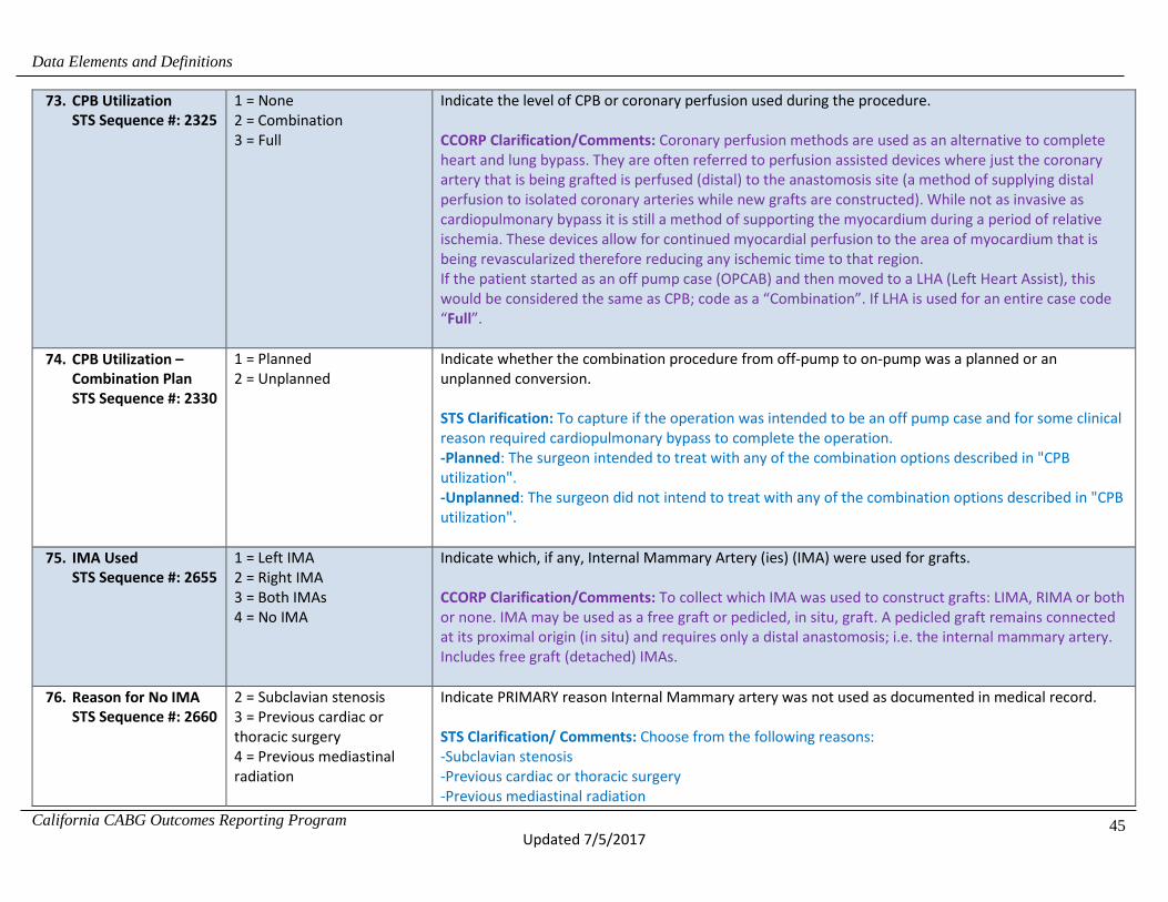
Data Elements and Definitions
California CABG Outcomes Reporting Program Updated 7/5/2017
45
73. CPB Utilization STS Sequence #: 2325
1 = None 2 = Combination 3 = Full
Indicate the level of CPB or coronary perfusion used during the procedure. CCORP Clarification/Comments: Coronary perfusion methods are used as an alternative to complete heart and lung bypass. They are often referred to perfusion assisted devices where just the coronary artery that is being grafted is perfused (distal) to the anastomosis site (a method of supplying distal perfusion to isolated coronary arteries while new grafts are constructed). While not as invasive as cardiopulmonary bypass it is still a method of supporting the myocardium during a period of relative ischemia. These devices allow for continued myocardial perfusion to the area of myocardium that is being revascularized therefore reducing any ischemic time to that region. If the patient started as an off pump case (OPCAB) and then moved to a LHA (Left Heart Assist), this would be considered the same as CPB; code as a “Combination”. If LHA is used for an entire case code “Full”.
74. CPB Utilization – Combination Plan STS Sequence #: 2330
1 = Planned 2 = Unplanned
Indicate whether the combination procedure from off-pump to on-pump was a planned or an unplanned conversion. STS Clarification: To capture if the operation was intended to be an off pump case and for some clinical reason required cardiopulmonary bypass to complete the operation. -Planned: The surgeon intended to treat with any of the combination options described in "CPB utilization". -Unplanned: The surgeon did not intend to treat with any of the combination options described in "CPB utilization".
75. IMA Used STS Sequence #: 2655
1 = Left IMA 2 = Right IMA 3 = Both IMAs 4 = No IMA
Indicate which, if any, Internal Mammary Artery (ies) (IMA) were used for grafts. CCORP Clarification/Comments: To collect which IMA was used to construct grafts: LIMA, RIMA or both or none. IMA may be used as a free graft or pedicled, in situ, graft. A pedicled graft remains connected at its proximal origin (in situ) and requires only a distal anastomosis; i.e. the internal mammary artery. Includes free graft (detached) IMAs.
76. Reason for No IMA STS Sequence #: 2660
2 = Subclavian stenosis 3 = Previous cardiac or thoracic surgery 4 = Previous mediastinal radiation
Indicate PRIMARY reason Internal Mammary artery was not used as documented in medical record. STS Clarification/ Comments: Choose from the following reasons: -Subclavian stenosis -Previous cardiac or thoracic surgery -Previous mediastinal radiation

Data Elements and Definitions
California CABG Outcomes Reporting Program Updated 7/5/2017
46
5 = Emergent or salvage procedure 6 = No (bypassable) LAD disease 7 = Other
-Emergent or salvage procedure -No (BYPASSABLE) LAD disease - This can include clean LAD, diffusely diseased LAD or other condition resulting in the LAD not being bypassed -Other – The National Quality Forum (NQF) AND CCORP do not consider this exclusion for measure purposes. Example: The physician did not use an IMA because of paralysis of the right hemi diaphragm. This does not fit into any of the category choices. – Code as “other” and it is not an acceptable exclusion for IMA usage. STS FAQ 10/2016: How should exclusion be coded for the patient with AV fistula in the left arm for dialysis? Code subclavian stenosis. STS FAQ 10/2016: The physician did not use an IMA because of paralysis of the right hemi diaphragm. This does not fit into any of the category choices. What do you suggest I do and will this count against us? Answer: Code as “other”, and it is not an acceptable exclusion for IMA usage. STS FAQ 10/2016: How should trauma to the IMA be coded in the exclusions? Answer: This should be coded as “other”.
77. Valve STS Sequence #: 2125
1 = Yes 2 = No
Indicate whether a surgical procedure was done on the Aortic, Mitral, Tricuspid or Pulmonic valves CCORP Clarification/Comments: includes valve replacements and/or repairs.
78. Aortic Valve STS Sequence #: 3390
2 = No 3 = Yes, planned 4 = Yes, unplanned due to surgical complication 5 = Yes, unplanned due to unsuspected disease or anatomy
Indicate whether an aortic valve procedure was performed. CCORP Clarification/Comments: Include all AV procedures (aortic valve replacement, resuspension or repair- see below) done during this surgery.

Data Elements and Definitions
California CABG Outcomes Reporting Program Updated 7/5/2017
47
79. Aortic Valve Procedure STS Sequence #: 3395
1 = Replacement 2 = Repair/Reconstruction 3 = Root Replacement with valved conduit (Bentall) 5 = Resuspension AV without replacement of ascending aorta 6 = Resuspension AV with replacement of ascending aorta 7 = Apico-aortic conduit (Aortic valve bypass) 8 = Autograft with pulmonary valve (Ross Procedure) 9 = Homograft root replacement 10 = Valve sparing root reimplantation (David) 11 = Valve sparing root remodeling (Yacoub) 13 = Replacement AV and insertion aortic non-valved conduit in supra-coronary position 14 = Replacement AV and major root reconstruction/debridement with valved conduit 15 = Valve sparing root reconstruction (Florida Sleeve)
Indicate the type of procedure that was performed on the aortic valve and/or ascending aorta.

Data Elements and Definitions
California CABG Outcomes Reporting Program Updated 7/5/2017
48
80. Mitral Valve STS Sequence #: 3495
2 = No 3 = Yes, planned 4 = Yes, unplanned due to surgical complication 5 = Yes, unplanned due to unsuspected disease or anatomy
Indicate whether a mitral valve procedure was performed.
81. Mitral Valve Procedure STS Sequence #: 3500
1 = Repair 2 = Replacement
Indicate the type of procedure that was performed on the mitral valve.
82. Tricuspid Valve STS Sequence #: 3640
2 = No 3 = Yes, planned 4 = Yes, unplanned due to surgical complication 5 = Yes, unplanned due to unsuspected disease or anatomy
Indicate whether a tricuspid valve procedure was performed.
83. Tricuspid Procedure STS Sequence #: 3645
2 = Annuloplasty only 3 = Replacement 4 = Reconstruction with annuloplasty 5 = Reconstruction without annuloplasty 6 = Valvectomy
Indicate the type of procedure that was performed on the tricuspid valve.

Data Elements and Definitions
California CABG Outcomes Reporting Program Updated 7/5/2017
49
84. Pulmonic Valve STS Sequence #: 3685
2 = No 3 = Yes, planned 4 = Yes, unplanned due to surgical complication 5 = Yes, unplanned due to unsuspected disease or anatomy
Indicate whether a pulmonic valve procedure was performed.
85. Pulmonic Procedure STS Sequence #: 3690
2 = Replacement 3 = Reconstruction 4 = Valvectomy
Indicate the type of procedure that was performed on the pulmonic valve.
86. Reoperation for Bleed STS Sequence #: 4755
1 = Yes 2 = No
Indicate whether the patient was re-explored for mediastinal bleeding with or without tamponade either in the ICU or returned to the operating room. CCORP Clarification/Comments: Requires reopening the chest for bleeding. STS: Do not capture reopening of the chest or situations of excessive bleeding that occur prior to the patient leaving the operating room at the time of the primary procedure. Do not include medically (non-operatively) treated excessive post-operative bleeding/tamponade events. The patient must return to the operating room suite for surgical intervention. Include patients that return to an O.R. suite or equivalent O.R. environment (i.e., ICU setting) as identified by your institution, that require surgical re-intervention to investigate/correct bleeding/tamponade. Include only those bleeding/tamponade interventions that pertain to the mediastinum or thoracic cavity.
87. Reintervention – Graft Occlusion STS Sequence #: 4770
2 = No 3 = Yes, surgical 4 = Yes, PCI
Indicate whether the patient returned to the operating room or the cath lab for intervention of coronary graft occlusion due to acute closure, thrombosis, technical or embolic origin. STS Clarification: Only capture surgical or cath lab interventions that occur during the hospitalization. Q: Previously, only returns to the OR were counted as reoperation. Are cath lab procedures for graft occlusion now counted? A: Yes, if PCI was performed for graft occlusion due to thrombosis, acute closure, emboli or technical issues code yes to this field. The patient after a CAB requires a return to the cath lab for an intervention in the native coronary artery, is this coded as Reop graft occlusion?

Data Elements and Definitions
California CABG Outcomes Reporting Program Updated 7/5/2017
50
Answer: NO, this should be coded as Reop Other Cardiac. If a native vessel requires intervention or reintervention post op, code as Reop Other Cardiac, since this is different from graft occlusion.
88. Deep Sternal Infection/ Mediastinitis STS Sequence #: 4700
2 = No 3 = Yes, within 30 days of procedure 4 = Yes, >30 days after procedure, but during hospitalization for surgery
Indicate whether a deep sternal wound infection or mediastinitis was diagnosed within 30 days of the procedure or any time during the hospitalization for surgery. STS Clarification: The STS Composite scores weigh deep sternal wound infection and mediastinitis the same.
89. Neuro – Stroke Permanent STS Sequence #: 4810
2 = No 3 = Yes, hemorrhagic 4 = Yes, embolic 5 = Yes, undetermined type
Indicate whether the patient has a postoperative stroke and the type of stroke (i.e., any confirmed neurological deficit of abrupt onset caused by a disturbance in blood supply to the brain) that did not resolve within 24 hours. STS Clarification/Comments: Neurological deficits such as confusion, delirium and/or encephalopatic (anoxic or metabolic) events are not to be coded in this field. Q: A patient had a Coronary Artery Bypass (CAB) and Carotid Artery Endarterectomy (CEA) done by a cardiac surgeon and a vascular surgeon. The patient had a stroke, and it was documented in the notes that it was from the CEA. How should this be coded? A: The stroke is coded as a postoperative complication of the CAB. Q: How should the following be coded? The patient was being sedated, but stopped withdrawing to painful stimuli on one side. A neuro consult suggested a CVA on the left side and ordered a CT Scan. The patient expired later on the same day as the consult before the test could be performed to determine if a CVA has occurred. A: This neurologic deficit would be coded as Stroke Permanent.
90. Pulm – Ventilation Prolonged STS Sequence #: 4835
1 = Yes 2 = No
Indicate whether the patient had prolonged post-operative pulmonary ventilation > 24.0 hours. The hours of postoperative ventilation time include OR exit until extubation, plus any additional hours following reintubation. Include (but not limited to) causes such as ARDS, pulmonary edema, and/or any patient requiring mechanical ventilation > 24 hours postoperatively.

Data Elements and Definitions
California CABG Outcomes Reporting Program Updated 7/5/2017
51
CCORP Clarification/Comments: Postoperative period begins when patient leaves the O.R. A total of 24 hours, include initial and additional hours of mechanical ventilation. Do not include the hours ventilated if a patient returns to the operating room suite and requires re intubation as part of general anesthesia. TIME is calculated from the point of leaving the O.R. and NOT when patient was initially intubated. STS Clarification: To calculate total hours, include initial and additional hours of mechanical ventilation. Extended ventilation may include, but is not limited to, the specific definitional reasons. Example: If a major stroke or coma occurred that required ventilation for life support, code as prolonged if greater than 24 hours. Do not include the hours ventilated if a patient returns to the operating room suite and requires re-intubation as part of general anesthesia. Q: How should one code when a patient is ventilated prior to cardiac surgery? A: Do not code as a complication unless the hours ventilated post-op are > 24 hours. Q: A patient has been long-term ventilator dependent PRIOR to his CABG. Six months prior to the current hospitalization, the patient suffered multiple complications, including a tracheostomy, from disease processes and non-cardiac surgery. How should this be coded? A: Due to the language in the definition (...any patient requiring mechanical ventilation > 24 hours postoperatively) and for consistent coding, you will need to code the prolonged ventilation field for this patient as "Yes." Hopefully, the acuity of this patient will be captured in the co-morbidities/risk factors. Q: How does one code if a patient is extubated five hours after surgery and reintubated during the same hospital stay for an additional 20 hours? A: Count a total of 24 hours, including initial and additional hours of mechanical ventilation. For this example, code “Yes” to Prolonged Ventilation.
91. Renal – Renal Failure STS Sequence #: 4870
1 = Yes 2 = No
Indicate whether the patient had acute renal failure or worsening renal function resulting in ONE OR BOTH of the following: A) Increase in serum creatinine level 3.0 x greater than baseline, or serum creatinine level ≥4 mg/dL, Acute rise must be at least 0.5 mg/dl B) A new requirement for dialysis postoperatively. STS Clarification: The postoperative creatinine will be used to evaluate renal function according to the RIFLE criteria. The Acute Dialysis Quality Initiative, a multidisciplinary collaboration, defined a range of

Data Elements and Definitions
California CABG Outcomes Reporting Program Updated 7/5/2017
52
acute renal dysfunction called the RIFLE classification system. It is used to define grades of severity based on objective measurements. STS will use the underlined serum creatinine values to analyze post-op renal function. GFR and urine output will not be included at this time. Renal Failure criteria are highlighted. Classifications of Loss and End-stage disease are beyond the current scope of follow-up. See RIFLE description below. Risk (R) - Increase in serum creatinine level X 1.5 or decrease in GFR by 25%, or UO <0.5 mL/kg/h for 6 hours Injury (I) - Increase in serum creatinine level X 2.0 or decrease in GFR by 50%, or UO <0.5 mL/kg/h for 12 hours Failure (F) ‐ Increase in serum creatinine level X 3.0, or serum creatinine level ≥4 mg/dL with at least a 0.5 mg/dl rise, or decrease in GFR by 75%,; UO <0.3 mL/kg/h for 24 hours, or anuria for 12 hours Loss (L) - Persistent ARF, complete loss of kidney function >4 weeks End-stage kidney disease (E) - Loss of kidney function >3 months Reference: http://ccforum.com/content/8/4/R204 The analysis exclusion criteria for renal failure excludes those with preop dialysis or preop creatinine ≥ 4.0. STS FAQ 10/2016: Patient had initially normal creatinine, had MI with cardiogenic shock, shock liver, and ARF requiring HD preoperatively. Had perma-cath placed with HD. Preop creatinine peaked at 4.76. Two weeks later creatinine down to 1.05. Then had CAB. Post op creatinine tripled and received one dialysis session. Would this patient be considered as renal failure postoperatively? Answer: Because the patient was not undergoing dialysis at the time of hospitalization for the index procedure and the preoperative creatinine was normal (1.05), code yes, the patient does have renal failure postoperatively with a creatinine 3X baseline (4.76) and dialysis.
92. Renal – Dialysis Requirement STS Sequence #: 4875
1 = Yes 2 = No
Indicate whether the patient had a new requirement for dialysis postoperatively, which may include hemodialysis, peritoneal dialysis. STS Clarification/Comments: May include either hemo or peritoneal dialysis. This includes a onetime need for dialysis as well as implementation of longer term therapy. If the patient was on preoperative peritoneal dialysis and moved to hemodialysis postoperatively, this does not constitute a worsening of the condition and should not be coded as an event. Continuous Veno-Venous Hemofiltration) (CVVH,

Data Elements and Definitions
California CABG Outcomes Reporting Program Updated 7/5/2017
53
CVVH-D) and Continuous Renal Replacement Therapy (CRRT) should be coded here as “Yes.” (Code Ultra filtration as “No”, it is captured in a separate field.) If preoperative dialysis = “yes”, you may automatically code post op renal failure and dialysis ‘no”.
93. Other – A Fib STS Sequence #: 4930
1 = Yes 2 = No
Indicate whether the patient experienced atrial fibrillation/flutter (AF) requiring treatment. Exclude patients who were in A Fib at the start of surgery. STS Clarification: Include any episode of A Fib lasting longer than one hour and/or requiring treatment. Capture event(s) in all patients who were not in A Fib at the start of surgery. Q: A patient is on beta blockers post-op and is titrating each day to give higher doses. The second post-op day the patient has a two hour run of A Fib. During this run of A Fib, the beta blocker is increased or an extra dose of beta blocker is given. Should this be coded as post-op A Fib? A: This is considered a post-op A Fib event. Q: A patient is on a protocol preoperatively; the patient then goes in to atrial fibrillation (AF) post-operatively and the protocol is not adjusted. Does this qualify as post-op A Fib? A: If the patient was in sinus rhythm and then develops AF postoperatively, this should be coded “Yes” as a post op event. CCORP Clarification/Comments: Count as post op A fib any episodes lasting > =1 hour regardless of treatment unless patient was in A fib at time of induction of anesthesia. STS FAQ 07/2016 : Prior to admission, pt is in NSR. In the operative report section, “Findings at Operation: Paroxysmal afib. The pt developed afib intraoperatively." This is noted after CPB was commenced and the pt was observed to have a large LAA. Consent is for AVR only, but a modified MAZE (LAA and PVI) is also performed unexpectedly. 1. Is it best to capture the afib post op or pre op? Intraop Afib wouldn’t be in either the preop or postop section. The patient was in NSR preop. 2. Modified MAZE was unexpected. Does this get captured somewhere? If so, then which element? Answer: Unless the patient has atrial fibrillation post operatively, it is neither preoperative nor postoperative complication. You do not need to code the LAA as an unexpected procedure. Code the modified maze for the PVI and also code the LAA. This is NOT postop Afib.
Data Elements and Definitions
California CABG Outcomes Reporting Program Updated 7/5/2017
54
94. Facility Identification Number CCORP-specific variable
The six-digit facility identification number assigned to a hospital by the Office of Statewide Health Planning and Development (OSHPD), as defined in Section 97170.
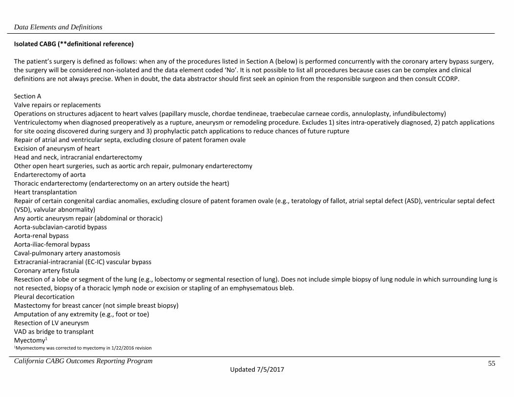
Data Elements and Definitions
California CABG Outcomes Reporting Program Updated 7/5/2017
55
Isolated CABG (**definitional reference) The patient’s surgery is defined as follows: when any of the procedures listed in Section A (below) is performed concurrently with the coronary artery bypass surgery, the surgery will be considered non-isolated and the data element coded ‘No’. It is not possible to list all procedures because cases can be complex and clinical definitions are not always precise. When in doubt, the data abstractor should first seek an opinion from the responsible surgeon and then consult CCORP. Section A Valve repairs or replacements Operations on structures adjacent to heart valves (papillary muscle, chordae tendineae, traebeculae carneae cordis, annuloplasty, infundibulectomy) Ventriculectomy when diagnosed preoperatively as a rupture, aneurysm or remodeling procedure. Excludes 1) sites intra-operatively diagnosed, 2) patch applications for site oozing discovered during surgery and 3) prophylactic patch applications to reduce chances of future rupture Repair of atrial and ventricular septa, excluding closure of patent foramen ovale Excision of aneurysm of heart Head and neck, intracranial endarterectomy Other open heart surgeries, such as aortic arch repair, pulmonary endarterectomy Endarterectomy of aorta Thoracic endarterectomy (endarterectomy on an artery outside the heart) Heart transplantation Repair of certain congenital cardiac anomalies, excluding closure of patent foramen ovale (e.g., teratology of fallot, atrial septal defect (ASD), ventricular septal defect (VSD), valvular abnormality) Any aortic aneurysm repair (abdominal or thoracic) Aorta-subclavian-carotid bypass Aorta-renal bypass Aorta-iliac-femoral bypass Caval-pulmonary artery anastomosis Extracranial-intracranial (EC-IC) vascular bypass Coronary artery fistula Resection of a lobe or segment of the lung (e.g., lobectomy or segmental resection of lung). Does not include simple biopsy of lung nodule in which surrounding lung is not resected, biopsy of a thoracic lymph node or excision or stapling of an emphysematous bleb. Pleural decortication Mastectomy for breast cancer (not simple breast biopsy) Amputation of any extremity (e.g., foot or toe) Resection of LV aneurysm VAD as bridge to transplant Myectomy1
1Myomectomy was corrected to myectomy in 1/22/2016 revision

Data Elements and Definitions
California CABG Outcomes Reporting Program Updated 7/5/2017
56
If a procedure listed in Section B is performed concurrently with the coronary artery bypass surgery, the surgery will be considered an isolated CABG and the data element coded ‘Yes’ (unless a procedure listed in section A is performed during the same surgery). These particular procedures are listed because the Office has received frequent questions regarding their coding.
Section B Transmyocardial laser revascularization (TMR) Pericardiectomy and excision of lesions of heart Repair/restoration of the heart or pericardium. Coronary endarterectomy Pacemakers Internal cardiac defibrillators (ICDs) Fem-fem cardiopulmonary bypass (a form of cardiopulmonary bypass that should not be confused with aortofemoral bypass surgery listed in Section A) Thymectomy Thyroidectomy Maze procedures, surgical or catheter Plication of LV aneurysm Impella Responsible Surgeon Name (**definitional reference): “Responsible surgeon” means the principle surgeon who performs a coronary artery bypass procedure. The first and last name collected should exactly match the name assigned to the license number issued by the California Medical Board. The middle initial collected should match the first letter of the middle name assigned to the license number issued by the California Medical Board. Example: if a surgeon’s middle name is Harry, the middle initial should be reported as ‘H’. NOTE: do not include period (.). If a trainee performs this procedure, then the responsible surgeon is the physician responsible for supervising this procedure performed by the trainee. In situations in which a responsible surgeon cannot otherwise be determined, the responsible surgeon is the surgeon who bills for the coronary artery bypass procedure.

Data Elements and Definitions
California CABG Outcomes Reporting Program Updated 7/5/2017
57
Isolated CABG (revised definition) for Jan 1 2016
Was the surgery an Isolated CABG, CABG + Valve, or other Non-Isolated CABG?
Valid Values
1=Isolated CABG 3= CABG + Valve 4=Other Non-Isolated CABG
Definition Type of CABG Exclusions from Isolated CABG:
• Valve repairs or replacements • Operations on structures adjacent to heart valves (papillary muscle, chordae tendineae, traebeculae carneae cordis, annuloplasty, infundibulectomy) • Ventriculectomy when diagnosed preoperatively as a rupture, aneurysm or remodeling procedure. Excludes 1) sites intra-operatively diagnosed, 2) patch
applications for site oozing discovered during surgery and 3) prophylactic patch applications to reduce chances of future rupture • Repair of atrial and ventricular septa, excluding closure of patent foramen ovale • Excision of aneurysm of heart • Head and neck, intracranial endarterectomy • Other open heart surgeries, such as aortic arch repair, pulmonary endarterectomy • Endarterectomy of aorta • Thoracic endarterectomy (endarterectomy on an artery outside the heart) • Carotid endarterectomy • Heart transplantation • Repair of certain congenital cardiac anomalies, excluding closure of patent foramen ovale (e.g., teratology of fallot, atrial septal defect (ASD), ventricular septal
defect (VSD), valvular abnormality) • Any aortic aneurysm repair (abdominal or thoracic) • Aorta-subclavian-carotid bypass • Aorta-renal bypass • Aorta-iliac-femoral bypass • Caval-pulmonary artery anastomosis

Data Elements and Definitions
California CABG Outcomes Reporting Program Updated 7/5/2017
58
• Extracranial-intracranial (EC-IC) vascular bypass • Coronary artery fistula • Resection of a lobe or segment of the lung (e.g., lobectomy or segmental resection of lung). Does not include simple biopsy of lung nodule in which
surrounding lung is not resected, biopsy of a thoracic lymph node or excision or stapling of an emphysematous bleb. • Pleural decortication • Mastectomy for breast cancer (not simple breast biopsy) • Amputation of any extremity (e.g., foot or toe) • Resection of LV aneurysm • Ventricular Assist Device (VAD) as bridge to transplant • Septal myectomy with hypertrophic obstructive cardiomyopathy • Full open mazes • Repair of aortic dissection
Clarification for Isolated CABG CABG + Valve
CABG + Valve includes all CABG cases with aortic valve replacement (AVR), mitral valve replacement (MVR), mitral valve repair (MVRepair) and AVR +MVR/MVRepair
Exclusions from CABG + Valve:
• Pulmonic Valve Procedure • Tricuspid Valve Procedure • Ventriculectomy when diagnosed preoperatively as a rupture, aneurysm or remodeling procedure. Excludes 1) sites intra-operatively diagnosed, 2) patch
applications for site oozing discovered during surgery and 3) prophylactic patch applications to reduce chances of future rupture • Repair of atrial and ventricular septa, excluding closure of patent foramen ovale • Excision of aneurysm of heart • Head and neck, intracranial endarterectomy • Other open heart surgeries, such as aortic arch repair, pulmonary endarterectomy • Endarterectomy of aorta • Thoracic endarterectomy (endarterectomy on an artery outside the heart) • Carotid endarterectomy • Heart transplantation • Repair of congenital cardiac anomalies, such as tetralogy of fallot, atrial septal defect (ASD), ventricular septal defect or other complex anomaly • Any aortic aneurysm repair (abdominal or thoracic) • Repair of aortic dissection
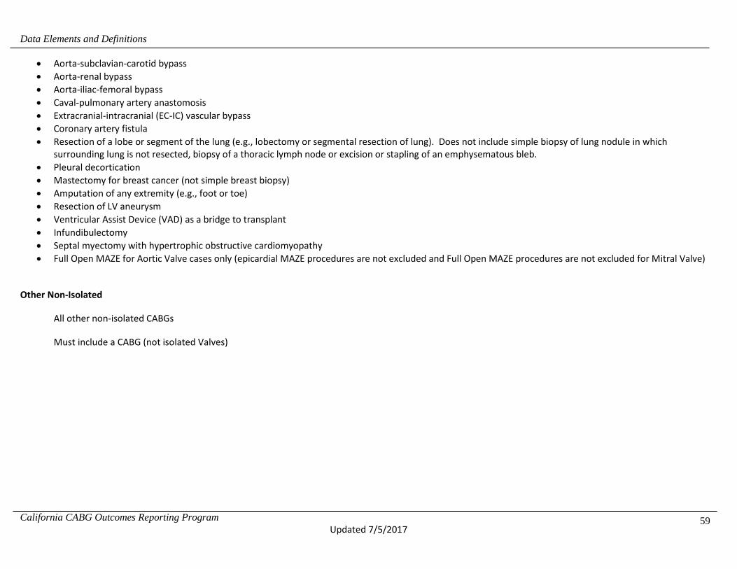
Data Elements and Definitions
California CABG Outcomes Reporting Program Updated 7/5/2017
59
• Aorta-subclavian-carotid bypass • Aorta-renal bypass • Aorta-iliac-femoral bypass • Caval-pulmonary artery anastomosis • Extracranial-intracranial (EC-IC) vascular bypass • Coronary artery fistula • Resection of a lobe or segment of the lung (e.g., lobectomy or segmental resection of lung). Does not include simple biopsy of lung nodule in which
surrounding lung is not resected, biopsy of a thoracic lymph node or excision or stapling of an emphysematous bleb. • Pleural decortication • Mastectomy for breast cancer (not simple breast biopsy) • Amputation of any extremity (e.g., foot or toe) • Resection of LV aneurysm • Ventricular Assist Device (VAD) as a bridge to transplant • Infundibulectomy • Septal myectomy with hypertrophic obstructive cardiomyopathy • Full Open MAZE for Aortic Valve cases only (epicardial MAZE procedures are not excluded and Full Open MAZE procedures are not excluded for Mitral Valve)
Other Non-Isolated
All other non-isolated CABGs
Must include a CABG (not isolated Valves)