molecular morphogenesis of t-cell acute...
TRANSCRIPT
Chapter 1
Molecular Morphogenesis of T-Cell Acute Leukemia
Michael Litt, Bhavita Patel, Ying Li, Yi Qiu andSuming Huang
Additional information is available at the end of the chapter
http://dx.doi.org/10.5772/55144
1. Introduction
Many molecular alterations are involved in the morphogenesis of T-cell acute leukemia (T-ALL), classified as lymphoblastic leukemia/lymphoma by the World Health Organization. T-ALL is a malignant disease of the thymocytes which accounts for approximately 15% ofpediatric acute lymphoblastic leukemia (ALL) and 20-25% of adult ALL. Frequently, it presentswith a high tumor load accompanied by rapid disease progression. About 30% of T-ALL casesrelapse within the first two years following diagnosis with long term remission in 70-80% ofchildren and 40% of adults [1]-[4]. This poor prognosis is a consequent of our insufficientknowledge of the molecular mechanisms underlying abnormal T-cell pathogenesis. Under‐standing the abnormal molecular changes associated with T-ALL biology will provide us withthe tools for better diagnosis and treatment of lymphoblastic leukemia. Recent improvementsin genome-wide profiling methods have identified several genetic aberrations which areassociated with T-ALL pathogenesis. For simplification these molecular changes can beseparated into 4 different groups: chromosome aberrations, gene mutations, gene expressionprofiles, and epigenetic alterations. This chapter will discuss these molecular changes in depth.
2. T-cell development
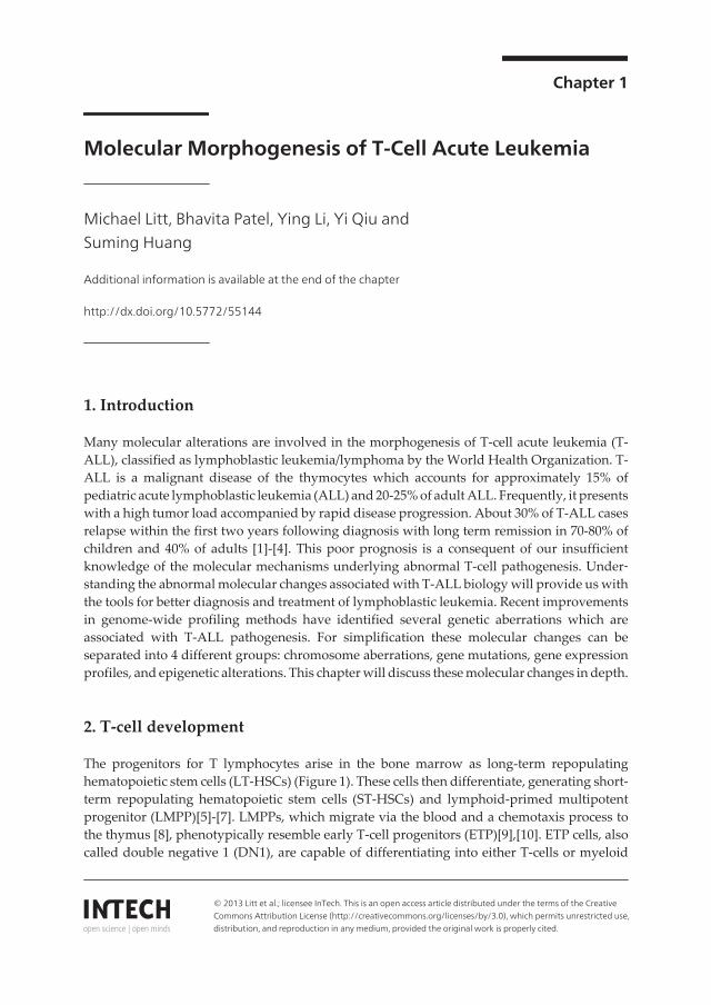
The progenitors for T lymphocytes arise in the bone marrow as long-term repopulatinghematopoietic stem cells (LT-HSCs) (Figure 1). These cells then differentiate, generating short-term repopulating hematopoietic stem cells (ST-HSCs) and lymphoid-primed multipotentprogenitor (LMPP)[5]-[7]. LMPPs, which migrate via the blood and a chemotaxis process tothe thymus [8], phenotypically resemble early T-cell progenitors (ETP)[9],[10]. ETP cells, alsocalled double negative 1 (DN1), are capable of differentiating into either T-cells or myeloid
© 2013 Litt et al.; licensee InTech. This is an open access article distributed under the terms of the CreativeCommons Attribution License (http://creativecommons.org/licenses/by/3.0), which permits unrestricted use,distribution, and reproduction in any medium, provided the original work is properly cited.
cells and phenotypically belong to a CD3-CD4-/lowCD8-CD25-CD44-KIT+ (Figures 1 and 2). IfETP cells commit to the T-cell lineage they progress to double negative 2 (DN2), followed bydouble negative 3 (DN3) and finally to double negative 4 (DN4) T-cell development stages.This process starts with the downregulation of c-KIT receptor resulting in the cell surfacephenotype CD4-CD8-CD25+CD44- for DN2 cells, next CD44 is lost for a cell surface phenotypeof CD4-CD8-CD25+CD44- for DN3 cells, and finally CD25 is lost for a cell surface phenotype ofCD4-CD8-CD25-CD44- for DN4 cells (Figures 1 and 2) [9],[11]-[13]. This differentiation fromETP to DN4 cells occurs within the thymus in intimate contact with the epithelial stromal cells,which express Notch ligands, essential growth factors (interleukin-7), and morphogens (sonichedgehog proteins) important for T-cell development. Before differentiation into doublepositive cells (DP) which have the cell surface phenotype CD4+CD8+, DN4 cells lose theirdependence on Notch ligand, interleukin-7 and sonic hedgehog (Shh) [14],[15]. Once they areDP cells, they undergo positive and negative selection. Following selection, αβ T-cell receptor(TCR)+ T cells migrate from the thymus to secondary lymphoid organs to manifest theirimmune function. These mature cells are single positive (SP) with the cell surface phenotypeof either CD4+ or CD8+ [9],[11].
Figure 1. Stages in T-cell development. The different regions of the adult thymic lobule are indicated to the rights. Theprogression of hematopoietic stem cells (HSC), multipotent progenitors (MPP), and the common lymphoid progeni‐tors (CLPs) are shown to the left in the bone marrow. Lymphoid progenitors migrated through the blood to the thy‐mus. The migration and differentiation from immigrant precursor to early T-cell precursors (ETP), to double negative(DN), to double positive (DP), and to single positive (SP) stages is illustrated within the distinct microenvironments ofthe thymus. Complete commitment to the T-cell lineage is indicated with a line between the DN2b and DN3a stages. βor γδ selection is indicated between the DN3a and DN3b stages. This figure is modified form Aifnatis 2008 and Roth‐enberg 2008 [9],[11]
T-Cell Leukemia - Characteristics, Treatment and Prevention2
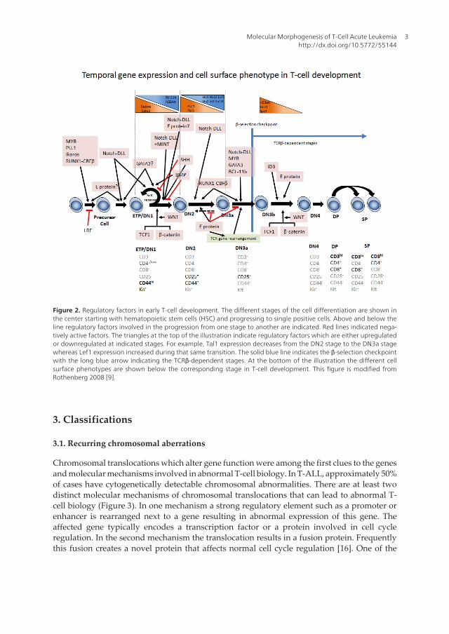
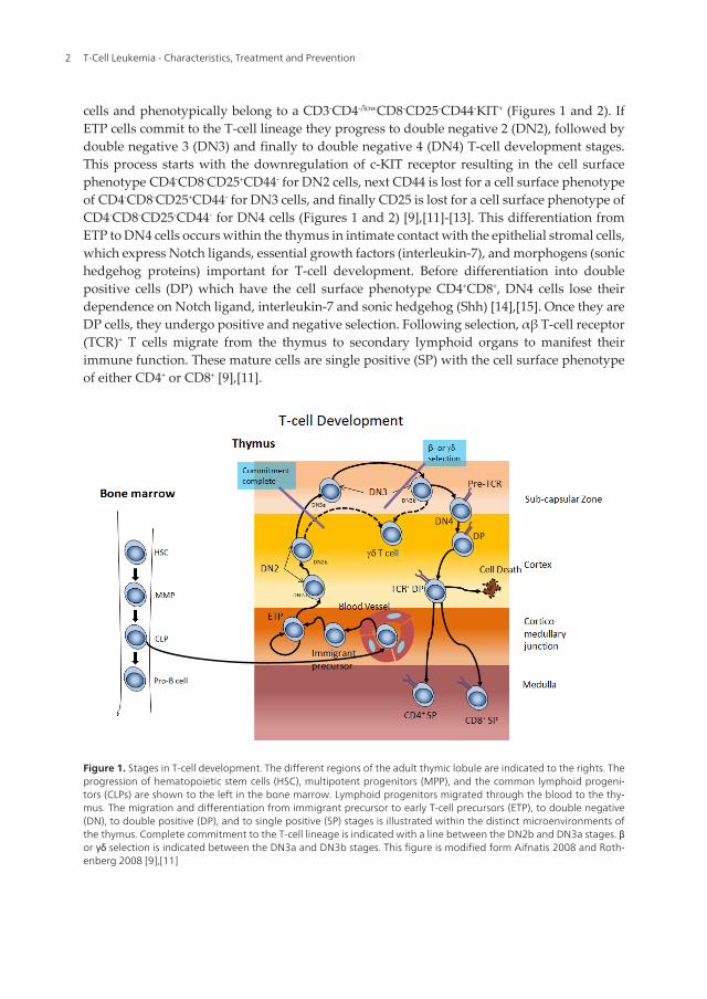
Figure 2. Regulatory factors in early T-cell development. The different stages of the cell differentiation are shown inthe center starting with hematopoietic stem cells (HSC) and progressing to single positive cells. Above and below theline regulatory factors involved in the progression from one stage to another are indicated. Red lines indicated nega‐tively active factors. The triangles at the top of the illustration indicate regulatory factors which are either upregulatedor downregulated at indicated stages. For example, Tal1 expression decreases from the DN2 stage to the DN3a stagewhereas Lef1 expression increased during that same transition. The solid blue line indicates the β-selection checkpointwith the long blue arrow indicating the TCRβ-dependent stages. At the bottom of the illustration the different cellsurface phenotypes are shown below the corresponding stage in T-cell development. This figure is modified fromRothenberg 2008 [9].
3. Classifications
3.1. Recurring chromosomal aberrations
Chromosomal translocations which alter gene function were among the first clues to the genesand molecular mechanisms involved in abnormal T-cell biology. In T-ALL, approximately 50%of cases have cytogenetically detectable chromosomal abnormalities. There are at least twodistinct molecular mechanisms of chromosomal translocations that can lead to abnormal T-cell biology (Figure 3). In one mechanism a strong regulatory element such as a promoter orenhancer is rearranged next to a gene resulting in abnormal expression of this gene. Theaffected gene typically encodes a transcription factor or a protein involved in cell cycleregulation. In the second mechanism the translocation results in a fusion protein. Frequentlythis fusion creates a novel protein that affects normal cell cycle regulation [16]. One of the
Molecular Morphogenesis of T-Cell Acute Leukemiahttp://dx.doi.org/10.5772/55144
3
hallmark features of T-ALL is translocations involving T-cell receptor genes, which areobserved in majority of T-ALL patients. The bulk of these recurring aberrations involve strongtranscriptional regulator elements from the T-cell receptor (TCR) genes being juxtaposed withgenes encoding transcription factors. These alterations are frequently caused by erroneousV(D)J recombination events during T-cell development. Overall these chromosomal abnor‐malities lead to aberrant gene expression and proteins that alter normal growth, differentia‐tion, and survival of T-cells and their precursors.
Figure 3. Two mechanisms of aberrant activities caused by chromosomal translocations. A. A strong promoter or en‐hancer is rearranged next to a proto-oncogene resulting in abnormal expression of the proto-oncogene. The TCR locielements and recurring gene targets involved in T-cell leukemogenesis are indicated to the left. B. Chromosomal rear‐rangement between two transcription factors result in a chimeric transcription factor with oncogenic activity. Recur‐ring gene fusions in T-cell leukemogenesis are indicated in the center below the arrow.
T-Cell Leukemia - Characteristics, Treatment and Prevention4
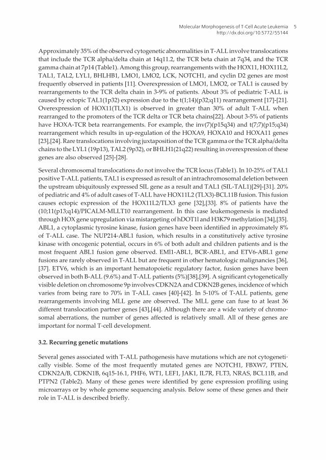
Approximately 35% of the observed cytogenetic abnormalities in T-ALL involve translocationsthat include the TCR alpha/delta chain at 14q11.2, the TCR beta chain at 7q34, and the TCRgamma chain at 7p14 (Table1). Among this group, rearrangements with the HOX11, HOX11L2,TAL1, TAL2, LYL1, BHLHB1, LMO1, LMO2, LCK, NOTCH1, and cyclin D2 genes are mostfrequently observed in patients [11]. Overexpression of LMO1, LMO2, or TAL1 is caused byrearrangements to the TCR delta chain in 3-9% of patients. About 3% of pediatric T-ALL iscaused by ectopic TAL1(1p32) expression due to the t(1;14)(p32;q11) rearrangement [17]-[21].Overexpression of HOX11(TLX1) is observed in greater than 30% of adult T-ALL whenrearranged to the promoters of the TCR delta or TCR beta chains[22]. About 3-5% of patientshave HOXA-TCR beta rearrangements. For example, the inv(7)(p15q34) and t(7;7)(p15;q34)rearrangement which results in up-regulation of the HOXA9, HOXA10 and HOXA11 genes[23],[24]. Rare translocations involving juxtaposition of the TCR gamma or the TCR alpha/deltachains to the LYL1 (19p13), TAL2 (9p32), or BHLH1(21q22) resulting in overexpression of thesegenes are also observed [25]-[28].
Several chromosomal translocations do not involve the TCR locus (Table1). In 10-25% of TAL1positive T-ALL patients, TAL1 is expressed as result of an intrachromosomal deletion betweenthe upstream ubiquitously expressed SIL gene as a result and TAL1 (SIL-TAL1)[29]-[31]. 20%of pediatric and 4% of adult cases of T-ALL have HOX11L2 (TLX3)-BCL11B fusion. This fusioncauses ectopic expression of the HOX11L2/TLX3 gene [32],[33]. 8% of patients have the(10;11(p13;q14)/PICALM-MLLT10 rearrangement. In this case leukemogenesis is mediatedthrough HOX gene upregulation via mistargeting of hDOT1l and H3K79 methylation [34],[35].ABL1, a cytoplasmic tyrosine kinase, fusion genes have been identified in approximately 8%of T-ALL case. The NUP214-ABL1 fusion, which results in a constitutively active tyrosinekinase with oncogenic potential, occurs in 6% of both adult and children patients and is themost frequent ABL1 fusion gene observed. EMl1-ABL1, BCR-ABL1, and ETV6-ABL1 genefusions are rarely observed in T-ALL but are frequent in other hematologic malignancies [36],[37]. ETV6, which is an important hematopoietic regulatory factor, fusion genes have beenobserved in both B-ALL (9.6%) and T-ALL patients (5%)[38],[39]. A significant cytogeneticallyvisible deletion on chromosome 9p involves CDKN2A and CDKN2B genes, incidence of whichvaries from being rare to 70% in T-ALL cases [40]-[42]. In 5-10% of T-ALL patients, generearrangements involving MLL gene are observed. The MLL gene can fuse to at least 36different translocation partner genes [43],[44]. Although there are a wide variety of chromo‐somal aberrations, the number of genes affected is relatively small. All of these genes areimportant for normal T-cell development.
3.2. Recurring genetic mutations
Several genes associated with T-ALL pathogenesis have mutations which are not cytogeneti‐cally visible. Some of the most frequently mutated genes are NOTCH1, FBXW7, PTEN,CDKN2A/B, CDKN1B, 6q15-16.1, PHF6, WT1, LEF1, JAK1, IL7R, FLT3, NRAS, BCL11B, andPTPN2 (Table2). Many of these genes were identified by gene expression profiling usingmicroarrays or by whole genome sequencing analysis. Below some of these genes and theirrole in T-ALL is described briefly.
Molecular Morphogenesis of T-Cell Acute Leukemiahttp://dx.doi.org/10.5772/55144
5

Recurring Translocations in T-ALL
TCR Rearrangements Non-TCR Rerrangements
Gene Rearrangemen
t
Frequency Gene Rearrangement Frequency
TAL1 t(1;14)
(p32;q11)
t(1;7)(p32;q34)
~3 of T-ALL TAL1 STIL-TAL1 (1p32 deletion) 12-25% T-
ALL
TAL2 t(7;9)(q34;q32) rare HOXA PICALM-MLLT10 (t(10;11)
(p13;q14))
MLL-MLLT1 (t(11;19)
(q23;p13))
SET-NUP214 9q34 deletions
LMO1 t(11;14)
(p15;q11)
t(7;11)
(q34;p15)
6-8% of T-ALL ABL1 EML1-ABL1 (t(9:14)
(q34;q32))
BCR-ABL1 (t(9;22)(q34;q11))
ETV6-ABl1 (t(9;12)(q34;p13))
NUP214-ABL1
8% T-ALL
for ABL1
6% T-ALL
for NUP214
LMO2 t(11;14)
(p13;q11)
t(7;11)
(q34;p13)
11p13 deletions
ETV6 ETV6-JAK2 (t(9;12)(p24;p13)
ETV6-ARNT (t(1;12)(q21;p13)
Rare
HOX11 t(10;14)
(q24;q11)
t(7;10)
(q34;q24)
30% of T-ALL
HOX11L2 t(5;14)
(q35;q32)
20% Childhood
T-ALL
4% Adult T-ALL
HOXA Inv(7)(p15q34)
t(7;7)(p15;q34)
LYL1 t(7;19)
(q34;p13)
rare
Table 1. Table of recurring translocation involved in T-ALL. The rearrangements are divided into those involving TCRand non-TCR loci.
T-Cell Leukemia - Characteristics, Treatment and Prevention6
Recurring genetic alterations in T-ALL
Gene Alteration Frequency
Notch1 Sequence mutations ~50% of T-ALL
FBW7 Sequence mutations ~20% of T-ALL
PTEN Deletions/Sequence mutations 6-8% of T-ALL
CDKN2A/B Deletions 30-70% of T-ALL
CDKN1B Deletions/Sequence mutations 12% of T-ALL
6q15-16.1 Deletions 12% of T-ALL
PHF6 Deletions/Sequence mutations 16% of childhood T-ALL38% of adult T-ALL
WT1 Frameshift mutations 13% childhood T-ALL12% of adult T-ALL
LEF1 Focal deletions/sequence mutations 15% of childhood T-ALL
JAK1 Sequence mutations 18% of adult T-ALL
IL7R Gain of function mutation 9% of T-ALL
FLT3 Internal tandem duplication 4% of adult T-ALL3% of childhood T-ALL
NRAS Sequence mutations 10% childhood T-ALL
BCL11 Deletions/Sequence mutations 9% of all T-ALL case16% of T-ALL cases with HOX11overexpression
PTPN2 Deletion 6% of T-ALL
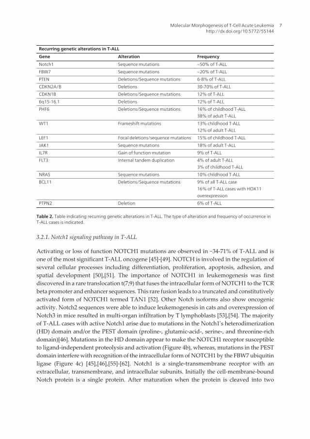
Table 2. Table indicating recurring genetic alterations in T-ALL. The type of alteration and frequency of occurrence inT-ALL cases is indicated.
3.2.1. Notch1 signaling pathway in T-ALL
Activating or loss of function NOTCH1 mutations are observed in ~34-71% of T-ALL and isone of the most significant T-ALL oncogene [45]-[49]. NOTCH is involved in the regulation ofseveral cellular processes including differentiation, proliferation, apoptosis, adhesion, andspatial development [50],[51]. The importance of NOTCH1 in leukemogenesis was firstdiscovered in a rare translocation t(7;9) that fuses the intracellular form of NOTCH1 to the TCRbeta promoter and enhancer sequences. This rare fusion leads to a truncated and constitutivelyactivated form of NOTCH1 termed TAN1 [52]. Other Notch isoforms also show oncogenicactivity. Notch2 sequences were able to induce leukemogenesis in cats and overexpression ofNotch3 in mice resulted in multi-organ infiltration by T lymphoblasts [53],[54]. The majorityof T-ALL cases with active Notch1 arise due to mutations in the Notch1's heterodimerization(HD) domain and/or the PEST domain (proline-, glutamic-acid-, serine-, and threonine-richdomain)[46]. Mutations in the HD domain appear to make the NOTCH1 receptor susceptibleto ligand-independent proteolysis and activation (Figure 4b), whereas, mutations in the PESTdomain interfere with recognition of the intracellular form of NOTCH1 by the FBW7 ubiquitinligase (Figure 4c) [45],[46],[55]-[62]. Notch1 is a single-transmembrane receptor with anextracellular, transmembrane, and intracellular subunits. Initially the cell-membrane-boundNotch protein is a single protein. After maturation when the protein is cleaved into two
Molecular Morphogenesis of T-Cell Acute Leukemiahttp://dx.doi.org/10.5772/55144
7
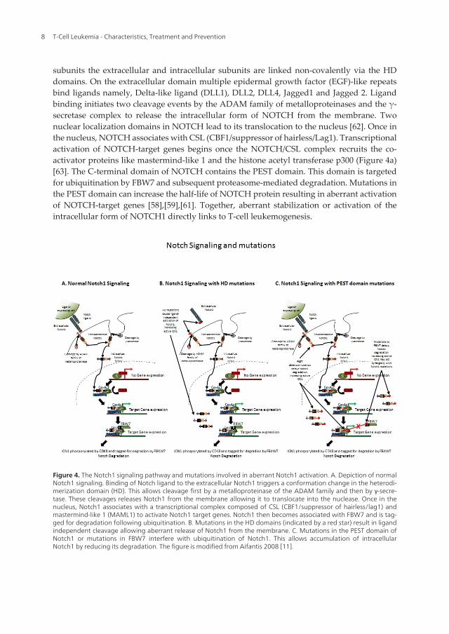
subunits the extracellular and intracellular subunits are linked non-covalently via the HDdomains. On the extracellular domain multiple epidermal growth factor (EGF)-like repeatsbind ligands namely, Delta-like ligand (DLL1), DLL2, DLL4, Jagged1 and Jagged 2. Ligandbinding initiates two cleavage events by the ADAM family of metalloproteinases and the γ-secretase complex to release the intracellular form of NOTCH from the membrane. Twonuclear localization domains in NOTCH lead to its translocation to the nucleus [62]. Once inthe nucleus, NOTCH associates with CSL (CBF1/suppressor of hairless/Lag1). Transcriptionalactivation of NOTCH-target genes begins once the NOTCH/CSL complex recruits the co-activator proteins like mastermind-like 1 and the histone acetyl transferase p300 (Figure 4a)[63]. The C-terminal domain of NOTCH contains the PEST domain. This domain is targetedfor ubiquitination by FBW7 and subsequent proteasome-mediated degradation. Mutations inthe PEST domain can increase the half-life of NOTCH protein resulting in aberrant activationof NOTCH-target genes [58],[59],[61]. Together, aberrant stabilization or activation of theintracellular form of NOTCH1 directly links to T-cell leukemogenesis.
Figure 4. The Notch1 signaling pathway and mutations involved in aberrant Notch1 activation. A. Depiction of normalNotch1 signaling. Binding of Notch ligand to the extracellular Notch1 triggers a conformation change in the heterodi‐merization domain (HD). This allows cleavage first by a metalloproteinase of the ADAM family and then by γ-secre‐tase. These cleavages releases Notch1 from the membrane allowing it to translocate into the nuclease. Once in thenucleus, Notch1 associates with a transcriptional complex composed of CSL (CBF1/suppressor of hairless/lag1) andmastermind-like 1 (MAML1) to activate Notch1 target genes. Notch1 then becomes associated with FBW7 and is tag‐ged for degradation following ubiquitination. B. Mutations in the HD domains (indicated by a red star) result in ligandindependent cleavage allowing aberrant release of Notch1 from the membrane. C. Mutations in the PEST domain ofNotch1 or mutations in FBW7 interfere with ubiquitination of Notch1. This allows accumulation of intracellularNotch1 by reducing its degradation. The figure is modified from Aifantis 2008 [11].
T-Cell Leukemia - Characteristics, Treatment and Prevention8
Because NOTCH1 plays a significant role in T-cell leukemogenesis, its regulation has beenstudied extensively. Nearly 40% of Notch-responsive genes are regulators of cell metabolismand protein biosynthesis [64]. c-MYC, a master regulator of multiple biosynthesis andmetabolic pathways, is a direct transcriptional target of Notch1. Notch1 binding sites in theMYC promoter have been shown to be important for MYC expression in T-ALL [64]-[67].Constitutively active Notch1 was shown to activate the NF-κB pathway [68], an importantregulator of cell survival, cell cycle, cell adhesion and cell migration. This activation can occurby the direct transcriptional activation of Relb and Nfkb2 as well as via a Notch1 and IKKcomplex interaction. Another Notch1 target is PTEN (phosphatase and tension homologue).PTEN is negatively regulated by Notch1 through the activity of HES1 and MYC, resulting inthe deregulation of the PI3K-AKT metabolic pathway [69]. Finally, Notch1 is also involved inthe regulation of the NFAT signaling pathway, where it regulates the pathway by alteringexpression of calcineurin, a calcium-activated phosphatase [70]. Overall, these findingsemphasize the role of Notch1 in inducing T-cell leukemogenesis through multiple cellsignaling pathways capable of regulating cell survival, proliferation and metabolism.
As mentioned above, FBW7 (F-box and WD repeat domain containing 7), an E3 ubiquitin ligaselocated on chromosome 4q31.3, is observed to be mutated in T-ALL with a frequency rangingfrom 8.6% to 16% [59],[61],[71]. FBW7 is part of the SCF complex (SKP1-Cullin-1-F box proteincomplex), which can target MYC, JUN, cyclin E, and Notch1 for ubiquitination coupledproteosomal degradation [60]. The WD40 domain of FBW7 contains a degron-binding pocketdomain. This domain recognizes phosphothreonine in the consensus sequence I/L/P-T-P-X-X-S/E of protein substrates. Roughly 20% of T-ALL patients have mutations in FBW7 thatdestroys the degron-binding pocket. Moreover, the degron sequence of Notch1 (LTPSPES)located in the distal portion of its PEST domain is found to be mutated in T-ALL, thus extendingNotch1 half-life and altering downstream signaling cascades. Interestingly, T-ALL patientsfrequently have mutations in both the FBW7 degron binding pocket as well as in the Notch1degron sequence (Figure 4c) [58],[59],[61]. These combined mutations elevate intracellularNotch1 activity and therefore, enhances leukemia manifestation. Current studies suggestFBW7 mutations induce T-cell leukemogenesis by disrupting Notch1 regulation.
PTEN (phosphatase and tensin homolog deleted on chromosome 10) is deleted or mutated in6-8% of T-ALL cases. The major substrate of PTEN is PIP3 (phosphatidylinositol-3,4,5-triphosphate). PTEN activity prevents the accumulation of PIP3, thus limiting or terminatingactivation of a cascade of PI3K-dependent signaling molecules. The expression of PTEN hasbeen shown to be negatively regulated by Notch1. PTEN appears to be required for optimalnegative selection in the thymus. Loss of PTEN is characterized by overexpression of the c-myc oncogene and induction of lymphomagenesis within the thymus [69],[72]. ThereforePTEN appears to be an important tumor-suppressor involved in T-cell leukemogenesis.
3.2.2. Cell cycle, apoptosis, and transcription regulators in T-ALL
Deletions in CDKN2A and CDKN2B are significant secondary abnormities in pediatric T-ALL.Loss of the tumor suppressor CDKN2A/B expression is observed in 30-70% of T-ALL casesand can occur due to chromosomal translocation, promoter hypermethylation, somatic
Molecular Morphogenesis of T-Cell Acute Leukemiahttp://dx.doi.org/10.5772/55144
9

mutation, or gene deletions [40],[42]. CDKN2A and CDKN2B are located adjacent on chro‐mosome 9p21. CDKN2A encodes p16INK4a(cyclin-dependent kinase inhibitor)/p14ARFwhileCDKN2B encodes p15INKb. These genes block cell division during the G1/S phase of the cellcycle by inhibiting cyclin/CDK-4/6 complexes [73],[74]. The principle mode of CDKN2Ainactivation occurs via genomic deletions which can usually be detected by FISH [41]. Loss offunction of CDKN1B (cyclin-dependent kinase inhibitor 1B) gene, located on 12p13.2, havebeen observed in 12% of T-ALL cases [75]. Similar to CDKN2A and CDKN2B, CDKN1B actsas a tumor suppressor. Inactivation of CDKN1B leads to overexpression of D-cyclins, therebyinhibiting the cells ability to maintain quiescence in G0. Therefore, CDKN2/B and CDKN1Bplay an important role in abnormal T-cell biology by regulating cell cycle progression.
12% of pediatric T-ALL cases have deletion in 6q15-16.1 [75]. The single most down regulatedgene in this region is caspase 8 associated protein 2 (CASP8AP2). Deletion of CASP8AP2probably interferes with Fas-mediated apoptosis. In gene expression profiling study, loss ofCASP8AP2 was not observed in any pre-B-ALL samples [75], indicating deletions to 6q15-16.1maybe a hallmark of T-ALL.
The X-linked plant homeodomain (PHD) finger 6 (PHF6) gene has inactivating mutations in16% of pediatric and 38% of adult primary T-ALL cases [76]. Mutations in PHF6 are limited tomale T-ALL cases. Consequently, this gene may be responsible for the increased incidence ofT-ALL cases in males. Loss of expression of the PHF6 gene was associated with leukemiadriven by abnormal expression of the homeobox transcription factor oncogenes. PHF6 geneencodes a protein with two plant homeodomain-like zinc finger domains. A recent studydemonstrated that PHF6 copurifies with the nucleosome remodeling and deacetylation(NuRD) complex, implicating its role in chromatin regulation [77].
The WT1 (Wilms tumor) tumor suppressor gene is mutated in 13.2% of pediatric and 11.7% ofadult T-ALL cases [78],[79]. The WT1 is known to be a transcriptional activator of the eryth‐ropoietin gene. Loss of WT1 expression results in diminished erythropoietin receptor (EpoR)expression in hematopoietic progenitors, suggesting that activation of the EpoR gene by Wt1is an important mechanism in normal hematopoiesis [80]. WT1 mutations are frequentlyprevalent in T-ALL cases harboring chromosomal rearrangements associated with abnormalexpression of the homeobox transcription oncogenes, HOX11, HOX11L2, and HOXA9 [79].This suggests that the recurrent genetic mutations in WT1 are associated with abnormal HOXgene expression in T-ALL period
Lymphoid enhance factor 1 (LEF1) gene is mutated in 15% of pediatric T-ALL cases [81].Inactivation of LEF1 was associated with increased expression of MYC and MYC targets, agene expression signature consistent with developmental arrest at a cortical stage of T-celldifferentiation. Interestingly, T-ALL cases with LEF1 mutation lacked overexpression of TAL1,HOX11, HOX11L2 and HOXA genes suggesting that LEF1 acts via different molecularpathways in T-cell leukemogenesis. In fact, The LEF family of DNA-binding transcriptionfactors interacts with nuclear β-catenin in the WNT signaling pathway. The loss of LEF1 mayresult in the relief of transcriptional repression of MYC in T-ALL cases. It was reported thatLEF1 probably contributes to T-ALL pathogenesis by acting in concert with NOTCH1 to
T-Cell Leukemia - Characteristics, Treatment and Prevention10

promote up-regulation of MYC expression. In this case LEF1 also relieves transcriptionalrepression of MYC to allow its maximum overexpression by Notch1 [81].
3.2.3. JAK/STAT signaling pathway in T-ALL
About 18% of adult and 2% of pediatric T-ALL cases have activating mutations in the JanusKinase 1 (JAK1) [38]. The JAK family (JAK1, JAK2, JAK3, and TYK2) function as signaltransducers to control cell proliferation, survival, and differentiation. They are nonrecep‐tor tyrosine kinases that associate with cytokine receptors to phosphorylate tyrosine residuesof the target proteins. This process regulates the recruitment and activation of STAT proteins.The JAK/STAT signaling cascade is an important regulator of normal T-cell development.Each JAK family member associates with a different subset of cytokine receptors. JAK1regulates the class II cytokine receptors as well as receptors that use the gp130 or γc receptorsubunit. These class of cytokine receptors are involved in controlling lymphoid develop‐ment [82],[83]. The majority of the JAK1 kinase mutations observed in T-ALL cases resultin unregulated tyrosine kinase activity. T-ALL cases with mutations in JAK1 appear to beassociated with different T-ALL subgroups than patients harboring aberrant expressions ofthe homeobox transcription factors HOX11 and HOX11L2 [38]. JAK1 is involved in theregulation of both interleukin 7 receptor (IL7R) and protein tyrosine phosphatase non-receptor type 2 (PTPN2) [84],[85].
The interleukin 7 receptor (IL7R) has a gain-of-function mutation in exon 6 in 9% of T-ALL cases [85]. Several lines of evidence suggest IL7R plays an important role in T-cellleukemogenesis. IL-7 and IL7R signaling are essential for normal T-cell development.Deficiency of IL-7 and IL7R in mice caused reduction of non-functional T cells and showedan early block in thymocyte development [86]-[89]. Loss of IL7R function also results insevere combined immunodeficiency in humans [90]. Increased expression of IL7R wasassociated with spontaneous thymic lymphomas in mice. Furthermore, Notch1 has beenshown to transcriptionally upregulate IL7R receptor gene [91]. Mutations in exon 6 of IL7Rpromotes de novo formation of intermolecular disulfide bonds between IL7R mutantsubunits, which triggers constitutive activation of tyrosine kinase JAK1 regardless ofregulation by IL-7, γc, or JAK3. Gene expression profiles for IL7R mutations are generallyassociated with the T-ALL subgroup harboring HOX11L2 rearrangements and HOXAderegulation [85].
Inactivation of protein tyrosine phosphatase non-receptor type 2 (PTPN2) gene is ob‐served in ~6% of T-ALL cases [84],[92]. PTPN2 encodes a tyrosine phosphatase, located onchromosome 18p11.3-11.2, that negatively regulates JAK/STAT pathway and NUP214-ABL1 kinase activity. Loss of PTPN2 results in activation of the JAK/STAT pathway andincreased T-cell proliferation by cytokines. Unlike JAK1 mutations, deletions in PTPN2 geneappear to be restricted to T-ALL cases which specifically overexpress HOX11 [84]. There‐fore mutations in PTPN2 probably play a role in T-cell leukemogenesis by deregulatingtyrosine kinase signaling.
Activating mutations in the FMS-like tyrosine kinase 3 (FLT3) gene are amongst the mostcommon genetic aberrations in acute myeloid leukemia [93]-[95]. In T-ALL, FLT3 mutations
Molecular Morphogenesis of T-Cell Acute Leukemiahttp://dx.doi.org/10.5772/55144
11
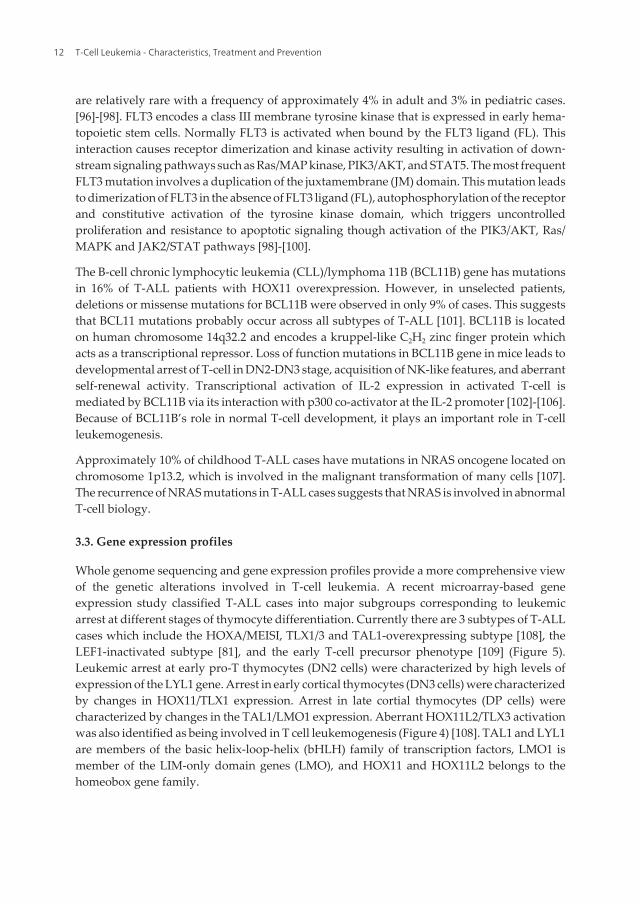
are relatively rare with a frequency of approximately 4% in adult and 3% in pediatric cases.[96]-[98]. FLT3 encodes a class III membrane tyrosine kinase that is expressed in early hema‐topoietic stem cells. Normally FLT3 is activated when bound by the FLT3 ligand (FL). Thisinteraction causes receptor dimerization and kinase activity resulting in activation of down‐stream signaling pathways such as Ras/MAP kinase, PIK3/AKT, and STAT5. The most frequentFLT3 mutation involves a duplication of the juxtamembrane (JM) domain. This mutation leadsto dimerization of FLT3 in the absence of FLT3 ligand (FL), autophosphorylation of the receptorand constitutive activation of the tyrosine kinase domain, which triggers uncontrolledproliferation and resistance to apoptotic signaling though activation of the PIK3/AKT, Ras/MAPK and JAK2/STAT pathways [98]-[100].
The B-cell chronic lymphocytic leukemia (CLL)/lymphoma 11B (BCL11B) gene has mutationsin 16% of T-ALL patients with HOX11 overexpression. However, in unselected patients,deletions or missense mutations for BCL11B were observed in only 9% of cases. This suggeststhat BCL11 mutations probably occur across all subtypes of T-ALL [101]. BCL11B is locatedon human chromosome 14q32.2 and encodes a kruppel-like C2H2 zinc finger protein whichacts as a transcriptional repressor. Loss of function mutations in BCL11B gene in mice leads todevelopmental arrest of T-cell in DN2-DN3 stage, acquisition of NK-like features, and aberrantself-renewal activity. Transcriptional activation of IL-2 expression in activated T-cell ismediated by BCL11B via its interaction with p300 co-activator at the IL-2 promoter [102]-[106].Because of BCL11B’s role in normal T-cell development, it plays an important role in T-cellleukemogenesis.
Approximately 10% of childhood T-ALL cases have mutations in NRAS oncogene located onchromosome 1p13.2, which is involved in the malignant transformation of many cells [107].The recurrence of NRAS mutations in T-ALL cases suggests that NRAS is involved in abnormalT-cell biology.
3.3. Gene expression profiles
Whole genome sequencing and gene expression profiles provide a more comprehensive viewof the genetic alterations involved in T-cell leukemia. A recent microarray-based geneexpression study classified T-ALL cases into major subgroups corresponding to leukemicarrest at different stages of thymocyte differentiation. Currently there are 3 subtypes of T-ALLcases which include the HOXA/MEISI, TLX1/3 and TAL1-overexpressing subtype [108], theLEF1-inactivated subtype [81], and the early T-cell precursor phenotype [109] (Figure 5).Leukemic arrest at early pro-T thymocytes (DN2 cells) were characterized by high levels ofexpression of the LYL1 gene. Arrest in early cortical thymocytes (DN3 cells) were characterizedby changes in HOX11/TLX1 expression. Arrest in late cortial thymocytes (DP cells) werecharacterized by changes in the TAL1/LMO1 expression. Aberrant HOX11L2/TLX3 activationwas also identified as being involved in T cell leukemogenesis (Figure 4) [108]. TAL1 and LYL1are members of the basic helix-loop-helix (bHLH) family of transcription factors, LMO1 ismember of the LIM-only domain genes (LMO), and HOX11 and HOX11L2 belongs to thehomeobox gene family.
T-Cell Leukemia - Characteristics, Treatment and Prevention12

Figure 5. Gene subtypes resulting in differentiation arrest at specific stages of T-cell development. The illustrationshows the progression of T-cell development from the double negative stages to the mature single positive stage. Thecolored rectangles indicates stages of leukemic arrest. Overexpression of LYL, HOX11, TAL1, and HOXA lead to differ‐entation arrest at the double negative stage, early cortical stage, late cortical stage, and positive selections stage, re‐spectively. Loss of Lef1 expression results in early cortical leukemic arrest. The table below indicates the molecularsubtypes leading to differentiation arrest at specific stages of T-cell development and the molecular subtypes occur‐ring across all the stages of T-cell development.
Recently whole genome sequencing of early T-cell precursor acute lymphoblastic leukemia(ETP-ALL) identified several genes involved in abnormal T-cell biology [10]. 15% of T-ALLcases are ETP-ALL. Phenotypically ETP-ALL is negative for the cell surface markers CD1a andCD8, has little to no expression of CD5, and has aberrant expression of myeloid and hemato‐poietic stem cell markers. This study performed whole genome sequencing on 12 children withETP-ALL. The frequency of the mutations identified from these 12 cases was then accessed in94 cases of T-ALL. Of these 94 cases 52 cases had ETP and 42 had a non-ETP pediatric T-ALL.Even though an average of 1140 sequence mutations and 12 structural variations in the genomewere identified per ETP case, they were able to narrow down the number of affected genes to3 group and 3 novel genes (DNM2, ECT2L, and RELN). 67% of the cases were characterizedby activating mutations in genes involved in the regulation of cytokine receptor and RAS
Molecular Morphogenesis of T-Cell Acute Leukemiahttp://dx.doi.org/10.5772/55144
13
signaling. These genes included NRAS, KRAS, FLT3, IL7R, JAK3, JAK1, SH2B3 and BRAF.58% of the cases were characterized by inactivating lesions that disrupted hematopoieticdevelopment. These genes included GATA3, ETV6, RUNX1, IKZF1, and EP300. 48% of thecases were characterized by changes in histone modifying genes (EZH2, EED, SUZ12, SETD2,and EP300) [10]. From gene expression profiling and whole genome sequencing we arebeginning to obtain a more complete picture of the genes involved in abnormal T-cell biology.
MicroRNA expression profiling found 10 detectable miRNAs in human T-ALL cells, five ofthese miRNAs (miR-19b, miR-20a, miR-26a, miR-92, and miR223) were predicted to targettumor suppressors genes implicated in T-ALL [110]. These five miRNA's were able to accel‐erate leukemia development in a mouse model. Furthermore, it was shown that these fivemiRNAs produced overlapping and cooperative effects of the tumor suppressor genesIKAROS, PTEN, BIM, PHF6, NF1 and FBXW7 in T-ALL pathogenesis. miR223 appears to bethe most overexpressed miRNA in leukemia. These results indicate the important role thatmiRNA's play in abnormal T-cell biology.
3.4. Basic helix-loop-helix proteins
As mentioned early, some of the most common recurrent chromosomal aberrations inabnormal T-cell biology involved chromosomal translocations of the TCR gene to the basichelix-loop-helix (bHLH) genes (MYC, TAL1, TAL2, LYL1, bHLHB1), the cysteine-rich (LIM-domain) genes (LMO1, LMO2), or the homeodomain genes (HOX11/TLX1), HOX11L2/TLX3,members of the HOXA cluster) (Table1). The most common bHLH gene with aberrantexpression observed in T-ALL cases is the transcriptional regulator TAL1 (T-cell acutelymphocytic leukemia 1; also known as SCL). It was first identified in T-ALL patients with thet(1;14)(p32;q11) translocation [17],[18],[20],[21]. This chromosomal rearrangement, which isobserved in 3% of cases, causes ectopic TAL1 expression by placing TAL1 under control of theTCRδ oncogene [19]-[21],[111]. 12%-25% of T-ALL cases have a submicroscopic 90-kb deletionthat fuses the TAL1 coding sequence to the first exon of the SIL gene (SCL interrupting locus).This rearrangement leads to dysregulation of TAL1 expression [17],[29]-[31]. The majority ofT-ALL cases, up to 60%, show ectopic TAL1 expression with no detectable TAL1 generearrangements [112]. Gene expression profiling has shown that ectopic expression of TAL1results in leukemic arrest in late cortical thymocytes (Figure 5) [108]. These results show thatactivation of TAL1 gene is required for the leukemic phenotype of T-cells.
The TAL1 gene, located on chromosome 1p32, encodes a class II basic helix-loop-helix factor[113]. The protein binds DNA as a heterodimer with the ubiquitously expressed class I bHLHgenes known as E-proteins such as E2A or HEB. These heterodimers recognize an E boxsequence (CANNTG)[114]. TAL1 positively and negatively modulates transcription of targetsgene as a large complex consisting of an E-protein, the LIM-only proteins LMO1/2, GATA1/2,Ldb1, and other associated coregulators. This complex usually binds a composite DNAelements containing an E box and a GATA-binding site separated by 9 or 10 bp (Figure 6) [115]-[117]. It was shown recently that in T-ALL cells TAL1, GATA-3, LMO1, and RUNX1 togetherform a core transcription regulatory circuit to reinforce and stabilize the TAL1-directedleukemogenic program [118].
T-Cell Leukemia - Characteristics, Treatment and Prevention14
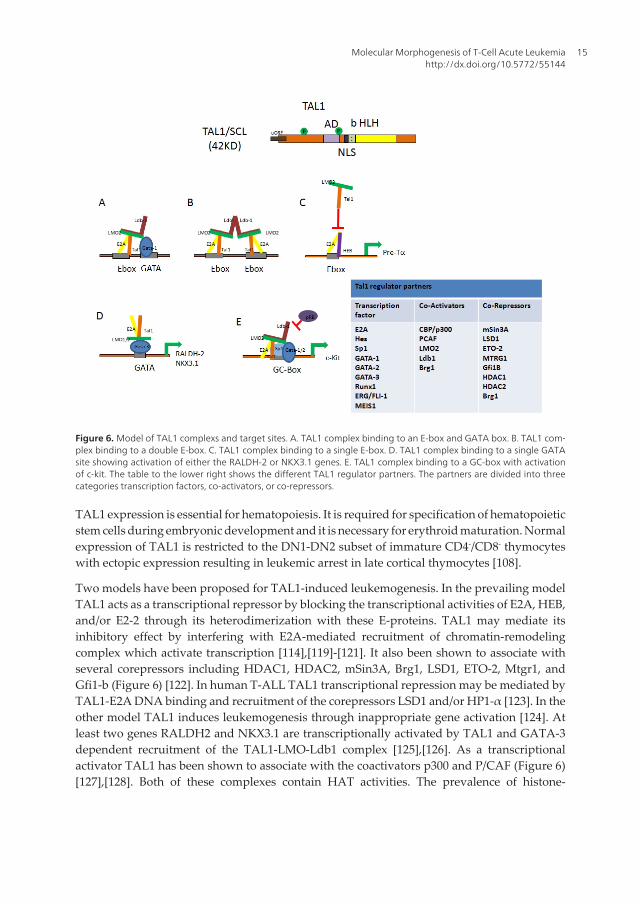
Figure 6. Model of TAL1 complexs and target sites. A. TAL1 complex binding to an E-box and GATA box. B. TAL1 com‐plex binding to a double E-box. C. TAL1 complex binding to a single E-box. D. TAL1 complex binding to a single GATAsite showing activation of either the RALDH-2 or NKX3.1 genes. E. TAL1 complex binding to a GC-box with activationof c-kit. The table to the lower right shows the different TAL1 regulator partners. The partners are divided into threecategories transcription factors, co-activators, or co-repressors.
TAL1 expression is essential for hematopoiesis. It is required for specification of hematopoieticstem cells during embryonic development and it is necessary for erythroid maturation. Normalexpression of TAL1 is restricted to the DN1-DN2 subset of immature CD4-/CD8- thymocyteswith ectopic expression resulting in leukemic arrest in late cortical thymocytes [108].
Two models have been proposed for TAL1-induced leukemogenesis. In the prevailing modelTAL1 acts as a transcriptional repressor by blocking the transcriptional activities of E2A, HEB,and/or E2-2 through its heterodimerization with these E-proteins. TAL1 may mediate itsinhibitory effect by interfering with E2A-mediated recruitment of chromatin-remodelingcomplex which activate transcription [114],[119]-[121]. It also been shown to associate withseveral corepressors including HDAC1, HDAC2, mSin3A, Brg1, LSD1, ETO-2, Mtgr1, andGfi1-b (Figure 6) [122]. In human T-ALL TAL1 transcriptional repression may be mediated byTAL1-E2A DNA binding and recruitment of the corepressors LSD1 and/or HP1-α [123]. In theother model TAL1 induces leukemogenesis through inappropriate gene activation [124]. Atleast two genes RALDH2 and NKX3.1 are transcriptionally activated by TAL1 and GATA-3dependent recruitment of the TAL1-LMO-Ldb1 complex [125],[126]. As a transcriptionalactivator TAL1 has been shown to associate with the coactivators p300 and P/CAF (Figure 6)[127],[128]. Both of these complexes contain HAT activities. The prevalence of histone-
Molecular Morphogenesis of T-Cell Acute Leukemiahttp://dx.doi.org/10.5772/55144
15
modifying enzymes in TAL1 complexes suggests that one function of TAL1 is to regulatechromatin states of its target genes.
TAL1 and the lymphoblastic leukemia-derived sequence 1 (LYL1) share 90% sequence identityin their bHLH motif [26]. Like TAL1, LYL1 role in leukemogenesis was discovered by studyingchromosomal rearrangements. It is expressed by adult hematopoietic cells and is overex‐pressed in T-ALL. Gene expression profiling showed that overexpression of LYL1 resulted inleukemic arrest at pro T-cell (Double negative) stage of T-cell differentiation (Figure 5) [108].In mouse embryos LYL1 and TAL1 expression overlaps in hematopoietic development,developing vasculature and endocardium. At the molecular level LYL1 controls expression ofseveral genes involved in the maturation and stabilization of the newly formed blood vessels[129]. Therefore, bHLH proteins play an important role in abnormal T-cell biology.
3.5. LIM domain proteins
Aberrant expression of the LMO1 and LMO2 proteins is observed in 45% of T-ALL cases. Thediscovery of the LMO1 and LMO2 genes adjacent to the chromosomal translocations t(11;14)(p15q11) and t(11;14)(p13;q11) was the first indication that these proteins were involved in T-cell leukemogenesis [130]-[132]. The LMO family (LMO1, LMO2, LMO3, and LMO4) encodesgenes that have two cysteine-rich zinc coordinating LIM domains. The LIM domain is foundin a variety of proteins including the homeodomain-containing transcription factors, kinases,and adaptors. Despite the presence of 2 zinc finger motifs, LMO1 and LMO2 genes do notappear to bind DNA. Instead the LMO proteins probably act as scaffolding protein to formmultiprotein complex through their interaction with the LIM domain binding protein 1 (LDB1)(Figure 6) [116].
Leukemogenesis by aberrant expression of LMO1 or LMO2 is thought to occur via twomechanisms. In the first mechanism aberrant expression or abnormal LMO proteins forms adysfunctional multiprotein complexes that alters the expression of the target genes by directlybinding to their promoters [133]-[136]. In the second mechanism abnormal LMO1 or LMO2complexes displace the LMO4 complex. This results in arrest of T-cell development at the DPstage [137].
LMO2 function is necessary for normal T-cell development. In fact, LMO2 has been shown tointeract with several factors involved in aberrant T-cell biology. As mentioned above TAL1 mayregulate its target genes through the TAL1-LMO-Ldb1 complex (Figure 6). Ectopic expres‐sion of LMO1 and LMO2 leads to accumulation of immature DN T cells in mice with subse‐quent leukemia manifestation with a long latency, suggesting the role of LMO is important forthe development of tumors but is not self-sufficient [26],[138],[139]. Ectopic expression of bothTAL1 and LMO1 in mice accelerated the progression to leukemogenesis (Figure 7). In this casethymic expression of the TAL1 and LMO1 oncogenes induced expansion of the ETP/DN1 toDN4 population and lead to T-ALL in ~120 days. The acquisition of a Notch1 gain-of-functionmutation was proposed to be the rationale behind this increase in leukemia penetrance. In fact,thymic expression of all three oncogenes Notch1, TAL1 and LMO1 induced T-ALL with highpenetrance in 31 days, the time necessary for clonal expansion (Figure 7) [140]. These studiessuggest that aberrant LMO proteins are key players in abnormal T-cell biology.
T-Cell Leukemia - Characteristics, Treatment and Prevention16
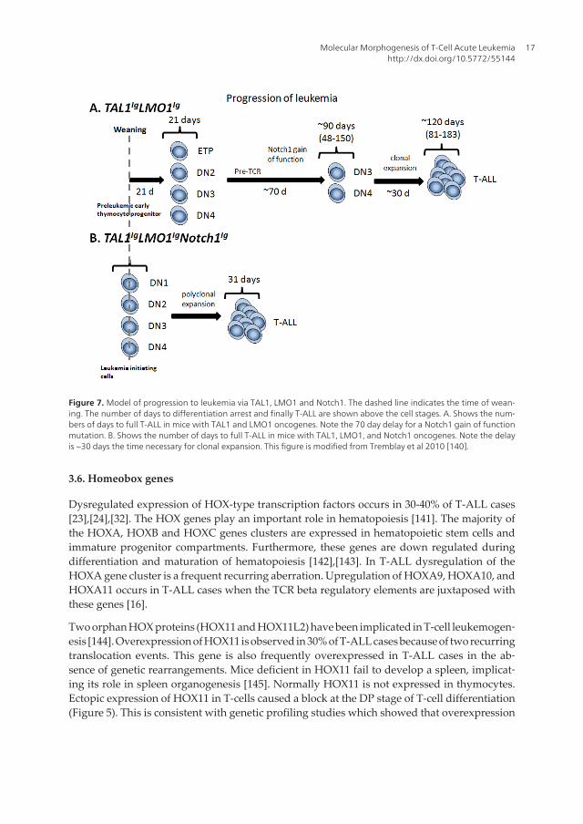
Figure 7. Model of progression to leukemia via TAL1, LMO1 and Notch1. The dashed line indicates the time of wean‐ing. The number of days to differentiation arrest and finally T-ALL are shown above the cell stages. A. Shows the num‐bers of days to full T-ALL in mice with TAL1 and LMO1 oncogenes. Note the 70 day delay for a Notch1 gain of functionmutation. B. Shows the number of days to full T-ALL in mice with TAL1, LMO1, and Notch1 oncogenes. Note the delayis ~30 days the time necessary for clonal expansion. This figure is modified from Tremblay et al 2010 [140].
3.6. Homeobox genes
Dysregulated expression of HOX-type transcription factors occurs in 30-40% of T-ALL cases[23],[24],[32]. The HOX genes play an important role in hematopoiesis [141]. The majority ofthe HOXA, HOXB and HOXC genes clusters are expressed in hematopoietic stem cells andimmature progenitor compartments. Furthermore, these genes are down regulated duringdifferentiation and maturation of hematopoiesis [142],[143]. In T-ALL dysregulation of theHOXA gene cluster is a frequent recurring aberration. Upregulation of HOXA9, HOXA10, andHOXA11 occurs in T-ALL cases when the TCR beta regulatory elements are juxtaposed withthese genes [16].
Two orphan HOX proteins (HOX11 and HOX11L2) have been implicated in T-cell leukemogen‐esis [144]. Overexpression of HOX11 is observed in 30% of T-ALL cases because of two recurringtranslocation events. This gene is also frequently overexpressed in T-ALL cases in the ab‐sence of genetic rearrangements. Mice deficient in HOX11 fail to develop a spleen, implicat‐ing its role in spleen organogenesis [145]. Normally HOX11 is not expressed in thymocytes.Ectopic expression of HOX11 in T-cells caused a block at the DP stage of T-cell differentiation(Figure 5). This is consistent with genetic profiling studies which showed that overexpression
Molecular Morphogenesis of T-Cell Acute Leukemiahttp://dx.doi.org/10.5772/55144
17
of HOX11 results in leukemic arrest at early cortical thymocytes stage (Figure 5) [108]. Overex‐pression of HOX11 in hematopoietic stems cells of mice developed T-cell leukemia. However,the long latency of tumorigenesis suggests other genetic abnormalities are required [146]-[148].It should be noted that nearly all HOX11 T-ALL cases have activating NOTCH1 mutation. Ithas been proposed that HOX11 binding to the Groucho-related TLE corepressor was necessa‐ry for maximal transcriptional regulation of Notch1-responsive genes. This suggests that HOX11and Notch1 may synergistically regulate transcription in T-ALL [149].
3.7. Epigenetic modifications
Aberrant changes in DNA methylation and histone modifications occur frequently in allcancers. Estimates vary but studies suggest that there are approximately 100 epigeneticchanges for every DNA based genetic mutation. Consequently epigenetic modifications willalmost certainly play an important role in T-cell leukemogenesis.
Comparative genomic hybridization data of T-ALL primary samples has shown recurrentdeletions in 25% of T-ALL cases in EZH2 and SUZ12 genes. These genes are members of thepolycomb repressor complex 2 (PRC2) and involved in establishing the repressive H3K27me3mark. Activation of Notch1 was shown to cause the loss of the H3K27me3 mark by antago‐nizing the activity of PRC2. This data implicates histone modifications and PRC2 as importantregulatory factors in T-cell leukemogenesis [150].
The CpG island methylator phenotype (CIMP) has been used to characterize T-ALL patients.The CIMP+ phenotype has a large number of hypermethylated genes with the CIMP- havinga low number of hypermethylated genes. Analysis of the methylation status of 20 genes, themajority of which are implicated in abnormal T-cell biology, in 61 pediatric T-ALL patientsand 11 healthy children showed a difference in the CIMP pattern. On average patients had 2.4hypermethylated loci where none of the normal individual's loci where hypermethylated[151]. Therefore changes in the patterns of CpG island methylation at critical genes can beassociated with specific tumorigenesis and consequently may be playing an important role inT-cell leukemogenesis.
4. Summary
Although there are a large number of genes involved in the molecular morphogenesis of T-cell leukemogenesis, many of the genes act through related pathways. This has helped usclarify the different genetic subtypes of T-ALL improving our risk stratification of T-ALL.Furthermore understanding the different genetic subtypes is allowing for personalizedchemotherapy. Powerful new tools such as next-generation sequencing aid in identifying morerelevant recurring lesions in leukemogenesis. This is resulting in the development of bettertherapeutic agents and methods. Because of improved supportive care, better risk stratificationand personalized chemotherapy the 5-year survival of pediatric acute lymphoblastic leukemiahas increase to 85% [152]. Even though we have made significant progress in the understanding
T-Cell Leukemia - Characteristics, Treatment and Prevention18

of the molecular morphogenesis of T-ALL there are still significant gaps in our knowledge ofthe genes involved in leukemogenesis.
Acknowledgements
We are grateful to members of the Huang laboratory for their suggestions and comments. Wealso apologize to those whose work could not be cited due to space constraints. This work wassupported by grants from the National Institute of Health (R01 HL090589 and R01 HL091929to SH; 5T32CA9126-34 to BP: R01HL095674 to YQ).
Author details
Michael Litt1, Bhavita Patel2, Ying Li2, Yi Qiu3,4 and Suming Huang1,3
1 Medical Education Center, Ball State University, Muncie, IN, USA
2 Department of Bichemistry and Molecular Biology, University of Florida, College of Medi‐cine, Gainesville, FL, USA
3 Shands Cancer Center, University of Florida, College of Medicine, Gainesville, FL, USA
4 Anatomy and Cell Biology, University of Florida, College of Medicine, Gainesville, FL,USA
References
[1] Pui CH, Relling MV, Downing JR. Acute lymphoblastic leukemia. N Engl J Med.2004;350(15):1535-1548. Prepublished on 2004/04/09 as DOI 10.1056/NEJMra023001.
[2] van Grotel M, Meijerink JP, Beverloo HB, et al. The outcome of molecular-cytogeneticsubgroups in pediatric T-cell acute lymphoblastic leukemia: a retrospective study ofpatients treated according to DCOG or COALL protocols. Haematologica. 2006;91(9):1212-1221. Prepublished on 2006/09/08 as DOI.
[3] Iacobucci I, Papayannidis C, Lonetti A, Ferrari A, Baccarani M, Martinelli G. Cytoge‐netic and molecular predictors of outcome in acute lymphocytic leukemia: recentdevelopments. Curr Hematol Malig Rep. 2012;7(2):133-143. Prepublished on 2012/04/25as DOI 10.1007/s11899-012-0122-5.
[4] Kraszewska MD, Dawidowska M, Szczepanski T, Witt M. T-cell acute lymphoblasticleukaemia: recent molecular biology findings. Br J Haematol. 2012;156(3):303-315.Prepublished on 2011/12/08 as DOI 10.1111/j.1365-2141.2011.08957.x.
Molecular Morphogenesis of T-Cell Acute Leukemiahttp://dx.doi.org/10.5772/55144
19

[5] Akashi K, Reya T, Dalma-Weiszhausz D, Weissman IL. Lymphoid precursors. CurrOpin Immunol. 2000;12(2):144-150. Prepublished on 2000/03/14 as DOI.
[6] Blom B, Spits H. Development of human lymphoid cells. Annu Rev Immunol.2006;24:287-320. Prepublished on 2006/03/23 as DOI 10.1146/annurev.immunol.24.021605.090612.
[7] Boehm T, Bleul CC. Thymus-homing precursors and the thymic microenvironment.Trends Immunol. 2006;27(10):477-484. Prepublished on 2006/08/22 as DOI 10.1016/j.it.2006.08.004.
[8] Scimone ML, Aifantis I, Apostolou I, von Boehmer H, von Andrian UH. A multistepadhesion cascade for lymphoid progenitor cell homing to the thymus. Proc Natl AcadSci U S A. 2006;103(18):7006-7011. Prepublished on 2006/04/28 as DOI 10.1073/pnas.0602024103.
[9] Rothenberg EV, Moore JE, Yui MA. Launching the T-cell-lineage developmentalprogramme. Nat Rev Immunol. 2008;8(1):9-21. Prepublished on 2007/12/22 as DOI10.1038/nri2232.
[10] Zhang J, Ding L, Holmfeldt L, et al. The genetic basis of early T-cell precursor acutelymphoblastic leukaemia. Nature. 2012;481(7380):157-163. Prepublished on 2012/01/13as DOI 10.1038/nature10725.
[11] Aifantis I, Raetz E, Buonamici S. Molecular pathogenesis of T-cell leukaemia andlymphoma. Nat Rev Immunol. 2008;8(5):380-390. Prepublished on 2008/04/19 as DOI10.1038/nri2304.
[12] Naito T, Tanaka H, Naoe Y, Taniuchi I. Transcriptional control of T-cell development.Int Immunol. 2011;23(11):661-668. Prepublished on 2011/09/29 as DOI 10.1093/intimm/dxr078.
[13] von Boehmer H, Aifantis I, Gounari F, et al. Thymic selection revisited: how essentialis it? Immunol Rev. 2003;191:62-78. Prepublished on 2003/03/05 as DOI.
[14] Di Santo JP, Radtke F, Rodewald HR. To be or not to be a pro-T? Curr Opin Immunol.2000;12(2):159-165. Prepublished on 2000/03/14 as DOI.
[15] El Andaloussi A, Graves S, Meng F, Mandal M, Mashayekhi M, Aifantis I. Hedgehogsignaling controls thymocyte progenitor homeostasis and differentiation in thethymus. Nat Immunol. 2006;7(4):418-426. Prepublished on 2006/03/07 as DOI 10.1038/ni1313.
[16] Look AT. Oncogenic transcription factors in the human acute leukemias. Science.1997;278(5340):1059-1064. Prepublished on 1997/11/14 as DOI.
[17] Aplan PD, Lombardi DP, Ginsberg AM, Cossman J, Bertness VL, Kirsch IR. Disruptionof the human SCL locus by "illegitimate" V-(D)-J recombinase activity. Science.1990;250(4986):1426-1429. Prepublished on 1990/12/07 as DOI.
T-Cell Leukemia - Characteristics, Treatment and Prevention20
[18] Begley CG, Aplan PD, Davey MP, et al. Demonstration of delta rec-pseudo J alpharearrangement with deletion of the delta locus in a human stem-cell leukemia. J ExpMed. 1989;170(1):339-342. Prepublished on 1989/07/01 as DOI.
[19] Carroll AJ, Crist WM, Link MP, et al. The t(1;14)(p34;q11) is nonrandom and restrictedto T-cell acute lymphoblastic leukemia: a Pediatric Oncology Group study. Blood.1990;76(6):1220-1224. Prepublished on 1990/09/15 as DOI.
[20] Chen Q, Cheng JT, Tasi LH, et al. The tal gene undergoes chromosome translocation inT cell leukemia and potentially encodes a helix-loop-helix protein. EMBO J. 1990;9(2):415-424. Prepublished on 1990/02/01 as DOI.
[21] Finger LR, Kagan J, Christopher G, et al. Involvement of the TCL5 gene on humanchromosome 1 in T-cell leukemia and melanoma. Proc Natl Acad Sci U S A. 1989;86(13):5039-5043. Prepublished on 1989/07/01 as DOI.
[22] Bergeron J, Clappier E, Radford I, et al. Prognostic and oncogenic relevance of TLX1/HOX11 expression level in T-ALLs. Blood. 2007;110(7):2324-2330. Prepublished on2007/07/05 as DOI 10.1182/blood-2007-04-079988.
[23] Soulier J, Clappier E, Cayuela JM, et al. HOXA genes are included in genetic andbiologic networks defining human acute T-cell leukemia (T-ALL). Blood. 2005;106(1):274-286. Prepublished on 2005/03/19 as DOI 10.1182/blood-2004-10-3900.
[24] Speleman F, Cauwelier B, Dastugue N, et al. A new recurrent inversion, inv(7)(p15q34),leads to transcriptional activation of HOXA10 and HOXA11 in a subset of T-cell acutelymphoblastic leukemias. Leukemia. 2005;19(3):358-366. Prepublished on 2005/01/28 asDOI 10.1038/sj.leu.2403657.
[25] Graux C, Cools J, Michaux L, Vandenberghe P, Hagemeijer A. Cytogenetics andmolecular genetics of T-cell acute lymphoblastic leukemia: from thymocyte to lym‐phoblast. Leukemia. 2006;20(9):1496-1510. Prepublished on 2006/07/11 as DOI 10.1038/sj.leu.2404302.
[26] Mellentin JD, Smith SD, Cleary ML. lyl-1, a novel gene altered by chromosomaltranslocation in T cell leukemia, codes for a protein with a helix-loop-helix DNAbinding motif. Cell. 1989;58(1):77-83. Prepublished on 1989/07/14 as DOI.
[27] Wang J, Jani-Sait SN, Escalon EA, et al. The t(14;21)(q11.2;q22) chromosomal translo‐cation associated with T-cell acute lymphoblastic leukemia activates the BHLHB1 gene.Proc Natl Acad Sci U S A. 2000;97(7):3497-3502. Prepublished on 2000/03/29 as DOI.
[28] Xia Y, Brown L, Yang CY, et al. TAL2, a helix-loop-helix gene activated by the (7;9)(q34;q32) translocation in human T-cell leukemia. Proc Natl Acad Sci U S A. 1991;88(24):11416-11420. Prepublished on 1991/12/25 as DOI.
[29] Bash RO, Crist WM, Shuster JJ, et al. Clinical features and outcome of T-cell acutelymphoblastic leukemia in childhood with respect to alterations at the TAL1 locus: aPediatric Oncology Group study. Blood. 1993;81(8):2110-2117. Prepublished on1993/04/15 as DOI.
Molecular Morphogenesis of T-Cell Acute Leukemiahttp://dx.doi.org/10.5772/55144
21

[30] Breit TM, Mol EJ, Wolvers-Tettero IL, Ludwig WD, van Wering ER, van Dongen JJ. Site-specific deletions involving the tal-1 and sil genes are restricted to cells of the T cellreceptor alpha/beta lineage: T cell receptor delta gene deletion mechanism affectsmultiple genes. J Exp Med. 1993;177(4):965-977. Prepublished on 1993/04/01 as DOI.
[31] Brown L, Cheng JT, Chen Q, et al. Site-specific recombination of the tal-1 gene is acommon occurrence in human T cell leukemia. EMBO J. 1990;9(10):3343-3351. Prepub‐lished on 1990/10/01 as DOI.
[32] Bernard OA, Busson-LeConiat M, Ballerini P, et al. A new recurrent and specific cryptictranslocation, t(5;14)(q35;q32), is associated with expression of the Hox11L2 gene in Tacute lymphoblastic leukemia. Leukemia. 2001;15(10):1495-1504. Prepublished on2001/10/06 as DOI.
[33] Su XY, Della-Valle V, Andre-Schmutz I, et al. HOX11L2/TLX3 is transcriptionallyactivated through T-cell regulatory elements downstream of BCL11B as a result of thet(5;14)(q35;q32). Blood. 2006;108(13):4198-4201. Prepublished on 2006/08/24 as DOI10.1182/blood-2006-07-032953.
[34] Grossmann V, Bacher U, Kohlmann A, et al. EZH2 mutations and their association withPICALM-MLLT10 positive acute leukaemia. Br J Haematol. 2012;157(3):387-390.Prepublished on 2012/01/13 as DOI 10.1111/j.1365-2141.2011.08986.x.
[35] Okada Y, Jiang Q, Lemieux M, Jeannotte L, Su L, Zhang Y. Leukaemic transformationby CALM-AF10 involves upregulation of Hoxa5 by hDOT1L. Nat Cell Biol. 2006;8(9):1017-1024. Prepublished on 2006/08/22 as DOI 10.1038/ncb1464.
[36] Hagemeijer A, Graux C. ABL1 rearrangements in T-cell acute lymphoblastic leukemia.Genes Chromosomes Cancer. 2010;49(4):299-308. Prepublished on 2010/01/15 as DOI10.1002/gcc.20743.
[37] Zipfel PA, Zhang W, Quiroz M, Pendergast AM. Requirement for Abl kinases in T cellreceptor signaling. Curr Biol. 2004;14(14):1222-1231. Prepublished on 2004/07/23 as DOI10.1016/j.cub.2004.07.021.
[38] Flex E, Petrangeli V, Stella L, et al. Somatically acquired JAK1 mutations in adult acutelymphoblastic leukemia. J Exp Med. 2008;205(4):751-758. Prepublished on 2008/03/26 asDOI 10.1084/jem.20072182.
[39] Otsubo K, Kanegane H, Eguchi M, et al. ETV6-ARNT fusion in a patient with childhoodT lymphoblastic leukemia. Cancer Genet Cytogenet. 2010;202(1):22-26. Prepublished on2010/09/02 as DOI 10.1016/j.cancergencyto.2010.07.121.
[40] Fizzotti M, Cimino G, Pisegna S, et al. Detection of homozygous deletions of the cyclin-dependent kinase 4 inhibitor (p16) gene in acute lymphoblastic leukemia and associa‐tion with adverse prognostic features. Blood. 1995;85(10):2685-2690. Prepublished on1995/05/15 as DOI.
[41] Sulong S, Moorman AV, Irving JA, et al. A comprehensive analysis of the CDKN2Agene in childhood acute lymphoblastic leukemia reveals genomic deletion, copy
T-Cell Leukemia - Characteristics, Treatment and Prevention22

number neutral loss of heterozygosity, and association with specific cytogeneticsubgroups. Blood. 2009;113(1):100-107. Prepublished on 2008/10/08 as DOI 10.1182/blood-2008-07-166801.
[42] Yamada Y, Hatta Y, Murata K, et al. Deletions of p15 and/or p16 genes as a poor-prognosis factor in adult T-cell leukemia. J Clin Oncol. 1997;15(5):1778-1785. Prepub‐lished on 1997/05/01 as DOI.
[43] Daser A, Rabbitts TH. Extending the repertoire of the mixed-lineage leukemia geneMLL in leukemogenesis. Genes Dev. 2004;18(9):965-974. Prepublished on 2004/05/11 asDOI 10.1101/gad.1195504.
[44] Turkmen S, Timmermann B, Bartels G, et al. Involvement of the MLL gene in adult T-lymphoblastic leukemia. Genes Chromosomes Cancer. 2012. Prepublished on 2012/08/29as DOI 10.1002/gcc.21996.
[45] O'Neil J, Calvo J, McKenna K, et al. Activating Notch1 mutations in mouse models ofT-ALL. Blood. 2006;107(2):781-785. Prepublished on 2005/09/17 as DOI 10.1182/blood-2005-06-2553.
[46] Weng AP, Ferrando AA, Lee W, et al. Activating mutations of NOTCH1 in human Tcell acute lymphoblastic leukemia. Science. 2004;306(5694):269-271. Prepublished on2004/10/09 as DOI 10.1126/science.1102160.
[47] Asnafi V, Buzyn A, Le Noir S, et al. NOTCH1/FBXW7 mutation identifies a largesubgroup with favorable outcome in adult T-cell acute lymphoblastic leukemia (T-ALL): a Group for Research on Adult Acute Lymphoblastic Leukemia (GRAALL)study. Blood. 2009;113(17):3918-3924. Prepublished on 2008/12/26 as DOI 10.1182/blood-2008-10-184069.
[48] Breit S, Stanulla M, Flohr T, et al. Activating NOTCH1 mutations predict favorable earlytreatment response and long-term outcome in childhood precursor T-cell lymphoblas‐tic leukemia. Blood. 2006;108(4):1151-1157. Prepublished on 2006/04/15 as DOI 10.1182/blood-2005-12-4956.
[49] Larson Gedman A, Chen Q, Kugel Desmoulin S, et al. The impact of NOTCH1, FBW7and PTEN mutations on prognosis and downstream signaling in pediatric T-cell acutelymphoblastic leukemia: a report from the Children's Oncology Group. Leukemia.2009;23(8):1417-1425. Prepublished on 2009/04/03 as DOI 10.1038/leu.2009.64.
[50] Artavanis-Tsakonas S. The molecular biology of the Notch locus and the fine tuning ofdifferentiation in Drosophila. Trends Genet. 1988;4(4):95-100. Prepublished on1988/04/01 as DOI.
[51] Sambandam A, Maillard I, Zediak VP, et al. Notch signaling controls the generationand differentiation of early T lineage progenitors. Nat Immunol. 2005;6(7):663-670.Prepublished on 2005/06/14 as DOI 10.1038/ni1216.
Molecular Morphogenesis of T-Cell Acute Leukemiahttp://dx.doi.org/10.5772/55144
23
[52] Ellisen LW, Bird J, West DC, et al. TAN-1, the human homolog of the Drosophila notchgene, is broken by chromosomal translocations in T lymphoblastic neoplasms. Cell.1991;66(4):649-661. Prepublished on 1991/08/23 as DOI.
[53] Bellavia D, Checquolo S, Campese AF, Felli MP, Gulino A, Screpanti I. Notch3: fromsubtle structural differences to functional diversity. Oncogene. 2008;27(38):5092-5098.Prepublished on 2008/09/02 as DOI 10.1038/onc.2008.230.
[54] Rohn JL, Lauring AS, Linenberger ML, Overbaugh J. Transduction of Notch2 in felineleukemia virus-induced thymic lymphoma. J Virol. 1996;70(11):8071-8080. Prepublish‐ed on 1996/11/01 as DOI.
[55] Artavanis-Tsakonas S, Rand MD, Lake RJ. Notch signaling: cell fate control and signalintegration in development. Science. 1999;284(5415):770-776. Prepublished on1999/04/30 as DOI.
[56] Gupta-Rossi N, Le Bail O, Gonen H, et al. Functional interaction between SEL-10, an F-box protein, and the nuclear form of activated Notch1 receptor. J Biol Chem.2001;276(37):34371-34378. Prepublished on 2001/06/27 as DOI 10.1074/jbc.M101343200.
[57] Hubbard EJ, Wu G, Kitajewski J, Greenwald I. sel-10, a negative regulator of lin-12activity in Caenorhabditis elegans, encodes a member of the CDC4 family of proteins.Genes Dev. 1997;11(23):3182-3193. Prepublished on 1998/02/12 as DOI.
[58] Maser RS, Choudhury B, Campbell PJ, et al. Chromosomally unstable mouse tumourshave genomic alterations similar to diverse human cancers. Nature. 2007;447(7147):966-971. Prepublished on 2007/05/23 as DOI 10.1038/nature05886.
[59] O'Neil J, Grim J, Strack P, et al. FBW7 mutations in leukemic cells mediate NOTCHpathway activation and resistance to gamma-secretase inhibitors. J Exp Med.2007;204(8):1813-1824. Prepublished on 2007/07/25 as DOI 10.1084/jem.20070876.
[60] Tetzlaff MT, Yu W, Li M, et al. Defective cardiovascular development and elevatedcyclin E and Notch proteins in mice lacking the Fbw7 F-box protein. Proc Natl Acad SciU S A. 2004;101(10):3338-3345. Prepublished on 2004/02/10 as DOI 10.1073/pnas.0307875101.
[61] Thompson BJ, Buonamici S, Sulis ML, et al. The SCFFBW7 ubiquitin ligase complex asa tumor suppressor in T cell leukemia. J Exp Med. 2007;204(8):1825-1835. Prepublishedon 2007/07/25 as DOI 10.1084/jem.20070872.
[62] Guruharsha KG, Kankel MW, Artavanis-Tsakonas S. The Notch signalling system:recent insights into the complexity of a conserved pathway. Nat Rev Genet. 2012;13(9):654-666. Prepublished on 2012/08/08 as DOI 10.1038/nrg3272.
[63] Barrick D, Kopan R. The Notch transcription activation complex makes its move. Cell.2006;124(5):883-885. Prepublished on 2006/03/15 as DOI 10.1016/j.cell.2006.02.028.
[64] Palomero T, Lim WK, Odom DT, et al. NOTCH1 directly regulates c-MYC and activatesa feed-forward-loop transcriptional network promoting leukemic cell growth. Proc Natl
T-Cell Leukemia - Characteristics, Treatment and Prevention24

Acad Sci U S A. 2006;103(48):18261-18266. Prepublished on 2006/11/23 as DOI 10.1073/pnas.0606108103.
[65] Klinakis A, Szabolcs M, Politi K, Kiaris H, Artavanis-Tsakonas S, Efstratiadis A. Mycis a Notch1 transcriptional target and a requisite for Notch1-induced mammarytumorigenesis in mice. Proc Natl Acad Sci U S A. 2006;103(24):9262-9267. Prepublishedon 2006/06/06 as DOI 10.1073/pnas.0603371103.
[66] Sharma VM, Calvo JA, Draheim KM, et al. Notch1 contributes to mouse T-cell leukemiaby directly inducing the expression of c-myc. Mol Cell Biol. 2006;26(21):8022-8031.Prepublished on 2006/09/07 as DOI 10.1128/MCB.01091-06.
[67] Weng AP, Millholland JM, Yashiro-Ohtani Y, et al. c-Myc is an important direct targetof Notch1 in T-cell acute lymphoblastic leukemia/lymphoma. Genes Dev. 2006;20(15):2096-2109. Prepublished on 2006/07/19 as DOI 10.1101/gad.1450406.
[68] Vilimas T, Mascarenhas J, Palomero T, et al. Targeting the NF-kappaB signalingpathway in Notch1-induced T-cell leukemia. Nat Med. 2007;13(1):70-77. Prepublishedon 2006/12/19 as DOI 10.1038/nm1524.
[69] Palomero T, Sulis ML, Cortina M, et al. Mutational loss of PTEN induces resistance toNOTCH1 inhibition in T-cell leukemia. Nat Med. 2007;13(10):1203-1210. Prepublishedon 2007/09/18 as DOI 10.1038/nm1636.
[70] Medyouf H, Alcalde H, Berthier C, et al. Targeting calcineurin activation as a thera‐peutic strategy for T-cell acute lymphoblastic leukemia. Nat Med. 2007;13(6):736-741.Prepublished on 2007/05/23 as DOI 10.1038/nm1588.
[71] Kox C, Zimmermann M, Stanulla M, et al. The favorable effect of activating NOTCH1receptor mutations on long-term outcome in T-ALL patients treated on the ALL-BFM2000 protocol can be separated from FBXW7 loss of function. Leukemia. 2010;24(12):2005-2013. Prepublished on 2010/10/15 as DOI 10.1038/leu.2010.203.
[72] Gutierrez A, Sanda T, Grebliunaite R, et al. High frequency of PTEN, PI3K, and AKTabnormalities in T-cell acute lymphoblastic leukemia. Blood. 2009;114(3):647-650.Prepublished on 2009/05/22 as DOI 10.1182/blood-2009-02-206722.
[73] Drexler HG. Review of alterations of the cyclin-dependent kinase inhibitor INK4 familygenes p15, p16, p18 and p19 in human leukemia-lymphoma cells. Leukemia. 1998;12(6):845-859. Prepublished on 1998/06/25 as DOI.
[74] Ruas M, Peters G. The p16INK4a/CDKN2A tumor suppressor and its relatives. BiochimBiophys Acta. 1998;1378(2):F115-177. Prepublished on 1998/11/21 as DOI.
[75] Remke M, Pfister S, Kox C, et al. High-resolution genomic profiling of childhood T-ALL reveals frequent copy-number alterations affecting the TGF-beta and PI3K-AKTpathways and deletions at 6q15-16.1 as a genomic marker for unfavorable earlytreatment response. Blood. 2009;114(5):1053-1062. Prepublished on 2009/05/02 as DOI10.1182/blood-2008-10-186536.
Molecular Morphogenesis of T-Cell Acute Leukemiahttp://dx.doi.org/10.5772/55144
25
[76] Van Vlierberghe P, Palomero T, Khiabanian H, et al. PHF6 mutations in T-cell acutelymphoblastic leukemia. Nat Genet. 2010;42(4):338-342. Prepublished on 2010/03/17 asDOI 10.1038/ng.542.
[77] Todd MA, Picketts DJ. PHF6 Interacts with the Nucleosome Remodeling and Deace‐tylation (NuRD) Complex. J Proteome Res. 2012;11(8):4326-4337. Prepublished on2012/06/23 as DOI 10.1021/pr3004369.
[78] Renneville A, Kaltenbach S, Clappier E, et al. Wilms tumor 1 (WT1) gene mutations inpediatric T-cell malignancies. Leukemia. 2010;24(2):476-480. Prepublished on 2009/10/23as DOI 10.1038/leu.2009.221.
[79] Tosello V, Mansour MR, Barnes K, et al. WT1 mutations in T-ALL. Blood. 2009;114(5):1038-1045. Prepublished on 2009/06/06 as DOI 10.1182/blood-2008-12-192039.
[80] Dame C, Kirschner KM, Bartz KV, Wallach T, Hussels CS, Scholz H. Wilms tumorsuppressor, Wt1, is a transcriptional activator of the erythropoietin gene. Blood.2006;107(11):4282-4290. Prepublished on 2006/02/10 as DOI 10.1182/blood-2005-07-2889.
[81] Gutierrez A, Sanda T, Ma W, et al. Inactivation of LEF1 in T-cell acute lymphoblasticleukemia. Blood. 2010;115(14):2845-2851. Prepublished on 2010/02/04 as DOI 10.1182/blood-2009-07-234377.
[82] Schindler C, Levy DE, Decker T. JAK-STAT signaling: from interferons to cytokines. JBiol Chem. 2007;282(28):20059-20063. Prepublished on 2007/05/16 as DOI 10.1074/jbc.R700016200.
[83] Yamaoka K, Saharinen P, Pesu M, Holt VE, 3rd, Silvennoinen O, O'Shea JJ. The Januskinases (Jaks). Genome Biol. 2004;5(12):253. Prepublished on 2004/12/04 as DOI 10.1186/gb-2004-5-12-253.
[84] Kleppe M, Tousseyn T, Geissinger E, et al. Mutation analysis of the tyrosine phospha‐tase PTPN2 in Hodgkin's lymphoma and T-cell non-Hodgkin's lymphoma. Haemato‐logica. 2011;96(11):1723-1727. Prepublished on 2011/07/28 as DOI 10.3324/haematol.2011.041921.
[85] Zenatti PP, Ribeiro D, Li W, et al. Oncogenic IL7R gain-of-function mutations inchildhood T-cell acute lymphoblastic leukemia. Nat Genet. 2011;43(10):932-939.Prepublished on 2011/09/06 as DOI 10.1038/ng.924.
[86] Fry TJ, Mackall CL. The many faces of IL-7: from lymphopoiesis to peripheral T cellmaintenance. J Immunol. 2005;174(11):6571-6576. Prepublished on 2005/05/21 as DOI.
[87] Jiang Q, Li WQ, Aiello FB, et al. Cell biology of IL-7, a key lymphotrophin. CytokineGrowth Factor Rev. 2005;16(4-5):513-533. Prepublished on 2005/07/06 as DOI 10.1016/j.cytogfr.2005.05.004.
T-Cell Leukemia - Characteristics, Treatment and Prevention26
[88] Peschon JJ, Morrissey PJ, Grabstein KH, et al. Early lymphocyte expansion is severelyimpaired in interleukin 7 receptor-deficient mice. J Exp Med. 1994;180(5):1955-1960.Prepublished on 1994/11/01 as DOI.
[89] von Freeden-Jeffry U, Vieira P, Lucian LA, McNeil T, Burdach SE, Murray R. Lympho‐penia in interleukin (IL)-7 gene-deleted mice identifies IL-7 as a nonredundantcytokine. J Exp Med. 1995;181(4):1519-1526. Prepublished on 1995/04/01 as DOI.
[90] Puel A, Ziegler SF, Buckley RH, Leonard WJ. Defective IL7R expression inT(-)B(+)NK(+) severe combined immunodeficiency. Nat Genet. 1998;20(4):394-397.Prepublished on 1998/12/08 as DOI 10.1038/3877.
[91] Gonzalez-Garcia S, Garcia-Peydro M, Martin-Gayo E, et al. CSL-MAML-dependentNotch1 signaling controls T lineage-specific IL-7R{alpha} gene expression in earlyhuman thymopoiesis and leukemia. J Exp Med. 2009;206(4):779-791. Prepublished on2009/04/08 as DOI 10.1084/jem.20081922.
[92] Kleppe M, Lahortiga I, El Chaar T, et al. Deletion of the protein tyrosine phosphatasegene PTPN2 in T-cell acute lymphoblastic leukemia. Nat Genet. 2010;42(6):530-535.Prepublished on 2010/05/18 as DOI 10.1038/ng.587.
[93] Abu-Duhier FM, Goodeve AC, Wilson GA, Care RS, Peake IR, Reilly JT. Identificationof novel FLT-3 Asp835 mutations in adult acute myeloid leukaemia. Br J Haematol.2001;113(4):983-988. Prepublished on 2001/07/10 as DOI.
[94] Nakao M, Yokota S, Iwai T, et al. Internal tandem duplication of the flt3 gene found inacute myeloid leukemia. Leukemia. 1996;10(12):1911-1918. Prepublished on 1996/12/01as DOI.
[95] Yamamoto Y, Kiyoi H, Nakano Y, et al. Activating mutation of D835 within theactivation loop of FLT3 in human hematologic malignancies. Blood. 2001;97(8):2434-2439. Prepublished on 2001/04/06 as DOI.
[96] Mansur MB, Emerenciano M, Splendore A, Brewer L, Hassan R, Pombo-de-OliveiraMS. T-cell lymphoblastic leukemia in early childhood presents NOTCH1 mutationsand MLL rearrangements. Leuk Res. 2010;34(4):483-486. Prepublished on 2009/07/28 asDOI 10.1016/j.leukres.2009.06.026.
[97] Nakao M, Janssen JW, Erz D, Seriu T, Bartram CR. Tandem duplication of the FLT3gene in acute lymphoblastic leukemia: a marker for the monitoring of minimal residualdisease. Leukemia. 2000;14(3):522-524. Prepublished on 2000/03/17 as DOI.
[98] Van Vlierberghe P, Meijerink JP, Stam RW, et al. Activating FLT3 mutations in CD4+/CD8- pediatric T-cell acute lymphoblastic leukemias. Blood. 2005;106(13):4414-4415.Prepublished on 2005/12/06 as DOI 10.1182/blood-2005-06-2267.
[99] Paietta E, Ferrando AA, Neuberg D, et al. Activating FLT3 mutations in CD117/KIT(+)T-cell acute lymphoblastic leukemias. Blood. 2004;104(2):558-560. Prepublished on2004/03/27 as DOI 10.1182/blood-2004-01-0168.
Molecular Morphogenesis of T-Cell Acute Leukemiahttp://dx.doi.org/10.5772/55144
27

[100] Williams AB, Nguyen B, Li L, et al. Mutations of FLT3/ITD confer resistance to multipletyrosine kinase inhibitors. Leukemia. 2012. Prepublished on 2012/08/04 as DOI 10.1038/leu.2012.191.
[101] Gutierrez A, Kentsis A, Sanda T, et al. The BCL11B tumor suppressor is mutated acrossthe major molecular subtypes of T-cell acute lymphoblastic leukemia. Blood.2011;118(15):4169-4173. Prepublished on 2011/09/01 as DOI 10.1182/blood-2010-11-318873.
[102] Dik WA, Pike-Overzet K, Weerkamp F, et al. New insights on human T cell develop‐ment by quantitative T cell receptor gene rearrangement studies and gene expressionprofiling. J Exp Med. 2005;201(11):1715-1723. Prepublished on 2005/06/02 as DOI10.1084/jem.20042524.
[103] Huang X, Shen Q, Chen S, et al. Gene expression profiles in BCL11B-siRNA treatedmalignant T cells. J Hematol Oncol. 2011;4:23. Prepublished on 2011/05/18 as DOI10.1186/1756-8722-4-23.
[104] Kastner P, Chan S, Vogel WK, et al. Bcl11b represses a mature T-cell gene expressionprogram in immature CD4(+)CD8(+) thymocytes. Eur J Immunol. 2010;40(8):2143-2154.Prepublished on 2010/06/15 as DOI 10.1002/eji.200940258.
[105] Przybylski GK, Dik WA, Wanzeck J, et al. Disruption of the BCL11B gene throughinv(14)(q11.2q32.31) results in the expression of BCL11B-TRDC fusion transcripts andis associated with the absence of wild-type BCL11B transcripts in T-ALL. Leukemia.2005;19(2):201-208. Prepublished on 2005/01/26 as DOI 10.1038/sj.leu.2403619.
[106] Robbins SH, Walzer T, Dembele D, et al. Novel insights into the relationships betweendendritic cell subsets in human and mouse revealed by genome-wide expressionprofiling. Genome Biol. 2008;9(1):R17. Prepublished on 2008/01/26 as DOI 10.1186/gb-2008-9-1-r17.
[107] Yokota S, Nakao M, Horiike S, et al. Mutational analysis of the N-ras gene in acutelymphoblastic leukemia: a study of 125 Japanese pediatric cases. Int J Hematol.1998;67(4):379-387. Prepublished on 1998/08/08 as DOI.
[108] Ferrando AA, Neuberg DS, Staunton J, et al. Gene expression signatures define noveloncogenic pathways in T cell acute lymphoblastic leukemia. Cancer Cell. 2002;1(1):75-87.Prepublished on 2002/06/28 as DOI.
[109] Coustan-Smith E, Mullighan CG, Onciu M, et al. Early T-cell precursor leukaemia: asubtype of very high-risk acute lymphoblastic leukaemia. Lancet Oncol. 2009;10(2):147-156. Prepublished on 2009/01/17 as DOI 10.1016/S1470-2045(08)70314-0.
[110] Mavrakis KJ, Van Der Meulen J, Wolfe AL, et al. A cooperative microRNA-tumorsuppressor gene network in acute T-cell lymphoblastic leukemia (T-ALL). Nat Genet.2011;43(7):673-678. Prepublished on 2011/06/07 as DOI 10.1038/ng.858.
[111] Begley CG, Aplan PD, Denning SM, Haynes BF, Waldmann TA, Kirsch IR. The geneSCL is expressed during early hematopoiesis and encodes a differentiation-related
T-Cell Leukemia - Characteristics, Treatment and Prevention28

DNA-binding motif. Proc Natl Acad Sci U S A. 1989;86(24):10128-10132. Prepublishedon 1989/12/01 as DOI.
[112] Bash RO, Hall S, Timmons CF, et al. Does activation of the TAL1 gene occur in a majorityof patients with T-cell acute lymphoblastic leukemia? A pediatric oncology groupstudy. Blood. 1995;86(2):666-676. Prepublished on 1995/07/15 as DOI.
[113] Cantor AB, Orkin SH. Transcriptional regulation of erythropoiesis: an affair involvingmultiple partners. Oncogene. 2002;21(21):3368-3376. Prepublished on 2002/05/29 as DOI10.1038/sj.onc.1205326.
[114] Hsu HL, Wadman I, Baer R. Formation of in vivo complexes between the TAL1 andE2A polypeptides of leukemic T cells. Proc Natl Acad Sci U S A. 1994;91(8):3181-3185.Prepublished on 1994/04/12 as DOI.
[115] Anantharaman A, Lin IJ, Barrow J, et al. Role of helix-loop-helix proteins duringdifferentiation of erythroid cells. Mol Cell Biol. 2011;31(7):1332-1343. Prepublished on2011/02/02 as DOI 10.1128/MCB.01186-10.
[116] El Omari K, Hoosdally SJ, Tuladhar K, et al. Structure of the leukemia oncogene LMO2:implications for the assembly of a hematopoietic transcription factor complex. Blood.2011;117(7):2146-2156. Prepublished on 2010/11/16 as DOI 10.1182/blood-2010-07-293357.
[117] Lecuyer E, Hoang T. SCL: from the origin of hematopoiesis to stem cells and leukemia.Exp Hematol. 2004;32(1):11-24. Prepublished on 2004/01/17 as DOI.
[118] Sanda T, Lawton LN, Barrasa MI, et al. Core Transcriptional Regulatory CircuitControlled by the TAL1 Complex in Human T Cell Acute Lymphoblastic Leukemia.Cancer Cell. 2012;22(2):209-221. Prepublished on 2012/08/18 as DOI 10.1016/j.ccr.2012.06.007.
[119] Hsu HL, Cheng JT, Chen Q, Baer R. Enhancer-binding activity of the tal-1 oncoproteinin association with the E47/E12 helix-loop-helix proteins. Mol Cell Biol. 1991;11(6):3037-3042. Prepublished on 1991/06/01 as DOI.
[120] Park ST, Sun XH. The Tal1 oncoprotein inhibits E47-mediated transcription. Mecha‐nism of inhibition. J Biol Chem. 1998;273(12):7030-7037. Prepublished on 1998/04/18 asDOI.
[121] Voronova AF, Lee F. The E2A and tal-1 helix-loop-helix proteins associate in vivo andare modulated by Id proteins during interleukin 6-induced myeloid differentiation.Proc Natl Acad Sci U S A. 1994;91(13):5952-5956. Prepublished on 1994/06/21 as DOI.
[122] Kassouf MT, Hughes JR, Taylor S, et al. Genome-wide identification of TAL1's func‐tional targets: insights into its mechanisms of action in primary erythroid cells. GenomeRes. 2010;20(8):1064-1083. Prepublished on 2010/06/23 as DOI 10.1101/gr.104935.110.
Molecular Morphogenesis of T-Cell Acute Leukemiahttp://dx.doi.org/10.5772/55144
29
[123] Hu X, Li X, Valverde K, et al. LSD1-mediated epigenetic modification is required forTAL1 function and hematopoiesis. Proc Natl Acad Sci U S A. 2009;106(25):10141-10146.Prepublished on 2009/06/06 as DOI 10.1073/pnas.0900437106.
[124] Palomero T, Odom DT, O'Neil J, et al. Transcriptional regulatory networks downstreamof TAL1/SCL in T-cell acute lymphoblastic leukemia. Blood. 2006;108(3):986-992.Prepublished on 2006/04/20 as DOI 10.1182/blood-2005-08-3482.
[125] Kusy S, Gerby B, Goardon N, et al. NKX3.1 is a direct TAL1 target gene that mediatesproliferation of TAL1-expressing human T cell acute lymphoblastic leukemia. J ExpMed. 2010;207(10):2141-2156. Prepublished on 2010/09/22 as DOI 10.1084/jem.20100745.
[126] Ono Y, Fukuhara N, Yoshie O. TAL1 and LIM-only proteins synergistically induceretinaldehyde dehydrogenase 2 expression in T-cell acute lymphoblastic leukemia byacting as cofactors for GATA3. Mol Cell Biol. 1998;18(12):6939-6950. Prepublished on1998/11/20 as DOI.
[127] Huang S, Qiu Y, Shi Y, Xu Z, Brandt SJ. P/CAF-mediated acetylation regulates thefunction of the basic helix-loop-helix transcription factor TAL1/SCL. EMBO J.2000;19(24):6792-6803. Prepublished on 2000/12/16 as DOI 10.1093/emboj/19.24.6792.
[128] Huang S, Qiu Y, Stein RW, Brandt SJ. p300 functions as a transcriptional coactivator forthe TAL1/SCL oncoprotein. Oncogene. 1999;18(35):4958-4967. Prepublished on1999/09/22 as DOI 10.1038/sj.onc.1202889.
[129] Giroux S, Kaushik AL, Capron C, et al. lyl-1 and tal-1/scl, two genes encoding closelyrelated bHLH transcription factors, display highly overlapping expression patternsduring cardiovascular and hematopoietic ontogeny. Gene Expr Patterns. 2007;7(3):215-226. Prepublished on 2006/11/23 as DOI 10.1016/j.modgep.2006.10.004.
[130] Boehm T, Baer R, Lavenir I, et al. The mechanism of chromosomal translocation t(11;14)involving the T-cell receptor C delta locus on human chromosome 14q11 and atranscribed region of chromosome 11p15. EMBO J. 1988;7(2):385-394. Prepublished on1988/02/01 as DOI.
[131] Boehm T, Foroni L, Kaneko Y, Perutz MF, Rabbitts TH. The rhombotin family ofcysteine-rich LIM-domain oncogenes: distinct members are involved in T-cell translo‐cations to human chromosomes 11p15 and 11p13. Proc Natl Acad Sci U S A. 1991;88(10):4367-4371. Prepublished on 1991/05/15 as DOI.
[132] Royer-Pokora B, Loos U, Ludwig WD. TTG-2, a new gene encoding a cysteine-richprotein with the LIM motif, is overexpressed in acute T-cell leukaemia with the t(11;14)(p13;q11). Oncogene. 1991;6(10):1887-1893. Prepublished on 1991/10/01 as DOI.
[133] Osada H, Grutz G, Axelson H, Forster A, Rabbitts TH. Association of erythroidtranscription factors: complexes involving the LIM protein RBTN2 and the zinc-fingerprotein GATA1. Proc Natl Acad Sci U S A. 1995;92(21):9585-9589. Prepublished on1995/10/10 as DOI.
T-Cell Leukemia - Characteristics, Treatment and Prevention30

[134] Valge-Archer V, Forster A, Rabbitts TH. The LMO1 and LDB1 proteins interact inhuman T cell acute leukaemia with the chromosomal translocation t(11;14)(p15;q11).Oncogene. 1998;17(24):3199-3202. Prepublished on 1999/01/01 as DOI 10.1038/sj.onc.1202353.
[135] Valge-Archer VE, Osada H, Warren AJ, et al. The LIM protein RBTN2 and the basichelix-loop-helix protein TAL1 are present in a complex in erythroid cells. Proc Natl AcadSci U S A. 1994;91(18):8617-8621. Prepublished on 1994/08/30 as DOI.
[136] Wadman IA, Osada H, Grutz GG, et al. The LIM-only protein Lmo2 is a bridgingmolecule assembling an erythroid, DNA-binding complex which includes the TAL1,E47, GATA-1 and Ldb1/NLI proteins. EMBO J. 1997;16(11):3145-3157. Prepublished on1997/06/02 as DOI 10.1093/emboj/16.11.3145.
[137] Grutz GG, Bucher K, Lavenir I, Larson T, Larson R, Rabbitts TH. The oncogenic T cellLIM-protein Lmo2 forms part of a DNA-binding complex specifically in immature Tcells. EMBO J. 1998;17(16):4594-4605. Prepublished on 1998/08/26 as DOI 10.1093/emboj/17.16.4594.
[138] Chervinsky DS, Zhao XF, Lam DH, Ellsworth M, Gross KW, Aplan PD. Disordered T-cell development and T-cell malignancies in SCL LMO1 double-transgenic mice:parallels with E2A-deficient mice. Mol Cell Biol. 1999;19(7):5025-5035. Prepublished on1999/06/22 as DOI.
[139] Larson RC, Lavenir I, Larson TA, et al. Protein dimerization between Lmo2 (Rbtn2) andTal1 alters thymocyte development and potentiates T cell tumorigenesis in transgenicmice. EMBO J. 1996;15(5):1021-1027. Prepublished on 1996/03/01 as DOI.
[140] Tremblay M, Tremblay CS, Herblot S, et al. Modeling T-cell acute lymphoblasticleukemia induced by the SCL and LMO1 oncogenes. Genes Dev. 2010;24(11):1093-1105.Prepublished on 2010/06/03 as DOI 10.1101/gad.1897910.
[141] Argiropoulos B, Humphries RK. Hox genes in hematopoiesis and leukemogenesis.Oncogene. 2007;26(47):6766-6776. Prepublished on 2007/10/16 as DOI 10.1038/sj.onc.1210760.
[142] Gehring WJ, Qian YQ, Billeter M, et al. Homeodomain-DNA recognition. Cell.1994;78(2):211-223. Prepublished on 1994/07/29 as DOI.
[143] Owens BM, Hawley RG. HOX and non-HOX homeobox genes in leukemic hemato‐poiesis. Stem Cells. 2002;20(5):364-379. Prepublished on 2002/09/28 as DOI 10.1634/stemcells.20-5-364.
[144] Su X, Drabkin H, Clappier E, et al. Transforming potential of the T-cell acute lympho‐blastic leukemia-associated homeobox genes HOXA13, TLX1, and TLX3. GenesChromosomes Cancer. 2006;45(9):846-855. Prepublished on 2006/06/29 as DOI 10.1002/gcc.20348.
[145] Roberts CW, Shutter JR, Korsmeyer SJ. Hox11 controls the genesis of the spleen.Nature. 1994;368(6473):747-749. Prepublished on 1994/04/21 as DOI 10.1038/368747a0.
Molecular Morphogenesis of T-Cell Acute Leukemiahttp://dx.doi.org/10.5772/55144
31

[146] Dixon DN, Izon DJ, Dagger S, et al. TLX1/HOX11 transcription factor inhibits differ‐entiation and promotes a non-haemopoietic phenotype in murine bone marrow cells.Br J Haematol. 2007;138(1):54-67. Prepublished on 2007/06/09 as DOI 10.1111/j.1365-2141.2007.06626.x.
[147] Hawley RG, Fong AZ, Lu M, Hawley TS. The HOX11 homeobox-containing gene ofhuman leukemia immortalizes murine hematopoietic precursors. Oncogene. 1994;9(1):1-12. Prepublished on 1994/01/01 as DOI.
[148] Hawley RG, Fong AZ, Reis MD, Zhang N, Lu M, Hawley TS. Transforming functionof the HOX11/TCL3 homeobox gene. Cancer Res. 1997;57(2):337-345. Prepublished on1997/01/15 as DOI.
[149] Riz I, Hawley TS, Luu TV, Lee NH, Hawley RG. TLX1 and NOTCH coregulatetranscription in T cell acute lymphoblastic leukemia cells. Mol Cancer. 2010;9:181.Prepublished on 2010/07/14 as DOI 10.1186/1476-4598-9-181.
[150] Ntziachristos P, Tsirigos A, Van Vlierberghe P, et al. Genetic inactivation of thepolycomb repressive complex 2 in T cell acute lymphoblastic leukemia. Nat Med.2012;18(2):298-301. Prepublished on 2012/01/13 as DOI 10.1038/nm.2651.
[151] Kraszewska MD, Dawidowska M, Larmonie NS, et al. DNA methylation pattern isaltered in childhood T-cell acute lymphoblastic leukemia patients as compared withnormal thymic subsets: insights into CpG island methylator phenotype in T-ALL.Leukemia. 2012;26(2):367-371. Prepublished on 2011/08/13 as DOI 10.1038/leu.2011.208.
[152] Pui CH, Mullighan CG, Evans WE, Relling MV. Pediatric acute lymphoblastic leuke‐mia: where are we going and how do we get there? Blood. 2012;120(6):1165-1174.Prepublished on 2012/06/26 as DOI 10.1182/blood-2012-05-378943.
T-Cell Leukemia - Characteristics, Treatment and Prevention32