oligogalacturonide signals plants: large fragments … defensesignals inplants: ... (1, 3)...
TRANSCRIPT
Proc. Natl. Acad. Sci. USAVol. 92, pp. 4145-4149, May 1995Colloquium Paper
This paper was presented at a colloquium entitled "Self-Defense by Plants: Induction and Signalling Pathways,"organized by Clarence A. Ryan, Christopher J. Lamb, Andre T. Jagendorf, and Pappachan E. Kolattukudy, heldSeptember 15-17, 1994, by the National Academy of Sciences, in Irvine, CA.
Oligogalacturonide defense signals in plants: Large fragmentsinteract with the plasma membrane in vitro
(polygalacturonic acid/pectin/tomato/protein phosphorylation/pp34)
PHILIPPE REYMOND, SUZANNE GRUNBERGER, KALANETHEE PAUL, MATHIAS MULLER*, AND EDWARD E. FARMERtUniversite de Lausanne, Institut de Biologie et de Physiologie Vegetales, Batiment de Biologie, 1015 Lausanne, Switzerland
ABSTRACT Oligogalacturonides are plant cell wall-derived regulatory molecules which stimulate defense geneexpression during pathogenesis. In vitro, these compoundsenhance the phosphorylation of an -34-kDa protein (pp34) inpurified plasma membranes from potato and tomato leaves.We now show that polygalacturonate-enhanced phosphoryla-tion of pp34 occurs in plasma membranes purified fromtomato roots, hypocotyls, and stems and from undifferenti-ated potato cells. Furthermore, a similar phosphorylation isdetected in leaf plasma membranes from soybean, a plantdistantly related to tomato. Purified oligogalacturonides 13 toat least 26 residues long stimulate pp34 thiophosphorylationin vitro. This stimulation pattern differs from the induction ofmany known defense responses in vivo, where a narrowerrange of smaller fragments, between approximately 10 and 15residues long, are active. On the basis of these differences wesuggest that observed effects of applied exogenous oligogalac-turonides on defense responses may not necessarily reflect thesituation during pathogenesis. The cell wall could act as abarrier to many exogenous oligo- and polygalacturonides aswell as other large regulatory ligands.
The plant cell wall is a source of regulatory molecules whichare capable of controlling defense and developmental pro-cesses (1, 2). Perhaps the best-characterized class of plant cellwall-derived signals is the oligogalacturonides (OGAs), ho-mopolymers of a-1,4-linked D-galacturonic acid. These mole-cules derive from a parent polysaccharide, homogalacturonan,which resides in the pectic matrix. OGAs are thought to bereleased by degradative enzymes during pathogenesis. Thesemolecules were first shown to stimulate accumulation ofphytoalexin (1, 3) and proteinase inhibitor (4), but subsequentstudies have revealed a wider variety of effects of exogenousoligogalacturonides. For example, these molecules can stimu-late the formation of flowers and inhibit the formation of rootsin tobacco thin cell layers (5) and inhibit root initiation/development on tobacco leaf explants (6). These effects are, inmost cases, dependent on the degree of polymerization (DP)-i.e., OGAs must have an approximate length of 10 to 14 to bebiologically active. This largely unexplained feature of OGAsignaling deserves further study, as does signal transduction forOGA-stimulated responses.
Relatively little is known about signal transduction for OGAresponses. OGAs stimulate ion flux (7, 8) and an oxidativeburst in which G proteins may participate (9). There is evidencethat linolenic acid hydroperoxide is a necessary intermediate inthe OGA-stimulated expression of proteinase inhibitor genes in
The publication costs of this article were defrayed in part by page chargepayment. This article must therefore be hereby marked "advertisement" inaccordance with 18 U.S.C. §1734 solely to indicate this fact.
tomato leaves (10). These genes, however, do not obey the "DPrule," and there is as yet no indication of how general therequirement for fatty acid hydroperoxidation is for other OGA-responsive genes.We have reported the OGA-stimulated in vitro phosphory-
lation of a small family of leaf plasma membrane-associatedproteins from tomato and potato (11, 12). These results wouldbe consistent with a role for protein phosphorylation in theplant's response to pectin-derived cell wall fragments. Othergroups have also reported results strongly implicating proteinphosphorylation/dephosphorylation in the induction of plantdefense responses (13-16).
Following the initial report (11) of the OGA-stimulated invitro phosphorylation of plasma membrane-associated pro-teins, the phosphorylation of one of these proteins of molec-ular mass -34 kDa ("pp34") has been investigated in somedetail (12). The phosphorylation of pp34 shows some featuresthat suggest that it might be somehow involved in OGA signaltransduction. For example, enhanced pp34 thiophosphoryla-tion so far appears to be specific for a-D-1,4-linked galactu-ronic acid polymers and is only weakly stimulated by a-L-1,4-guluronic acid, a stereochemically close relative of thesepolymers (12). Additionally, the concentrations of a tomatoleaf-derived oligogalacturonic acid fraction required to stim-ulate pp34 phosphorylation are in the range of those necessaryto stimulate a number of biological responses to this ligand(17). A chain length of OGA of approximately 14-15 wasshown to be required to stimulate pp34 thiophosphorylation invitro (12). This length does not appear to correlate with thelength required to stimulate any known defensive or develop-mental response.We chose to investigate the degree of polymerization de-
pendence of pp34 phosphorylation in more detail, using quan-titative analysis by phosphoimaging. Additionally, we studiedthe distribution of OGA-stimulated phosphorylation of pp34throughout the vegetative tissues of tomato and in other,nonsolanaceous, species.
METHODSPlant Materials. Two-week-old tomato (Lycopersicon escu-
lentum cv. Bonny Best) plants were grown as described (12).Maize plants (Zea mays cv. LG11) were grown for 7 days in agreenhouse with a 17-h daylength and a minimum daytimetemperature of 25°C. Three-week-old soybean (Glycine maxvar. Maple Arrow) plants were grown under similar conditions
Abbreviations: OGA, oligogalacturonide; PGA, polygalacturonate;DP, degree of polymerization.*Present address: Sinsheimer Laboratories, University of California,Santa Cruz, CA 95064.tTo whom reprint requests should be addressed.
4145

4146 Colloquium Paper: Reymond et al
in a growth chamber. Potato cell suspension cultures (derivedfrom Solanum tuberosum cv. Bintje meristem tissue) weremaintained in MS medium (Sigma; containing 2,4-dichloro-phenoxyacetic acid at 3 mg-liter-1 and kinetin at 0.1 mg.liter-1)in low light [z60 /LE.cm-2sec-1 (1 einstein, E, = 1 mol ofphotons)] at 22°C with a weekly cycle.
In Vitro Thiophosphorylation. In vitro thiophosphorylationof plasma membrane fractions was according to the protocolof Farmer et al. (12). The specific activity of the adenosine5'-[y-[35S]thio]triphosphate (DuPont/NEN) was adjusted to23.5 TBq-mmol-1 by dilution with unlabeled adenosine 5'-[y-thio]triphosphate. The concentration of ATP used in the invitro thiophosphorylation assays was 0.25 /xM. After electro-phoresis in 10% polyacrylamide gels containing SDS, gels weredried and analyzed by autoradiography or phosphoimaging,taking care to work in the region of linearity. OGAs are notdegraded in the thiophosphorylation assay (K.P. and E.E.F.,unpublished results). Western blotting using a polyclonalantibody to pp34 was carried out as described by Jacinto et al.(18).
Preparation of Membrane Fractions. Plasma membranesfrom tomato, soybean, and maize leaves were prepared aspreviously described (12), using a dextran/polyethylene glycoltwo-phase system with both polymers at a concentration of5.9% (wt/vol). Unless otherwise indicated we used plasmamembranes purified by one two-phase partition ("U1").Plasma membranes purified by two consecutive two-phasepartitions were referred to as "U2" and were used for data inTable 1 and Fig. 3.OGAs. OGAs were prepared from sodium polygalactur-
onate (sodium PGA; Sigma) by the procedure of Spiro et al.(19) with modification. We simplified the anion exchangechromatography step on Q-Sepharose. Sodium PGA (4 g) wasdigested for 8 h at 24°C in 200 ml of 20 mM NaOAc, pH 5.0,containing 1 mg of bovine serum albumin, with 30 units ofendopolygalacturonase from Fusarium moniliforme (a giftfrom C. Bergmann, University of Georgia). After digestion thePGA solution was autoclaved and selectively precipitated (19).The pellet was resuspended in 50 mM ammonium formate andexhaustively dialyzed against 300mM ammonium formate, pH6.5, in molecular weight 2000 cut-off dialysis tubing (Spectra-Por). The dialysate was loaded onto a 2.2 x 36 cm Q-Sepharosecolumn preequilibrated in 300 mM ammonium formate, pH6.5 at room temperature (22-25°C). The column was initiallywashed with 200 ml of 300 mM ammonium formate, pH 6.5,at a flow rate of 5 ml/min. The column was then developedwith a 600-ml linear gradient of 300 mM to 750 mM ammo-nium formate, pH 6.5. Fractions of 10 ml were collected andquantitated by uronic acid analysis (20). Depending on the DPrange of OGAs required, fractions early in the gradient(smaller oligouronides) or late in the gradient were pooled.For example, fractions 20-40,41-48, and 49-52 were enrichedin OGAs of DPs ranging from 4 to 12, 6 to 19, and 12 to 30,respectively.OGA pools from Q-Sepharose chromatography were fur-
ther resolved on a semipreparative CarboPac PA-100 (Dionex)column of 9 x 250 mm according to Spiro et al. (19). Thepulsed amperometric detection monitor was set at E1 = + 150mV; E2 = +700 mV; E3 = -300 mV; T1 = 480 msec; T2 = 120msec; T3 = 360 msec; sensitivity was set at 300 nA. Each peakfrom this column was collected and the degree of polymer-ization of the OGAs it contained was estimated by comparisonof retention times with standards of known length as well as byuronide (20) and reducing-end assays (21). After exhaustivedialysis against water (19) oligouronides were analyzed forpurity on an analytical Carbo-Pac PA-1 column (Dionex) andby gel electrophoresis in gels containing 15% acrylamide and0.75% bisacrylamide in 89 mM Tris base/89 mM boric acid/2 mM Na2EDTA and run and stained as described (12).Oligouronides were estimated to be approximately 85% pure
as judged by gel electrophoresis as well as by pulsed ampero-metric detection on HPLC. It should be noted that othercontaminants invisible to these two techniques may also bepresent in the samples. Purified oligouronides were stored at-20°C frozen in water. When OGA concentrations are givenas molarities these values are calculated for potassium saltsexpected to be the principal salt species in the fractions used.Tomato leaf PGA and fractionated pectin were gifts fromC. A. Ryan (Washington State University, Pullman) and are,respectively, the "TFA-PIIF" and "G50-PIIF" referred to byBishop et al. (4). Citrus pectin was from Sigma.
RESULTS AND INTERPRETATION
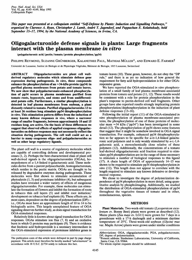
Polygalacturonide-Stimulated pp34 Phosphorylation IsDistributed Throughout the Vegetative Tissues of Tomato.Polygalacturonides and OGAs were shown previously to stim-ulate the in vitro phosphorylation of an -34-kDa protein(pp34) in plasma membrane from tomato and potato leaves(11, 12). The tomato protein migrates as if its mass were about32.5 kDa but is termed "pp34" for simplicity since it isimmunologically and biochemically related to potato pp34(18). To extend our information on the phosphorylation ofpp34 we first decided to look at the distribution of polygalac-turonide-stimulated thiophosphorylation in plasma membranefractions from throughout the vegetative tissues of tomato.Plasma membrane fractions from leaf, hypocotyl, stem, androot were prepared from 14- to 15-day-old tomato plants. Fig.1A shows that the in vitro thiophosphorylation of pp34 intomato leaf, hypocotyl, stem, and root plasma membrane isstimulated by tomato leaf PGA. In each case the most pro-nounced effect of the polygalacturonide was on pp34 thio-phosphorylation, with little effect on "background" phosphor-ylations. These results provide an indication that most of thevegetative tissues of tomato might be OGA sensitive. Thepresence of 0.2 ,tg of a-D-galacturonic acid (Fig. 1A) did notenhance root plasma membrane pp34 thiophosphorylation,indicating that the action of tomato leaf PGA was not simplydue to an ionic strength effect. The OGA-enhanced phosphor-ylation of pp34 from root plasma membrane may be a firstindication that one or more biological activities of OGAs exist
AL H S R
-- + + -- CkDa i t i
106
50
B L H S R
FIG. 1. Distribution of a PGA-responsive phosphoprotein, pp34(arrowhead), in plasma membranes from the vegetative tissues oftomato. (A) In vitro thiophosphorylation of plasma membrane (2 ,gof protein) from leaf (L), hypocotyl (H), stem (S), and root (R) in theabsence (-) or presence (+) of 0.2 j,g of tomato leaf PGA. In the rootpanel c is a sample thiophosphorylated in the presence of 0.2 ,ug ofgalacturonic acid. (B) Western blot of tomato plasma membranes (10,tg of protein) probed with anti-pp34 polyclonal antibody showingcross-reactivity in the -32-kDa (arrowhead) region.
Proc. Natl. Acad. Sci. USA 92 (1995)
Proc. NatL Acad Sci. USA 92 (1995) 4147
in the tomato root. Western blot analysis of leaf, hypocotyl,stem, and root plasma membrane fractions (Fig. 1B) revealeda cross-reacting polypeptide at -32 kDa, a position corre-sponding to the position of tomato pp34.The age of the tomato plants from which leaves were used
to prepare plasma membranes had no significant effect on invitro pp34 thiophosphorylation (data not shown). Thus pp34phosphorylation is apparently not related to a specific devel-opmental stage of the expanding leaf. Previous studies on awide variety of plants have shown that different vegetativetissues are OGA responsive. These organs include leaves (4),stem tissue (22), thin cell layers (5), seedlings (cotyledon andhypocotyl) (3), and cell suspension cultures (see ref. 17 forreview). It now appears that tissues sensitive to PGA-derivedsignals exist throughout the vegetative parts of plants. This istrue of the responses of intact tissues to OGA and to the in vitrophosphorylation of pp34.
Polygalacturonide-Stimulated pp34 Phosphorylation Occursin Soybean LeafPlasma Membranes.We extended our search forPGA-stimulated pp34 thiophosphorylation to two other species,namely soybean and maize. These plants were chosen for thefollowing reasons. OGAs induce phytoalexin accumulation insoybean cotyledons and hypocotyls (1), and this species is one ofthe most highly studied plants in terms of defense responses.Maize is a graminaceous monocot widely diverged from tomatoor soybean. So far as we are aware no reports of OGA-induceddefense gene expression have been published for these plants. Amaize defense gene is induced in response to wounding andfungal infection (23), but the authors give no indication of thenature of the elicitor(s) involved in this defense mechanism.
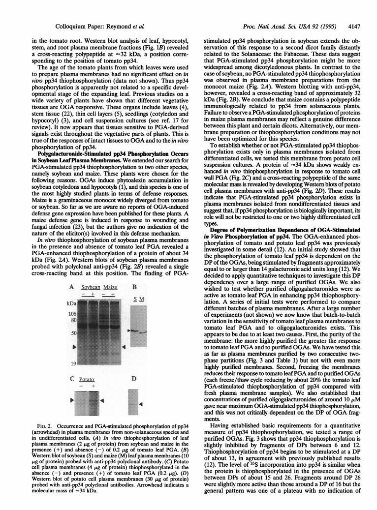
In vitro thiophosphorylation of soybean plasma membranesin the presence and absence of tomato leaf PGA revealed aPGA-enhanced thiophosphorylation of a protein of about 34kDa (Fig. 24). Western blots of soybean plasma membranesprobed with polyclonal anti-pp34 (Fig. 2B) revealed a singlecross-reacting band at this position. The finding of PGA-
A Soybean Maize+ +
kDa
10O
C Potato-+
B
SM
4
D
·--··:-'
FIG. 2. Occurrence and PGA-stimulated phosphorylation of pp34(arrowhead) in plasma membranes from non-solanaceous species andin undifferentiated cells. (A) In vitro thiophosphorylation of leafplasma membranes (2 tag of protein) from soybean and maize in thepresence (+) and absence (-) of 0.2 ,tg of tomato leaf PGA. (B)Western blot of soybean (S) and maize (M) leafplasma membranes (10tag of protein) probed with anti-pp34 polyclonal antibody. (C) Potatocell plasma membranes (4 ,ig of protein) thiophosphorylated in theabsence (-) and presence (+) of tomato leaf PGA (0.2 ,tg). (D)Western blot of potato cell plasma membranes (30 ,tg of protein)probed with anti-pp34 polyclonal antibodies. Arrowhead indicates amolecular mass of -34 kDa.
stimulated pp34 phosphorylation in soybean extends the ob-servation of this response to a second dicot family distantlyrelated to the Solanaceae: the Fabaceae. These data suggestthat PGA-stimulated pp34 phosphorylation might be morewidespread among dicotyledonous plants. In contrast to thecase of soybean, no PGA-stimulated pp34 thiophosphorylationwas observed in plasma membrane preparations from themonocot maize (Fig. 2A). Western blotting with anti-pp34,however, revealed a cross-reacting band of approximately 32kDa (Fig. 2B). We conclude that maize contains a polypeptideimmunologically related to pp34 from solanaceous plants.Failure to observe a PGA-stimulated phosphorylation ofproteinsin maize plasma membranes may reflect a genuine differencebetween this plant and certain dicots. Alternatively, our mem-brane preparation or thiophosphorylation conditions may nothave been optimized for this species.To establish whether or not PGA-stimulated pp34 thiophos-
phorylation exists only in plasma membranes isolated fromdifferentiated cells, we tested this membrane from potato cellsuspension cultures. A protein of -34 kDa shows weakly en-hanced in vitro thiophosphorylation in response to tomato cellwall PGA (Fig. 2C) and a cross-reacting polypeptide of the samemolecular mass is revealed by developing Western blots of potatocell plasma membranes with anti-pp34 (Fig. 2D). These resultsindicate that PGA-stimulated pp34 phosphorylation exists inplasma membranes isolated from nondifferentiated tissues andsuggest that, if pp34 phosphorylation is biologically important, itsrole will not be restricted to one or two highly differentiated celltypes.
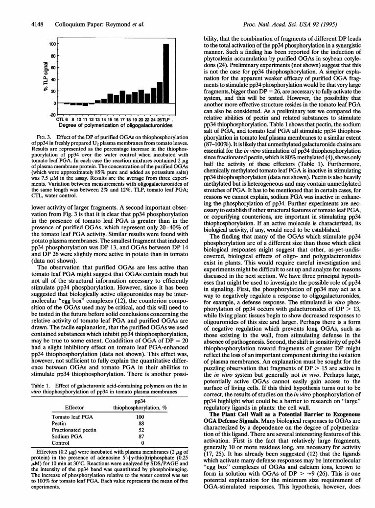
Degree of Polymerization Dependence of OGA-Stimulatedin Vitro Phosphorylation of pp34. The OGA-enhanced phos-phorylation of tomato and potato leaf pp34 was previouslyinvestigated in some detail (12). An initial study showed thatthe phosphorylation of tomato leaf pp34 is dependent on theDP of the OGAs, being stimulated by fragments approximatelyequal to or larger than 14 galacturonic acid units long (12). Wedecided to apply quantitative techniques to investigate this DPdependency over a large range of purified OGAs. We alsowished to test whether purified oligogalacturonides were asactive as tomato leaf PGA in enhancing pp34 thiophosphory-lation. A series of initial tests were performed to comparedifferent batches of plasma membranes. After a large numberof experiments (not shown) we now know that batch-to-batchvariation in the sensitivity of tomato leaf plasma membranes totomato leaf PGA and to oligogalacturonides exists. Thisappears to be due to at least two causes. First, the purity of themembrane: the more highly purified the greater the responseto tomato leaf PGA and to purified OGAs. We have tested thisas far as plasma membranes purified by two consecutive two-phase partitions (Fig. 3 and Table 1) but not with even morehighly purified membranes. Second, freezing the membranesreduces their response to tomato leaf PGA and to purified OGAs(each freeze/thaw cycle reducing by about 20% the tomato leafPGA-stimulated thiophosphorylation of pp34 compared withfresh plasma membrane samples). We also established thatconcentrations of purified oligogalacturonides of around 10 ,uMgave near maximum OGA-stimulated pp34 thiophosphorylation,and this was not critically dependent on the DP of OGA frag-ments.Having established basic requirements for a quantitative
measure of pp34 thiophosphorylation, we tested a range ofpurified OGAs. Fig. 3 shows that pp34 thiophosphorylation isslightly inhibited by fragments of DPs between 6 and 12.Thiophosphorylation of pp34 begins to be stimulated at a DPof about 13, in agreement with previously published results(12). The level of 35S incorporation into pp34 is similar whenthe protein is thiophosphorylated in the presence of OGAsbetween DPs of about 15 and 26. Fragments around DP 26were slightly more active than those around a DP of 16 but thegeneral pattern was one of a plateau with no indication of
Colloquium Paper: Reymond et al
4148 Colloquium Paper: Reymond et al
-aa?
-J
I--
100
80
60
40
20
-20CTL 6 8 10 11 12 13 14 15 16 17 18 19 20 22 24 26TLP.Degree of polymerization of oligogalacturonides
FIG. 3. Effect of the DP of purified OGAs on thiophosphorylationof pp34 in freshly prepared U2 plasma membranes from tomato leaves.Results are represented as the percentage increase in the thiophos-phorylation of pp34 over the water control when incubated withtomato leaf PGA. In each case the reaction mixtures contained 2 jigof plasma membrane protein. The concentration of the purified OGAs(which were approximately 85% pure and added as potassium salts)was 7.5 JLM in the assay. Results are the average from three experi-ments. Variation between measurements with oligogalacturonides ofthe same length was between 2% and 12%. TLP, tomato leaf PGA;CTL, water control.
lower activity of larger fragments. A second important obser-vation from Fig. 3 is that it is clear that pp34 phosphorylationin the presence of tomato leaf PGA is greater than in thepresence of purified OGAs, which represent only 20-40% ofthe tomato leaf PGA activity. Similar results were found withpotato plasma membranes. The smallest fragment that inducedpp34 phosphorylation was DP 13, and OGAs between DP 14and DP 26 were slightly more active in potato than in tomato(data not shown).The observation that purified OGAs are less active than
tomato leaf PGA might suggest that OGAs contain much butnot all of the structural information necessary to efficientlystimulate pp34 phosphorylation. However, since it has beensuggested that biologically active oligouronides may be inter-molecular "egg box" complexes (12), the counterion compo-sition of the OGAs used may be critical, and this will need tobe tested in the future before solid conclusions concerning therelative activity of tomato leaf PGA and purified OGAs aredrawn. The facile explanation, that the purified OGAs we usedcontained substances which inhibit pp34 thiophosphorylation,may be true to some extent. Coaddition of OGA of DP = 20had a slight inhibitory effect on tomato leaf PGA-enhancedpp34 thiophosphorylation (data not shown). This effect was,however, not sufficient to fully explain the quantitative differ-ence between OGAs and tomato PGA in their abilities tostimulate pp34 thiophosphorylation. There is another possi-Table 1. Effect of galacturonic acid-containing polymers on the invitro thiophosphorylation of pp34 in tomato plasma membranes
pp34Effector thiophosphorylation, %
Tomato leaf PGA 100Pectin 88Fractionated pectin 52Sodium PGA 87Control 0
Effectors (0.2 ,ig) were incubated with plasma membranes (2 /xg ofprotein) in the presence of adenosine 5'-[y-thio]triphosphate (0.25piM) for 10 min at 30°C. Reactions were analyzed by SDS/PAGE andthe intensity of the pp34 band was quantitated by phosphoimaging.The increase of phosphorylation relative to the water control was setto 100% for tomato leaf PGA. Each value represents the mean of fiveexperiments.
bility, that the combination of fragments of different DP leadsto the total activation of the pp34 phosphorylation in a synergisticmanner. Such a finding has been reported for the induction ofphytoalexin accumulation by purified OGAs in soybean cotyle-dons (24). Preliminary experiments (not shown) suggest that thisis not the case for pp34 thiophosphorylation. A simpler expla-nation for the apparent weaker efficacy of purified OGA frag-ments to stimulate pp34 phosphorylation would be thatvery largefragments, bigger thanDP = 26, are necessary to fully activate thesystem, and this will be tested. However, the possibility thatanother more effective structure resides in the tomato leaf PGAcan also be considered. As a preliminary test we compared therelative abilities of pectin and related substances to stimulatepp34 thiophosphorylation. Table 1 shows that pectin, the sodiumsalt of PGA, and tomato leaf PGA all stimulate pp34 thiophos-phorylation in tomato leaf plasma membranes to a similar extent(87-100%). It is likely that unmethylated galacturonide chains areessential for the in vitro stimulation of pp34 thiophosphorylationsince fractionated pectin, which is 80% methylated (4), shows onlyhalf the activity of these effectors (Table 1). Furthermore,chemically methylated tomato leaf PGA is inactive in stimulatingpp34 thiophosphorylation (data not shown). Pectin is also heavilymethylated but is heterogeneous and may contain unmethylatedstretches of PGA. It has to be mentioned that in certain cases, forreasons we cannot explain, sodium PGA was inactive in enhanc-ing the phosphorylation of pp34: Further experiments are nec-essary to establish if other structural features oftomato leafPGA,or copurifying counterions, are important in stimulating pp34thiophosphorylation. If an active molecule is characterized, itsbiological activity, if any, would need to be established.The finding that many of the OGAs which stimulate pp34
phosphorylation are of a different size than those which elicitbiological responses might suggest that other, as-yet-undis-covered, biological effects of oligo- and polygalacturonidesexist in plants. This would require careful investigation andexperiments might be difficult to set up and analyze for reasonsdiscussed in the next section. We have three principal hypoth-eses that might be used to investigate the possible role of pp34in signaling. First, the phosphorylation of pp34 may act as away to negatively regulate a response to oligogalacturonides,for example, a defense response. The stimulated in vitro phos-phorylation of pp34 occurs with galacturonides of DP > 13,while living plant tissues begin to show decreased responses tooligouronides of this size and larger. Perhaps there is a formof negative regulation which prevents long OGAs, such asthose existing in the wall, from stimulating defense in theabsence of pathogenesis. Second, the shift in sensitivity of pp34thiophosphorylation toward fragments of greater DP mightreflect the loss of an important component during the isolationof plasma membranes. An explanation must be sought for thepuzzling observation that fragments of DP > 15 are active inthe in vitro system but generally not in vivo. Perhaps large,potentially active OGAs cannot easily gain access to thesurface of living cells. If this third hypothesis turns out to becorrect, the results of studies on the in vitro phosphorylation ofpp34 highlight what could be a barrier to research on "large"regulatory ligands in plants: the cell wall.The Plant Cell Wall as a Potential Barrier to Exogenous
OGA Defense Signals. Many biological responses to OGAs arecharacterized by a dependence on the degree of polymeriza-tion of this ligand. There are several interesting features of thisactivation. First is the fact that relatively large fragments,generally 10 or more residues long, are necessary for activity(17, 25). It has already been suggested (12) that the ligandswhich activate many defense responses may be intermolecular"egg box" complexes of OGAs and calcium ions, known toform in solution with OGAs of DP > ~9 (26). This is onepotential explanation for the minimum size requirement ofOGA-stimulated responses. This hypothesis, however, does
Proc. NatL. Acad Sci. USA 92 (1995)
Proc. NatL Acad Sci USA 92 (1995) 4149
not explain why OGAs larger than a DP of about 14 tend to beinactive when applied to living tissues.We suggest that the permeability of the cell wall to larger
fragments may limit their access to the cell surface. In otherwords, if the cell wall were removed, plant tissues would besensitive to a greater range of OGAs. The undamaged cell wallmight then represent the case in which there is a window onlylarge enough to allow the passage of OGAs of DPs up to 14or 15. There are some indications in the literature that thismight be the case. Experiments on the sensitivity of protoplaststo OGAs and PGA have been conducted, and the results havebeen compared with those of similar experiments on walledcells (27, 28). It appears that PGA, in the presence of calcium,can exert strong effects on membrane potential, weak effectson cytosolic pH, and strong effects on cytosolic calcium levelsin protoplasts (27). Similar experiments on walled cells indi-cate that PGA is less active than on protoplasts (27). Theseresults suggest (to us) that the cell wall may exclude most of theexogenous PGA from contact with the plasma membrane.
If it is the cell wall that limits the size range of active,endogenous OGAs, this may have consequences in pathogen-esis. Pectinolytic enzymes operating within the plant cell wallwould release oligo- and polygalacturonides. As pathogenesisprogresses and cell wall degradation proceeds, larger andlarger fragments would have access to the cell surface, increas-ing the number of signal-active molecules available to activatedefense gene expression.More generally, it is possible that, due to the presence of the
cell wall, we may not easily detect the presence of highmolecular mass signal molecules in plants. In animals a numberof very large signals exist in the extracellular matrix. Theseinclude proteins such as fibronectin and complex moleculessuch as proteoglycans (29, 30). Similar molecules may deter-mine adhesion events in plants (31), but evidence for roles asregulators is not yet available. It is noticeable that most signalmolecules so far discovered in plants tend to be relativelysmall; there are few exceptions (for example, see ref. 32). It ispossible that in vitro studies in the absence of the cell wall mayhelp lead to the discovery of larger regulatory ligands in plants.We thank P. Albersheim, A. Darvill, C. Bergmann, and M. Spiro of
the Complex Carbohydrate Research Center, University of Georgia,for their generosity in providing endopolygalacturonase, OGA stan-dards, and purification protocols prior to publication. C.A. Ryan(Washington State University) provided tomato leaf PGA, and A.Buchala and J.-P. M6traux (University of Fribourg, Switzerland)provided potato cell suspension cultures. We thank B. Kiinstner formaintenance of plant material. This work was supported by SwissNational Science Foundation Grant 31-36472.92.
1. Darvill, A. G. & Albersheim, P. (1984) Annu. Rev. Plant Physiol.35, 243-275.
2. Ryan, C. A. & Farmer, E. E. (1991) Annu. Rev. Plant Physiol.Plant Mol. Biol. 42, 651-674.
3. West, C. A., Bruce, R. J. & Jin, D. F; (1984) in Structure, Functionand Biosynthesis of Plant Cell Walls, eds. Dugger, W. M. &
Bartinicki-Garcia, S. (Williams and Wilkins, Baltimore), pp.359-375.
4. Bishop, P. D., Makus, D., Pearce, G. & Ryan, C. A. (1984)J. Biol.Chem. 259, 13172-13177.
5. Mohnen, D., Eberhard, S., Marfa, V., Doubrava, N., Toubart, P.,Gollin, D. J., Gruber, T., Nuri, W., Albersheim, P. & Darvill,A. G. (1990) Development (Cambridge, U.K) 108, 191-201.
6. Bellincampi, D., Salvi, G., De Lorenzo, G. & Cervone, F. (1993)Plant J. 4, 207-213.
7. Thain, J. F., Doherty, H. M., Bowles, D.J. & Wildon, D. C.(1990) Plant Cell Environ. 13, 569-574.
8. Mathieu, Y., Kurkdjian, A., Xia, H., Guern, J., Koller, A., Spiro,M. D., O'Neill, M., Albersheim, P. & Darvill, A. (1991) Plant J.1, 333-343.
9. Legendre, L., Rueter, S., Heinstein, P. F. & Low, P. S. (1993)Plant Physiol. 102, 233-240.
10. Farmer, E. E., Caldelari, D., Pearce, G., Walker-Simmons, MK.& Ryan, C. A. (1994) Plant Physiol. 106, 337-342.
11. Farmer, E. E., Pearce, G. & Ryan, C. A. (1989) Proc. Natl. Acad.Sci. USA 86, 1539-1542.
12. Farmer, E. E., Moloshok, T. D., Saxton, M. J. & Ryan, C. A.(1991) J. Biol. Chem. 266, 3140-3145.
13. Dietrich, A., Mayer, J. E. & Hahlbrock, K. (1990) J. Biol. Chem.265, 6360-6365.
14. Felix, G., Grosskopf, D. G., Regenass, M. & Boiler, T. (1991)Proc. Natl. Acad. Sci. USA 88, 8831-8834.
15. Felix, G., Regenass, M., Spanu, P. & Boiler, T. (1994) Proc. Natl.Acad. Sci. USA 91, 952-956.
16. MacKintosh, C., Lyon, G. D. & MacKintosh, R. W. (1994) PlantJ. 5, 137-147.
17. Darvill, A., Augur, C., Bergmann, C., Carlson, R. W., Cheong,J.-J., Eberhard, S., Hahn, M. G., Lo, V.-M., Marfa, V., Meyer, B.,Mohnen, D., O'Neill, M.A., Spiro, M. D., van Halbeek, H.,Work, W. S. & Albersheim, P. (1992) Glycobiology 2, 181-198.
18. Jacinto, T., Farmer, E. E. & Ryan, C. A. (1993) PlantPhysiol. 103,1393-1397.
19. Spiro, M. D., Kates, K. A., Koller, A. G., O'Neill, M. A., Alber-sheim, P. & Darvill, A. G. (1993) Carbohydr. Res. 247, 9-20.
20. Blumenkrantz, N. & Asboe-Hansen, G. (1973)Anal. Biochem. 54,484-489.
21. Lever, M. (1972) Anal. Biochem. 47, 273-279.22. Branca, C., De Lorenzo, G. & Cervone, F. (1988) Physiol. Plant.
72, 499-504.23. Cordero, M. J., Raventos, D. & San Segundo, B. (1994) Plant J.
6, 141-150.24. Davis, K. R., Darvill, A. G., Albersheim, P. & Dell, A. (1986)
Plant Physiol. 80, 568-577.25. Ryan, C. A. (1988) Biochemistry 27, 8879-8883.26. Kohn, R. (1985) Pure Appl. Chem. 42, 371-397.27. Messiaen, J., Read, N. D., Van Cutsem, P. & Trewavas, A. J.
(1993) J. Cell Sci. 104, 365-371.28. Messiaen, J. & Van Cutsem, P. (1994) Plant Cell Physiol. 35,
677-689.29. Juliano, R. L. & Haskill, S. (1993)J. Cell Biol. 120, 577-585.30. Damsky, C. H. & Werb, Z. (1992) Curr. Opin. Cell Biol. 4,
772-781.31. Zhu, J.-K., Shi, J., Singh, U., Wyatt, S. E., Bressan, R.A.,
Hasegawa, P. M. & Carpita, N. C. (1993) Plant J. 3, 637-646.32. Hayashi, T. & Yoshida, K. (1988) Proc. Natl. Acad. Sci. USA 85,
2618-2622.
Colloquium Paper: Reymond et at