pediatric board review course - american …nysaap.org/blog/dermatology.pdf · pediatric board...
TRANSCRIPT
PEDIATRIC BOARD REVIEW COURSE
Dermatology
Julie V. Schaffer, M.D.
Associate Professor of Dermatology and Pediatrics
Director of Pediatric Dermatology
NYU School of Medicine

Vascular Tumors
• Infantile hemangioma
• Congenital hemangioma
– Rapidly involuting
(RICH)
– Non-involuting (NICH)
• Pyogenic granuloma
• Kaposiform
hemangioendothelioma
and tufted angioma
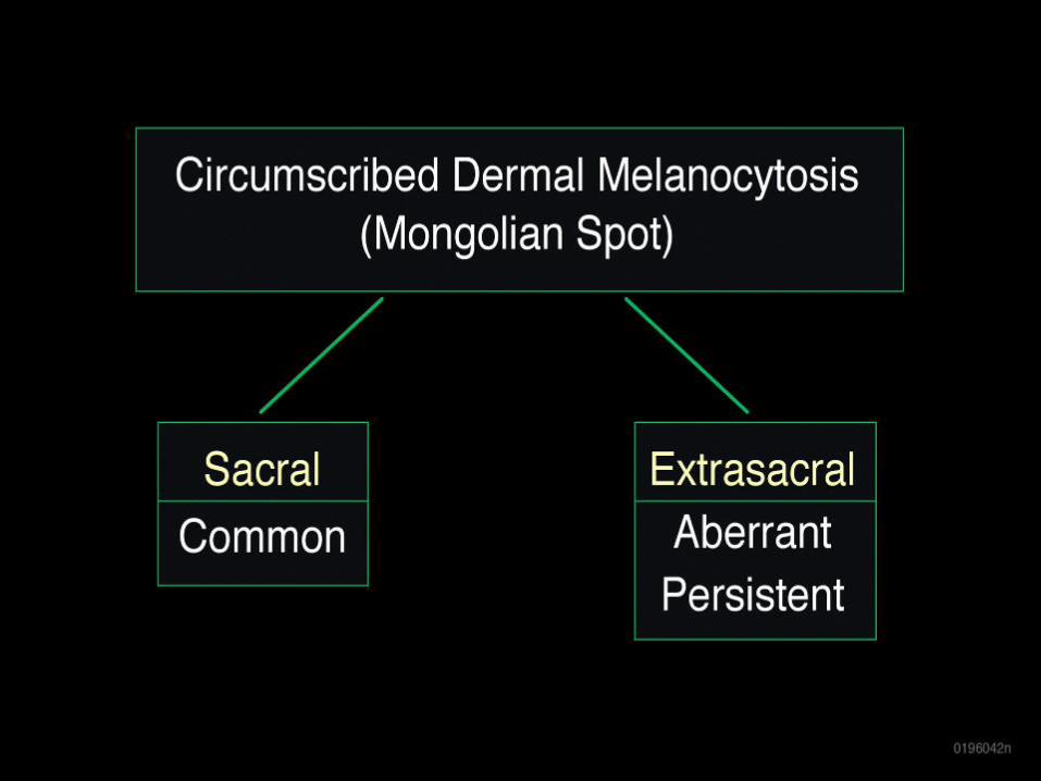
Vascular Malformations
Low flow
• Capillary (port wine stain)
• Venous
• Lymphatic
– Microcystic (lymphangioma)
– Macrocystic (cystic hygroma)
• Combined
High flow
• Arterial-venous (AFM, AVF)
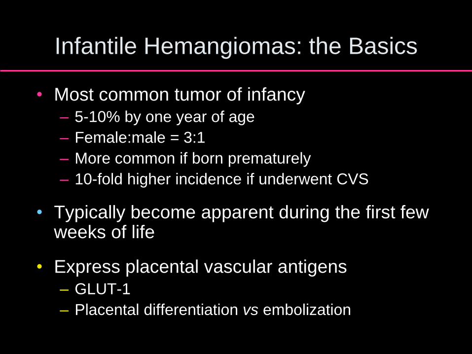
Infantile Hemangiomas: the Basics
• Most common tumor of infancy – 5-10% by one year of age
– Female:male = 3:1
– More common if born prematurely
– 10-fold higher incidence if underwent CVS
• Typically become apparent during the first few weeks of life
• Express placental vascular antigens – GLUT-1
– Placental differentiation vs embolization
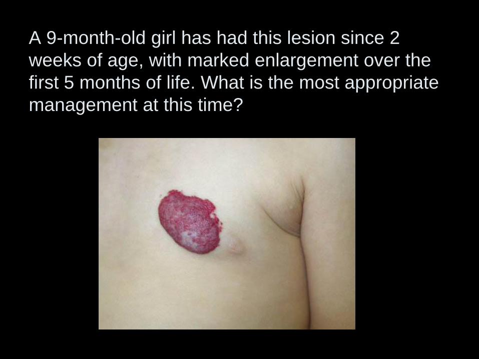
A 9-month-old girl has had this lesion since 2
weeks of age, with marked enlargement over the
first 5 months of life. What is the most appropriate
management at this time?
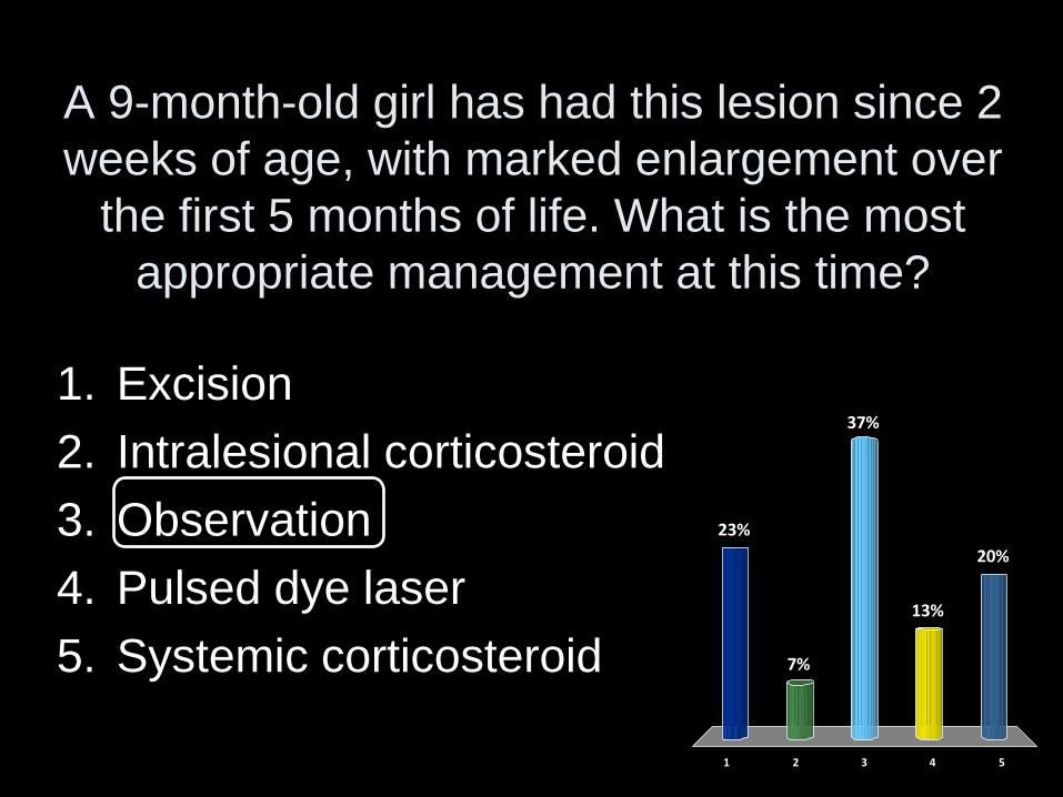
A 9-month-old girl has had this lesion since 2
weeks of age, with marked enlargement over
the first 5 months of life. What is the most
appropriate management at this time?
1 2 3 4 5
23%
7%
20%
13%
37%
1. Excision
2. Intralesional corticosteroid
3. Observation
4. Pulsed dye laser
5. Systemic corticosteroid

Natural History
• Proliferation – ‘Mark out territory’
early on, then volumetric growth
– Most growth complete by age 5 months
– Deep lesions tend to grow ~1 month longer
– Bright red
– Firm/rubbery
– Warm
• Involution – 30% by 3 years
– 50% by 5 years
– 90% by 9 years
– Dull red to gray
– Soft/spongy
– “Breaking apart”
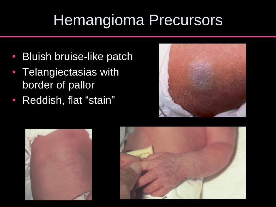
Hemangioma Precursors
• Bluish bruise-like patch
• Telangiectasias with
border of pallor
• Reddish, flat “stain”
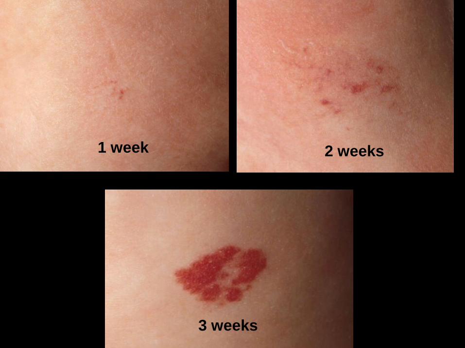
1 week 2 weeks
3 weeks
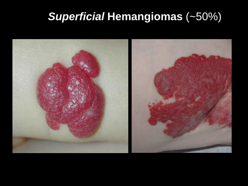
Superficial Hemangiomas (~50%)
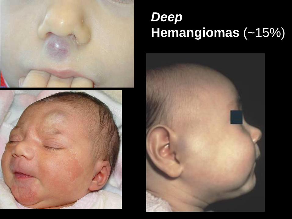
Deep
Hemangiomas (~15%)
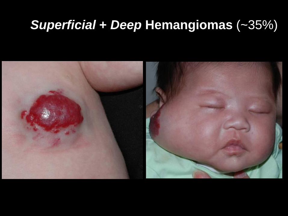
Superficial + Deep Hemangiomas (~35%)
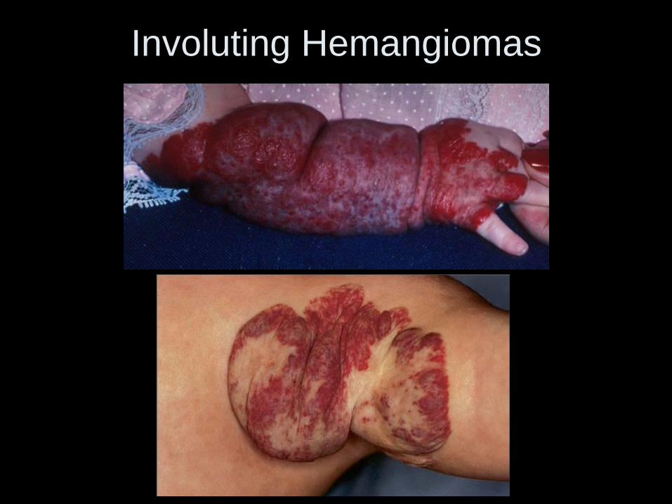
Involuting Hemangiomas
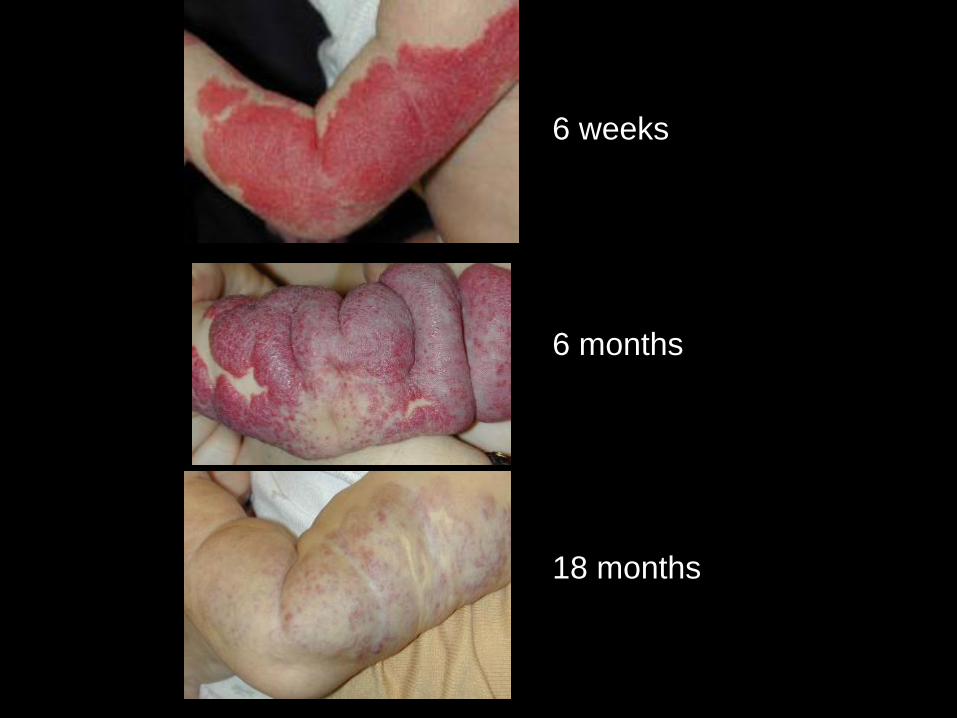
6 weeks
6 months
18 months
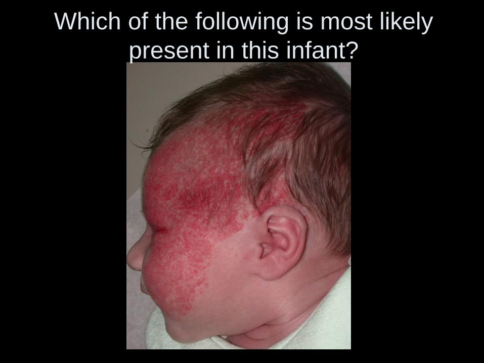
Which of the following is most likely
present in this infant?
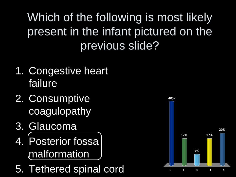
Which of the following is most likely
present in the infant pictured on the
previous slide?
1 2 3 4 5
40%
17%
20%
17%
7%
1. Congestive heart
failure
2. Consumptive
coagulopathy
3. Glaucoma
4. Posterior fossa
malformation
5. Tethered spinal cord
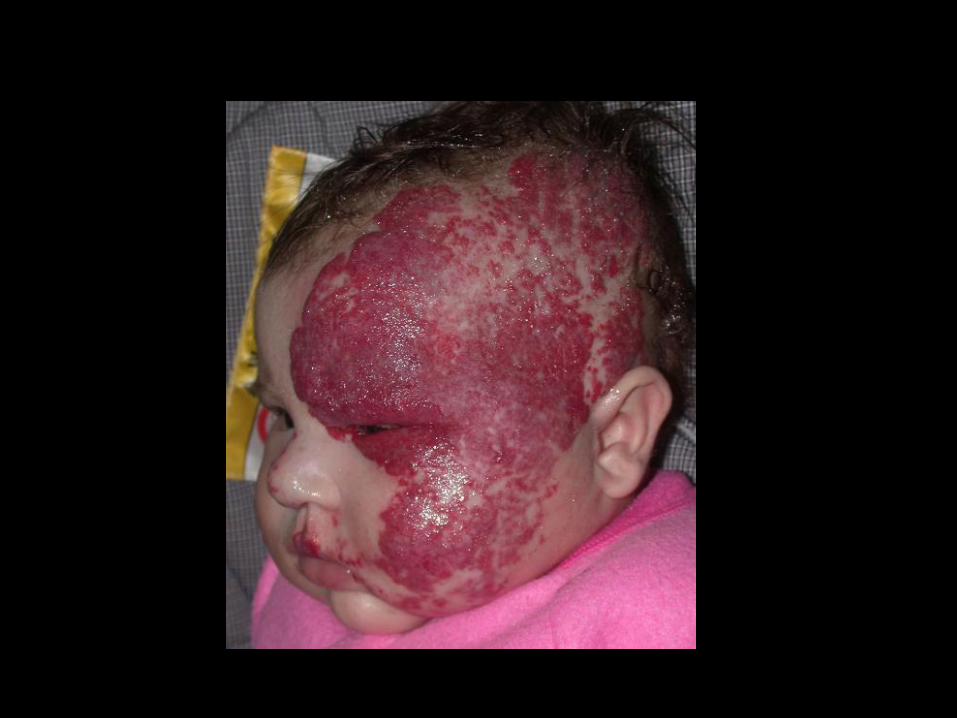
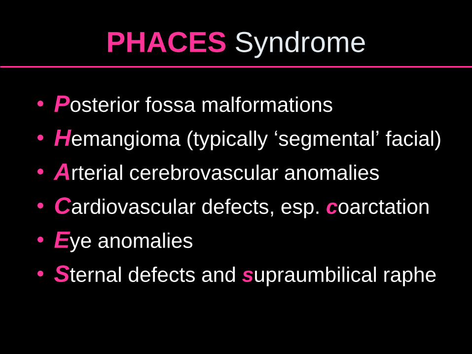
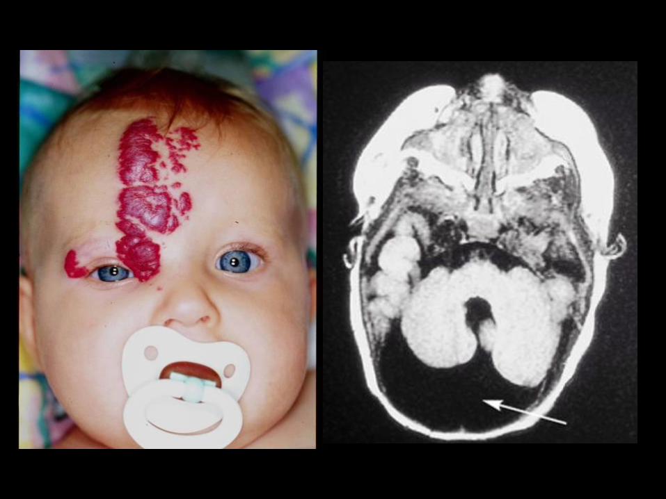
PHACES Syndrome
• Posterior fossa malformations
• Hemangioma (typically ‘segmental’ facial)
• Arterial cerebrovascular anomalies
• Cardiovascular defects, esp. coarctation
• Eye anomalies
• Sternal defects and supraumbilical raphe
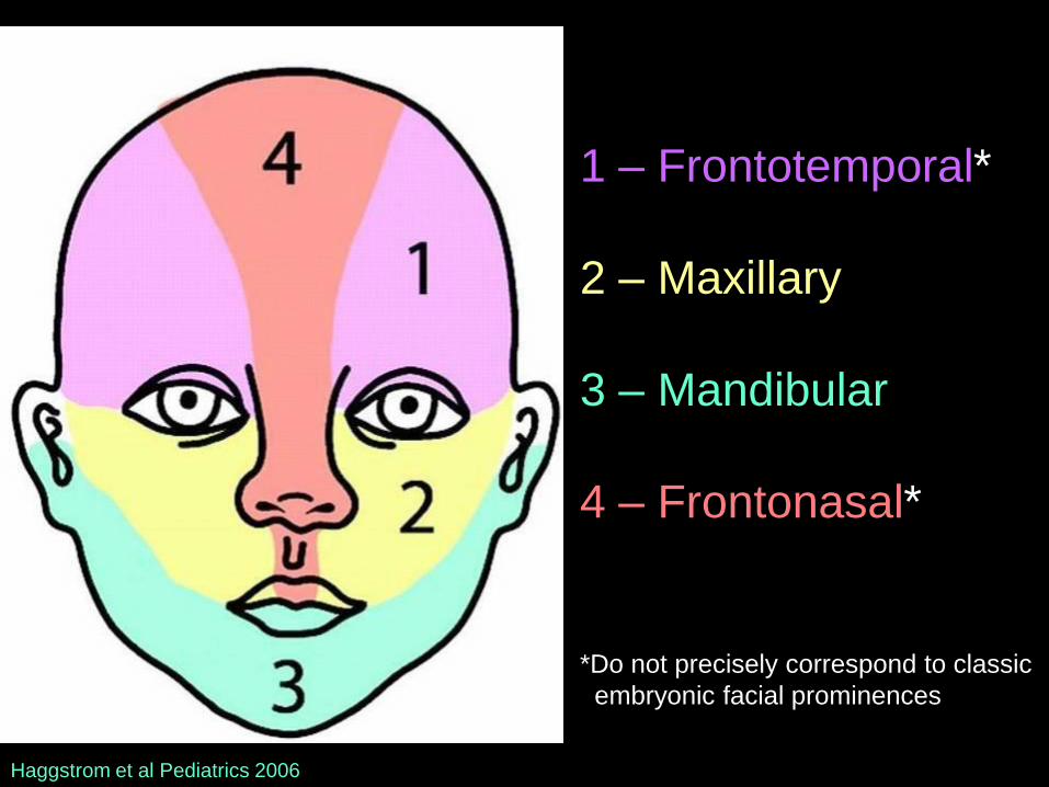
1 – Frontotemporal*
2 – Maxillary
3 – Mandibular
4 – Frontonasal*
*Do not precisely correspond to classic
embryonic facial prominences
Haggstrom et al Pediatrics 2006
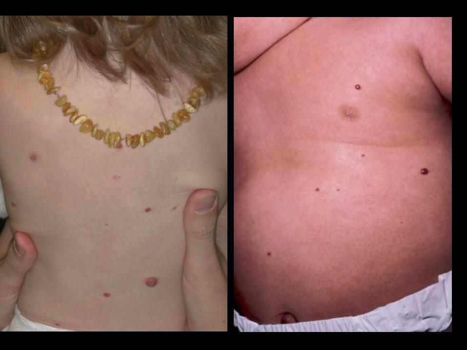
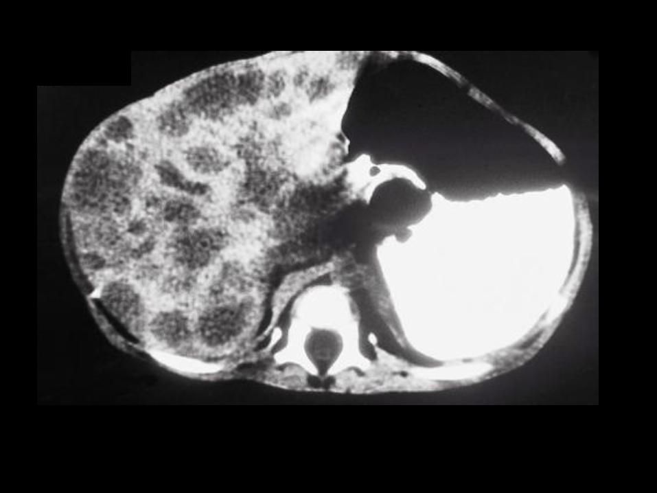
Neonatal Hemangiomatosis
• Multiple (~>5) hemangiomas – Usually small, superficial, “cherry-like”
• Benign (skin only) vs diffuse (skin + extracutaneous)
• Visceral involvement
– Liver > lungs, CNS, eyes, GI
– Hepatomegaly, high-output CHF, anemia

LOCATION,
LOCATION,
LOCATION
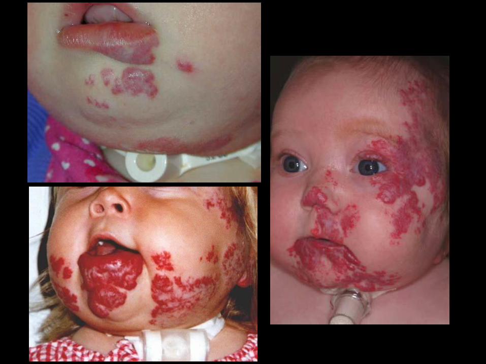
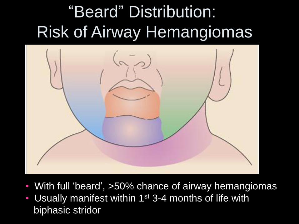
• With full ‘beard’, >50% chance of airway hemangiomas
• Usually manifest within 1st 3-4 months of life with
biphasic stridor
“Beard” Distribution:
Risk of Airway Hemangiomas
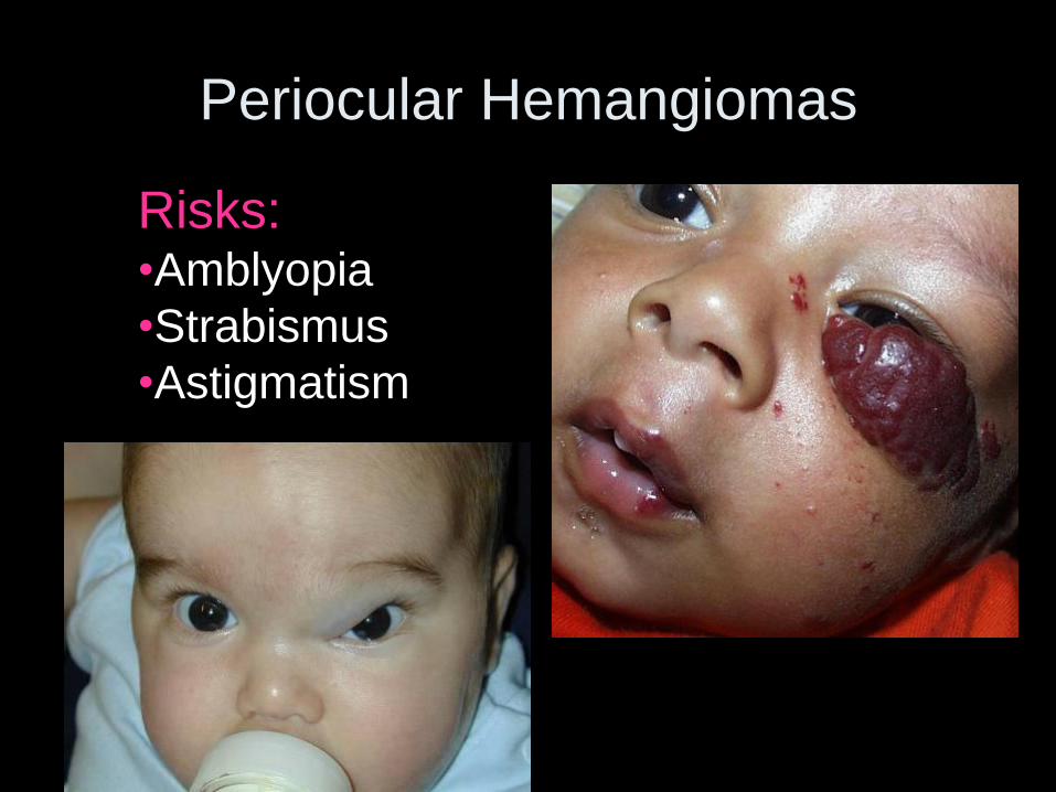
Risks: •Amblyopia
•Strabismus
•Astigmatism
Periocular Hemangiomas
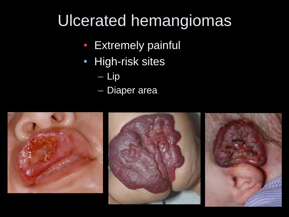
Ulcerated hemangiomas
• Extremely painful
• High-risk sites
– Lip
– Diaper area
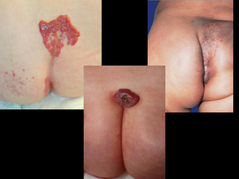
Midline Skin Lesions:
Markers of Spinal Dysraphism
• 75% of individuals with spinal dysraphism vs <3% overall
• High-risk markers:
– More than one type of skin lesion
– “Tails”
– Lipomas: most common marker overall
– Hypertrichosis (e.g. “faun tail”)
– Hemangiomas (risk > vascular stain)
– >5 mm/deep dimples above the gluteal cleft
– Aplasia cutis

Identifying High-Risk Hemangiomas
• Location
– Threat to vision or other vital functions
– Potential for disfigurement (esp. facial
lesions)
• Size/growth potential (depends on age)
• ‘Segmental’ subtype
• Ulceration
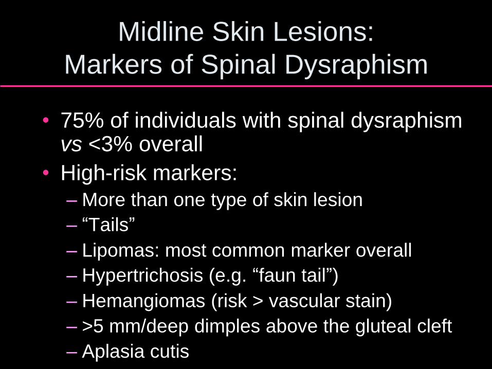
A Excision
B Intralesional corticosteroid
C Observation
D Propranolol
E Pulsed dye laser
Which of the following is the most appropriate
initial management of this 1-month-old’s
rapidly growing infantile hemangioma?

Hemangioma Treatment Options
• “Watchful waiting” / “Active nonintervention”
• Local wound care for ulceration
• Topical*, intralesional, or systemic corticosteroids
• Vincristine
• Interferon (risk spastic diplegia)
• Beta blockers – propranolol
• Pulsed dye laser*
• Excisional surgery
*Beneficial primarily for superficial lesions
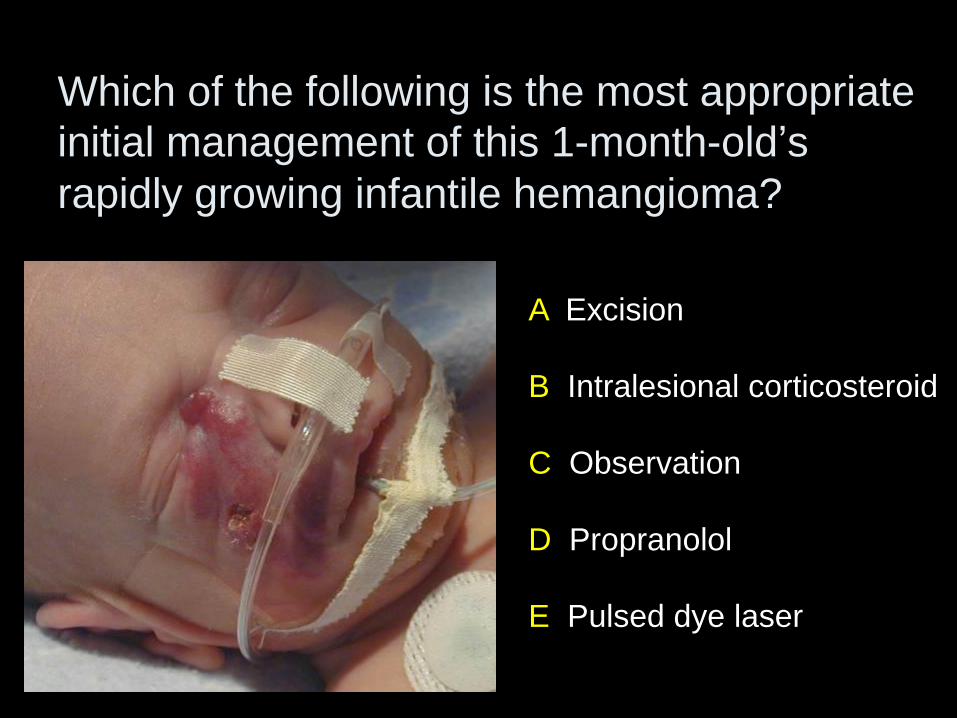
Vascular Tumors
Associated with
Kasabach-Merritt

Kasabach-Merritt Syndrome
• Vascular tumor + coagulopathy – Kaposiform hemangioendothelioma or tufted
angioma
– NOT classic infantile hemangiomas
• Rapidly enlarging, ecchymotic, indurated vascular mass
• Severe thrombocytopenia, DIC, microangiopathic hemolytic anemia
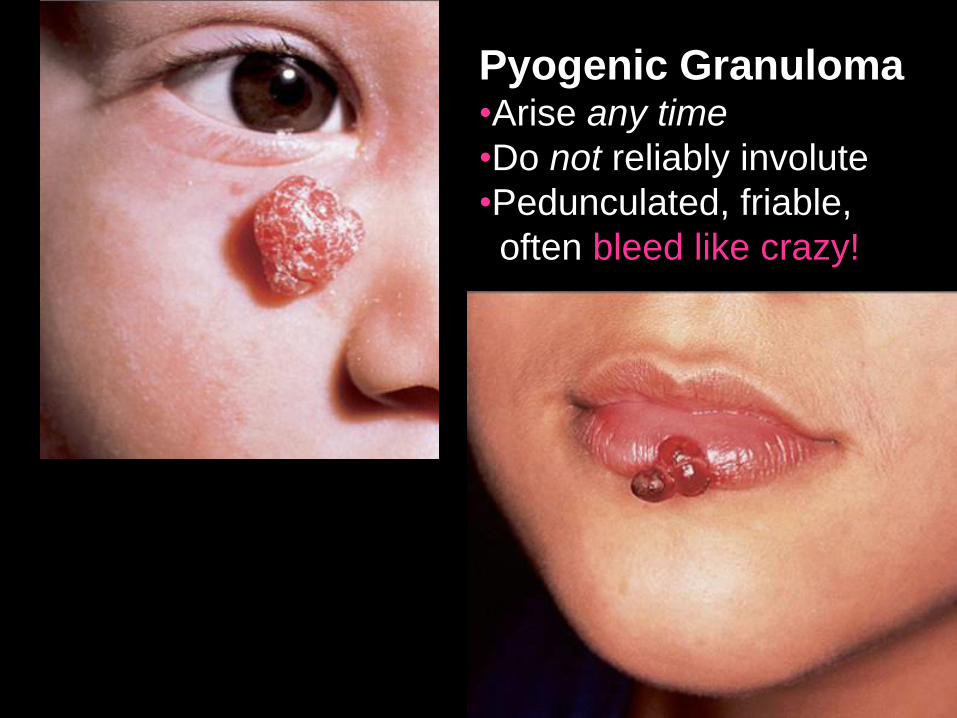
Pyogenic Granuloma •Arise any time
•Do not reliably involute
•Pedunculated, friable,
often bleed like crazy!
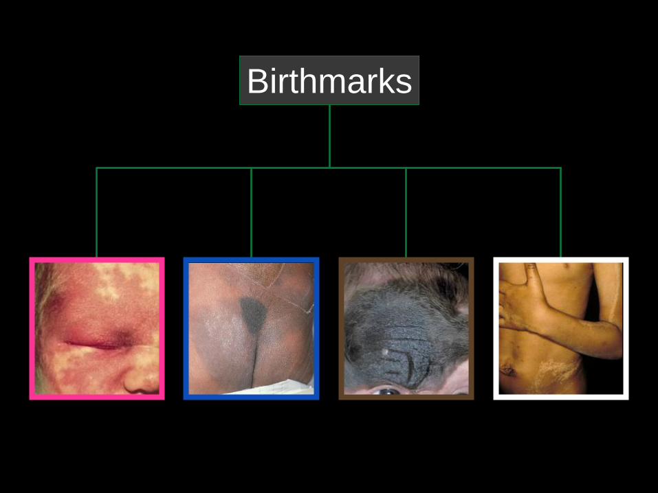
Birthmarks
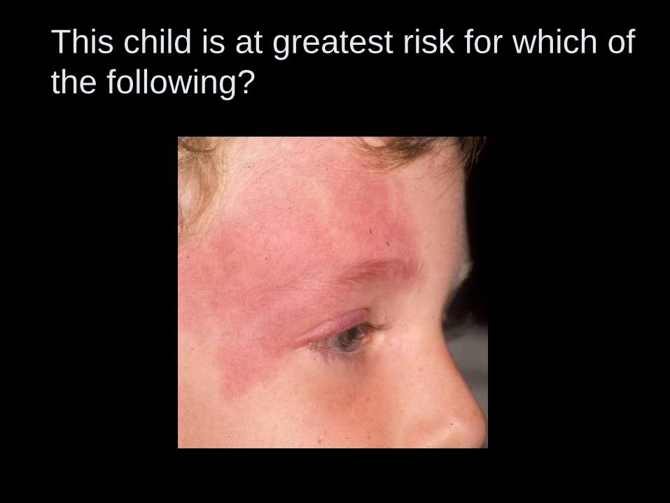
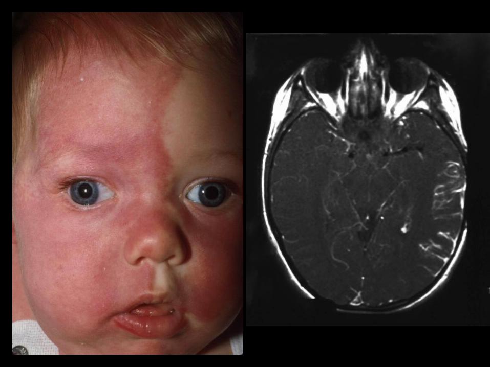
This child is at greatest risk for which of
the following?
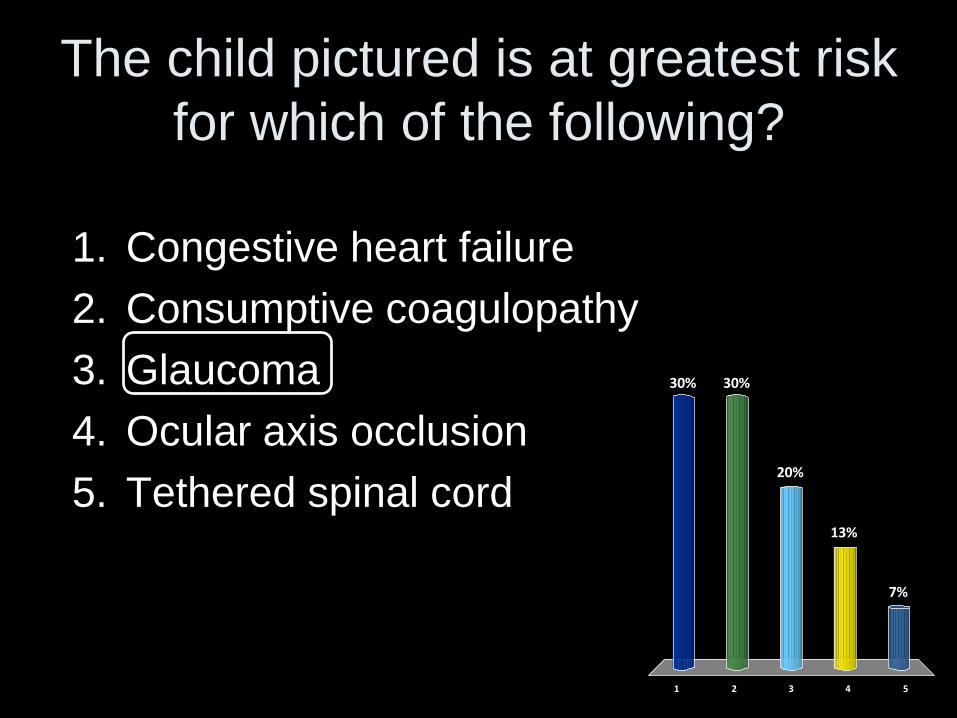
The child pictured is at greatest risk
for which of the following?
1 2 3 4 5
30% 30%
7%
13%
20%
1. Congestive heart failure
2. Consumptive coagulopathy
3. Glaucoma
4. Ocular axis occlusion
5. Tethered spinal cord
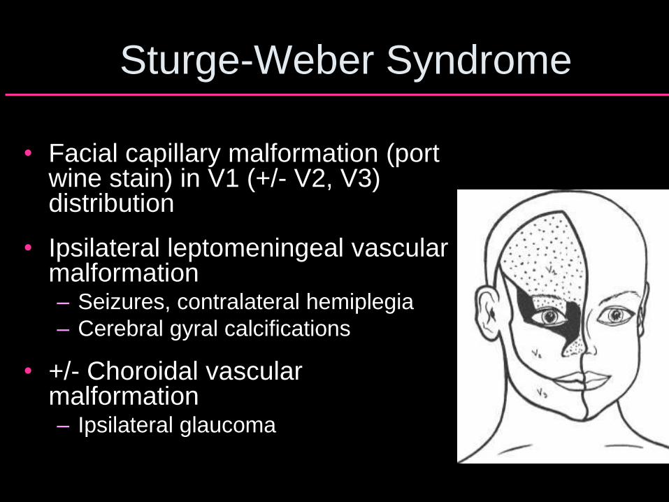
Sturge-Weber Syndrome
• Facial capillary malformation (port wine stain) in V1 (+/- V2, V3) distribution
• Ipsilateral leptomeningeal vascular malformation – Seizures, contralateral hemiplegia
– Cerebral gyral calcifications
• +/- Choroidal vascular malformation – Ipsilateral glaucoma
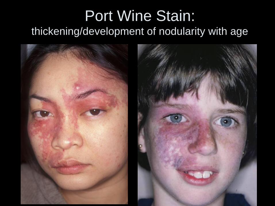
Port Wine Stain: thickening/development of nodularity with age
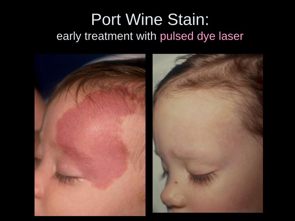
Port Wine Stain: early treatment with pulsed dye laser
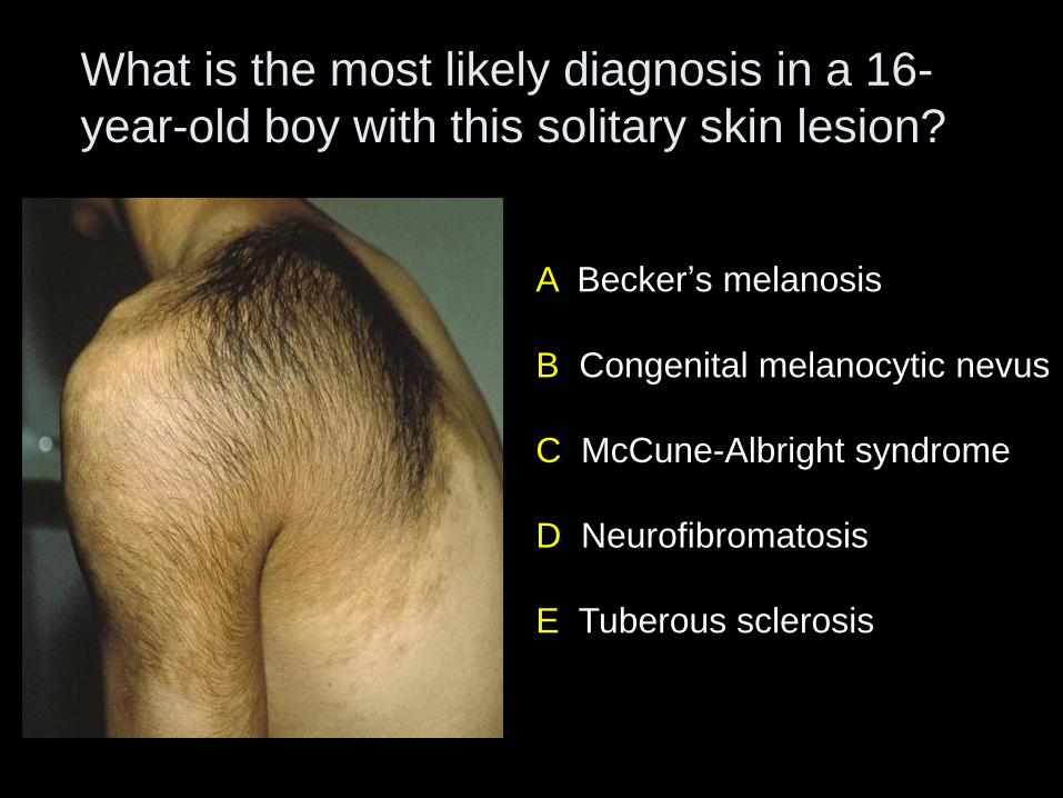
A Becker’s melanosis
B Congenital melanocytic nevus
C McCune-Albright syndrome
D Neurofibromatosis
E Tuberous sclerosis
What is the most likely diagnosis in a 16-
year-old boy with this solitary skin lesion?
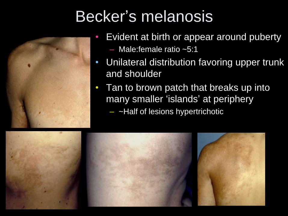
Becker’s melanosis • Evident at birth or appear around puberty
– Male:female ratio ~5:1
• Unilateral distribution favoring upper trunk
and shoulder
• Tan to brown patch that breaks up into
many smaller ‘islands’ at periphery
– ~Half of lesions hypertrichotic
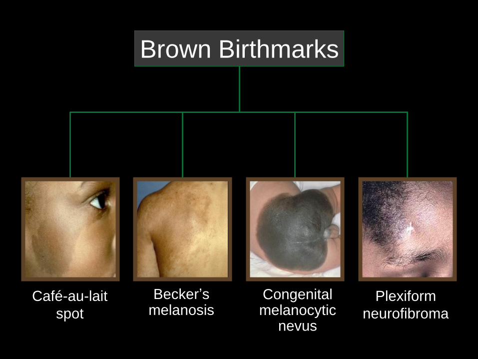
Brown Birthmarks
Plexiform
neurofibroma
Congenital melanocytic
nevus
Becker’s melanosis
Café-au-lait
spot
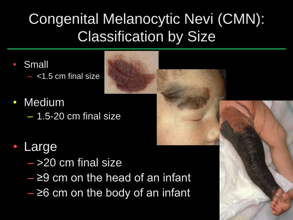
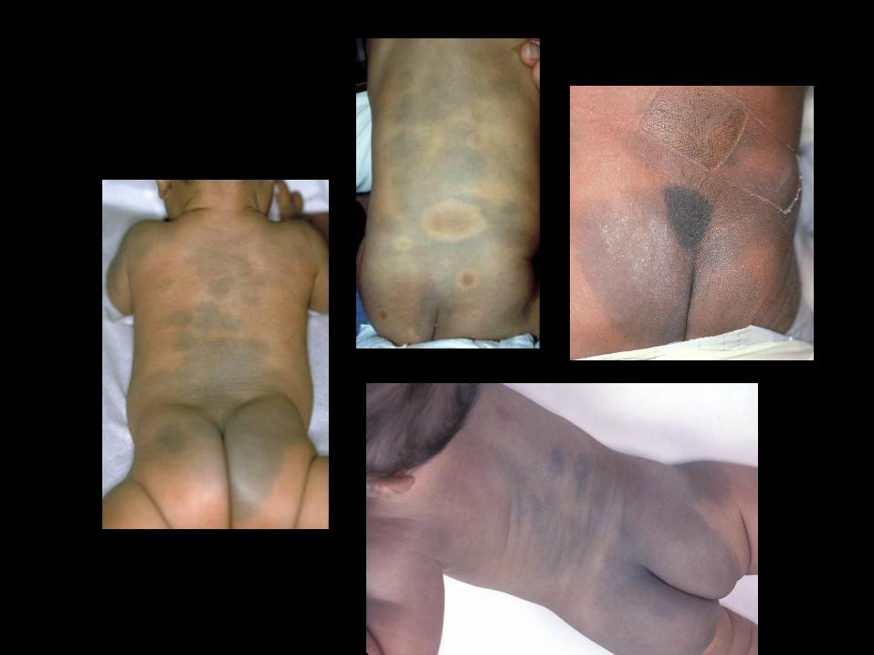
Congenital Melanocytic Nevi (CMN):
Classification by Size
• Small – <1.5 cm final size
• Medium – 1.5-20 cm final size
• Large
– >20 cm final size
– ≥9 cm on the head of an infant
– ≥6 cm on the body of an infant
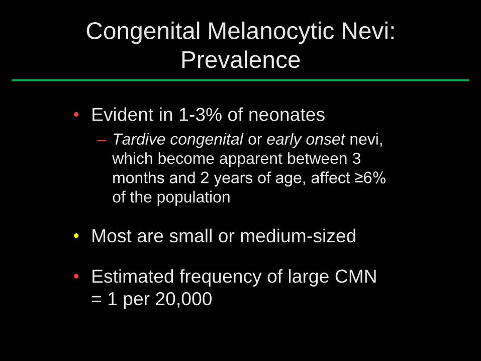
Congenital Melanocytic Nevi:
Prevalence
• Evident in 1-3% of neonates
– Tardive congenital or early onset nevi,
which become apparent between 3
months and 2 years of age, affect ≥6%
of the population
• Most are small or medium-sized
• Estimated frequency of large CMN
= 1 per 20,000
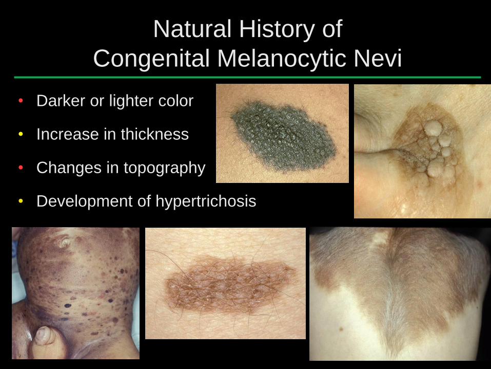
Natural History of
Congenital Melanocytic Nevi
• Darker or lighter color
• Increase in thickness
• Changes in topography
• Development of hypertrichosis
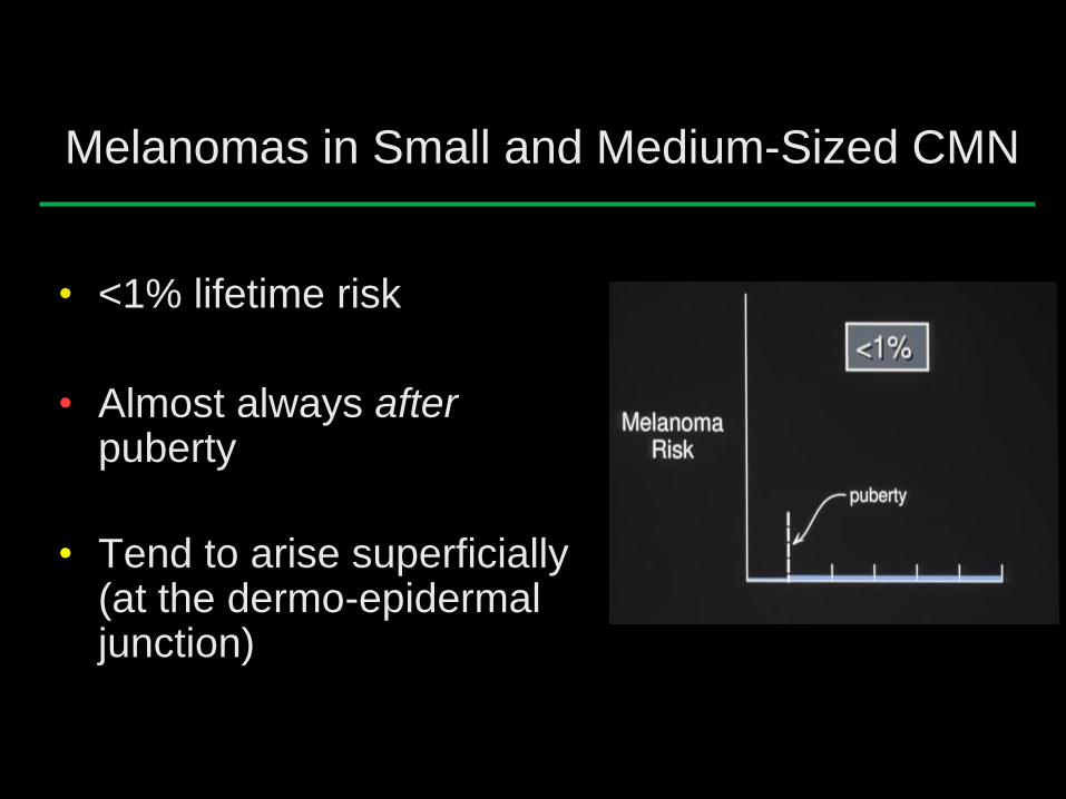
Melanomas in Small and Medium-Sized CMN
• <1% lifetime risk
• Almost always after puberty
• Tend to arise superficially (at the dermo-epidermal junction)
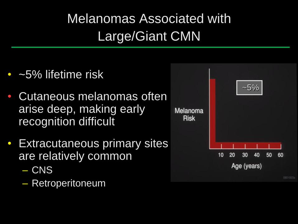
Melanomas Associated with
Large/Giant CMN
• ~5% lifetime risk
• Cutaneous melanomas often arise deep, making early recognition difficult
• Extracutaneous primary sites are relatively common – CNS
– Retroperitoneum
~5%
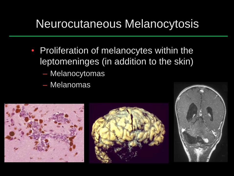
Neurocutaneous Melanocytosis
• Proliferation of melanocytes within the
leptomeninges (in addition to the skin)
– Melanocytomas
– Melanomas
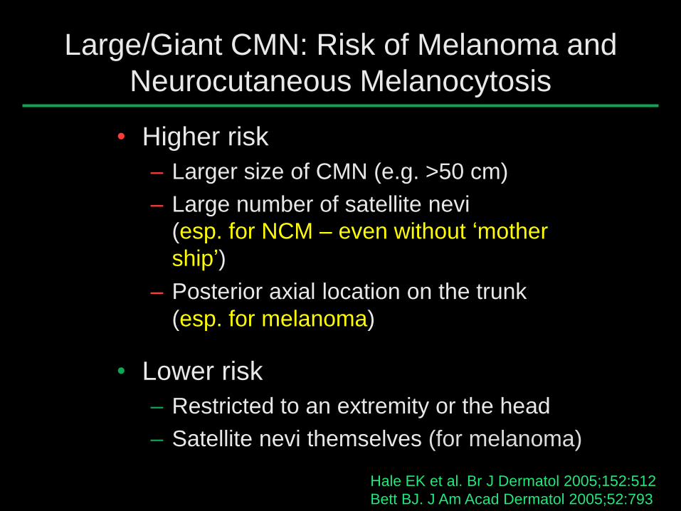
Large/Giant CMN: Risk of Melanoma and
Neurocutaneous Melanocytosis
• Higher risk
– Larger size of CMN (e.g. >50 cm)
– Large number of satellite nevi
(esp. for NCM – even without ‘mother
ship’)
– Posterior axial location on the trunk
(esp. for melanoma)
• Lower risk
– Restricted to an extremity or the head
– Satellite nevi themselves (for melanoma)
Hale EK et al. Br J Dermatol 2005;152:512
Bett BJ. J Am Acad Dermatol 2005;52:793

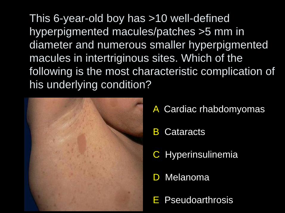
A Cardiac rhabdomyomas
B Cataracts
C Hyperinsulinemia
D Melanoma
E Pseudoarthrosis
This 6-year-old boy has >10 well-defined
hyperpigmented macules/patches >5 mm in
diameter and numerous smaller hyperpigmented
macules in intertriginous sites. Which of the
following is the most characteristic complication of
his underlying condition?
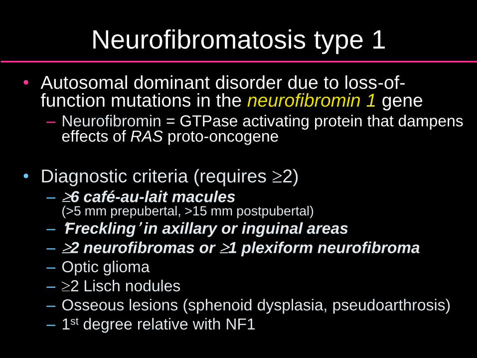
Neurofibromatosis type 1
• Autosomal dominant disorder due to loss-of-function mutations in the neurofibromin 1 gene – Neurofibromin = GTPase activating protein that dampens
effects of RAS proto-oncogene
• Diagnostic criteria (requires 2) – 6 café-au-lait macules
(>5 mm prepubertal, >15 mm postpubertal)
– ‘Freckling’ in axillary or inguinal areas
– 2 neurofibromas or 1 plexiform neurofibroma
– Optic glioma
– 2 Lisch nodules
– Osseous lesions (sphenoid dysplasia, pseudoarthrosis)
– 1st degree relative with NF1
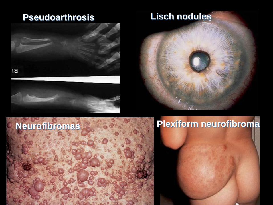
Lisch nodules
Neurofibromas Plexiform neurofibroma
Pseudoarthrosis
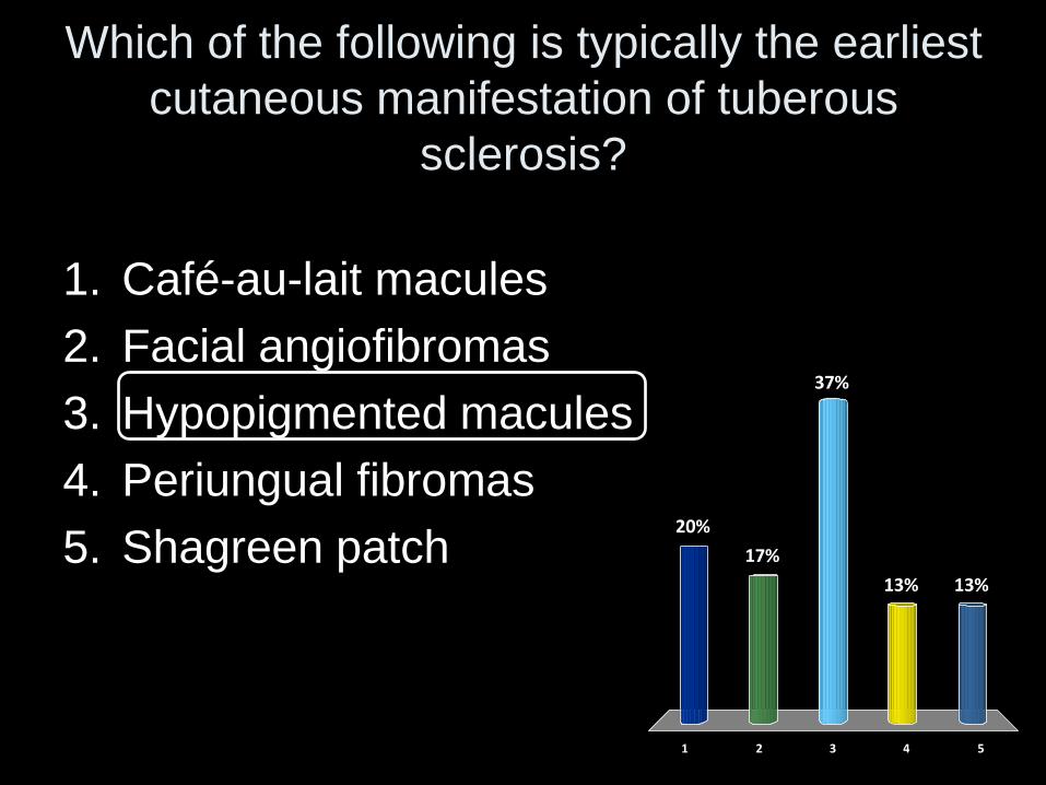
Which of the following is typically the earliest
cutaneous manifestation of tuberous
sclerosis?
1 2 3 4 5
20%
17%
13%13%
37%
1. Café-au-lait macules
2. Facial angiofibromas
3. Hypopigmented macules
4. Periungual fibromas
5. Shagreen patch
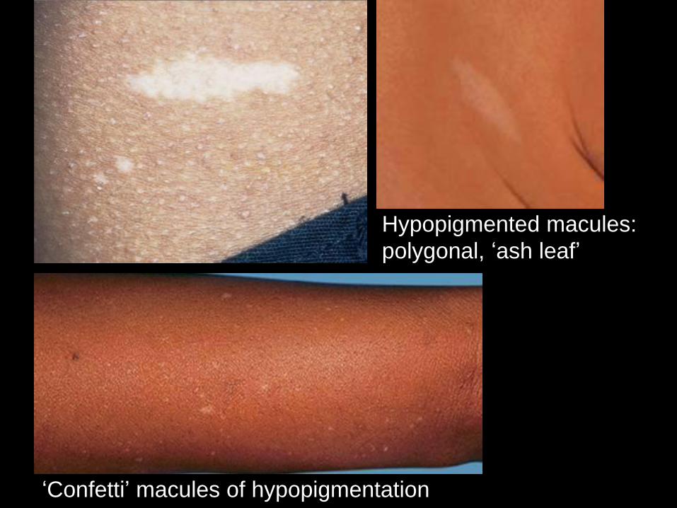
Hypopigmented macules:
polygonal, ‘ash leaf’
‘Confetti’ macules of hypopigmentation
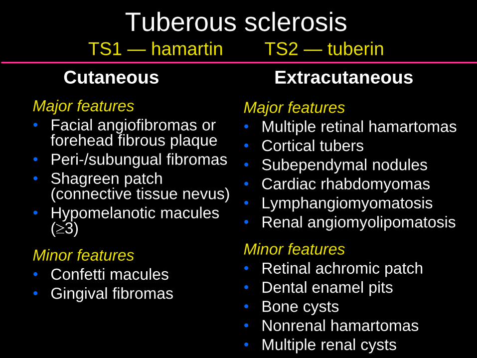
Tuberous sclerosis TS1 — hamartin TS2 — tuberin
Cutaneous
Major features
• Facial angiofibromas or forehead fibrous plaque
• Peri-/subungual fibromas
• Shagreen patch (connective tissue nevus)
• Hypomelanotic macules ( 3)
Minor features
• Confetti macules
• Gingival fibromas
Extracutaneous
Major features
• Multiple retinal hamartomas
• Cortical tubers
• Subependymal nodules
• Cardiac rhabdomyomas
• Lymphangiomyomatosis
• Renal angiomyolipomatosis
Minor features
• Retinal achromic patch
• Dental enamel pits
• Bone cysts
• Nonrenal hamartomas
• Multiple renal cysts
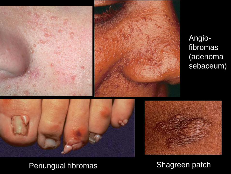
Angio-
fibromas
(adenoma
sebaceum)
Shagreen patch Periungual fibromas
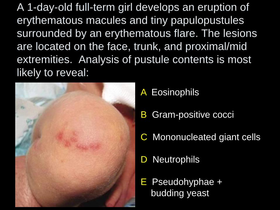
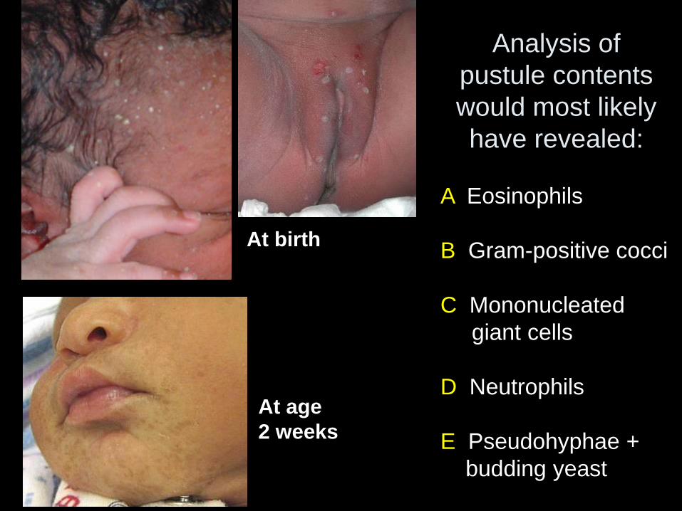
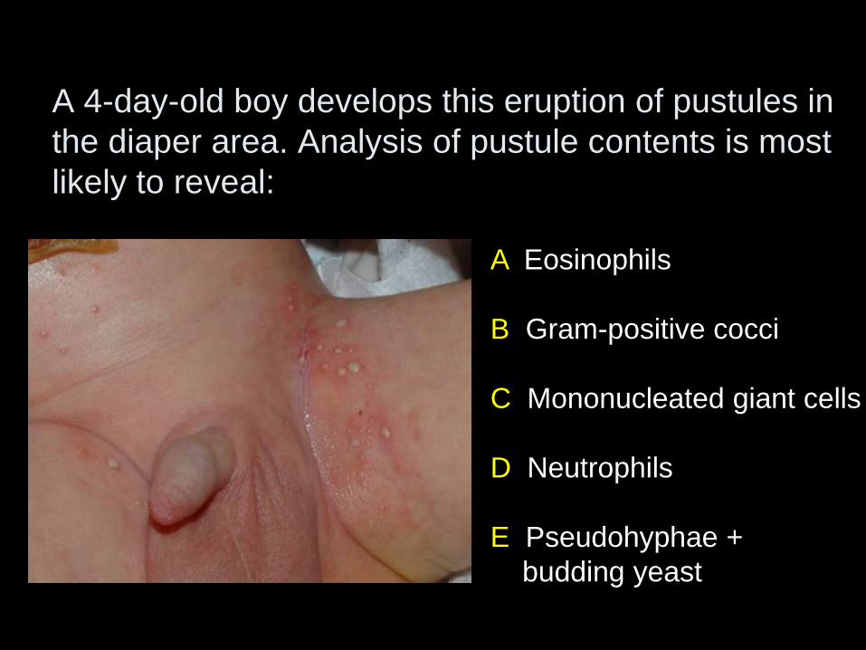
A Eosinophils
B Gram-positive cocci
C Mononucleated giant cells
D Neutrophils
E Pseudohyphae +
budding yeast
A 1-day-old full-term girl develops an eruption of
erythematous macules and tiny papulopustules
surrounded by an erythematous flare. The lesions
are located on the face, trunk, and proximal/mid
extremities. Analysis of pustule contents is most
likely to reveal:
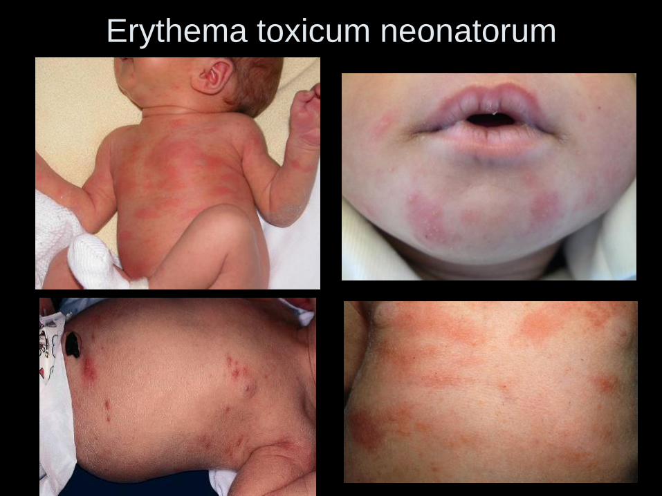
Erythema toxicum neonatorum
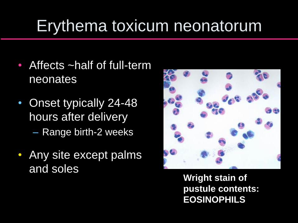
Erythema toxicum neonatorum
• Affects ~half of full-term
neonates
• Onset typically 24-48
hours after delivery
– Range birth-2 weeks
• Any site except palms
and soles Wright stain of
pustule contents:
EOSINOPHILS
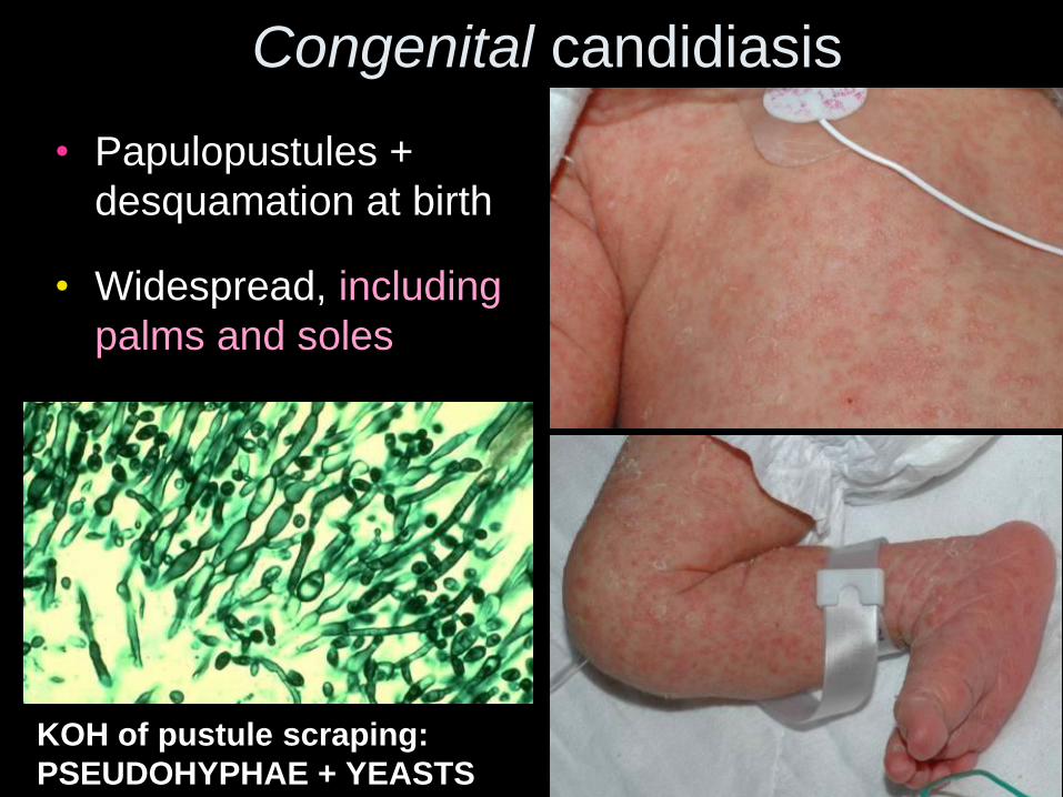
Congenital candidiasis
• Papulopustules +
desquamation at birth
• Widespread, including
palms and soles
KOH of pustule scraping:
PSEUDOHYPHAE + YEASTS
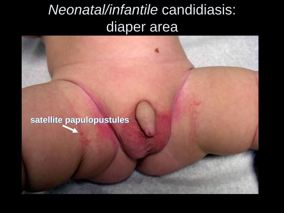
Neonatal/infantile candidiasis:
diaper area
satellite papulopustules
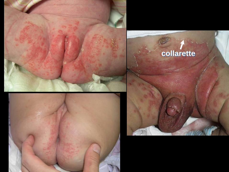
collarette
A Eosinophils
B Gram-positive cocci
C Mononucleated
giant cells
D Neutrophils
E Pseudohyphae +
budding yeast
Analysis of
pustule contents
would most likely
have revealed:
At birth
At age
2 weeks
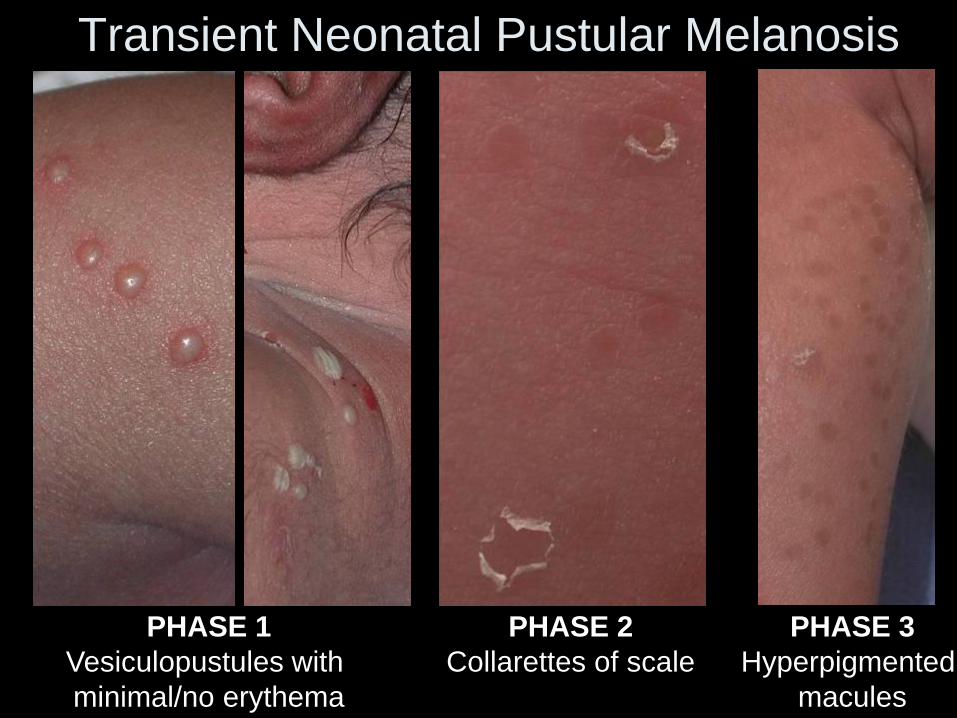
Transient Neonatal Pustular Melanosis
PHASE 1
Vesiculopustules with
minimal/no erythema
PHASE 2
Collarettes of scale
PHASE 3
Hyperpigmented
macules
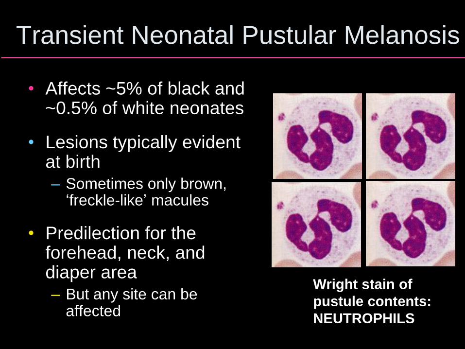
Transient Neonatal Pustular Melanosis
• Affects ~5% of black and ~0.5% of white neonates
• Lesions typically evident at birth – Sometimes only brown,
‘freckle-like’ macules
• Predilection for the forehead, neck, and diaper area – But any site can be
affected
Wright stain of
pustule contents:
NEUTROPHILS
A Eosinophils
B Gram-positive cocci
C Mononucleated giant cells
D Neutrophils
E Pseudohyphae +
budding yeast
A 4-day-old boy develops this eruption of pustules in
the diaper area. Analysis of pustule contents is most
likely to reveal:
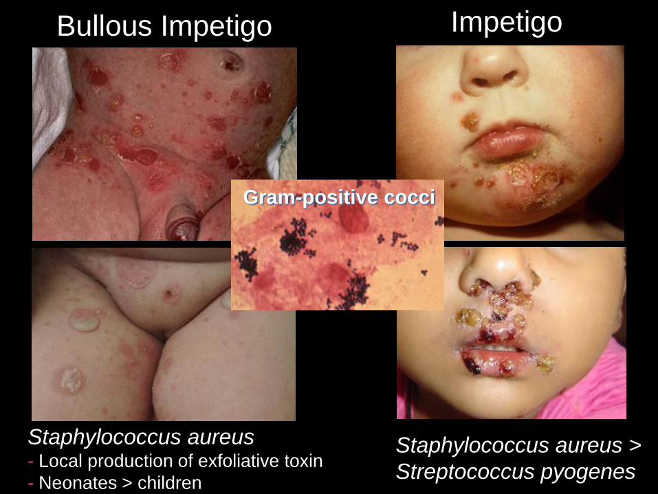
Bullous Impetigo
Staphylococcus aureus
- Local production of exfoliative toxin
- Neonates > children
Impetigo
Staphylococcus aureus >
Streptococcus pyogenes
Gram-positive cocci
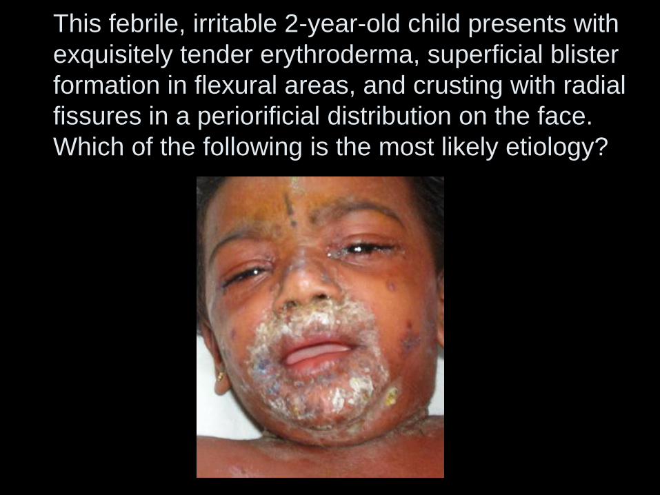
This febrile, irritable 2-year-old child presents with
exquisitely tender erythroderma, superficial blister
formation in flexural areas, and crusting with radial
fissures in a periorificial distribution on the face.
Which of the following is the most likely etiology?
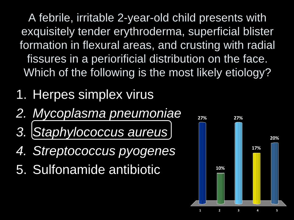
A febrile, irritable 2-year-old child presents with
exquisitely tender erythroderma, superficial blister
formation in flexural areas, and crusting with radial
fissures in a periorificial distribution on the face.
Which of the following is the most likely etiology?
1 2 3 4 5
27%
10%
20%
17%
27%
1. Herpes simplex virus
2. Mycoplasma pneumoniae
3. Staphylococcus aureus
4. Streptococcus pyogenes
5. Sulfonamide antibiotic
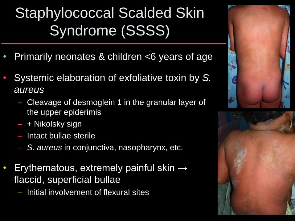
Staphylococcal Scalded Skin
Syndrome (SSSS)
• Primarily neonates & children <6 years of age
• Systemic elaboration of exfoliative toxin by S.
aureus
– Cleavage of desmoglein 1 in the granular layer of
the upper epiderimis
– + Nikolsky sign
– Intact bullae sterile
– S. aureus in conjunctiva, nasopharynx, etc.
• Erythematous, extremely painful skin →
flaccid, superficial bullae
– Initial involvement of flexural sites
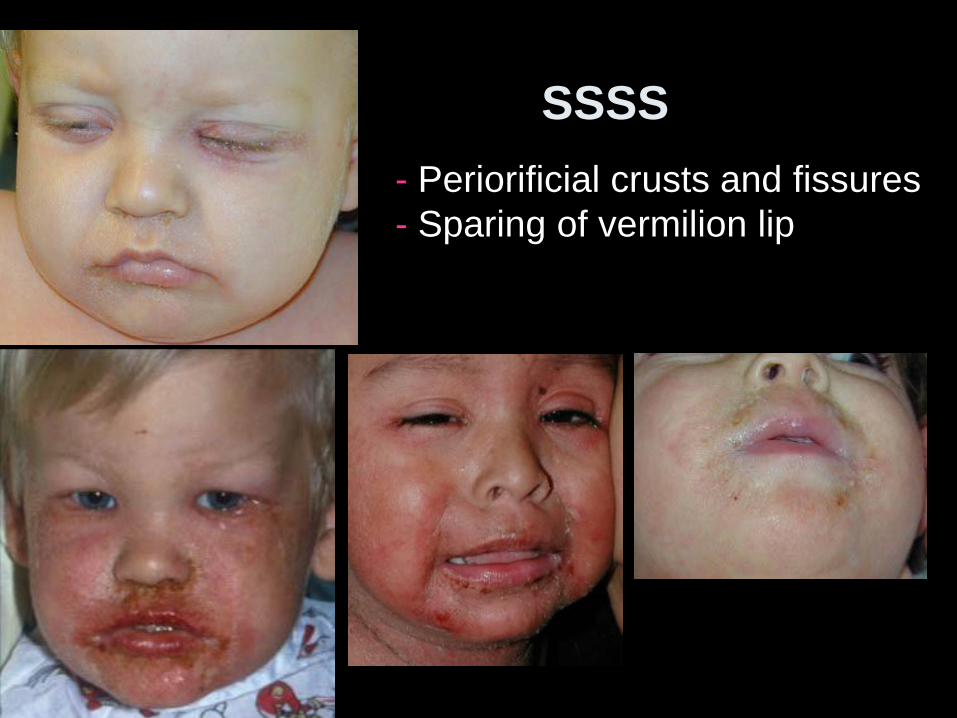
SSSS
- Periorificial crusts and fissures
- Sparing of vermilion lip
A Hepatitis B vaccination
B Herpes simplex virus
C Mycoplasma pneumoniae
D Streptococcus pyogenes
E Sulfonamide antibiotic
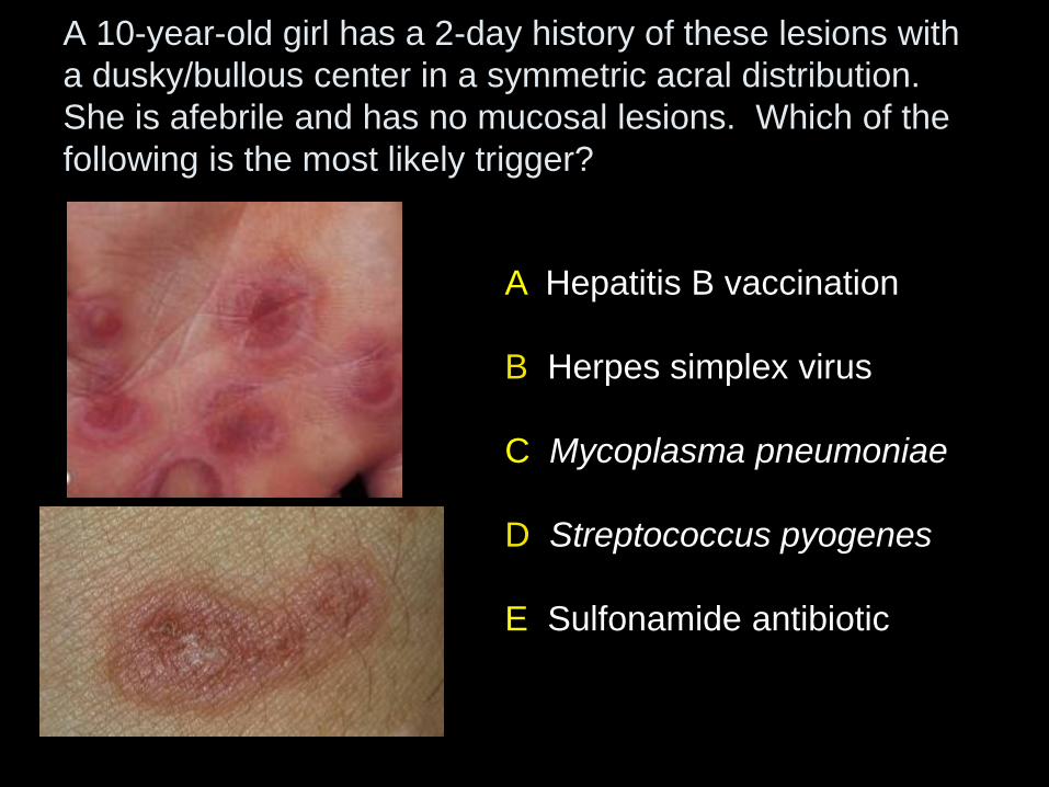
A 10-year-old girl has a 2-day history of these lesions with
a dusky/bullous center in a symmetric acral distribution.
She is afebrile and has no mucosal lesions. Which of the
following is the most likely trigger?
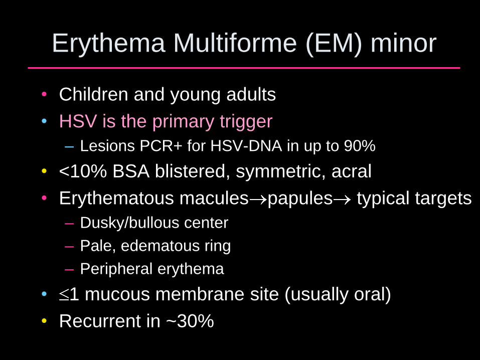
Erythema Multiforme (EM) minor
• Children and young adults
• HSV is the primary trigger
– Lesions PCR+ for HSV-DNA in up to 90%
• <10% BSA blistered, symmetric, acral
• Erythematous macules papules typical targets
– Dusky/bullous center
– Pale, edematous ring
– Peripheral erythema
• 1 mucous membrane site (usually oral)
• Recurrent in ~30%
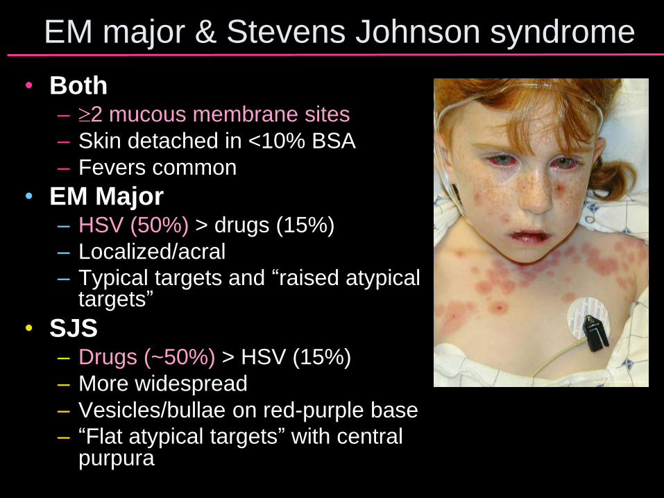
EM major & Stevens Johnson syndrome
• Both – 2 mucous membrane sites
– Skin detached in <10% BSA
– Fevers common
• EM Major – HSV (50%) > drugs (15%)
– Localized/acral
– Typical targets and “raised atypical targets”
• SJS – Drugs (~50%) > HSV (15%)
– More widespread
– Vesicles/bullae on red-purple base
– “Flat atypical targets” with central purpura
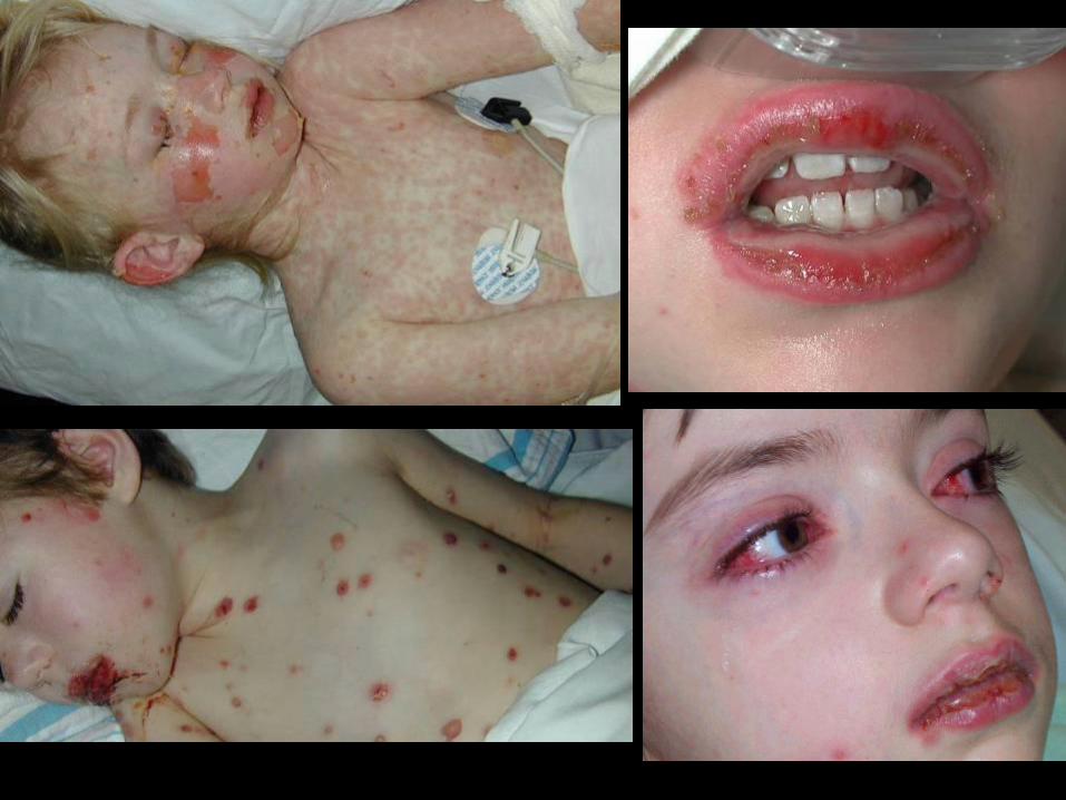

SJS & toxic epidermal necrolysis
• Onset typically 10 days to 3 weeks after starting a drug
• Discontinue suspect drugs immediately – Decreased mortality if stop drug early
– Increased mortality if drug has a long half-life
• Culprit drugs – TMP-SMX, long-acting sulfonamides
– Carbamazepine, phenytoin, phenobarbital
– Aminopenicillins
– NSAIDs
• Treatment with intravenous immunoglobulin
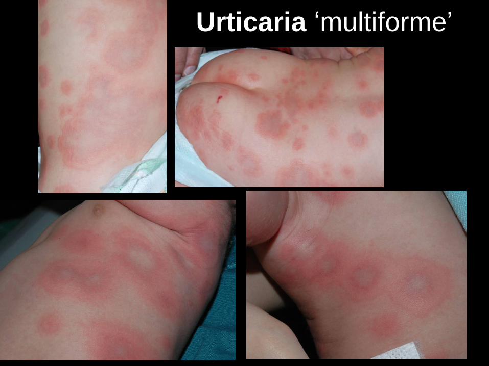
Urticaria ‘multiforme’
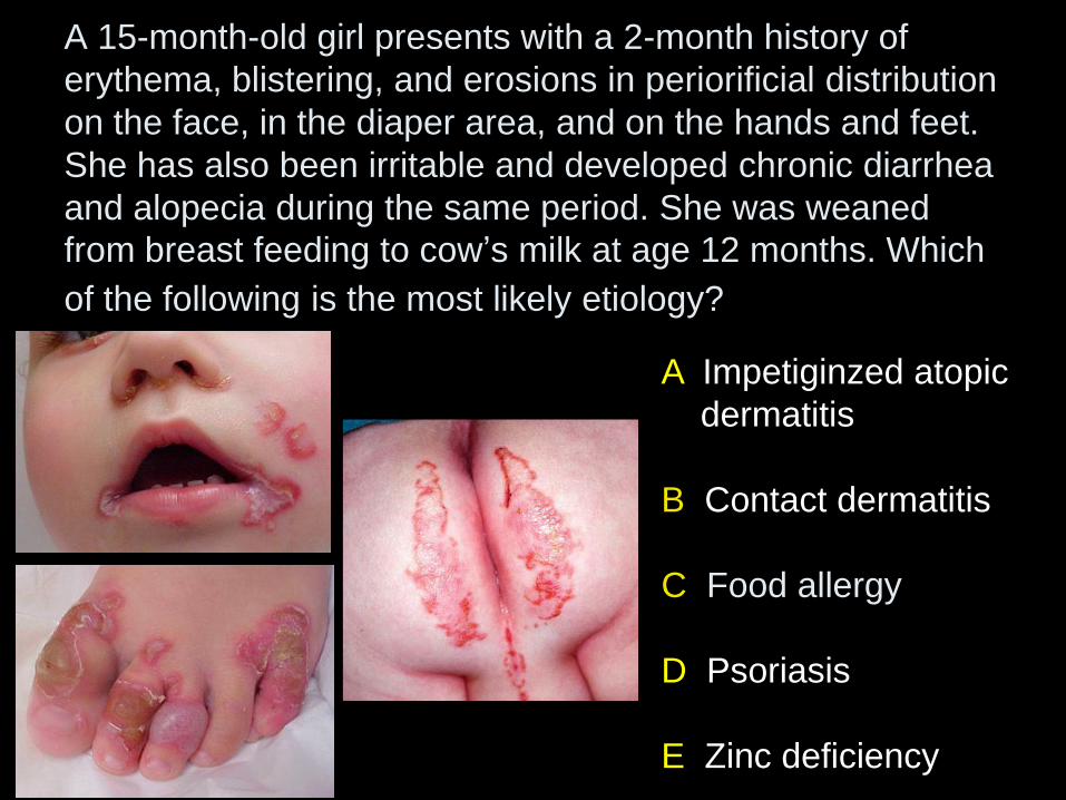
A 15-month-old girl presents with a 2-month history of
erythema, blistering, and erosions in periorificial distribution
on the face, in the diaper area, and on the hands and feet.
She has also been irritable and developed chronic diarrhea
and alopecia during the same period. She was weaned
from breast feeding to cow’s milk at age 12 months. Which
of the following is the most likely etiology?
A Impetiginzed atopic
dermatitis
B Contact dermatitis
C Food allergy
D Psoriasis
E Zinc deficiency
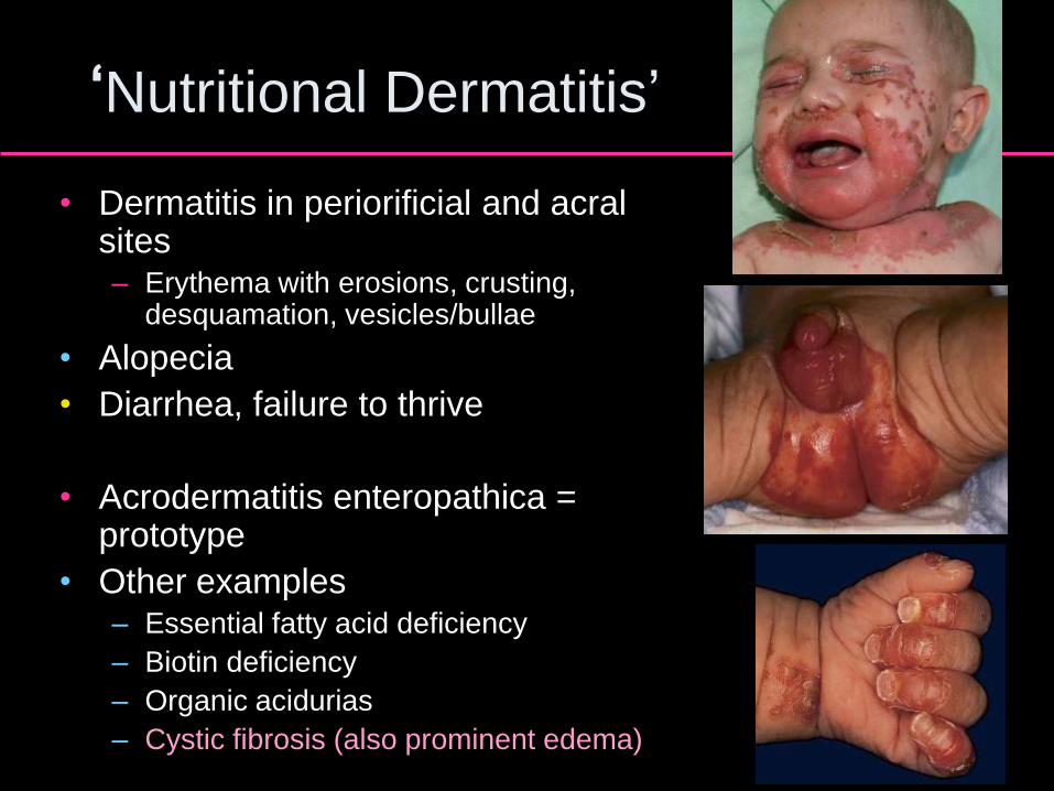
‘Nutritional Dermatitis’
• Dermatitis in periorificial and acral sites – Erythema with erosions, crusting,
desquamation, vesicles/bullae
• Alopecia
• Diarrhea, failure to thrive
• Acrodermatitis enteropathica = prototype
• Other examples – Essential fatty acid deficiency
– Biotin deficiency
– Organic acidurias
– Cystic fibrosis (also prominent edema)
Acrodermatitis Enteropathica
• Genetic form – mutations in SLC39A4 – Encodes intestinal zinc transporter
– Onset in first few weeks (or after weaning of zinc-enriched formula) if bottle fed
– Onset after weaning if breast fed
• Acquired forms – Low zinc level in breast milk
– TPN or prematurity without supplementation
– GI disorder or high-fiber diet impairing absorption
• Additional findings – Low plasma zinc
– Low serum alkaline phosphatase
– Irritability, stomatitis/glossitis, photophobia
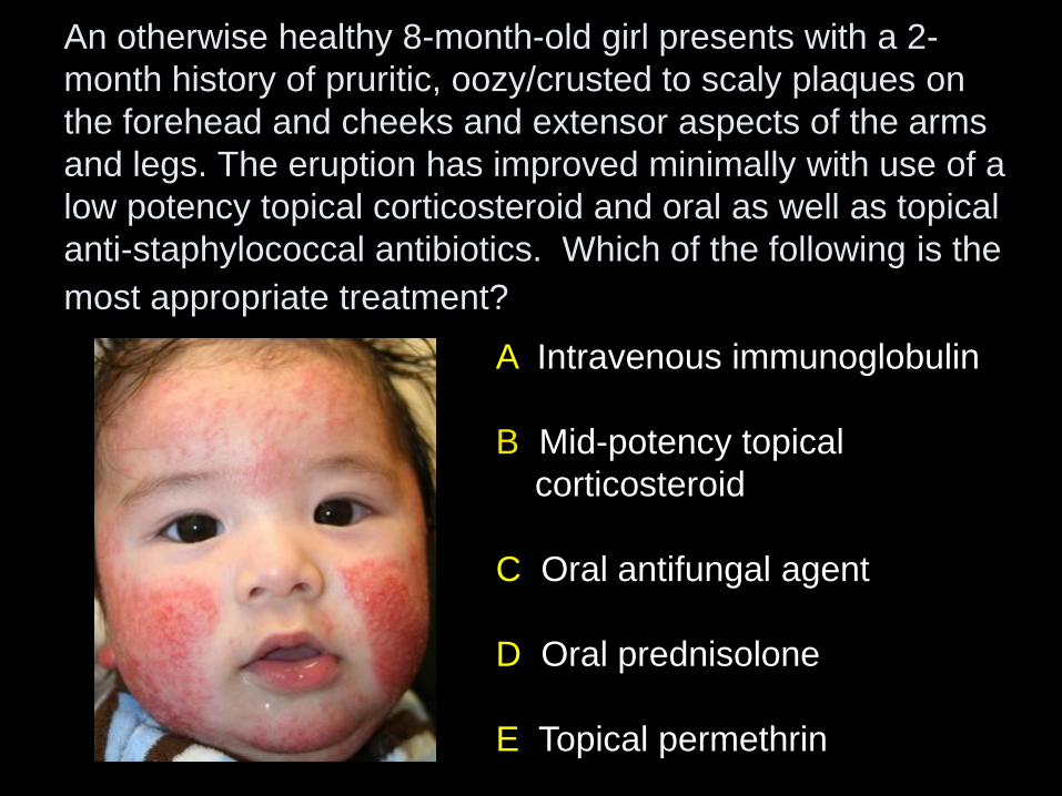
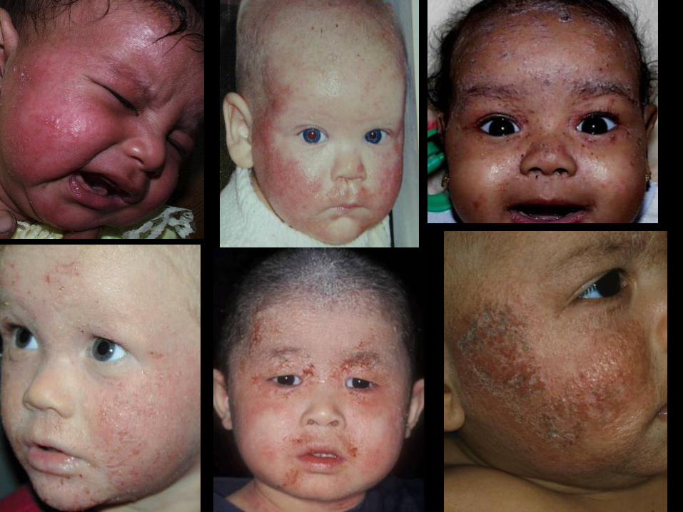
An otherwise healthy 8-month-old girl presents with a 2-
month history of pruritic, oozy/crusted to scaly plaques on
the forehead and cheeks and extensor aspects of the arms
and legs. The eruption has improved minimally with use of a
low potency topical corticosteroid and oral as well as topical
anti-staphylococcal antibiotics. Which of the following is the
most appropriate treatment?
A Intravenous immunoglobulin
B Mid-potency topical
corticosteroid
C Oral antifungal agent
D Oral prednisolone
E Topical permethrin
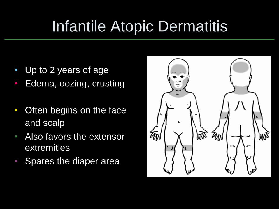
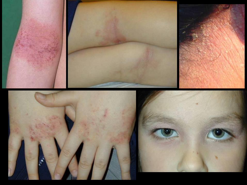
Infantile Atopic Dermatitis
• Up to 2 years of age
• Edema, oozing, crusting
• Often begins on the face
and scalp
• Also favors the extensor
extremities
• Spares the diaper area
Lehtonen E, et al. Pediatr Allergy Immunol 2003;14:405
Laughter D, et al. J Am Acad Dermatol 2000;43:649
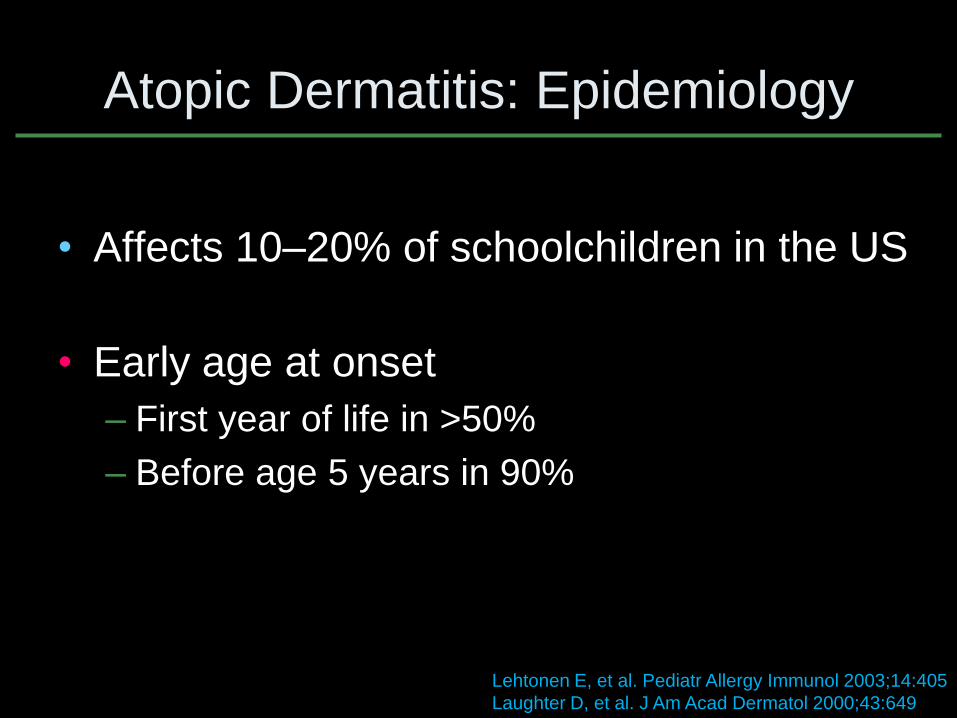
Atopic Dermatitis: Epidemiology
• Affects 10–20% of schoolchildren in the US
• Early age at onset
– First year of life in >50%
– Before age 5 years in 90%
Atopic Dermatitis: Disease Impact
• Intense pruritus/discomfort
• Skin infections – Staphylococcus aureus
– Herpes simplex virus (eczema herpeticum)
– Molluscum contagiosum
• Sleep disturbances
• Psychological distress
• Impaired social and school functioning
• Disrupted family dynamics Zuberbier T, et al. J Allergy Clin Immunol 2006;118:226 Brenninkmeijer EE, et al. Pediatr Dermatol 2009;26:14
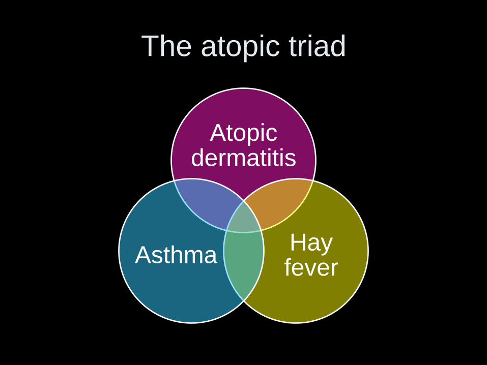
The atopic triad
Atopic dermatitis
Hay fever
Asthma
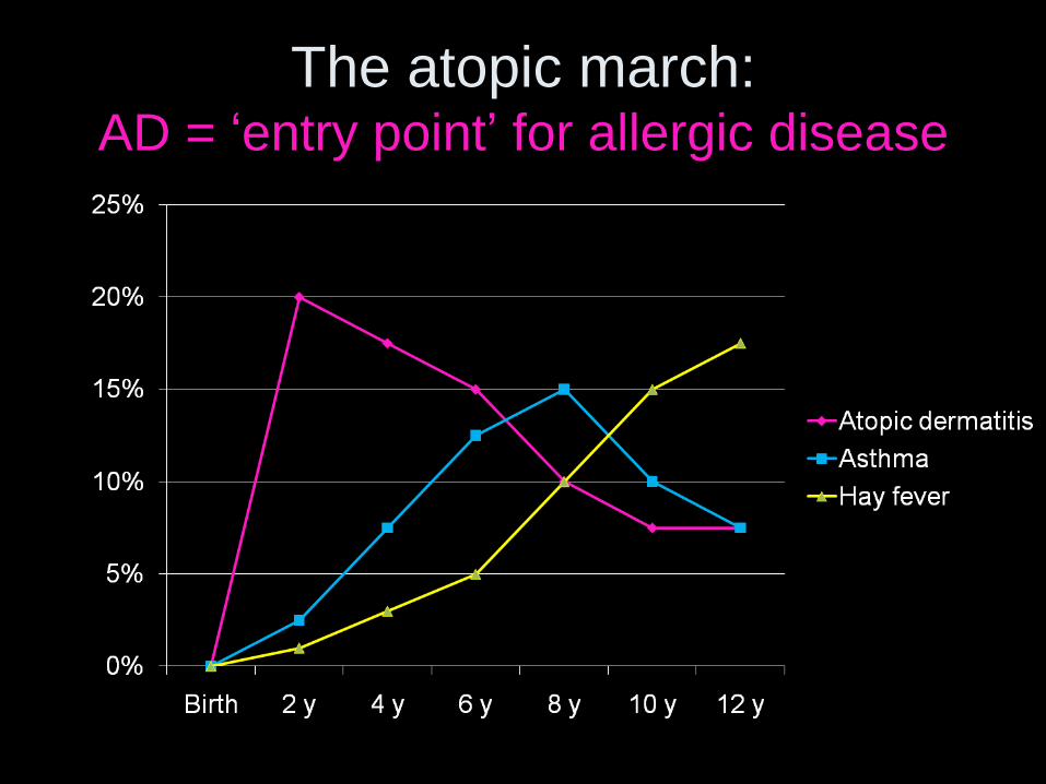
The atopic march: AD = ‘entry point’ for allergic disease
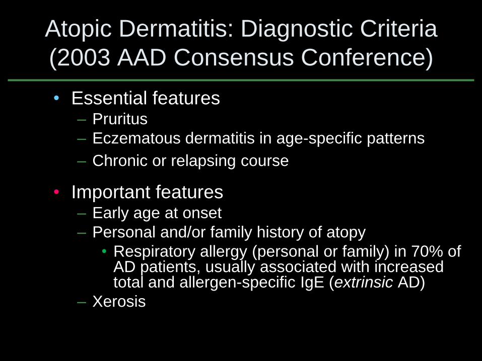
Atopic Dermatitis: Diagnostic Criteria
(2003 AAD Consensus Conference)
• Essential features – Pruritus
– Eczematous dermatitis in age-specific patterns
– Chronic or relapsing course
• Important features – Early age at onset
– Personal and/or family history of atopy
• Respiratory allergy (personal or family) in 70% of AD patients, usually associated with increased total and allergen-specific IgE (extrinsic AD)
– Xerosis
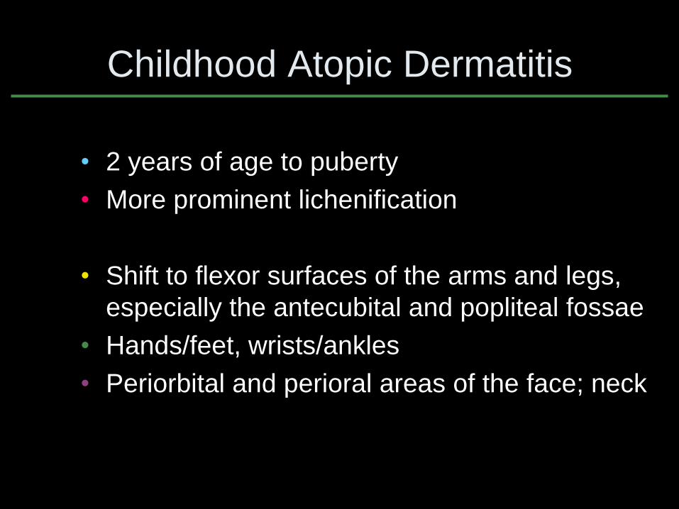
Childhood Atopic Dermatitis
• 2 years of age to puberty
• More prominent lichenification
• Shift to flexor surfaces of the arms and legs,
especially the antecubital and popliteal fossae
• Hands/feet, wrists/ankles
• Periorbital and perioral areas of the face; neck
Triggers/Exacerbating Factors for
Atopic Dermatitis
• Anxiety/stress
• Climate – Extremes of temperature (winter or summer) – Low humidity
• Irritants – Detergents, solvents, wool/other rough materials – Perspiration
• Infection – systemic (e.g. viral URI) or cutaneous
• Allergens - contact, inhaled, & food – Food allergies are a clinically significant trigger in only a
small minority of patients - ~1/3 of infants/young children with treatment-refractory, moderate to severe atopic dermatitis
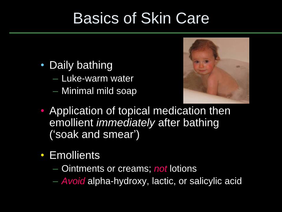
Basics of Skin Care
• Daily bathing – Luke-warm water
– Minimal mild soap
• Application of topical medication then emollient immediately after bathing (‘soak and smear’)
• Emollients – Ointments or creams; not lotions
– Avoid alpha-hydroxy, lactic, or salicylic acid
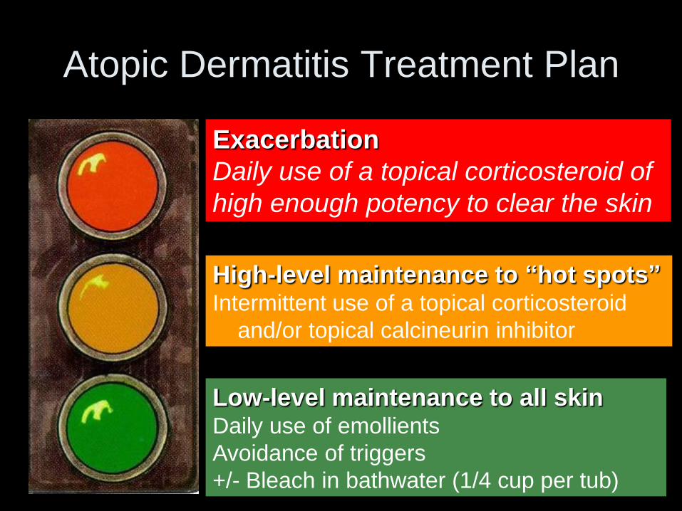
Atopic Dermatitis Treatment Plan
Exacerbation
Daily use of a topical corticosteroid of
high enough potency to clear the skin
Low-level maintenance to all skin Daily use of emollients
Avoidance of triggers
+/- Bleach in bathwater (1/4 cup per tub)
High-level maintenance to “hot spots” Intermittent use of a topical corticosteroid
and/or topical calcineurin inhibitor
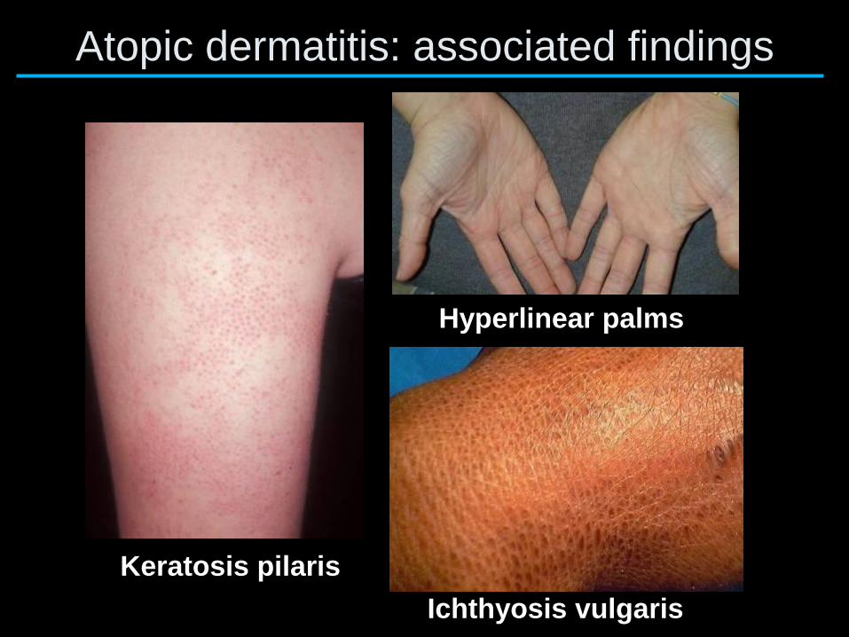
Atopic dermatitis: associated findings
Keratosis pilaris
Ichthyosis vulgaris
Hyperlinear palms
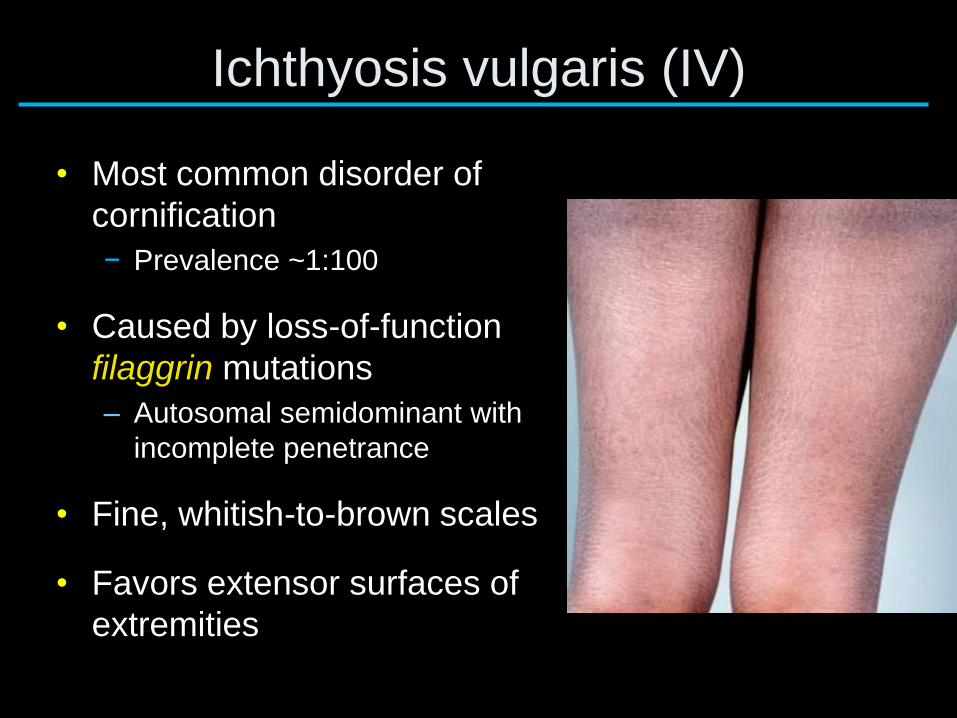
Ichthyosis vulgaris (IV)
• Most common disorder of
cornification
− Prevalence ~1:100
• Caused by loss-of-function
filaggrin mutations
– Autosomal semidominant with
incomplete penetrance
• Fine, whitish-to-brown scales
• Favors extensor surfaces of
extremities
Filaggrin and atopic dermatitis (AD)
• Loss-of-function filaggrin mutations – Same mutations in ichthyosis vulgaris and AD
– 20–50% in European/Asian children/adults with AD
– >50% if moderate to severe AD
– 5–10% in the general population (p<1x10⁻⁸)
• Overall odds ratio ≥4 – One of strongest known genetic factors for a
complex disease
A Folliculitis
B Gianotti-Crosti syndrome
C Molluscum contagiosium
D Papular urticaria
E Scabies
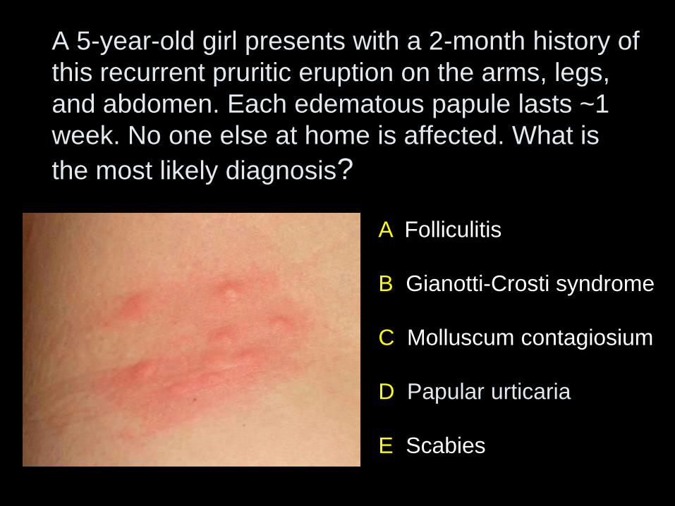
A 5-year-old girl presents with a 2-month history of
this recurrent pruritic eruption on the arms, legs,
and abdomen. Each edematous papule lasts ~1
week. No one else at home is affected. What is
the most likely diagnosis?
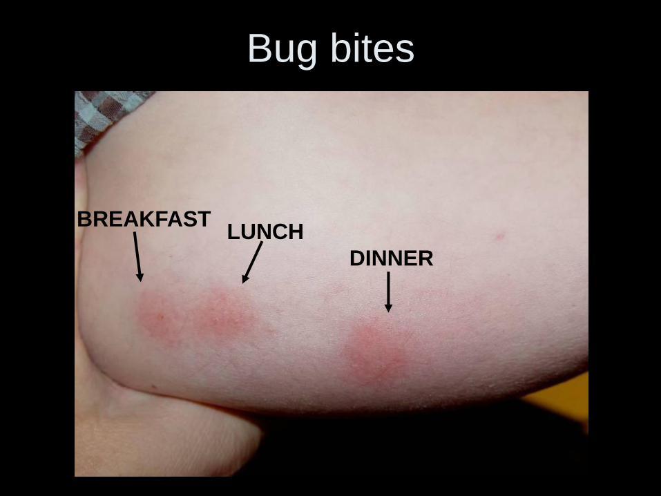

Insect Bite Reactions
• Pruritic, edematous, erythematous papules – Often grouped – ‘breakfast-lunch-dinner’ – at sites of bites
– Frequently excoriated and may become vesicular
– Disseminated papular urticaria can also occur
• Represents delayed-type hypersensitivity reaction – Individual lesions last days to weeks
– Depends on individual immune response, so often only one family member is affected
• Treatment – Topical corticosteroid of at least moderate potency
– Sedating antihistamines
• Prevention – Insect repellents/protective clothing for outdoor bugs (e.g.
mosquitoes)
– Treat affected animals/their environment (e.g. for fleas)
– Exterminate home (e.g. for bed bugs)
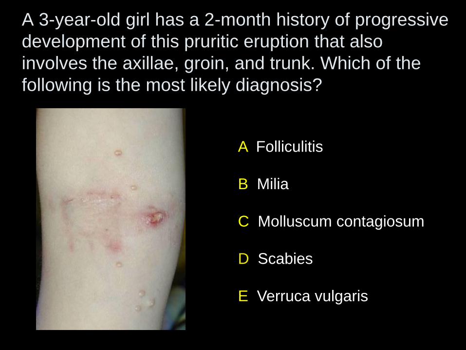
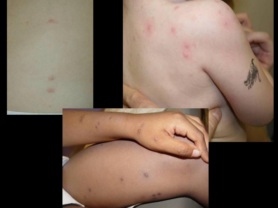
A 3-year-old girl has a 2-month history of progressive
development of this pruritic eruption that also
involves the axillae, groin, and trunk. Which of the
following is the most likely diagnosis?
A Folliculitis
B Milia
C Molluscum contagiosum
D Scabies
E Verruca vulgaris
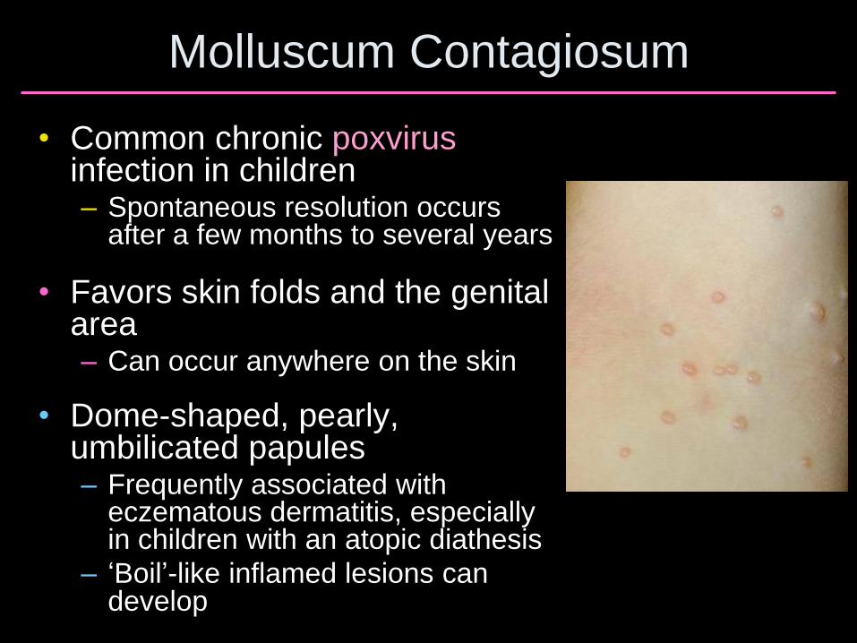
Molluscum Contagiosum
• Common chronic poxvirus infection in children – Spontaneous resolution occurs
after a few months to several years
• Favors skin folds and the genital area – Can occur anywhere on the skin
• Dome-shaped, pearly, umbilicated papules – Frequently associated with
eczematous dermatitis, especially in children with an atopic diathesis
– ‘Boil’-like inflamed lesions can develop
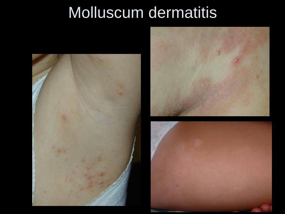
Molluscum dermatitis
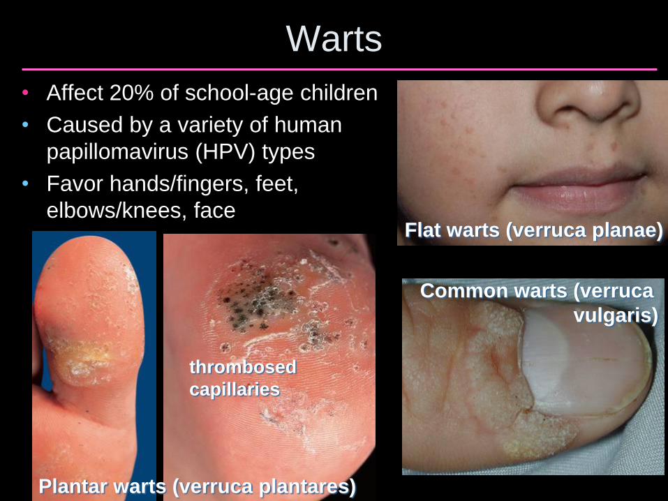
Warts
• Affect 20% of school-age children
• Caused by a variety of human
papillomavirus (HPV) types
• Favor hands/fingers, feet,
elbows/knees, face Flat warts (verruca planae)
Common warts (verruca
vulgaris)
Plantar warts (verruca plantares)
thrombosed
capillaries
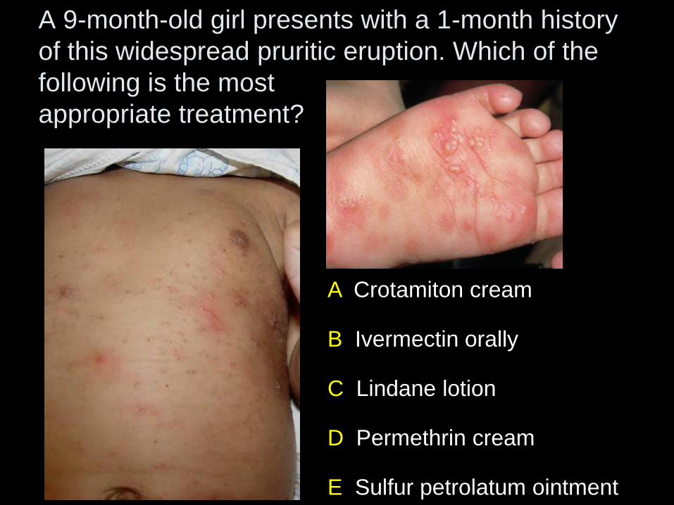
A Crotamiton cream
B Ivermectin orally
C Lindane lotion
D Permethrin cream
E Sulfur petrolatum ointment
A 9-month-old girl presents with a 1-month history
of this widespread pruritic eruption. Which of the
following is the most
appropriate treatment?
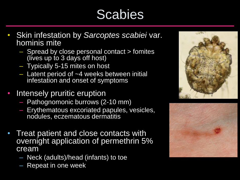
Scabies
• Skin infestation by Sarcoptes scabiei var. hominis mite – Spread by close personal contact > fomites
(lives up to 3 days off host)
– Typically 5-15 mites on host
– Latent period of ~4 weeks between initial infestation and onset of symptoms
• Intensely pruritic eruption – Pathognomonic burrows (2-10 mm)
– Erythematous excoriated papules, vesicles, nodules, eczematous dermatitis
• Treat patient and close contacts with overnight application of permethrin 5% cream – Neck (adults)/head (infants) to toe
– Repeat in one week
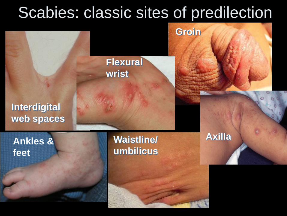
Scabies: classic sites of predilection
Interdigital
web spaces
Groin
Waistline/
umbilicus Ankles &
feet
Flexural
wrist
Axilla
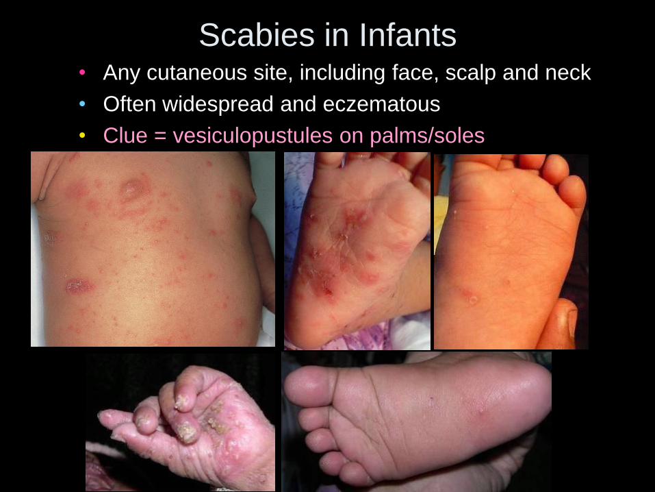
Scabies in Infants • Any cutaneous site, including face, scalp and neck
• Often widespread and eczematous
• Clue = vesiculopustules on palms/soles
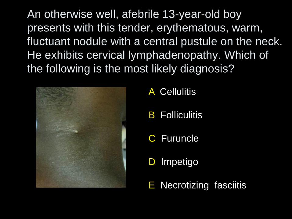
An otherwise well, afebrile 13-year-old boy
presents with this tender, erythematous, warm,
fluctuant nodule with a central pustule on the neck.
He exhibits cervical lymphadenopathy. Which of
the following is the most likely diagnosis?
A Cellulitis
B Folliculitis
C Furuncle
D Impetigo
E Necrotizing fasciitis
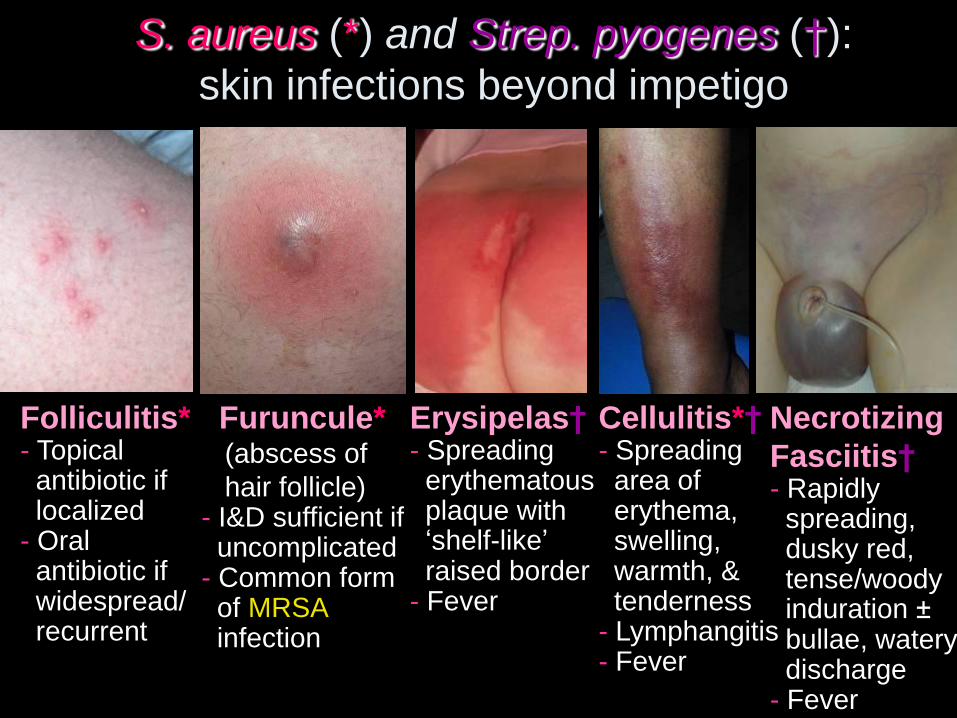
S. aureus (*) and Strep. pyogenes (†):
skin infections beyond impetigo
Folliculitis* - Topical antibiotic if localized - Oral antibiotic if widespread/ recurrent
Furuncule* (abscess of
hair follicle) - I&D sufficient if uncomplicated - Common form of MRSA infection
Erysipelas† - Spreading erythematous plaque with ‘shelf-like’ raised border - Fever
Cellulitis*† - Spreading area of erythema, swelling, warmth, & tenderness - Lymphangitis - Fever
Necrotizing
Fasciitis† - Rapidly spreading, dusky red, tense/woody induration ± bullae, watery discharge - Fever
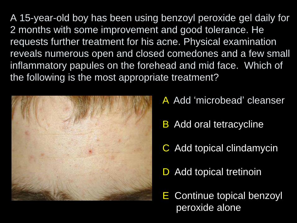
A Add ‘microbead’ cleanser
B Add oral tetracycline
C Add topical clindamycin
D Add topical tretinoin
E Continue topical benzoyl
peroxide alone
A 15-year-old boy has been using benzoyl peroxide gel daily for
2 months with some improvement and good tolerance. He
requests further treatment for his acne. Physical examination
reveals numerous open and closed comedones and a few small
inflammatory papules on the forehead and mid face. Which of
the following is the most appropriate treatment?
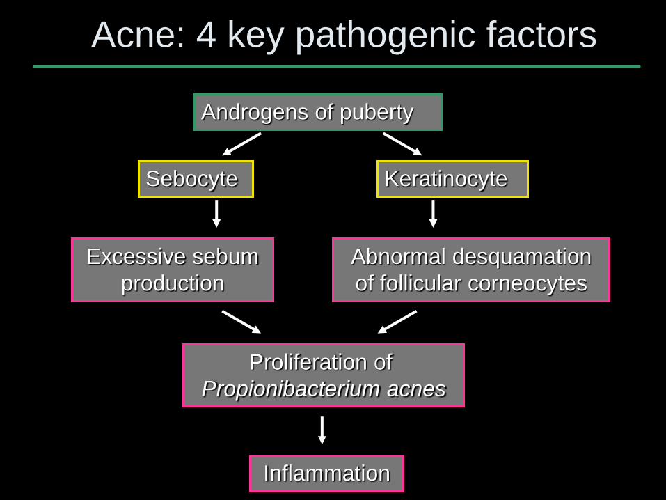
Acne: 4 key pathogenic factors
Androgens of puberty
Excessive sebum
production
Abnormal desquamation
of follicular corneocytes
Proliferation of
Propionibacterium acnes
Inflammation
Sebocyte Keratinocyte

Spectrum of efficacy of topical
anti-acne agents
Comedo-
lysis
↓ Sebum Anti-
microbial
Anti-
inflammatory
Tretinoin ++ - - (+)
Adapalene ++ - - +
Tazarotene ++ - - (+)
Clinda (+) - ++ +
Erythro (+) - ++ +
BPO + - +++ (+)
Azeleic acid + - + (+)
Sal acid (+) - - -
BPO = benzoyl peroxide

Acne: therapeutic ladder
• Mild primarily comedonal – Topical retinoid
• Mild primarily inflammatory – Topical antibiotic + BPO or
– Topical retinoid + topical antibiotic and/or BPO
• Moderate inflammatory (variable comedonal) – Oral antibiotic* + topical retinoid (+ BPO if >2 mos)
– For teen girls, consider adding oral contraceptive
• Severe nodulocystic/recalcitrant inflammatory – Oral retinoid (isotretinoin)
*1st line = doxycycline or minocycline
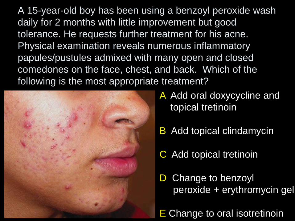
A 15-year-old boy has been using a benzoyl peroxide wash
daily for 2 months with little improvement but good
tolerance. He requests further treatment for his acne.
Physical examination reveals numerous inflammatory
papules/pustules admixed with many open and closed
comedones on the face, chest, and back. Which of the
following is the most appropriate treatment?
A Add oral doxycycline and
topical tretinoin
B Add topical clindamycin
C Add topical tretinoin
D Change to benzoyl
peroxide + erythromycin gel
E Change to oral isotretinoin

A Avoid offending allergen
B Moderately potent topical
corticosteroid
C Narrowband UVB
D Oral griseofulvin
E Topical selenium sulfide
A 16-year-old boy presents with a 3-month history of
this eruption on the upper trunk. The well-demarcated
hypopigmented macules coalesce centrally in areas of
involvement and fine white scale is evident upon
stretching the skin. Which of the following treatments is
most appropriate?
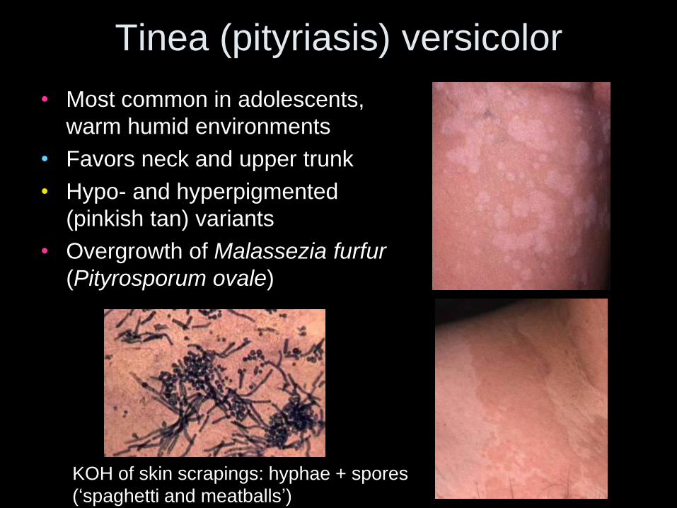
Tinea (pityriasis) versicolor
• Most common in adolescents,
warm humid environments
• Favors neck and upper trunk
• Hypo- and hyperpigmented
(pinkish tan) variants
• Overgrowth of Malassezia furfur
(Pityrosporum ovale)
KOH of skin scrapings: hyphae + spores
(‘spaghetti and meatballs’)
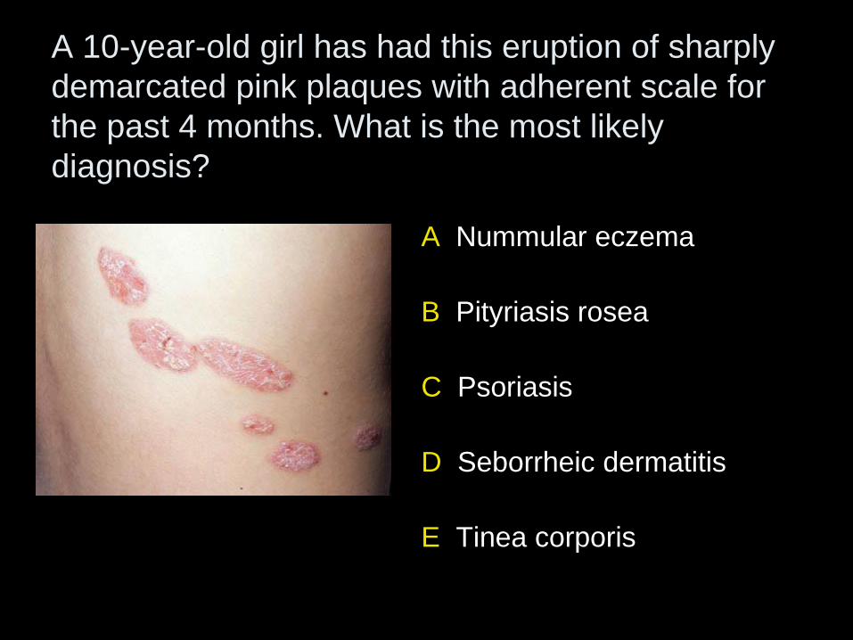
A 10-year-old girl has had this eruption of sharply
demarcated pink plaques with adherent scale for
the past 4 months. What is the most likely
diagnosis?
A Nummular eczema
B Pityriasis rosea
C Psoriasis
D Seborrheic dermatitis
E Tinea corporis
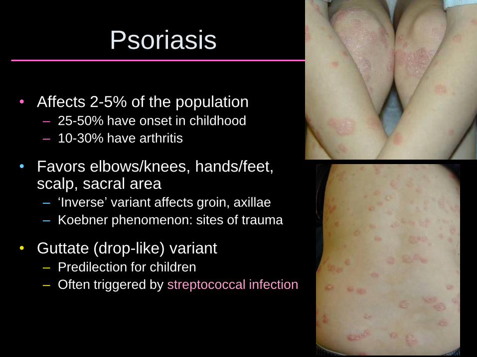
Psoriasis
• Affects 2-5% of the population – 25-50% have onset in childhood
– 10-30% have arthritis
• Favors elbows/knees, hands/feet, scalp, sacral area – ‘Inverse’ variant affects groin, axillae
– Koebner phenomenon: sites of trauma
• Guttate (drop-like) variant – Predilection for children
– Often triggered by streptococcal infection
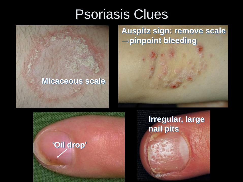
Psoriasis Clues
Micaceous scale
Auspitz sign: remove scale
→pinpoint bleeding
‘Oil drop’
Irregular, large
nail pits
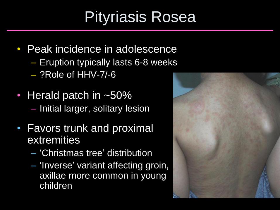
Pityriasis Rosea
• Peak incidence in adolescence – Eruption typically lasts 6-8 weeks
– ?Role of HHV-7/-6
• Herald patch in ~50% – Initial larger, solitary lesion
• Favors trunk and proximal extremities – ‘Christmas tree’ distribution
– ‘Inverse’ variant affecting groin, axillae more common in young children
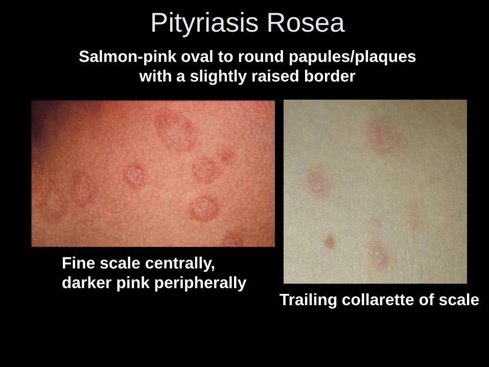
Pityriasis Rosea
Salmon-pink oval to round papules/plaques
with a slightly raised border
Fine scale centrally,
darker pink peripherally Trailing collarette of scale
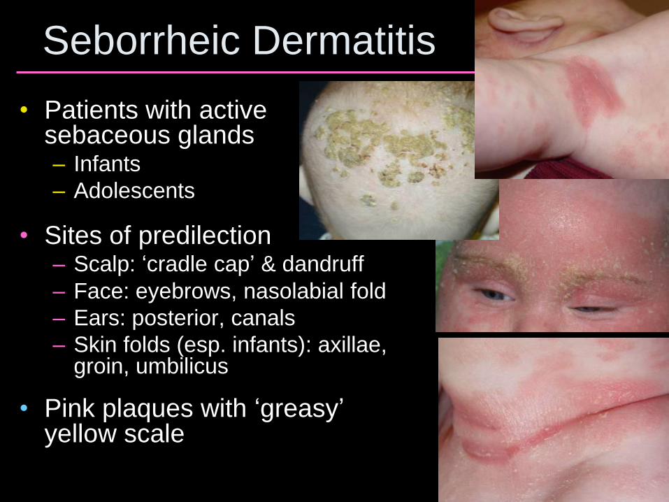
Seborrheic Dermatitis
• Patients with active sebaceous glands – Infants
– Adolescents
• Sites of predilection – Scalp: ‘cradle cap’ & dandruff
– Face: eyebrows, nasolabial fold
– Ears: posterior, canals
– Skin folds (esp. infants): axillae, groin, umbilicus
• Pink plaques with ‘greasy’ yellow scale
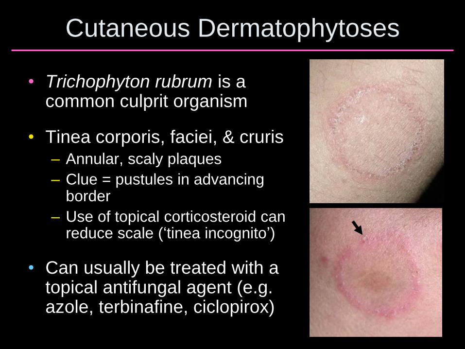
Cutaneous Dermatophytoses
• Trichophyton rubrum is a common culprit organism
• Tinea corporis, faciei, & cruris – Annular, scaly plaques
– Clue = pustules in advancing border
– Use of topical corticosteroid can reduce scale (‘tinea incognito’)
• Can usually be treated with a topical antifungal agent (e.g. azole, terbinafine, ciclopirox)
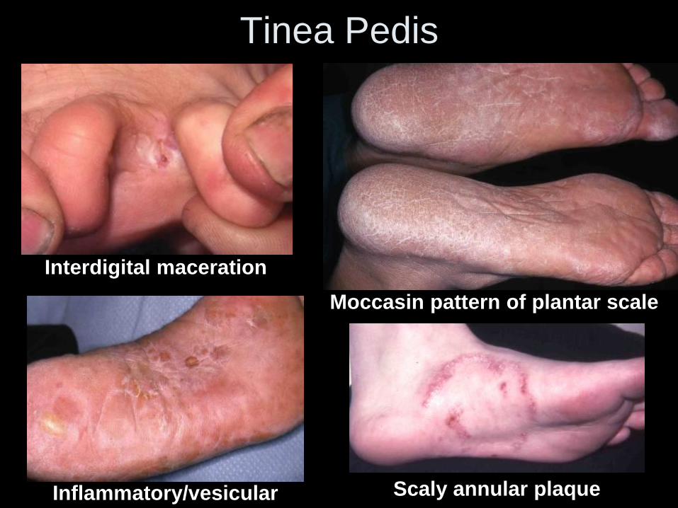
Tinea Pedis
Moccasin pattern of plantar scale
Interdigital maceration
Inflammatory/vesicular Scaly annular plaque
A Atopic dermatitis
B Contact dermatitis
C Nummular eczema
D Papular urticaria
E Scabies
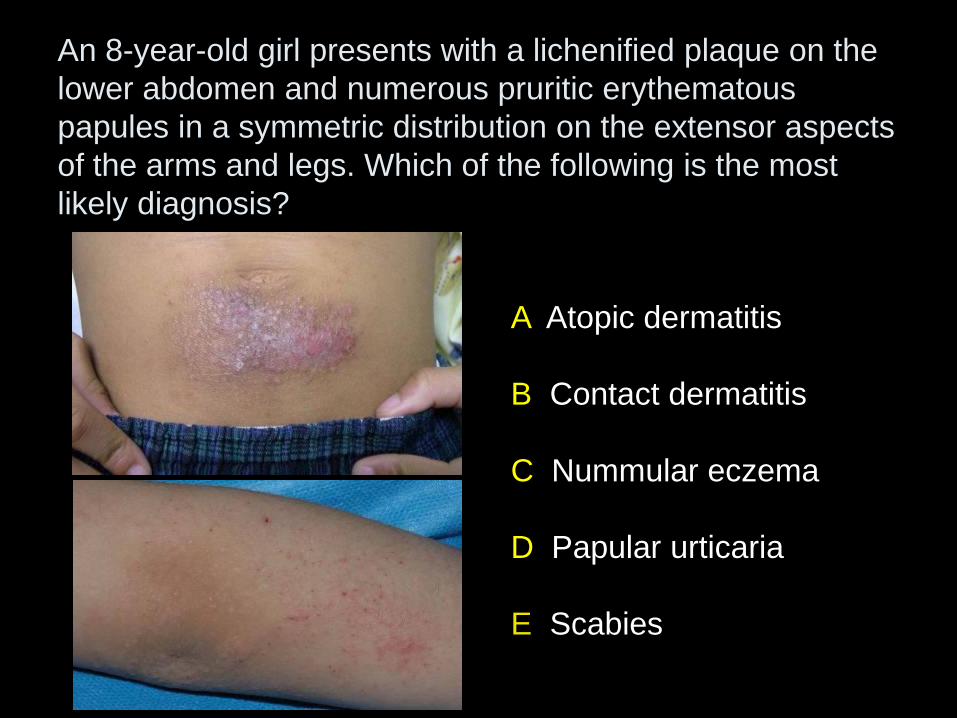
An 8-year-old girl presents with a lichenified plaque on the
lower abdomen and numerous pruritic erythematous
papules in a symmetric distribution on the extensor aspects
of the arms and legs. Which of the following is the most
likely diagnosis?
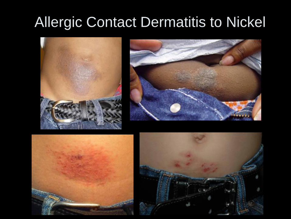
Allergic Contact Dermatitis to Nickel
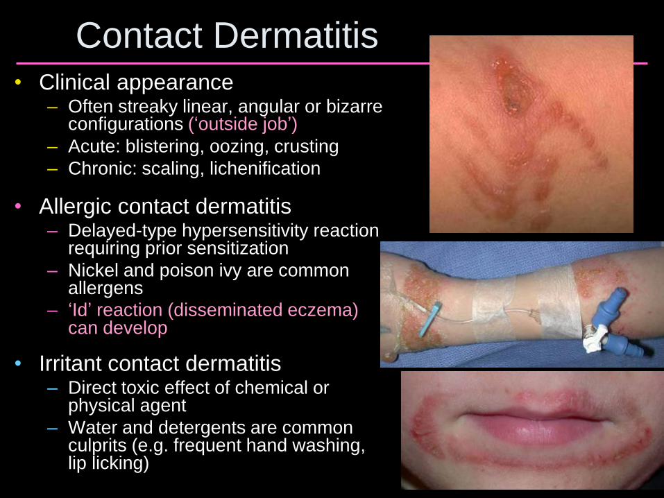
Contact Dermatitis • Clinical appearance
– Often streaky linear, angular or bizarre configurations (‘outside job’)
– Acute: blistering, oozing, crusting
– Chronic: scaling, lichenification
• Allergic contact dermatitis – Delayed-type hypersensitivity reaction
requiring prior sensitization
– Nickel and poison ivy are common allergens
– ‘Id’ reaction (disseminated eczema) can develop
• Irritant contact dermatitis – Direct toxic effect of chemical or
physical agent
– Water and detergents are common culprits (e.g. frequent hand washing, lip licking)
A Diphenhydramine topically
B Loratadine orally
C Pramoxine topically
D Prednisone orally for 2 weeks
E Triamcinolone cream topically
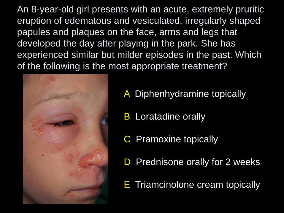
An 8-year-old girl presents with an acute, extremely pruritic
eruption of edematous and vesiculated, irregularly shaped
papules and plaques on the face, arms and legs that
developed the day after playing in the park. She has
experienced similar but milder episodes in the past. Which
of the following is the most appropriate treatment?
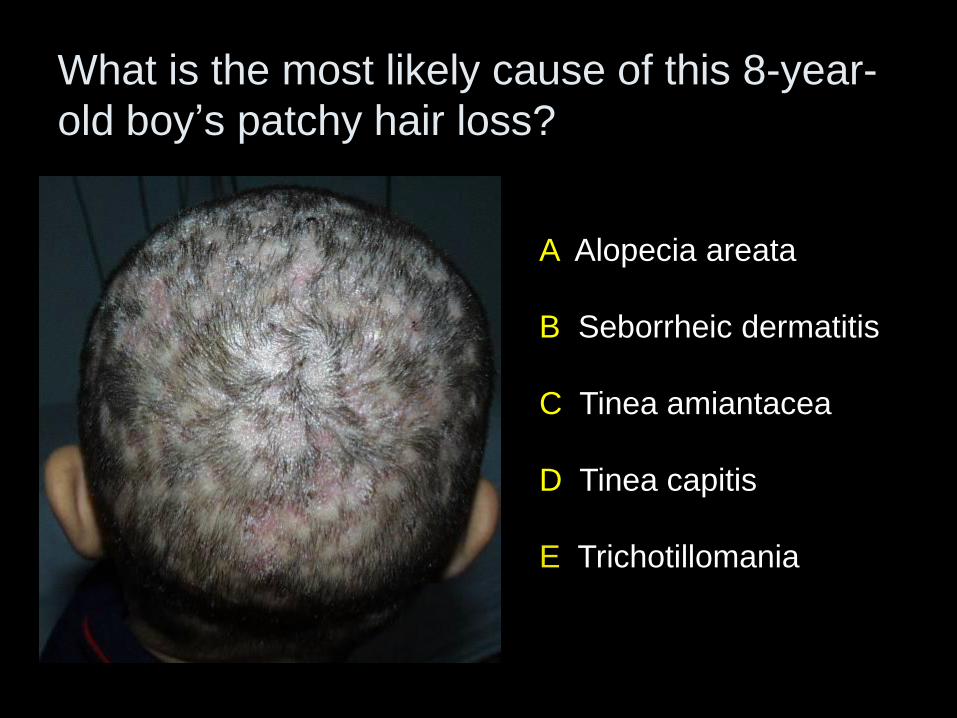
A Alopecia areata
B Seborrheic dermatitis
C Tinea amiantacea
D Tinea capitis
E Trichotillomania
What is the most likely cause of this 8-year-
old boy’s patchy hair loss?
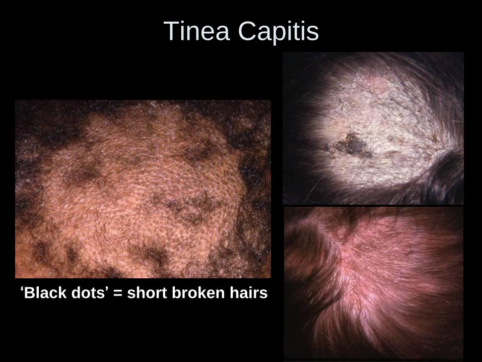
Tinea Capitis
‘Black dots’ = short broken hairs
A Cefazolin intravenously
B Griseofulvin orally
C Incision and drainage
D Ketoconazole topically
E Mupirocin topically
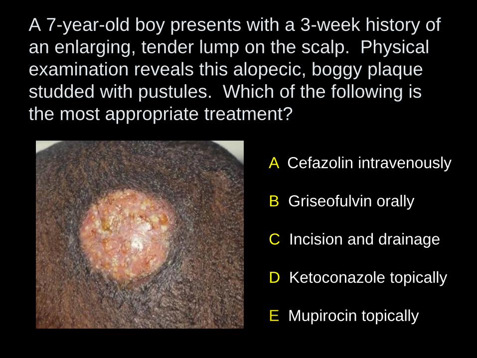
A 7-year-old boy presents with a 3-week history of
an enlarging, tender lump on the scalp. Physical
examination reveals this alopecic, boggy plaque
studded with pustules. Which of the following is
the most appropriate treatment?
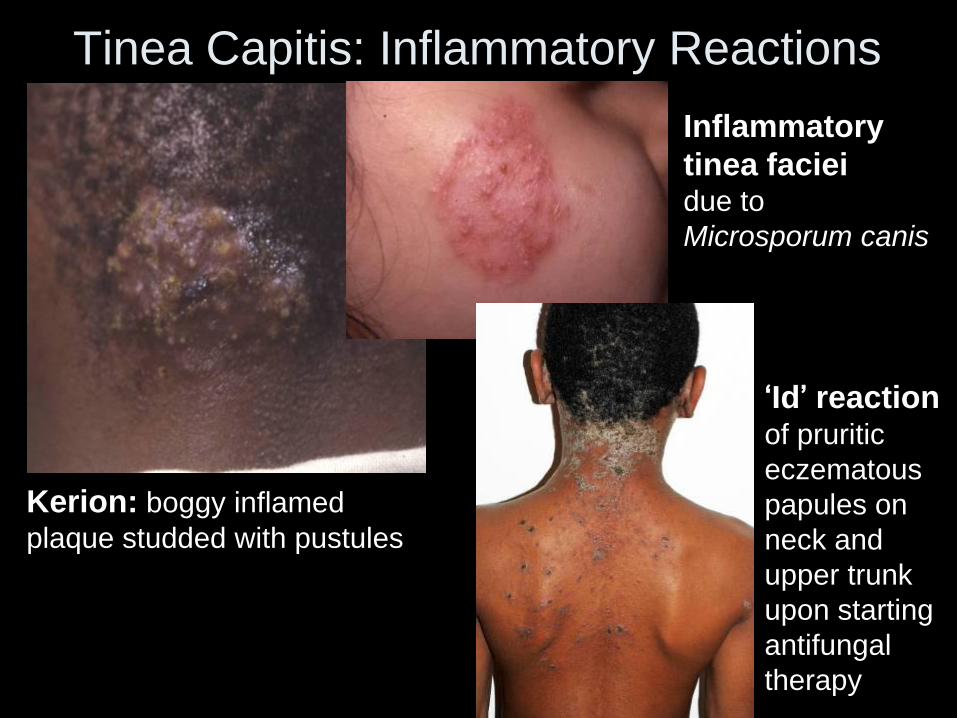
Tinea Capitis: Inflammatory Reactions
Kerion: boggy inflamed
plaque studded with pustules
Inflammatory
tinea faciei due to
Microsporum canis
‘Id’ reaction of pruritic
eczematous
papules on
neck and
upper trunk
upon starting
antifungal
therapy
Tinea Capitis • Trichophyton tonsurans (~95%) >>
Microsporum canis in US
• Favors prepubertal children – Predilection for those of African descent
– Always consider in school-age child with scaly scalp
– Posterior cervical lymphadenopathy often present
• Treatment – Requires ORAL antifungal agent: griseofulvin (20-25
mg/kg/day x 6-8 wks) or terbinafine (~5-8 mg/kg x 3-4 wks; T. tonsurans only)
– Perform fungal culture prior to initiating therapy
– Selenium sulfide or ketoconazole shampoo (patient and household contacts) to prevent spread
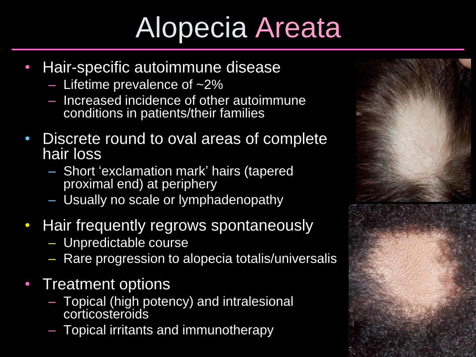
Alopecia Areata
• Hair-specific autoimmune disease – Lifetime prevalence of ~2%
– Increased incidence of other autoimmune conditions in patients/their families
• Discrete round to oval areas of complete hair loss – Short ‘exclamation mark’ hairs (tapered
proximal end) at periphery
– Usually no scale or lymphadenopathy
• Hair frequently regrows spontaneously – Unpredictable course
– Rare progression to alopecia totalis/universalis
• Treatment options – Topical (high potency) and intralesional
corticosteroids
– Topical irritants and immunotherapy
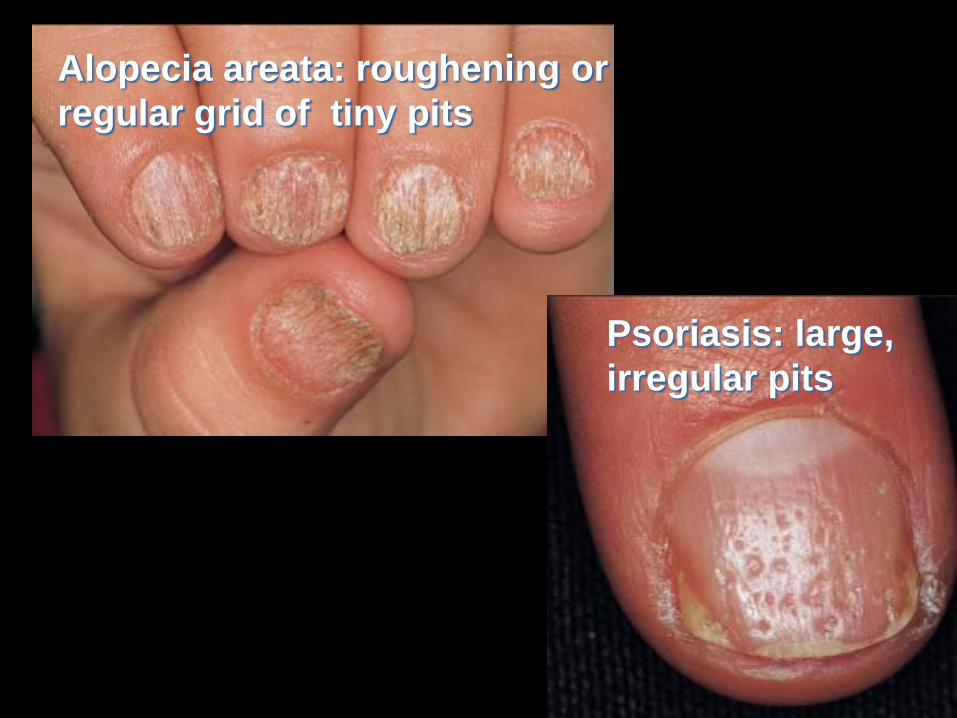
Psoriasis: large,
irregular pits
Alopecia areata: roughening or
regular grid of tiny pits
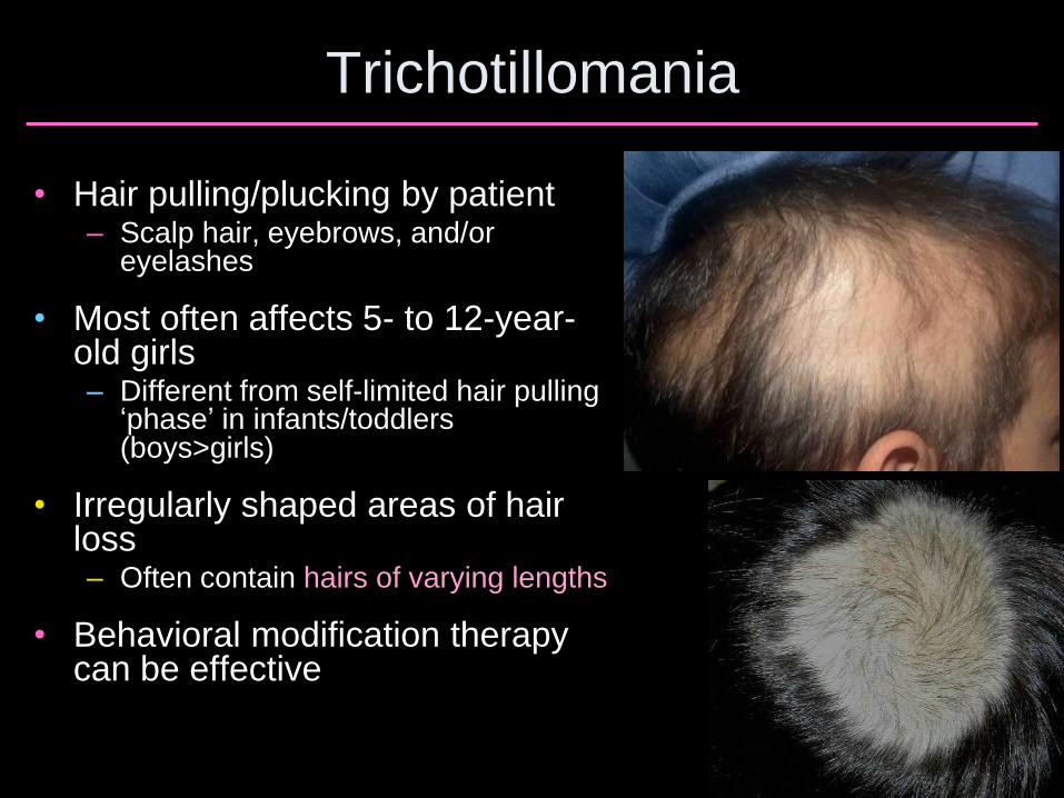
Trichotillomania
• Hair pulling/plucking by patient – Scalp hair, eyebrows, and/or
eyelashes
• Most often affects 5- to 12-year-old girls – Different from self-limited hair pulling
‘phase’ in infants/toddlers (boys>girls)
• Irregularly shaped areas of hair loss – Often contain hairs of varying lengths
• Behavioral modification therapy can be effective
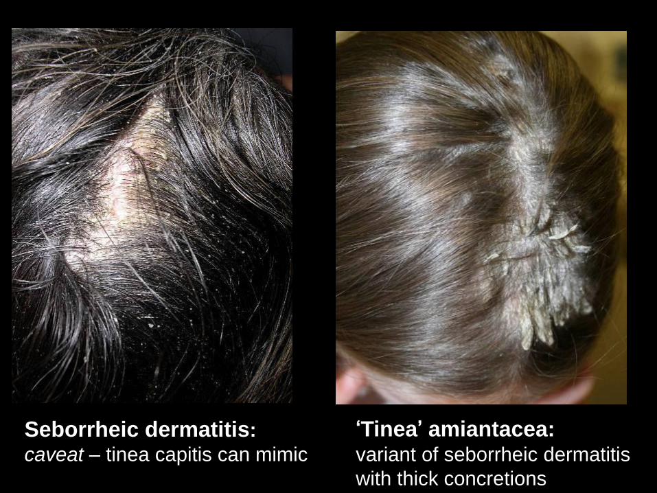
Seborrheic dermatitis:
caveat – tinea capitis can mimic
‘Tinea’ amiantacea: variant of seborrheic dermatitis
with thick concretions

Telogen Effluvium
• Period of excessive shedding of normal
telogen hairs
– DIFFUSE pattern of thinning
• Hair loss begins 2-3 months after
precipitating event such as:
– High fever/severe illness
– Severe psychological stress
• Complete recovery expected
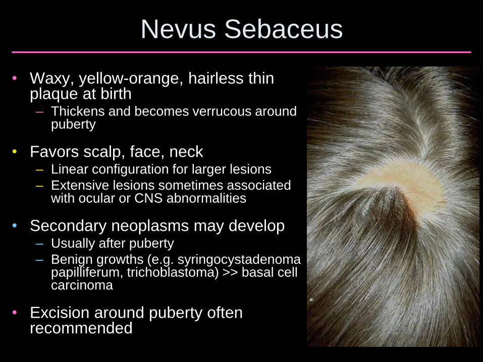
Nevus Sebaceus
• Waxy, yellow-orange, hairless thin plaque at birth – Thickens and becomes verrucous around
puberty
• Favors scalp, face, neck – Linear configuration for larger lesions
– Extensive lesions sometimes associated with ocular or CNS abnormalities
• Secondary neoplasms may develop – Usually after puberty
– Benign growths (e.g. syringocystadenoma papilliferum, trichoblastoma) >> basal cell carcinoma
• Excision around puberty often recommended
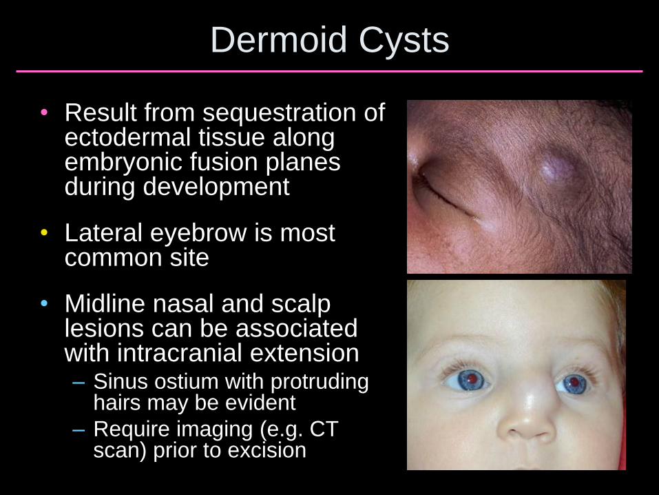
Dermoid Cysts
• Result from sequestration of ectodermal tissue along embryonic fusion planes during development
• Lateral eyebrow is most common site
• Midline nasal and scalp lesions can be associated with intracranial extension – Sinus ostium with protruding
hairs may be evident
– Require imaging (e.g. CT scan) prior to excision
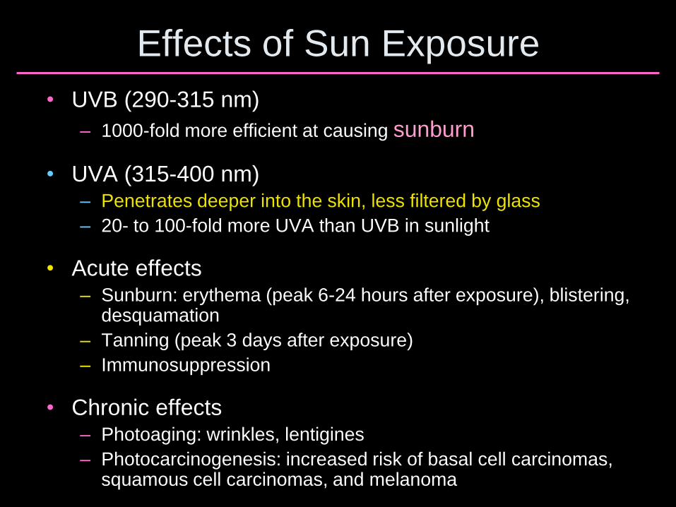
Effects of Sun Exposure
• UVB (290-315 nm)
– 1000-fold more efficient at causing sunburn
• UVA (315-400 nm) – Penetrates deeper into the skin, less filtered by glass
– 20- to 100-fold more UVA than UVB in sunlight
• Acute effects – Sunburn: erythema (peak 6-24 hours after exposure), blistering,
desquamation
– Tanning (peak 3 days after exposure)
– Immunosuppression
• Chronic effects – Photoaging: wrinkles, lentigines
– Photocarcinogenesis: increased risk of basal cell carcinomas, squamous cell carcinomas, and melanoma