pediatric chest (part 2)
TRANSCRIPT

PEDIATRIC MEDIASTINAL MASSES
Dr Mohit Goel
21 Nov. 2012

Paediatric Mediastinal Masses:

Lymphoma
Lymphoma is one of the most common mediastinal neoplasms and may affect any mediastinal location. Non-Hodgkin’s lymphoma usually manifests as generalised disease whereas Hodgkin’s disease tends to present as primary mediastinal lesions.
Hodgkin lymphoma
Hodgkin lymphoma in children is more common in the second decade of life. It can exhibit as multiple rounded soft tissue masses, a dominant bulky soft tissue mass, or a discrete or infiltrating thymic mass.
Lesions may comprise homogenous or heterogeneous soft tissue attenuation depending on the presence of necrosis, haemorrhage, or cystic degeneration. Foci of calcifications may be seen post-therapy.

The CT-images shows a large soft tissue mass in the anterior mediastinum, which arises in the thymus. There is associated paratracheal adenopathy (arrow).

Non-Hodgkin lymphoma
Non-Hodgkin disease in children occurs in the first and second decade of life.
It is associated with extranodal disease and has a greater predilection for noncontiguous and/or haematogenous spread to thoracic and distant nodal and extranodal sites.
Non-Hodgkin disease, in contrast to Hodgkin disease, often spares the thymus.
In this case, enlarged lymph nodes are seen in the right paratracheal , hilar and subcarinal areas without thymus involvement..
Thymic hyperplasia
In childhood, thymic hyperplasia is most often 'rebound' hyperplasia associated with chemotherapy, particularly therapy with corticosteroids.
The mechanism of hyperplasia is believed to be initial depletion of lymphocytes from the cortical portion of the gland due to high serum levels of glucocorticoids, followed by repopulation of the cortical lymphocytes when the cortisone levels return to normal.
On CT, hyperplasia appears as diffuse enlargement of the thymus, with preservation of the normal triangular shape.
CT, MRI of PET cannot differentiate rebound hyperplasia from infiltration of the thymus by tumor. The absence of other active disease and a gradual decrease in thymus size on serial CT's supports the diagnosis of rebound hyperplasia. The thymus usually returns to its normal size in 3 to 6 months.

Thymic Hyperplasia

THYMOMA
Thymoma is the commonest primary tumour of the anterior mediastinum. It occurs most frequently in adults older than 40 years and is rare in children and adolescents.
Thymoma appears as a well-defined, rounded or lobulated anterior-superior mediastinal mass anterior to the aortic root. The mass contains either homogenous or heterogeneous contents depending on the presence of haemorrhage, necrosis, or cyst formation.Calcific foci are seen on CT in a minority of patients


Thymic Carcinoma
Squamous cell and lymphoepithelioma-like carcinoma are the most common histological types. These occur most commonly in middle-aged adults. The appearance is of a large poorly defined infiltrative anterior mediastinalmass and it is commonly associated with pleural and pericardial effusions, and regional lymph node and distant metastasis.
Thymic carcinoma. CT shows large anterior mediastinal mass with ill-defined medial border. The superiorvena cava is compressed.

Thymolipoma
Thymolipoma is an uncommon benign slow growing neoplasm of the thymus gland composed of mature adipose cells and thymic tissue. It is typically a large soft anterior mediastinal mass and is able to conform to adjacent structures simulating cardiomegaly, lobar collapse, and diaphragmatic elevation.
Calcifications are absent. Thymolipoma does not have a capsule and does not have any mass effect.

Non-neoplastic thymic cyst may be congenital or acquired secondary to inflammation. It is seen as a well Circumscribed antero-superior Mediastinal mass with low attenuation contents. Typically, they are thin walled, homogeneous masses of near water attenuation
On CT the attenuation value may be higher than that of simple cysts when the contents are proteinaceous or hemorrhagic rather than serous.
The cystic mass may be uni- ormultiloculated and may show curvilinear calcification of the cystic wall or septa .
Non-neoplastic Thymic Cyst
Germ Cell Tumour
Germ-cell tumors are the most common cause of a fat containing lesions in the anterior mediastinum and the second most common cause of an anterior mediastinal mass in children.
Approximately 90 % are benign germ-cell tumors. Most arise in the thymus.
Mediastinal teratoma occurs in children and young adults with no sex predilection.
On CT, the teratoma appears as a multi-locular cystic tumour with walls of variable thickness. The combination of fluid, soft tissue, calcium, and fat attenuation in an anterior mediastinal mass is a highly specific finding that allows the prospective diagnosis of mature teratoma. Mature teratomas can be very large and still be benign.
A fat-fluid level produced by high lipid content in the cyst fluid is a rare but diagnostic sign.

Anterior mediastinal teratoma - A large heterogenous left anterior mediastinal mass containing soft tissue , fatty and calcific components.


Epicardial fat pad. (a) PA chest radiograph shows loss of the cardiac silhouette at the border of the right
side of the heart and an epicardial fat pad with relatively low density (arrow)

(b) CT scan shows the fat pad (arrow) as an area of homogeneous fat attenuation adjacent to the right border of the heart.

Right-sided retrosternal goiter. (a) PAchest
radiograph demonstrates a goiter (arrow) extending
into the middle mediastinum, obliterating
the right paratracheal stripe, and causing
deviation of the trachea to the left (black arrowhead).
Above the level of the clavicles, the margins of the mass are not sharp
(white arrowhead), indicating that the mass
has an anterior mediastinal component.
Posterior mediastinal masses above the level of the clavicles have an interface with lung and therefore typically have sharp, well-defined margins; in contrast, anterior
masses above the level of the clavicles do not have an interface with lung, so that their margins are not usually sharp.
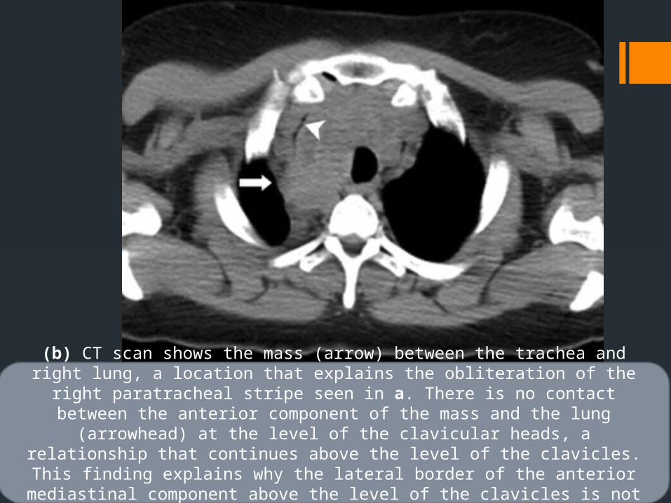
(b) CT scan shows the mass (arrow) between the trachea and right lung, a location that explains the obliteration of the right paratracheal stripe seen in a. There is no
contact between the anterior component of the mass and the lung (arrowhead) at the level of the clavicular heads, a relationship that continues above the level of the clavicles. This finding explains why the lateral border of the anterior mediastinal
component above the level of the clavicles is not sharp in a.

Cystic Hygroma/Lymphangioma
Cystic hygroma / lymphangioma is a benign proliferation of interconnecting lymphatic vessels and sacs that may grow in an infiltrative fashion. It typically affects infants younger than 6 months of age.
Mediastinal lymphangioma typically occurs in the superior aspect of the anterior mediastinum and is usually contiguous with a cervical or axillary component.
The mass usually appears as rounded, lobulated, multi-cystic tumour that can reach a massive size. It tends to surround and displace mediastinal structures and may infiltrate across tissue planes. The thin or thick septa may enhance minimally after contrast administration .
Due to its infiltrating nature, complete surgical resection may be difficult andclose follow-up is needed to check for recurrence.

Cystic hygroma in a 1-year-old baby boy with respiratory distressThe great vessels are encased and displaced although they are all patent. There is no evidence of superior vena cava obstruction (arrows show the brachiocephalic veins).

Pericardial Cyst
It is seen as a well marginated , spherical, or teardrop shaped mass that characteristically abuts the heart, the anterior chest wall, and the diaphragm.
The right anterior cardiophrenic angle is the most common site.
A pericardial cyst is typically shown as a unilocular, non-enhancing mass with water attenuation contents and an imperceptible wall .

.
Middle Mediastinal masses
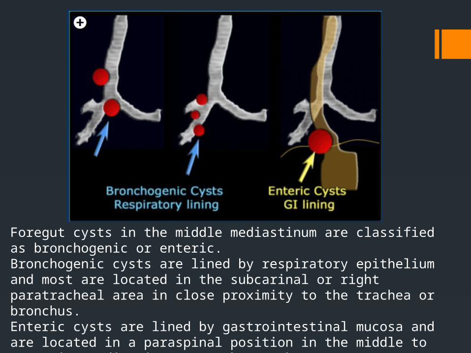
Foregut cysts in the middle mediastinum are classified as bronchogenic or enteric. Bronchogenic cysts are lined by respiratory epithelium and most are located in the subcarinal or right paratracheal area in close proximity to the trachea or bronchus. Enteric cysts are lined by gastrointestinal mucosa and are located in a paraspinal position in the middle to posterior mediastinum near the esophagus

BRONCHOGENIC CYST
They are developmental lesions that result from abnormal ventral budding of the tracheobronchial tree between the 26th and 40th days of gestation.
• LocationMediastinal location is more common than pulmonary
o Mediastinal 65-90% Majority in the middle mediastinum Typically para tracheal, carinal, or hilar Pericarinal most commono Pulmonary: Majority in the medial third of the lungs, More frequent in the lower lobes
Typically do not communicate with airway and do not contain air, Air presence indicates infection.
CT Findings• NECTo Homogeneous well circumscribed lesiono Cyst contents variable: Water to proteinaceouso Hence CT attenuation is variable
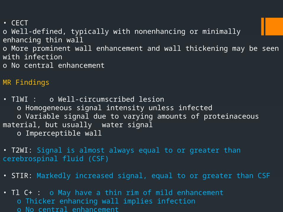
• CECTo Well-defined, typically with nonenhancing or minimally enhancing thin wallo More prominent wall enhancement and wall thickening may be seen with infectiono No central enhancement
MR Findings
• TlWI : o Well-circumscribed lesiono Homogeneous signal intensity unless infectedo Variable signal due to varying amounts of proteinaceous material, but usually water signalo Imperceptible wall
• T2WI: Signal is almost always equal to or greater than cerebrospinal fluid (CSF)
• STIR: Markedly increased signal, equal to or greater than CSF
• Tl C+ : o May have a thin rim of mild enhancemento Thicker enhancing wall implies infectiono No central enhancement

(Left) Axial T2WI MR shows homogeneous, well circumscribed ovoid mass (arrow) with signal greater than CSF (curved arrow).
(Right) AP radiograph shows large, smooth, homogeneous, left retrocardiac parenchymal mass (arrows).

Enteric foregut cyst
The images show a well defined lesion of water attenuation in the lower mediastinum in close proximity to the esophagus, which is typical for an enteric foregut cyst.

Posterior Mediastinal masses
Posterior mediastinal masses are of neural origin in approximately 95 % of cases and may arise from sympathetic ganglion cells (neuroblastoma, ganglioneuroblastoma or ganglioneuroma) or from nerve sheaths (neurofibroma or schwannoma). In the first decade of life they are usually malignant, most commonly neuroblastoma. In the second decade or life they are usually benign (ganglioneuroma, neurofibroma, rarely schwanoma).

• Malignant thoracic tumor of primitive neural crest cells
• Tendency to invade into spinal canal via neuroforamina
NEUROBLASTOMA
Pathology
• Most commonly arises from the adrenal gland but can arise anywhere along sympathetic chain, including posterior mediastinum
• Third most common pediatric malignancy behind leukemia and central nervous system tumors
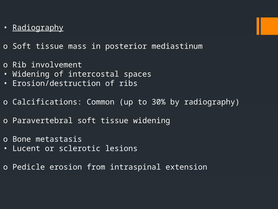
• Radiography
o Soft tissue mass in posterior mediastinum
o Rib involvement• Widening of intercostal spaces• Erosion/destruction of ribs
o Calcifications: Common (up to 30% by radiography)
o Paravertebral soft tissue widening
o Bone metastasis• Lucent or sclerotic lesions
o Pedicle erosion from intraspinal extension

CT Findings
• Posterior mediastinal mass, more commonly in inferior mediastinum but can occur in superior mediastinum/cervical region
• Mass often heterogeneous from necrosis, hemorrhage
• Calcification seen on CT in up to 85%
MR Findings
• Heterogeneous in signal and contrast-enhancement• Tends to be high in signal on T2Wl / low in signal on TlWI
Ultrasonographic Findings
o Heterogeneously echogenic mass


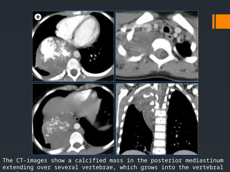
The CT-images show a calcified mass in the posterior mediastinum extending over several vertebrae, which grows into the vertebral canal.

THANK YOU