pediatric hepatology potpourri - kansasaap.org · 9/3/2012 1 pediatric hepatology potpourri lana...
TRANSCRIPT
9/3/2012
1
Pediatric Hepatology Potpourri
Lana Hattar, MD
Pediatric Gastroenterology, Hepatology & Nutrition
Clinical Assistant Professor
KUMC - Wichita School of Medicine
Disclosure
• I have no relevant financial relationships with the manufacturer(s) of any commercial product(s) and/or provider of commercial services discussed in this CME activity
• I do not intend to discuss an unapproved/investigative use of a commercial product/device in my presentation
Objectives
• To be able to recognize & manage patients with serious liver diseases that present with jaundice
• To be able to recognize & manage patients with serious liver diseases that present with hepatitis
9/3/2012
2
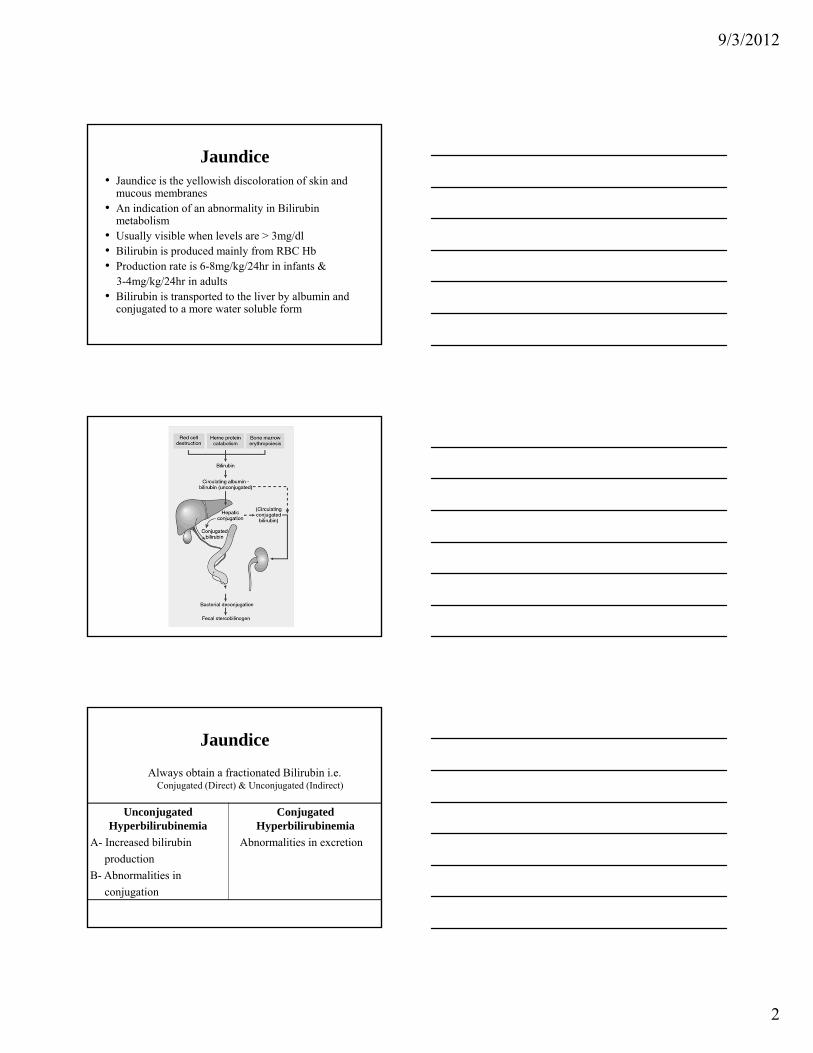
Jaundice• Jaundice is the yellowish discoloration of skin and
mucous membranes • An indication of an abnormality in Bilirubin
metabolism• Usually visible when levels are > 3mg/dl• Bilirubin is produced mainly from RBC Hb• Production rate is 6-8mg/kg/24hr in infants &
3-4mg/kg/24hr in adults• Bilirubin is transported to the liver by albumin and
conjugated to a more water soluble form
Jaundice
Always obtain a fractionated Bilirubin i.e. Conjugated (Direct) & Unconjugated (Indirect)
Unconjugated Hyperbilirubinemia
A- Increased bilirubin
production
B- Abnormalities in
conjugation
Conjugated Hyperbilirubinemia
Abnormalities in excretion
9/3/2012
3
Unconjugated HyperbilirubinemiaIncreased Bilirubin Production
• Blood group incompatibility• RBC hemoglobinopathies• Hematomas• Polycythemia• Drugs
Unconjugated HyperbilirubinemiaConjugation Abnormalities
• Breast Feeding Jaundice (Physiologic Jaundice)
• Breast Milk Jaundice
• Hypothyroidism
• Infants of diabetic mothers
• Medications and drugs
Unconjugated HyperbilirubinemiaConjugation Abnormalities
Criggler Najjar Syndrome
Hereditary deficiency of bilirubin glucuronosyl transferase (UGT1A1 gene)
• Type 1: Very high unconjugated bilirubin levels (20-40mg/dl). No response to phenobarbital
• Type 2: Medium elevation in unconjugated bilirubin levels (10-20mg/dl). Responds to phenobarbital
9/3/2012
4
Unconjugated HyperbilirubinemiaConjugation Abnormalities
Gilbert Syndrome
• Most commonly presents in adolescents and adults
• Unconjugated bilirubin increases with fasting and illness
• ALT, AST, GGT, AlkP must be normal
• Treatment: None. Education
Conjugated HyperbilirubinemiaExcretion Abnormalities
Location of defect is the key to diagnosis (hepatocyte, caniliculis, bile ducts)
• Inborn errors of metabolism(Dubin Johnson Syndrome, Rotor Syndrome)
• Cholestasis syndrome (PFICs 1, 2, 3)• Biliray Atresia• Choledochal cyst• Cholelithiasis
Other Causes of Jaundice• Metabolic abnormalities
A1AT Deficiency, CF, Wilson’s, Tyrosinemia, Galactosemia
• InfectionsHep A, B, C, CMV, EBV, Sepsis, UTI (newborn)
• DrugsAcetaminophen, Erythromycin, Valproate, Tetracycline, Statins, Insulin sensitizing agents
• ToxinsKava Kava, Amanita, heavy metals
• TPN
9/3/2012
5
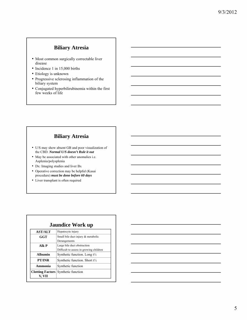
Biliary Atresia
• Most common surgically correctable liver disease
• Incidence 1 in 15,000 births• Etiology is unknown• Progressive sclerosing inflammation of the
biliary system• Conjugated hyperbilirubinemia within the first
few weeks of life
Biliary Atresia
• U/S may show absent GB and poor visualization of the CBD. Normal U/S doesn’t Rule it out
• May be associated with other anomalies i.e. Asplenia/polysplenia
• Dx: Imaging studies and liver Bx
• Operative correction may be helpful (Kasai procedure) must be done before 60 days
• Liver transplant is often required
Jaundice Work upAST/ALT Hepatocyte injury
GGT Small bile duct injury & metabolic
Derangements
Alk P Large bile duct obstruction
Difficult to assess in growing children
Albumin Synthetic function. Long t½
PT/INR Synthetic function. Short t½
Ammonia Synthetic function
Clotting Factors V, VII
Synthetic function

9/3/2012
6
Jaundice Work up
• Abdominal U/S
• CT scan
• MRCP
• ERCP
• Liver Biopsy
Biliary Atresia Abdominal US, liver biopsy, cholangiogram
Choledochal cyst Abdominal U/S
A1AT deficiency A1AT level and phenotype
IEOM Newborn screen (galactosemia)
Urine succinylacetone (tyrosinemia)
Serum amino acids, urine organic acids
TORCH Urine CMV culture, TORCH titers
UTI Urine Cx
Cystic Fibrosis Newborn screen, sweat chloride test
Alagille Syndrome Echocardiogram (if murmur present), spine film,
ophthalmology exam, liver biopsy
Hypothyroidism Newborn screen, TSH, total and free T4
Pan-hypopitutirism TSH, total and free T4, early a.m. cortisol,glucose,
brain MRI (assess pituitary gland)
9/3/2012
7
Treatment
Depending on the cause
• Adequate nutrition
• Ursodeoxycholic acid
(15-30 mg/kg/day divided BID)
• Fat soluble vitamins: ADEK
• Treatment for pruritus: Diphenhydramine, Hydroxyzine, Rifampin
Take Home Message
Always obtain a fractionated BilirubinAny Jaundice on DOL 1 is abnormalAny Conjugated Hyperbilirubinemia is
abnormalEvaluation of Jaundice requires assessment of
other aspects of liver function
Aminotransferases
• AST (SGOT): Found in liver, skeletal muscle, heart, kidney, brain, pancreas, lungs, WBC & RBC
Present in both mitochondria (80%) & Cytosol (20%)
• ALT (SGPT): Localized to cytosol of hepatocytes only, relatively liver specific
9/3/2012
8
Elevated Aminotransferases
• Elevation: ≥ 1.5 X upper limit of normal
• Minimal elevation: NASH
• Marked elevation:Acute toxic injury i.e. Acetaminophen
Ischemic/Hypoxic
Acute viral disease
Alcoholic hepatitis
Causes of elevated aminotransferasesHepatic
• Medications/Toxins
• Viral: Hepatitis A, B, C, EBV, CMV
• A1AT deficiency
• Autoimmune hepatitis
• Wilson’s disease
• Hemochromatosis
• Steatosis/NASH
Causes of elevated aminotransferasesNon-Hepatic
• Other AID: Celiac, IBD, SLE
• Thyroid disease
• Hemolysis
• Myopathy
• Acquired muscle disease
• Strenuous exercise
9/3/2012
9
History & Physical
• Symptoms: Fever, jaundice, abdominal
distension, pruritus, bleeding, fatigue
• FHx: Liver, AID (e.g. IBD, Celiac)
• PMHx: Blood transfusion, IV drugs, Foreign
travel
• MedsHx: Herbal, Acetaminophen, Minocycline
• PE: HSM, liver edge, jaundice, ascites, portal
HTN
Evaluation
• Drug/Toxin screen (Acetaminophen)
• Viral Serology (Hepatitis A, B, C, CMV, EBV, HSV,
HIV, Adenovirus)
• Autoimmune markers: ANA, Total IgG,
Anti-LKM, Anti-SM, Anti-F-Actin
• A1AT Pi typing
• Serum Ceruloplasmin, Serum copper, 24hr
urine copper collection
• Serum Iron, Ferritin, TIBC
Evaluation
• Muscle: CK, Aldolase
• Other AID: Celiac panel
• Systemic: TSH/Free T4, ESR, CBC/diff
9/3/2012
10
Evaluation
• Ab U/S (Liver, GB/Biliary system, Pancreas, Spleen)
• Doppler of hepatic vessels
• Ab CT scan
• ERCP
• MRCP
• Liver Biopsy(Histology, special stains, viral inclusions, presence & degree
of fibrosis)
Alpha One Antitrypsin Deficiency
• AD with incidence of 1:800 live births
• 10-15% of CLD in children & adults
• Predisposes to lung & liver disease
• PiMM: normal (95%). PiZZ/PiSZ: severe deficiency, PiMZ: intermediate
• Clinically: First noted in the newborn period
• Treatment: None. Avoid smoking
Supportive care. Liver transplant
Autoimmune Hepatitis
• Overall incidence 2:100,000• 60-75% female, 40% <20yrs (10-14yrs)• Wide range of presentation• Two types
Type 1: ANA, SMA, F-Actin, P-ANCAType 2: LKM, Anti-liver cytosol
• Biopsy: Interface hepatitis, mononuclear infiltrate,piecemeal necrosis, plasma cells
• Treatment: Prednisone, AZA, MMF, CSA, Anti-TNF, IVIG, IL-2 ab, Rituximab, Liver Transplant
9/3/2012
11
Wilson’s Disease(Hepatolenticular Degeneration)
• Rare AR with prevalence of 1:30,000• Etiology: decreased hepatocellular excretion of
copper into bile• Variable presentation
Classic 3: Liver disease, low Ceruloplasmin,Kaiser-Fleischer rings
• TreatmentChelating agents (D-Pencillamine, Trientine, Tetrathiomolybdate)
Zinc, anti-oxidents, dietary avoidance, liver transplant
NAFLD/NASH• NAFLD is the most common cause of liver disease in
childhood and adolescence
• A spectrum of liver pathologyA- Isolated steatosis or macrovesicular fat
accumulation within hepatocytes without inflammation
B- NASH: fat accumulation associated withinflammation and/or evidence of cellularinjury
C- Cirrhosis
NAFLD/NASH
• Predisposes to type 2 DM, HTN & dyslipidemia
• True Prevalence ?? (9.6 % in ages 2-19 yrs)
• Factors associated with increased risk
Obesity (10% are of healthy weight)
Male sex
Older age
Hispanic
Asian (esp. of Chinese and Filipino descent)
9/3/2012
12
Pathogenesis
Obesity (visceral adiposity) is correlated with dyslipidemia and increased insulin resistance
• Two hit hypothesis:Insulin resistance leads to hepatic
steatosisOxidative injury required for
progression to necroinflammatory steatohepatitis
Pathogenesis
• Presence of obesity in NAFLD increases risk of development of fibrosis 3 X
• Adolescent males more likely to develop NAFLD secondary to greater degree of insulin resistance in adolescents compared to children and adults. Estrogen may be protective
Diagnosis
• Age 10-14 yrs
• Mostly asymptomatic, some may have vague RUQ pain
Acanthosis Nigricans >50%
Family Hx
• Initial labs: CBC, AST/ALT, GGT,
Fasting lipid/glucose/insulin
9/3/2012
13
Differential diagnosis• Infections: HBV, HCV• AIH• Wilson’s• A1AT• Drug induced liver injury: Prednisone, Amiodarone,
tetracycline, valproate, MTX
• Chronic TPN use• Nutritional deficiencies: Refeeding syndrome,
rapid weight loss/starvation• Following bypass surgery
ImagingHepatic U/S
Readily available & inexpensive Lack of sensitivity to milder degrees of
steatosis, operator dependent, inability to adequately quantify the degree of steatosis, inflammation or fibrosis
Hepatic MRIMore sensitive but more expensive
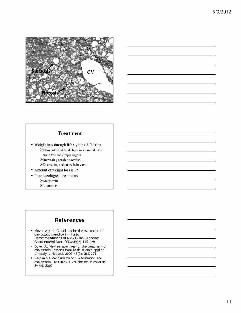
Liver Biopsy
• Dx: At least 5% of hepatocyte contain macrovesicular fat
• Types: Type I: Steatosis with ballooning
degeneration and perisinusoidal fibrosisType II: Steatosis with portal inflammation
and/or portal fibrosis without ballooning degeneration
• Presence & degree of fibrosis
9/3/2012
14
Treatment
• Weight loss through life style modificationElimination of foods high in saturated fats,
trans fats and simple sugars
Increasing aerobic exercise
Decreasing sedentary behaviors
• Amount of weight loss is ??
• Pharmacological treatmentsMetformin
Vitamin E
References
• Moyer V et al. Guidelines for the evaluation of cholestatic jaundice in infants: Recommendations of NASPGHAN. J pediatr Gastroenterol Nutr. 2004;39(2):115-128
• Boyer JL. New perspectives for the treatment of cholestasis: lessons from basic science applied clinically. J Hepatol. 2007;46(3): 365-371
• Karpen SJ. Mechanisms of bile formation and cholestasis. In: Suchy. Liver disease in children. 3rd ed. 2007
9/3/2012
15
References
• Suchy FJ. Liver disease in children. 3rd ed. 2007• Feldman et al. Sleisenger and Fordtran’s gastrointestinal
and liver disease. 9th ed. 2010• Bals R. Alpha one Antitrypsin deficiency. Best Pract Res
Clin Gastroenterology 2010;23(5):629-633• AASLD practice guidelines for Wilson’s disease.
Hepatology. 2008:47:6• Chai PF et al. Childhood autoimmune liver disease.
Indications and outcomes of liver transplantation. J Pediatr Gastroenterol Nutr. 2010;50:295-302
• Czaja AJ. Autoimmune liver disease. Curr Opin Gastroenterol 2005;21:293-299
Thank youQuestions ??