pediatric i.d. cases walking through your office door · subjective error in ... limited data for...
TRANSCRIPT

Pediatric I.D. Cases Walking Through Your Office Door
Stephen C. Eppes, M.D.
Christiana Care Health System
Sidney Kimmel Medical College at
Thomas Jefferson University

Disclosures

The Case of the Non-Lactose Fermenter
70 day old previously well infant
Two day history of diarrhea with a small amount of blood, feeding less well, and maximum recorded temp of 101o
Physical exam in office is normal
Stool studies are performed

The Case of the Non-Lactose Fermenter: Questions
a) Full sepsis work-up?
b) Hospitalization?
c) IV antibiotics?
d) PO antibiotics?

The Case of the Non-Lactose Fermenter
Sent home on no antibiotics with instructions for follow up
Next day lab reports stool is growing 4+ non-lactose fermenter
Clostridium difficile toxin assay is (+)
Phone call with family – condition unchanged, temps about 100o with 5 loose to watery stools in last 24 hours


Salmonella in Young Infants
The following day, Salmonella sp. identified by lab
Phone call indicates no change in status of child
What do you do now?

Salmonella in Young Infants
Well, if you didn’t do it before, take a good history as far as potential exposures (family members, food preparation, reptiles)
Recognize that young infants are at risk for extraintestinal spread
Begin PO antibiotic (e.g. TMP-SMX)
Check species identification and susceptibility
Monitor patient carefully

Salmonella in Young Infants
Should the baby have been empirically treated earlier?
Yes – especially if there was epidemiologic reason to suspect a bacterial pathogen (e.g. Salmonella, Shigella)
Possible downside to use of antibiotics –
(1) unnecessary antibiotic exposure
(2) prolonged carriage
(3) E. coli 0157:H7

Courtesy of Calmette and Guerin
5 y.o. Korean girl, recently moved to U.S.
Had BCG at one month of age
No known TB exposures
Asymptomatic, exam normal
PPD 8 mm
Chest radiograph showed: “suggestion of a small
infiltrate of bilateral suprahilar region"

Courtesy of Calmette and Guerin
a) Disregard 8 mm TST
b) Order IGRA
c) Repeat PPD in 1 year
d) Treat with INH for 9 months
e) Test family members
In addition to reviewing the CXR yourself you would:


Only the area of induration should be measured

Definitions of Positive TST in Pediatrics
Induration > 5 mm
Close contact with active TB
Suspected tuberculous disease Clinical, e.g. meningitis
Radiographic findings
Immunosuppressive conditions or therapies

Definitions of Positive TST in Pediatrics
Induration > 10 mm
Increased risk for disseminated disease < 4 years of age
Chronic illness, immunosuppression, or malnutrition
High risk of exposure to TB disease Born in high prevalence region
Travel to high prevalence region
Frequent exposure to high risk persons
Homeless, drug users, HIV, incarcerated
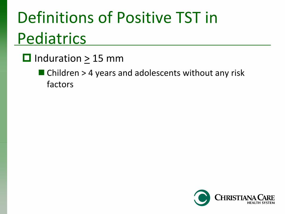
Definitions of Positive TST in Pediatrics Induration > 15 mm
Children > 4 years and adolescents without any risk factors

Tuberculin Skin Test (TST)
Advantages
Well studied
Treatment trials based on TST
Cheap
Disadvantages
2 visits
Lower specificity (BCG)
Requires precise placement and interpretation
Subjective error in interpretation
Reduced sensitivity in immunocompromised patients
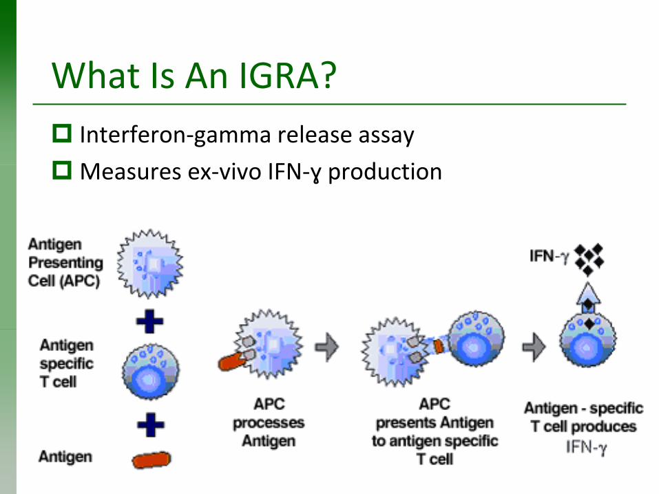
What Is An IGRA?
Interferon-gamma release assay
Measures ex-vivo IFN-ɣ production

IGRAs: Advantages Only one visit required
Results often within 24 hours
More reproducible results (compared with TST induration - often very subjective)
Not affected by prior BCG vaccination

IGRAs: Limitations
More labor-intensive
More expensive
Limited data for certain groups
Recently exposed to TB
Immunocompromised
Serial testing, e.g. health care providers
Young children

Straight from the AAP
“Some children who received BCG vaccine can have a false-positive TST result and LTBI is overestimated by the use of the TST in these circumstances.”
“The negative predictive value of IGRAs is not clear, but in general, if the IGRA result is negative and the TST is positive in an asymptomatic child, the diagnosis of LTBI is unlikely.”

AAP Revised Recommendations for Use of TST and IGRA in Children
TST preferred / IGRA acceptable
Children < 5 years of age

AAP Revised Recommendations for Use of TST and IGRA in Children
IGRA preferred, TST acceptable
Children > 5 yr who had BCG vaccine
Children > 5 yr unlikely to return for reading of TST

Patient # 37 on a Busy Monday
A 5 year old child has had nasal congestion, yellow discharge, cough and intermittent low grade fever (Tmax 100.8) for 12 days. Past medical history is unremarkable. Immunizations are current.
On exam he is non-ill appearing
Thick, yellow nasal discharge
Malodorous breath
Retracted TMs
Clear lungs

Patient # 37 on a Busy Monday
You would:
a. Treat with decongestants only
b. Treat with high dose amoxicillin
c. Treat with amox/clav (ES)
d. Treat with cefdinir

Patient # 37 on a Busy Monday
Acute bacterial sinusitis
New IDSA guidelines published 2012
Emphasis on accurate diagnosis:
Persistent / not improving (10 days)
Severe (> 3 days)
Worsening or “double-sickening” (> 3 days)
Chow AW, et al. Clin Infect Dis, 2012.

Acute Bacterial Rhinosinusitis
Antibiotic Recommendations from IDSA
Amox/clav (recommended by IDSA) 90 mg/kg/day divided 2X daily for children
10-14 days
2 g 2X daily for adults
5-7 days
Oral cephalosporin for non-type 1 reaction to penicillin
Levofloxacin for type 1 hypersensitivity
Acute Bacterial Rhinosinusitis
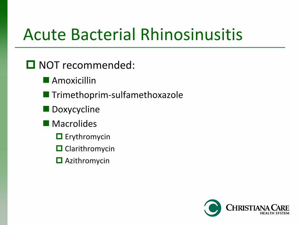
NOT recommended:
Amoxicillin
Trimethoprim-sulfamethoxazole
Doxycycline
Macrolides Erythromycin
Clarithromycin
Azithromycin

Acute Bacterial Rhinosinusitis
AAP Guidelines 2013
“Clinicians should not obtain imaging studies of any kind to distinguish acute bacterial sinusitis from viral URI, because they do not contribute to the diagnosis; however, a contrast-enhanced computed tomography scan of the paranasal sinuses should be obtained whenever a child is suspected of having orbital or central nervous system complications.”
“Amoxicillin with or without clavulanate is the first-line treatment of acute bacterial sinusitis.”

Taking a Good History
8 year old boy has had fever (to 104 degrees) for 2 days
Malaise and myalgias
Now presents with new skin findings


Taking a Good History
Exam reveals:
Talkative, not acutely ill in appearance
Multiple petechiae and purpura, mainly on extremities
Clear lungs, normal heart exam
Warmth over knees, tenderness to ROM

Taking a Good History
a) Social History
b) Family History
c) Immunization History
d) Medication History
e) Review of Systems
The pediatrician suspected the correct diagnosis on the basis of which part of the history:

Taking a Good History
a) Travel
b) Exposure to ill contacts
c) Pets in the home
d) Self-injurious behaviors
e) Flooring in the home
The pediatrician focused on what aspects of the Social History:
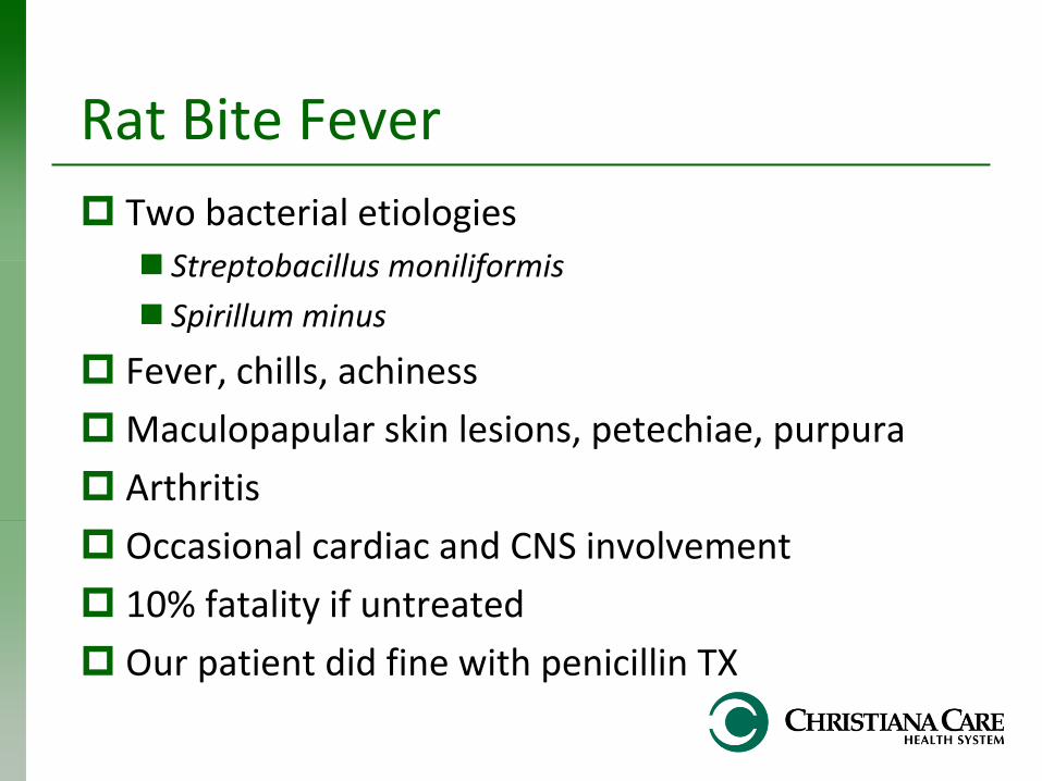
Rat Bite Fever
Two bacterial etiologies
Streptobacillus moniliformis
Spirillum minus
Fever, chills, achiness
Maculopapular skin lesions, petechiae, purpura
Arthritis
Occasional cardiac and CNS involvement
10% fatality if untreated
Our patient did fine with penicillin TX

It’s Always on Board Exams and Sometimes You See It in Real Life
3 day old infant born at 36 5/7 weeks to 33 y.o. Mexican-American mom Late fall, southeastern PA
Prenatal I.D. labs were negative, but HIV and GC/CT unknown
Gestational diabetes Delivered by cesarean section because of poor BPP, Apgars
3 and 8 On DOL 2 in well baby nursery at OSH, he developed
respiratory distress Metabolic acidosis, LFTs abnormal (ALT 200) CBC showed platelets of 88k and WBC of 4700 Intubated and transferred to CCHS

It’s Always on Board Exams and Sometimes You See It in Real Life
What antimicrobials would you start?
A) Ampicillin and gentamicin
B) Ampicillin, gentamicin and acyclovir
C) Ampicillin and cefotaxime
D) Vancomycin and piperacillin / tazobactam

It’s Always on Board Exams and Sometimes You See It in Real Life
Additional lab results:
Creatinine 1.5
CSF protein 919, WBC 3050 with segs, lymphs and monocytes
CSF Gram stain showed Gram positive rods (? Lactobacillus sp.)
Blood culture from OSH grew:

It’s Always on Board Exams and Sometimes You See It in Real Life
Additional lab results:
Creatinine 1.5
CSF protein 919, WBC 3050 with segs, lymphs and monocytes
CSF Gram stain showed Gram positive rods (? Lactobacillus sp.)
Blood culture from OSH grew:
Listeria monocytogenes

It’s Always on Board Exams: Listeria monocytogenes
Gram positive rod (sometimes Gram variable, often misidentified)
Usually associated with foodborne illness In pregnancy it is associated with
Spontaneous abortion Fetal death Preterm delivery
Early and late onset neonatal infection is associated with Sepsis Papular rash (“granulomatosis infantisepticum”) Meningitis (usually late onset, associated with 25% mortality
rate)
Treatment of choice: amp and gent

It’s Always on Board Exams: Listeria monocytogenes
Our patient:
Prolonged mechanical ventilation
EEG severely abnormal – burst suppression pattern Keppra, fosphenytoin, phenobarbital
Poor feeding
? Imaging
Age 5 weeks – pyloric stenosis

Figuring It Out
17 y.o. previously healthy male presented in August with 5 day history
Fever / chills
Headache / photophobia
Myalgias
Vomiting
Became dehydrated, required fluid administration, and was admitted to AIDHC
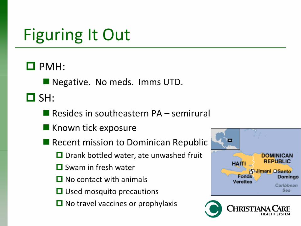
Figuring It Out
PMH:
Negative. No meds. Imms UTD.
SH:
Resides in southeastern PA – semirural
Known tick exposure
Recent mission to Dominican Republic Drank bottled water, ate unwashed fruit
Swam in fresh water
No contact with animals
Used mosquito precautions
No travel vaccines or prophylaxis

Figuring It Out
Physical examination
38.6 / 115 / 115/87 / pulse ox 100%
Photophobia, no conjunctivitis or papilledema
Heart, lungs and abdomen normal
Neuro exam unremarkable
Musculoskeletal exam normal
Skin without rashes or lesions

Figuring It Out: Initial Lab Results
CBC:
WBC 11,400 with neutrophil predominance
Platelets 145,000 with normal H/H
Creatinine 2.5, BUN 35
Bilirubin 1.9
AST 63, ALT 79
CPK normal
UA: Moderate bilirubin and 50-100 WBC

Figuring It Out
Hospital course:
Remained febrile
Additional lab tests sent
Chloroquine for 1.5 days (smears negative)
Hospital day 2 Conjunctival suffusion
Abdominal pain, hepatosplenomegaly
Intravenous doxycycline
Defervescence within 48 hr

Figuring It Out: What is in S.E. PA in the Summer?
Lyme disease
Ehrlichiosis / Anaplasmosis
Enteroviruses
Non-seasonal
Epstein-Barr virus
Adenoviruses

Connolly et al. Clinical Pediatrics, 2014.
Figuring It Out: What Can Returning Travelers Bring Home?
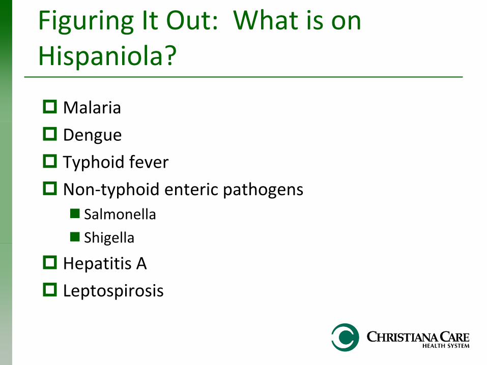
Figuring It Out: What is on Hispaniola?
Malaria
Dengue
Typhoid fever
Non-typhoid enteric pathogens
Salmonella
Shigella
Hepatitis A
Leptospirosis

Figuring It Out: Differential Diagnosis
Ehrlichiosis / Anaplasmosis
Malaria
Dengue
Leptospirosis

Leptospirosis
Etiology: Leptospira ictohemorrhagiae
Contact with animal urine, often from swimming
Early phase
Fever, headache, myalgias, conjunctivitis
Late phase
Immune mediated meningitis
Severe (Weil’s) disease Liver and kidney involvement, hemorrhage
Treatment: Penicillin G
Doxycycline
3rd generation cephalosporins
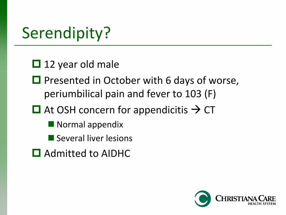
Serendipity?
12 year old male
Presented in October with 6 days of worse, periumbilical pain and fever to 103 (F)
At OSH concern for appendicitis CT
Normal appendix
Several liver lesions
Admitted to AIDHC

Serendipity?
Fever lasted 4 days
Drenching night sweats
No respiratory symptoms
No known lymph node swelling
No jaundice
No nausea, vomiting or diarrhea
Small for age but no weight loss
Abdominal pain improved after CT

Serendipity? Social History
Resides in rural Maryland with parents and 2 sisters
They own dogs, cats, chickens, goats, rabbits, pig and a cockatoo
Cleans the pens
“Always wears gloves”
No tobacco exposure
Attends public school

Serendipity? Physical Exam
T – 36.4, HR – 66, R – 20, BP 109/70
Alert, smiling, in no distress
Small for age
< 5th percentile for weight
Normal, including abdominal exam
Subsequently, 2x3 cm mass noted in right axilla


Serendipity? Lab Results
WBC 10.0, normal diff, Hg 12.7, platelets 458k
C-reactive protein normal
CMP normal
LDH normal
AFP normal
HCG negative
Serendipity? W/U for Infections
Blood cultures (aerobic / anaerobic) negative
EBV PCR negative
CMV EBV negative
Brucellosis antibodies negative
Histoplasma antibodies negative

Serendipity? Additional Imaging
Abdominal US – 3 hypoechoic lesions
Abdominal MRI – multiple liver lesions
“Highly suspicious for metastatic process”
PET scan – Hypermetabolic activity in 4 hepatic lesions, right axillary and subpectoral regions, and likely LUQ abnormality
CT neck and thorax – focal haziness in RLL, two 2 mm nodules in LLL and RML, and mildly enhanced soft tissue lesion in right axilla, normal neck, liver lesions as previously noted


Tissue is the Issue
Bone marrow biopsies – normal
Lymph node
Florid follicular hyperplasia
Nodal and perinodal granulomatous inflammation
Few foci of associated necrosis
Multiple special stains negative Gram
Silver
Acid fast
Warthin-Starry
Routine, fungal and AFB cultures (-)


Cat Scratch Disease
Nodal presentation in 90%
Inoculation lesion in 61%
“Atypical” presentations in 10%
Fever of unknown origin
Osteomyelitis
Hepatosplenic granulomas
Encephalitis
Ocular disease
Henoch-Schönlein purpura
If treatment required azithromycin
Also TMP-SMX, RIF, and gentamicin

Gastroenteritis – Not!
16 year old girl has 24 hour history of febrile illness beginning with vomiting and diarrhea
This morning, when getting off toilet, she became dizzy and fell, though no LOC
After the mother called for an appointment, she developed rash and her eyes looked red
In your office, vital signs included
Temp – 102.6
HR – 100
Resp – 28
BP – 94/58


Gastroenteritis – Not!
Assuming you take a complete history, you would particularly want to know:
a) Menstrual history
b) Exposure to ticks
c) History of sore throat and swollen cervical nodes
d) Recent travel

Gastroenteritis – Not!
You decide to admit her to the hospital. After appropriate cultures, empiric antibiotic therapy would be
a) Ceftriaxone
b) Doxycycline
c) Penicillin and clindamycin
d) Vancomycin and clindamycin

Gastroenteritis – Not!
Patient required multiple fluid boluses and was briefly on a dopamine infusion
She ultimately improved and was discharged home on clindamycin
When you see her back the following week, her exam was basically normal except for:


Gastroenteritis – Not!
Staphylococcal TSS
Mediated by TSST-1
MSSA >> MRSA
Menstrual – 50%
Non-menstrual Often minor cutaneous infection
Usually begins with GI symptoms

TSS: Clinical Findings
Fever > 102 (F)
Rash – erythroderma, followed by desquamation
Hypotension
Multisystem organ involvement (3 or more):
GI – Vomiting and diarrhea
Muscular – elevated CPK
Mucous membrane
Renal
Hepatic
Hematologic
CNS
TSS: Treatment
Fluids
Management of end organ dysfunction
Inotropic support if required
Vancomycin or anti-staphylococcal beta-lactam
PLUS clindamycin

Scary Sounding, But Often They Do OK
3 day old term infant noted on DOL 1 to have significant rash, resulting in admission to NICU
Maternal I.D. labs negative except for GBS, for which she received appropriate antibiotics
Vaginal delivery with meconium stained fluid but no chorioamnionitis
Mildly depressed at birth, but responded to stimulation
Blood culture obtained, started on ampicillin and gentamicin

Scary Sounding, But Often They Do OK
What did he have:
A) Neonatal HSV
B) Congenital varicella
C) Congenital candidiasis
D) Pustular melanosis
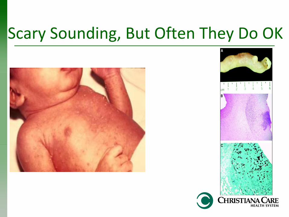
Scary Sounding, But Often They Do OK

Scary Sounding, But Often They Do OK Congenital Cutaneous Candidiasis
Usually present on DOL 1
Papules, pustules, vesicles
Often with palm / sole involvement
Helps differentiate from erythema toxicum
Funisitis is typical
Term infants usually do not have invasive infection
Topical antifungals often suffice
Preterm infants often have blood stream invasion and systemic illness
Systemic antifungal therapy required
Scary Sounding, But Often They Do OK Congenital Cutaneous Candidiasis
Our patient
Normal CBC
Negative blood culture
Negative urine culture
CXR – questionable mild infiltrate
Ophthalmologic exam – normal
Treated with brief course of fluconazole and did well

Ubiquitous, Uncanny, Understandable
15 year old female has a 1 week history of fever, fatigue, achiness and left sided abdominal pain
Throat pain was worst symptom
PMH – Gilbert’s
Admitted overnight (elsewhere) for IV hydration
Hg 9.3, platelets 138, T. bili 5.9, AST 192, ALT 185
Monospot negative
Admitted through ED to AIDHC

Ubiquitous, Uncanny, Understandable T – 36.8, P – 82, R – 20, BP – 106/51
General: alert, NAD
Skin: jaundiced, no rashes or lesions
HEENT: icteric conjunctivae, pharynx erythematous, absent tonsils
Neck: bilateral tender lymph nodes
Lungs: clear bilaterally
Heart: grade 1-2/6 SEM, normal rhythm, S1 and S2
Abdomen: mildly tender epigastrium, liver and spleen each 4 cm below costal margin
Musculoskeletal: normal

Ubiquitous, Uncanny, Understandable CBC: Hg 8.9, platelets 205, WBC 15.8 (29 segs, 15
bands, 46 lymphs, 8 monos, 2 atypical lymphs)
Reticulocytes: 11.1%
ESR 78, CRP 4.0
CMP: T. bili 5.5, AST 226, ALT 279
Rapid HIV: negative
Respiratory viral panel: rhino/entero
Throat culture: negative for GAS
Blood culture: negative
Abdominal US: hepatosplenomegaly, sludge in GB

Ubiquitous, Uncanny, Understandable
Remained febrile, but generally stable
H/H dropped to nadir of 7.3/20
4th hospital day – atypical lymphs 23%
Ubiquitous, Uncanny, Understandable
What did she have:
a) Ehrlichiosis
b) EBV-associated autoimmune hemolytic anemia
c) Typhoid fever
d) Leptospirosis

Diagnosis of EBV Infection
CBC
Atypical lymphocytosis
Heterophile (Monospot)
Highly specific
Variably sensitive
EBV-specific antibodies
IgM to VCA
IgG to VCA
EBNA
PCR to detect EBV genome

Months following EBV infection
Titer
IgG to VCA
IgM to
VCA
Anti-EA Anti-EBNA
D
R
Heterophile

Complications of EBV Infection in Normal Hosts Neurologic:
Encephalitis
Myelitis
Facial palsy
Guillain-Barré
Metamorphopsia
Splenic rupture
Secondary infections
Hematologic:
HLH
ITP
AHA
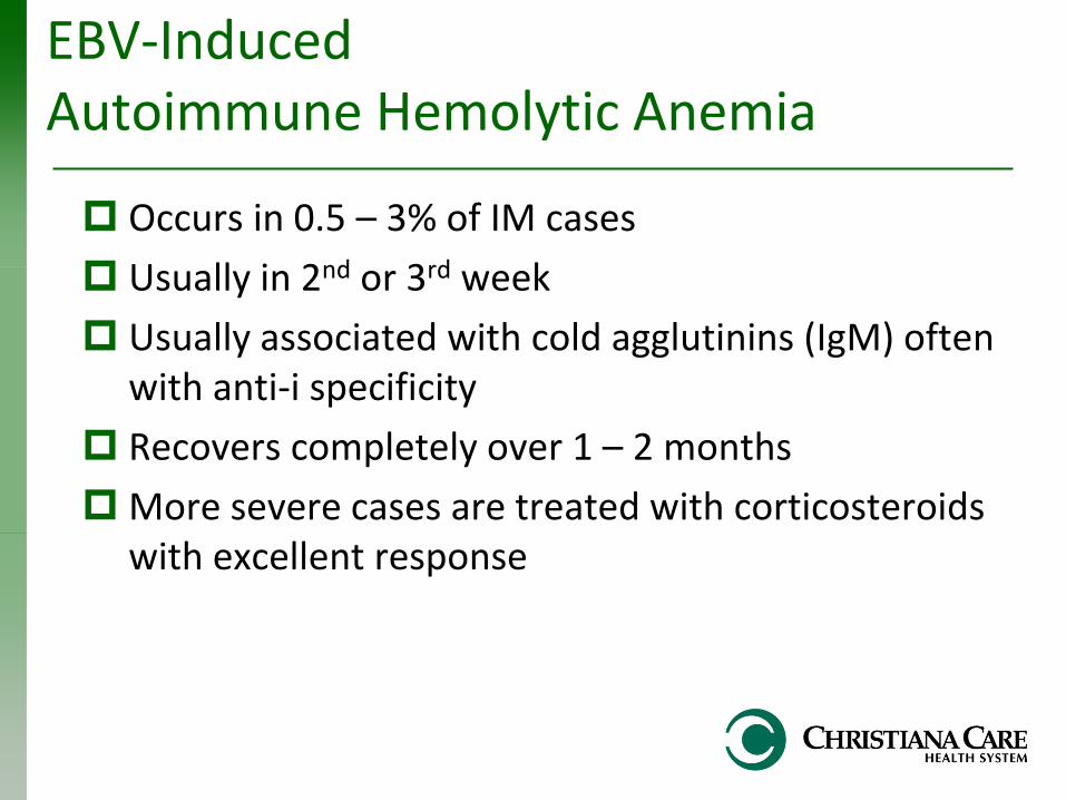
EBV-Induced Autoimmune Hemolytic Anemia
Occurs in 0.5 – 3% of IM cases
Usually in 2nd or 3rd week
Usually associated with cold agglutinins (IgM) often with anti-i specificity
Recovers completely over 1 – 2 months
More severe cases are treated with corticosteroids with excellent response
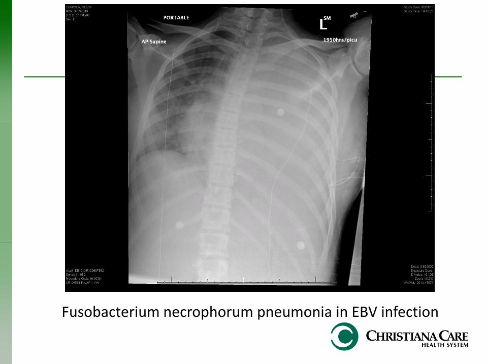
Fusobacterium necrophorum pneumonia in EBV infection