pediatric musculoskeletal imaging … system, and other rare indications exist as well: bone scan...
TRANSCRIPT
©2016 eviCore healthcare Pediatric Musculoskeletal Imaging Guidelines
PEDIATRIC MUSCULOSKELETAL IMAGING GUIDELINES
Version 18.0; Effective 03-18-2016
This version incorporates accepted revisions prior to 12/31/15
CPT® (Current Procedural Terminology) is a registered trademark of the American Medical Association (AMA). CPT® five digit codes, nomenclature and other data are copyright 2016 American Medical Association. All Rights Reserved. No fee schedules, basic units, relative values or related listings are included in the CPT® book. AMA does not directly or indirectly practice medicine or dispense medical services. AMA assumes no liability for the data contained herein or not contained herein.
eviCore healthcare Clinical Decision Support Tool Diagnostic Strategies: This tool addresses common symptoms and symptom complexes. Imaging requests for patients with atypical symptoms or clinical presentations that are not specifically addressed will require physician review. Consultation with the referring physician, specialist and/or patient’s Primary Care Physician (PCP) may provide additional insight.
V.18.0; Effective 3/18/2016 – Pediatric Musculoskeletal Imaging 2 of 28
PEDIATRIC MUSCULOSKELETAL IMAGING GUIDELINES
Pediatric MUSCULOSKELETAL Imaging Guidelines
PEDMS-1~General Guidelines 3
PEDMS-2~Fracture and Dislocation 10
PEDMS-3~Soft Tissue and Bone Masses 13
PEDMS-4~Limping Child 15
PEDMS-5~Developmental Dysplasia of the Hip 17
PEDMS-6~Avascular Necrosis (AVN) / Legg- Calvé-Perthes Disease / Idiopathic Osteonecrosis
19
PEDMS-7~Suspected Physical Child Abuse 21
PEDMS-8~Infection/Osteomyelitis 23
PEDMS-9~Foreign Body 23
PEDMS-10~Inflammatory Musculoskeletal Disease 24
PEDMS-11~Muscle/Tendon Unit Injuries 26
PEDMS-12~Osgood-Schlatter Disease 26
PEDMS-13~ Popliteal (Baker) Cyst 26
PEDMS-14~ Slipped Capital Femoral Epiphysis (SCFE) 27
PEDMS-15~Limb Length Discrepancy 28
PEDMS-16~ Congenital Anomalies of the Foot 28
V.18.0; Effective 3/18/2016 – Pediatric Musculoskeletal Imaging 3 of 28
PEDIATRIC MUSCULOSKELETAL IMAGING GUIDELINES
PEDMS-1~General Guidelines
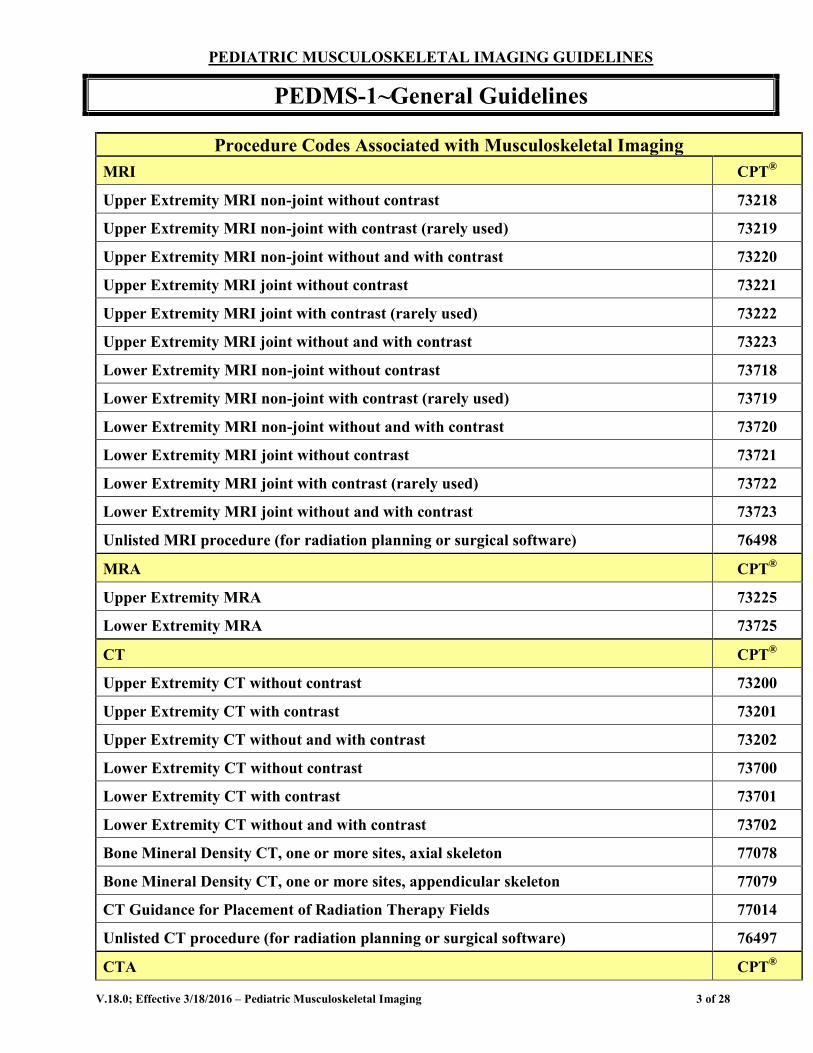
Procedure Codes Associated with Musculoskeletal Imaging MRI CPT®
Upper Extremity MRI non-joint without contrast 73218
Upper Extremity MRI non-joint with contrast (rarely used) 73219
Upper Extremity MRI non-joint without and with contrast 73220
Upper Extremity MRI joint without contrast 73221
Upper Extremity MRI joint with contrast (rarely used) 73222
Upper Extremity MRI joint without and with contrast 73223
Lower Extremity MRI non-joint without contrast 73718
Lower Extremity MRI non-joint with contrast (rarely used) 73719
Lower Extremity MRI non-joint without and with contrast 73720
Lower Extremity MRI joint without contrast 73721
Lower Extremity MRI joint with contrast (rarely used) 73722
Lower Extremity MRI joint without and with contrast 73723
Unlisted MRI procedure (for radiation planning or surgical software) 76498
MRA CPT®
Upper Extremity MRA 73225
Lower Extremity MRA 73725
CT CPT®
Upper Extremity CT without contrast 73200
Upper Extremity CT with contrast 73201
Upper Extremity CT without and with contrast 73202
Lower Extremity CT without contrast 73700
Lower Extremity CT with contrast 73701
Lower Extremity CT without and with contrast 73702
Bone Mineral Density CT, one or more sites, axial skeleton 77078
Bone Mineral Density CT, one or more sites, appendicular skeleton 77079
CT Guidance for Placement of Radiation Therapy Fields 77014
Unlisted CT procedure (for radiation planning or surgical software) 76497
CTA CPT®
V.18.0; Effective 3/18/2016 – Pediatric Musculoskeletal Imaging 4 of 28
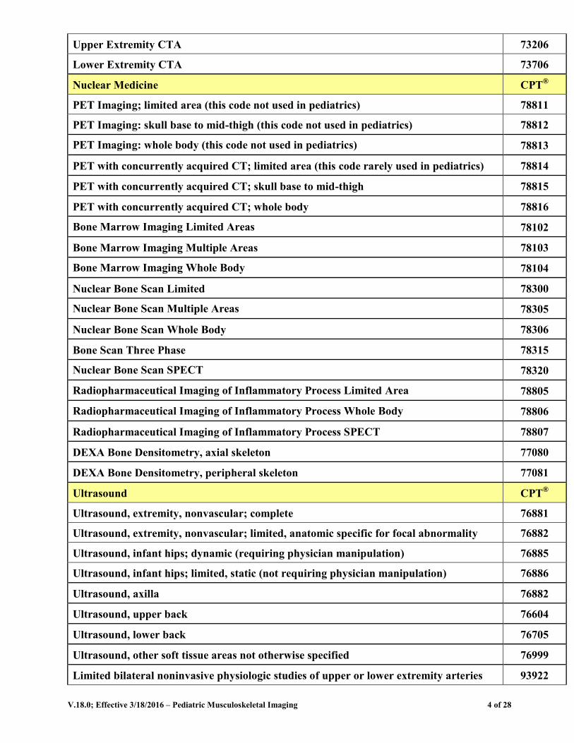
Upper Extremity CTA 73206
Lower Extremity CTA 73706
Nuclear Medicine CPT®
PET Imaging; limited area (this code not used in pediatrics) 78811
PET Imaging: skull base to mid-thigh (this code not used in pediatrics) 78812
PET Imaging: whole body (this code not used in pediatrics) 78813
PET with concurrently acquired CT; limited area (this code rarely used in pediatrics) 78814
PET with concurrently acquired CT; skull base to mid-thigh 78815
PET with concurrently acquired CT; whole body 78816
Bone Marrow Imaging Limited Areas 78102
Bone Marrow Imaging Multiple Areas 78103
Bone Marrow Imaging Whole Body 78104
Nuclear Bone Scan Limited 78300
Nuclear Bone Scan Multiple Areas 78305
Nuclear Bone Scan Whole Body 78306
Bone Scan Three Phase 78315
Nuclear Bone Scan SPECT 78320
Radiopharmaceutical Imaging of Inflammatory Process Limited Area 78805
Radiopharmaceutical Imaging of Inflammatory Process Whole Body 78806
Radiopharmaceutical Imaging of Inflammatory Process SPECT 78807
DEXA Bone Densitometry, axial skeleton 77080
DEXA Bone Densitometry, peripheral skeleton 77081
Ultrasound CPT®
Ultrasound, extremity, nonvascular; complete 76881
Ultrasound, extremity, nonvascular; limited, anatomic specific for focal abnormality 76882
Ultrasound, infant hips; dynamic (requiring physician manipulation) 76885
Ultrasound, infant hips; limited, static (not requiring physician manipulation) 76886
Ultrasound, axilla 76882
Ultrasound, upper back 76604
Ultrasound, lower back 76705
Ultrasound, other soft tissue areas not otherwise specified 76999
Limited bilateral noninvasive physiologic studies of upper or lower extremity arteries 93922
V.18.0; Effective 3/18/2016 – Pediatric Musculoskeletal Imaging 5 of 28
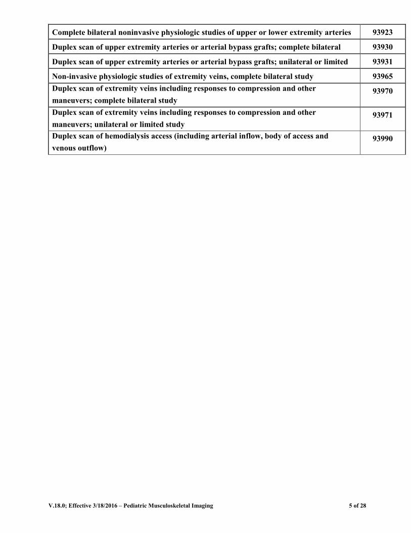
Complete bilateral noninvasive physiologic studies of upper or lower extremity arteries 93923
Duplex scan of upper extremity arteries or arterial bypass grafts; complete bilateral 93930
Duplex scan of upper extremity arteries or arterial bypass grafts; unilateral or limited 93931
Non-invasive physiologic studies of extremity veins, complete bilateral study 93965 Duplex scan of extremity veins including responses to compression and other maneuvers; complete bilateral study
93970
Duplex scan of extremity veins including responses to compression and other maneuvers; unilateral or limited study
93971
Duplex scan of hemodialysis access (including arterial inflow, body of access and venous outflow)
93990
V.18.0; Effective 3/18/2016 – Pediatric Musculoskeletal Imaging 6 of 28
PEDIATRIC MUSCULOSKELETAL IMAGING GUIDELINES
PEDMS-1~General Guidelines
PEDMS-1.1 Pediatric Musculoskeletal Imaging Age Considerations Many conditions affecting the musculoskeletal system in the pediatric population are different diagnoses than those occurring in the adult population. For those diseases which occur in both pediatric and adult populations, minor differences may exist in management due to patient age, comorbidities, and differences in disease natural history between children and adults. Patients age <18 years old should be imaged according to the Pediatric
Musculoskeletal Imaging Guidelines, and patients age ≥18 years should be imaged according to the Musculoskeletal Imaging Guidelines, except where directed otherwise by a specific guideline section.
PEDMS-1.2 Pediatric Musculoskeletal Imaging Appropriate Clinical Evaluation and Conservative Treatment A recent (within 60 days) face-to-face evaluation including a detailed history, physical
examination, appropriate laboratory studies, and basic imaging such as plain radiography or ultrasound should be performed prior to considering advanced imaging, unless the patient is undergoing guideline-supported scheduled follow-up imaging evaluation.
Plain x-ray should be done prior to advanced imaging for musculoskeletal conditions to rule out those situations that do not require advanced imaging, such as osteoarthritis, acute/healing fracture, osteomyelitis, and tumors of bone amenable to biopsy or radiation therapy (in known metastatic disease), etc.
o Even in soft tissue masses, plain x-rays are helpful in evaluating for calcium/bony deposits, e.g. myositis ossificans and invasion of bone
Provider-directed conservative care may include any or all of the following: R.I.C.E (rest, ice, compression, and elevation), NSAIDs (non-steroidal anti-inflammatory drugs), narcotic and non-narcotic analgesic medications, oral or injectable corticosteroids, viscosupplementation injections, a provider-directed home exercise program, cross-training, physical medicine, or immobilization by splinting/casting/bracing
These guidelines are based upon using advanced imaging to answer specific clinical questions that will affect patient management. Imaging is not indicated if the results will not affect patient management decisions. Standard medical practice would dictate continuing conservative therapy prior to advanced imaging in patients who are improving on current treatment programs.
V.18.0; Effective 3/18/2016 – Pediatric Musculoskeletal Imaging 7 of 28
Repeat imaging studies of the same body area are not necessary solely for return-to-play decisions.
Unless otherwise stated in a specific guideline section, repeat imaging studies of the same body area are not necessary unless there is evidence for progression of disease, new onset of disease, and/or documentation of how repeat imaging will affect patient management or treatment decisions.
PEDMS-1.3 Pediatric Musculoskeletal Imaging Modality General Considerations MRI
o MRI without contrast is the preferred modality for pediatric musculoskeletal imaging unless otherwise stated in a specific guideline section, as it is superior in imaging the soft tissues and can also define physiological processes in some instances, e.g. edema, loss of circulation (AVN), and increased vascularity (tumors)
o MRI without and with contrast is frequently recommended for evaluation of tumors, infection, post-operative evaluation, arthrography, and juvenile idiopathic arthritis, as described in the disease-specific guideline sections
o Due to the length of time for image acquisition and the need for stillness, anesthesia is required for almost all infants and young children (age <7 years), as well as older children with delays in development or maturity. In this patient population, MRI imaging sessions should be planned with a goal of avoiding a short-interval repeat anesthesia exposure due to insufficient information using the following considerations: MRI should always be performed without and with contrast unless
there is a specific contraindication to gadolinium use, since the patient already has intravenous access for anesthesia
If multiple body areas are supported by eviCore guidelines for the clinical condition being evaluated, MRI of all necessary body areas should be obtained concurrently in the same anesthesia session
o The presence of surgical hardware or implanted devices may preclude MRI, as magnetic field distortion may limit detail in adjacent structures
o The selection of best examination may require coordination between the provider and the imaging service
CT o CT without contrast is generally superior to MRI for imaging bone and joint
anatomy; thus it is useful for studying complex fractures (particularly of the joints, dislocations, and assessing delayed union or non-union of fractures, integration of bone graft material, if plain x-rays are equivocal CT should not be used to replace MRI in an attempt to avoid sedation
unless listed as a recommended study in a specific guideline section
V.18.0; Effective 3/18/2016 – Pediatric Musculoskeletal Imaging 8 of 28
o CT beam attenuation can result in “spray” artifact which can obscure adjacent details. This can occur with radiopaque material such as metal objects or dense bones.
o The selection of best examination may require coordination between the provider and the imaging service
Ultrasound o Ultrasound is frequently used to evaluate infants for hip dysplasia, to detect
and/or aspirate joint effusion, and as an initial evaluation of extremity soft tissue masses
o CPT codes vary by body area and presence or absence of Doppler imaging and are included in the table at the beginning of this guideline
Nuclear Medicine o Nuclear medicine studies are commonly used in evaluation of the peripheral
musculoskeletal system, and other rare indications exist as well: Bone scan (CPT® 78315 or 78320) is indicated for evaluation of
suspected loosening of orthopedic prostheses when recent plain x-ray is nondiagnostic
Nuclear medicine bone marrow imaging (CPT® 78102, 78103, or 78104) is indicated for detection of ischemic or infarcted regions in sickle cell disease
Triple phase bone scan (CPT® 78315) is indicated for evaluation of complex regional pain syndrome or reflex sympathetic dystrophy
Bone scan (CPT® 78300, 78305, 78306, 78315, or 78320) is indicated for evaluation of suspected frostbite
Bone scan (CPT® 78300, 78305, 78306, or 78320) is indicated for evaluation of Paget’s disease
3D Rendering o 3D Rendering indications in pediatric musculoskeletal imaging are identical
to those for adult patients. See MS-3~3D Rendering for imaging guidelines.
The guidelines listed in this section for certain specific indications are not intended to be all-inclusive; clinical judgment remains paramount and variance from these guidelines may be appropriate and warranted for specific clinical situations. References 1. ACR-ASER-SCBT-MP-SPR Practice Parameter for the Performance of Pediatric Computed
Tomography (CT), revised 2014, available at: http://www.acr.org/~/media/ACR/Documents/PGTS/guidelines/CT_Pediatric.pdf.
2. ACR -SPR Practice Parameter for the Performance and Interpretation of Pediatric Magnetic Resonance Imaging (MRI), revised 2014, available at: http://www.acr.org/~/media/ACR/Documents/PGTS/guidelines/MRI_Pediatric.pdf.
V.18.0; Effective 3/18/2016 – Pediatric Musculoskeletal Imaging 9 of 28
3. ACR-SPR-SSR Practice Parameter for the Performance of Radiography of the Extremities, amended 2014, available at: http://www.acr.org/~/media/ACR/Documents/PGTS/guidelines/Extremity_Radiography.pdf.
4. Ing C, DiMaggio C, Whitehouse A et al, Long-term Differences in Language and Cognitive Function After Childhood Exposure to Anesthesia, Pediatrics 2012;130:e476-e485.
5. Monteleone M, Khandji A, Cappell J et al, Anesthesia in Children: Perspectives From Nonsurgical Pediatric Specialists, J Neurosurg Anesthesiol 2014;26:396-398.
6. DiMaggio C, Sun LS, and Li G, Early Childhood Exposure to Anesthesia and Risk of Developmental and Behavioral Disorders in a Sibling Birth Cohort, Anesth Analg 2011; 113:1143-1151.
7. Hindorf C, Glatting G, Chiesa C et al. EANM Dosimetry Committee Guidelines for Bone Marrow and Whole Body Dosimetry, Eur J Nucl Med Mol Imaging 2010;37:1238-1250.
8. Donohoe KJ, Brown ML, Collier D, et al. Society of Nuclear Medicine procedure guideline for bone scintigraphy, version 3.0, approved June 20, 2003, available at: http://interactive.snm.org/docs/pg_ch34_0403.pdf.
9. Mehta RC and MA Wilson MA, Frostbite injury: prediction of tissue viability with triple-phase bone scanning, Radiology 1989; 170:511-514.
10. Drane WE, Myositis ossificans and the three phase bone scan, AJR Am J Roentgenol 1984; 142:179-180.
V.18.0; Effective 3/18/2016 – Pediatric Musculoskeletal Imaging 10 of 28
PEDIATRIC MUSCULOSKELETAL IMAGING GUIDELINES
PEDMS-2~Fracture and Dislocation
A recent (within 60 days) face-to-face evaluation including a detailed history, physical examination, and plain radiography should be performed prior to considering advanced imaging.
PEDMS-2.1 Acute Fracture Plain x-rays should be performed initially in any obvious or suspected acute fracture
or dislocation o If plain x-rays are positive, no further imaging is generally indicated except in
complex (comminuted or displaced) joint fractures where MRI or CT without contrast can be approved for preoperative planning
o 3D Rendering may sometime be indicated for complex fracture repairs. See MS-3~3D Rendering for imaging guidelines.
If plain x-rays are negative or equivocal for fracture, and fracture is still clinically suspected, CT or MRI without contrast is indicated if the results will determine immediate treatment decisions as documented by the treating physician
Bone scan is indicated for evaluation of suspected fracture when two x-rays are negative at least 10 days apart, using any of the following CPT code combinations:
o CPT® 78300, 78305, or 78306 as a single study o See PEDMS-2.5 Stress/Occult Fracture for bone scan indications
PEDMS-2.2 Joint Fracture CT can be approved in complex (comminuted or displaced) fractures involving a joint
for preoperative planning. CT can be approved when there is clinical concern for delayed union or non-union of
fracture or joint fusions on follow-up plain x-ray
PEDMS-2.3 Growth Plate Injuries (Salter-Harris fractures) These fractures can generally be diagnosed and managed adequately with plain x-ray If there is concern for delayed union or non-union of the bone, CT without contrast is
indicated
MRI without contrast is indicated for the evaluation of a suspected physeal bar in a healing fracture or other complication of a fracture involving the growth plate, which may result in abnormal growth
V.18.0; Effective 3/18/2016 – Pediatric Musculoskeletal Imaging 11 of 28
Compressive injuries of the growth plate (Salter-Harris I) injuries may be difficult to identify on plain films, and MRI without contrast is indicated for confirmation.
PEDMS-2.4 Osteochondral or Chondral Fractures, including Osteochondritis Dissecans If x-rays are negative and an osteochondral fracture is still suspected, or if x-ray or
clinical exam suggest an unstable osteochondral injury, either MRI without contrast, MR arthrogram, or CT arthrogram is indicated
If plain x-rays show a non-displaced osteochondral fragment, follow up imaging should be with plain x-rays. Advanced imaging is not necessary.
MRI without contrast or CT without contrast is indicated when healing cannot be adequately assessed on follow up plain x-rays. Imaging is also indicated for surgical planning.
PEDMS-2.5 Stress/Occult Fracture These fractures, almost always in weight bearing bones, can usually be adequately evaluated by history, physical exam, plain x-ray and bone scan. Plain x-rays should be performed before advanced imaging. Plain x-rays are often
negative initially but become positive at 4 weeks in stress fractures or 14 days in occult fractures.
Bone scan (CPT® 78315 or 78320) is indicated for evaluation of suspected stress fracture when two x-rays are negative at least 10 days apart
Periodic follow-up plain x-rays will usually show progressive healing o CT without contrast is indicated when there is clinical concern for non-union
If the initial plain x-ray or bone scan fails to establish a definitive diagnosis of stress fracture and hip, femur, or tibia stress fracture is suspected, MRI or CT without contrast is indicated without waiting 4 weeks or obtaining follow-up plain x-rays.
For all other suspected stress fractures, MRI or CT without contrast is indicated if follow-up plain x-rays are negative after 4 weeks of conservative therapy and stress fracture is still suspected.
PEDMS-2.6 Compartment Syndrome Advanced imaging is not indicated. Diagnosis is made clinically and by direct
measurement of compartment pressure and is a surgical emergency.
PEDMS-2.7 Physical Child Abuse See PEDMS-7~ Suspected Child Abuse for imaging guidelines
V.18.0; Effective 3/18/2016 – Pediatric Musculoskeletal Imaging 12 of 28
References 1. Taljanovic MS, Daffner RH, Weissman BN et al, Chronic Hip Pain, ACR Appropriateness Criteria
®,
2011:1-12. 2. Bruno MA, Weissman BN, Kransdorf MJ et al, Acute Hand and Wrist Trauma, ACR
Appropriateness Criteria®
, 2013:1-13. 3. Luchs JS, Flug JA, Weissman BN et al, Chronic Ankle Pain, ACR Appropriateness Criteria
®,
2012:1-11. 4. Daffner RH, Weissman BN, Appel M et al, Stress (Fatigue/Insufficiency) Fracture, Including
Sacrum, Excluding Other Vertebrae, ACR Appropriateness Criteria®
, 2011:1-11. 5. Borsa JJ, Peterson HA, Ehman RL, MR imaging of physeal bars, Radiology 1996; 199:683-687. 6. Karantanas AH, Sports Injuries in Children and Adolescents, Springer Verlag. (2011) p202-4 7. Ecklund K and Jaramillo D, Patterns of premature physeal arrest: MR imaging of 111 children, AJR
Am J Roentgenol 2002;178:967-972. 8. Meyer JS, Coley BD, Karmazyn B et al, Suspected Physical Abuse—Child, ACR Appropriateness
Criteria®
, 2012:1-11. 9. Christian CW, Crawford-Jakubiak JE, Flaherty EG et al, AAP Clinical Practice Guideline: The
Evaluation of Suspected Physical Child Abuse, Pediatrics 2015; 135:e1337-e1354.
V.18.0; Effective 3/18/2016 – Pediatric Musculoskeletal Imaging 13 of 28
PEDIATRIC MUSCULOSKELETAL IMAGING GUIDELINES
PEDMS-3~Soft Tissue and Bone Masses
PEDMS-3.1 General Considerations A recent (within 60 days) face-to-face evaluation including a detailed history, physical
examination, with detailed information on the mass (including location, size, duration, solid vs. cystic, fixed vs. not fixed to bone) should be performed prior to considering advanced imaging.
Evaluation by a surgical specialist is strongly recommended to help determine the most helpful advanced imaging studies for an individual patient.
Plain x-rays should be performed as initial imaging. This is true even for soft tissue masses that are clearly not directly associated with osseous structures. Details such as soft tissue calcification, presence or absence of phleboliths, radiographic density, and any effect on adjacent bone are all potentially significant plain film findings that may help better identify the etiology of the mass and determine the optimal modality and contrast level when advanced imaging is indicated.
If initial plain x-ray is negative, ultrasound (CPT® 76881 or 76882) can be approved to evaluate:
o Ill-defined masses or areas of swelling o Hematomas o Subcutaneous lipomas with inconclusive clinical examination o Lipomas in other locations o Masses that have been present and stable for ≥1 year o Vascular malformations (see PEDPVD-2Vascular and Lymphatic
Malformations for imaging guidelines) Advanced imaging is not indicated for the following entities:
o Ganglion cysts o Sebaceous cysts o Hematomas o Subcutaneous lipomas
MRI without or without and with contrast can be performed if surgery is planned.
Lipomas in other locations (not subcutaneous) should be evaluated by MRI without and with contrast or by ultrasound (CPT® 76881 or 76882)
V.18.0; Effective 3/18/2016 – Pediatric Musculoskeletal Imaging 14 of 28
PEDMS-3.2 Soft Tissue Mass with Negative X-ray MRI without and with contrast is indicated
o CT without or with contrast is indicated if MRI is contraindicated
PEDMS-3.3 Soft Tissue Mass with Calcification/Ossification on X-ray MRI without and with contrast is indicated
o CT without or with contrast is indicated if MRI is contraindicated
Bone scan (CPT® 78300, 78305, 78306, 78315, or 78320) is indicated for evaluation of suspected myositis ossificans
PEDMS-3.4 Mass Involving Bone (including lytic and blastic metastatic disease) Many benign bone tumors have a characteristic appearance on plain X-ray and
advanced imaging is not necessary unless one of the following applies: o Imaging requested for preoperative planning (MRI without and with contrast
and/or CT without and with contrast may be indicated) o MRI without and with contrast can be approved when the diagnosis is uncertain
based on plain X-ray appearance CT without or with contrast can be approved if MRI is contraindicated SPECT bone scan (CPT® 78320) can be approved if osteoid osteoma is
suspected Known benign bone tumors, Osteogenic Sarcoma, and Ewing Sarcoma Family of
Tumors should be imaged according to PEDONC-9 Bone Tumors. References 1. Morrison WB, Weissman BN, Kransdorf MJ et al, Primary Bone Tumors, ACR Appropriateness
Criteria®
, 2013:1-12. 2. Nickloes TA and Sutphin DD, Lipomas. Medscape, updated June 19, 2015.
http://emedicine.medscape.com/article/191233-overview. 3. Zoga AC, Weissman BN, Kransdorf MJ et al, Chronic Hip Pain, ACR Appropriateness Criteria
®,
2012:1-9. 4. Arndt CAS, Soft Tissue Sarcomas, Nelson Textbook of Pediatrics, eds Kliegman RM, Stanton BF,
Schor NF, St. Geme JW III, and Behrman RE, 19th edition 2011, pp 1760-1762. 5. Arndt CAS, Neoplasms of Bone, Nelson Textbook of Pediatrics, eds Kliegman RM, Stanton BF,
Schor NF, St. Geme JW III, and Behrman RE, 19th edition 2011, pp 1763-1768.
V.18.0; Effective 3/18/2016 – Pediatric Musculoskeletal Imaging 15 of 28
PEDIATRIC MUSCULOSKELETAL IMAGING GUIDELINES
PEDMS-4~Limping Child
PEDMS-4.1 General Evaluation of the Limping Child This guideline primarily applies to children under the age of 6 years. It may also be
applied to older children with pre-existing conditions who may not be able to communicate, such as a child with severe intellectual disability. Many of these cases will be urgent, because of the risk of adverse outcomes in delay of diagnosis.
A recent (within 60 days) face-to-face evaluation, including a detailed history and physical examination, should be performed, which will help determine any indication for advanced imaging. Based on this clinical evaluation, the most likely etiology should be determined, usually trauma, infection, or neither trauma nor infection.
PEDMS-4.2 Limping Child with Suspected Trauma Plain radiographs are indicated. For children under age 4 this may require X-rays of
the entire leg from hip to foot. If clinical suspicion is high for “toddler fracture” imaging may start with tibia/fibula radiographs, and if a fracture is demonstrated, additional imaging may not be required.
If initial radiographs are negative, but limping symptoms or avoidance of weight-bearing persist, follow-up radiographs in 7-10 days are indicated.
CT use is limited in the evaluation of the limping child with suspected trauma. Requests should be for Medical Director review.
MRI without contrast of the affected body area is indicated if plain films are negative and suspicion remains high for stress fractures or soft tissue injury.
Radionuclide bone scan (CPT® 78300, 78305, or 78306) may be indicated in setting of a non-focal exam, especially in younger and non-verbal children. Due to relatively high radiation exposure, bone scan is reserved for high suspicion cases with negative radiographs. It is a preferred examination in a child with implanted hardware or devices precluding MRI.
PEDMS-4.3 Limping Child with Suspected Infection Pain localized to hip:
o It is essential to exclude septic arthritis. Ultrasound of the hip (CPT® 76881 or 76882) is used to exclude hip joint effusion. If hip joint effusion is demonstrated, hip joint fluid aspiration should be
performed to distinguish infection from non-infectious etiologies. If no hip joint effusion is demonstrated, plain radiographs should be
obtained. If plain films are not diagnostic, MRI without or without and with
contrast is indicated. For unilateral hip use CPT® 73721 (without contrast) or CPT®
73723 (without and with contrast)
V.18.0; Effective 3/18/2016 – Pediatric Musculoskeletal Imaging 16 of 28
For bilateral hips use a single CPT® 73721 (without contrast) or CPT® 73723 (without and with contrast) and add modifier -50
Pain localized distal to hip: o Plain radiographs of the leg should be obtained. If these are not diagnostic,
MRI without contrast or without and with contrast of the affected body part is indicated.
Nonlocalized pain: o Plain radiographs of the spine, pelvis, and lower extremities may be necessary
to localize the abnormality. o If plain radiography is not diagnostic and suspicion for infection remains high,
whole body bone scan (CPT® 78306) or MRI without contrast or without and with contrast of the affected body area is indicated.
PEDMS-4.4 Limping Child with No Evidence of Trauma or Infection This differential diagnosis is quite broad.
o Transient (or toxic) synovitis of the hip: Ultrasound of the hip (CPT® 76881 or 76882) is the preferred initial
exam. If no hip effusion is demonstrated, plain radiographs should be
obtained. If a hip joint effusion is demonstrated, hip joint fluid aspiration is
indicated. This is usually performed with US guidance, though fluoroscopic guidance or blind aspiration may be required.
o Avascular Necrosis: See PEDMS-6~Avascular Necrosis (AVN/Legg-Calvé-Perthes Disease
o Juvenile Idiopathic Arthritis: See PEDMS-10.1~Juvenile Idiopathic Arthritis o Histiocytic Disorders: See PEDONC-18~Pediatric Histiocytic Disorders o Neoplasm: See PEDONC-1~General Guidelines, PEDONC-3~Pediatric
Leukemias, PEDONC-6, Neuroblastoma, PEDONC-8 Pediatric Soft Tissue Sarcomas, or PEDONC-9~Bone Tumors, Child abuse: See PEDMS-7~Suspected Child Abuse
References 1. Milla SS, Coley BD, Karmazyn B et al, Limping Child—Ages 0-5 Years, ACR Appropriateness
Criteria®
, 2012:1-11. 2. Herman MJ and Martinek M, The Limping Child, Pediatr Rev 2015; 36:184-197.
V.18.0; Effective 3/18/2016 – Pediatric Musculoskeletal Imaging 17 of 28
PEDIATRIC MUSCULOSKELETAL IMAGING GUIDELINES
PEDMS-5~Developmental Dysplasia of the Hip
Developmental dysplasia of the hip (DDH) was formerly known as congenital dislocation of the hip. DDH includes a spectrum of abnormalities including abnormal acetabular shape (dysplasia) and malposition of the femoral head ranging from mild subluxation, dislocatable hip to fixed dislocation. 60-80% of abnormalities are identified by physical exam, and more than 90% are identified by ultrasound. Treatment may involve placement in a Pavlik harness, casting, or surgery in extreme or refractory cases.
Screening studies: The routine use of ultrasound in screening neonates and infants without risk factors
for DDH is not recommended by the American Academy of Pediatrics and the American Academy of Orthopedic Surgeons.
Screening ultrasound (CPT®76885 or CPT®76886) is recommended for infants 4 to 6 weeks of age with one or more of the following risk factors:
o Breech presentation o Family history of DDH o Abnormal hip exam (e.g. positive Ortolani or Barlow maneuvers, asymmetric
thigh folds, shortening of the thigh observed on the dislocated side, limitation of hip abduction)
Indications for follow-up hip ultrasound (CPT®76885 or CPT®76886): o Type IIa hip was diagnosed on a previous hip ultrasound using the Graf method
and follow-up hip ultrasound is requested to confirm normal development Graf type IIa hip has an alpha angle (bony angle) between 50-59 degrees
in a child less than 3 months of age The overwhelming majority of these hips mature spontaneously, but
follow-up may be required to ensure that maturation has occurred Full description of the Graf classification can be found at:
http://radiopaedia.org/articles/ultrasound-classification-of-developmental-dysplasia-of-the-hip-1.
o Prior ultrasound demonstrates abnormal hip and treatment has been applied, such as a Pavlik harness or other device. Follow-up ultrasound is indicated to document effectiveness of treatment, to ensure the femoral head remains located in the acetabulum or to identify treatment failure. The usual interval for follow-up sonography is monthly, but earlier imaging is indicated for clinical suspicion of treatment failure, subluxation or dislocation of the hip.
V.18.0; Effective 3/18/2016 – Pediatric Musculoskeletal Imaging 18 of 28
MRI without contrast or CT without contrast is indicated to evaluate alignment following reduction. Children in casts or following surgery may require repeated advanced imaging to ensure the reduction remains satisfactory, or to assess incorporation of bone graft material.
o For unilateral hip MRI use CPT® 73721 o For bilateral hips MRI use a single CPT® 73721 and add modifier -50 o For unilateral hip CT use CPT® 73700 o For bilateral hips CT use a single CPT® 73700 and add modifier -50
Hip ultrasound is NOT indicated for the following: o Infants less than 2 weeks of age as hip laxity is normal after birth and usually
resolves spontaneously. o Infants older than 6 months of age as plain x-ray of the hips become more reliable
due to femoral head ossification and should be used in infants over 6 months of age.
o Type I, IIb, IIc, IId, and III hips diagnosed on a previous hip ultrasound using the Graf method. Type I hip is normal, and Type IIb, IIc, IId, and III require referral for treatment rather than follow-up imaging.
o Plain x-ray of the hips should be performed rather than ultrasound if there is a clinical suspicion for teratogenic dysplasia.
References 1. Dempsey ME, Karmazyn B, Coley BD et al, Developmental Dysplasia of the Hip--Child, ACR
Appropriateness Criteria®
, 2013:1-7. 2. American Academy of Orthopaedic Surgeons, Detection and Nonoperative Management of Pediatric
Developmental Dysplasia of the Hip in Infants Up to Six Months of Age: Evidence-Based Clinical Practice Guideline, First Edition, September 5, 2014.
3. Sankar WN, Horn BD, Wells L, and Dormans JP, Developmental Dysplasia of the Hip, Nelson
Textbook of Pediatrics, eds Kliegman RM, Stanton BF, Schor NF, St. Geme JW III, and Behrman RE, 19th edition 2011, pp 2356-2360.
4. Chin MS, Betz BW, and Halanski MA, Comparison of hip reduction using magnetic resonance imaging or computed tomography in hip dysplasia, J Pediatr Orthop 2011; 31:525-529.
V.18.0; Effective 3/18/2016 – Pediatric Musculoskeletal Imaging 19 of 28
PEDIATRIC MUSCULOSKELETAL IMAGING GUIDELINES
PEDMS-6~Avascular Necrosis (AVN) / Legg-Calvé-Perthes Disease / Idiopathic Osteonecrosis
Legg-Calvé-Perthes Disease (LCP) is idiopathic osteonecrosis (AVN) of the femoral head. This may occur in children when the femoral head loses its blood supply. It most commonly affects children between the ages of 4 and 8 (occasionally younger or older). Clinically, LCP is quite different than adult AVN since there is good healing potential of the femoral head, especially in younger children. Treatment is observation in mild cases and containment of the head within the acetabulum by abduction bracing or occasionally surgery in more severe cases.
A recent (within 60 days) face-to-face evaluation including a detailed history, physical examination, and plain radiography should be performed prior to considering advanced imaging, unless the patient is undergoing guideline-supported scheduled follow-up imaging evaluation.
PEDMS-6.1 Avascular Necrosis and Legg-Calvé-Perthes Disease Plain x-ray is the initial imaging study and may be all that is necessary for follow-up. If the diagnosis is uncertain on plain x-ray, hip MRI either without contrast or without
and with contrast is indicated o For unilateral hip use CPT® 73721 (without contrast) or CPT® 73723 (without
and with contrast) o For bilateral hips use a single CPT® 73721 (without contrast) or CPT® 73723
(without and with contrast) and add modifier -50 o If MRI is contraindicated or unavailable, any one of the following studies may
be approved in lieu of MRI: Nuclear bone scan (CPT® 78300, 78305, 78306, or 78320) Nuclear medicine bone marrow imaging (CPT® 78102, 78103, or 78104)
PEDMS-6.2 Osteonecrosis Osteonecrosis can occur in a number of conditions, including during treatment for
developmental dysplasia of the hip Patients with acute lymphoblastic leukemia, lymphoblastic lymphoma, or other
conditions with recurrent exposure to high dose corticosteroids and known or suspected osteonecrosis should be imaged according to guidelines in: PEDONC-3.2 Acute Lymphoblastic Leukemia.
Known or suspected osteonecrosis in long term cancer survivors should be imaged according to guidelines in: PEDONC-19.4 Osteonecrosis in Long Term Cancer Survivors.
V.18.0; Effective 3/18/2016 – Pediatric Musculoskeletal Imaging 20 of 28
References 1. Seeger LL, Daffner RH, Weissman BN et al, Avascular Necrosis (Osteonecrosis) of the Hip, ACR
Appropriateness Criteria®
, 2009:1-6. 2. Greene WB (Ed.). Essentials of Musculoskeletal Care. 3rd Ed. Rosemont, IL, American Academy of
Orthopaedic Surgeons, 2005, pp.900-902. 3. Dillman JR and Hernandez RJ, MRI of Legg-Calvé-Perthes Disease, AJR Am J Roentgenol 2009;
193:1394-1407. 4. Boutault JR, Baunin C, Bérard E et al, Diffusion MRI of the neck of the femur in Legg-Calvé-
Perthes disease: a preliminary study, Diagn Interv Imaging, 2013; 94:78-83. 5. Gough-Palmer A and McHugh K, Investigating hip pain in a well child, BMJ 2007; 334:1216-1217. 6. Hindorf C, Glatting G, Chiesa C et al. EANM Dosimetry Committee Guidelines for Bone Marrow
and Whole Body Dosimetry, Eur J Nucl Med Mol Imaging 2010;37:1238-1250.
V.18.0; Effective 3/18/2016 – Pediatric Musculoskeletal Imaging 21 of 28
PEDIATRIC MUSCULOSKELETAL IMAGING GUIDELINES
PEDMS-7~Suspected Physical Child Abuse
The suspicion of physical abuse of a child often requires imaging, both for clinical management and for forensic purposes. Every effort should be made to support reasonable requests for imaging in these children. Child abuse injuries may affect any organ or system. Fractures are common, but injuries may also include solid and hollow visceral organs, superficial and deep soft tissue injuries, or burns. Some fracture patterns are highly correlated with non-accidental mechanisms, such as the “classic metaphyseal lesion,” also known as a corner fracture or bucket handle fracture, but fractures may occur in any bone. Unsuspected fractures, multiple fractures at various stages of healing, or fractures of a configuration or distribution inconsistent with the history provided, may raise the suspicion for physical abuse. Skeletal Injury The radiographic skeletal survey is the primary imaging procedure for detecting
fractures, especially in children age 24 months or younger. In older children, skeletal survey may be indicated, but more tailored radiographic evaluation based on history and physical examination may be preferable to skeletal survey.
Bone scan (CPT® 78300, 78305, 78306, or 78320) is complimentary to plain radiographs, and may be used when the skeletal survey is negative but clinical suspicion remains high.
Suspected injury to the spine should usually first be evaluated with plain radiographs. CT without contrast and/or MRI without contrast or without and with contrast may be required for complete evaluation of osseous and soft tissue spine injuries. If requested for suspected or known physical abuse, both CT and MRI should be approved.
Head Injury CT Head without contrast (CPT® 70450) is indicated when there is clinical evidence
of head injury or when skull fracture of any age is detected on survey skull x-ray o CT Head without contrast (CPT® 70450) is also indicated when known or
suspected cervical trauma is present in a pediatric patient MRI Brain without contrast (CPT® 70551) or without and with contrast (CPT® 70553)
is indicated to further evaluate brain parenchymal injury, or in a child where the clinical signs of brain injury are not sufficiently explained by CT findings.
Infants may require advanced imaging even if no neurologic symptoms are detected due to the great potential morbidity of abusive head trauma.
V.18.0; Effective 3/18/2016 – Pediatric Musculoskeletal Imaging 22 of 28
Other Body Area Injuries CT should be performed with IV contrast unless an absolute contraindication exists.
Any of the following imaging studies are indicated for suspected injury to the abdomen or pelvis:
o Abdominal ultrasound (CPT® 76700) o Pelvic ultrasound (CPT® 76856) o CT Abdomen with contrast (CPT® 74160) o CT Pelvis with contrast (CPT® 72193) o CT Abdomen/Pelvis with contrast (CPT® 74177)
Any of the following imaging studies are indicated for suspected injury to the chest: o CT Chest without contrast (CPT® 71250) o CT Chest with contrast (CPT® 71260)
Screening of other children A skeletal survey, or other imaging, may be requested for siblings of abused children,
or for other household members under the age of two due to the high incidence of occult fractures in these children. All such requests should be approved.
References 1. Meyer JS, Coley BD, Karmazyn B et al, Suspected Physical Abuse—Child, ACR Appropriateness
Criteria®
, 2012:1-11. 2. Campbell KA, Olson LM, and Keenan HT, Critical Elements in the Medical Evaluation of
Suspected Physical Child Abuse, Pediatrics 2015; 136:35-43. 3. Christian CW, Crawford-Jakubiak JE, Flaherty EG et al, AAP Clinical Practice Guideline: The
Evaluation of Suspected Physical Child Abuse, Pediatrics 2015; 135:e1337-e1354.
V.18.0; Effective 3/18/2016 – Pediatric Musculoskeletal Imaging 23 of 28
PEDIATRIC MUSCULOSKELETAL IMAGING GUIDELINES
PEDMS-8~Infection/Osteomyelitis
Infection and osteomyelitis imaging indications in pediatric patients are identical to those for adult patients other than the limping child.
o See MS-9 Infection/Osteomyelitis for imaging guidelines other than in the limping child
o See PEDMS-4.3 Limping Child with Suspected Infection for imaging guidelines when limping is present
o See PEDMS-10~Inflammatory Musculoskeletal Disease for imaging guidelines for chronic recurrent multifocal osteomyelitis (CRMO, which is an autoimmune disease)
Any of the following studies are indicated for initial evaluation of suspected osteomyelitis as well as evaluation of response to treatment in established osteomyelitis:
o Bone scan (one of CPT® 78300, 78305, 78306, or 78315) o Nuclear Bone Marrow imaging (one of CPT® 78102, 78103, or 78104) o Radiopharmaceutical inflammatory imaging (one of CPT® 78805, 78806, or
78807)
Bone scan (one of CPT® 78300, 78305, 78306, or 78315) is indicated for initial evaluation of suspected osteomyelitis as well as evaluation of response to treatment in established osteomyelitis
References 1. Tuson CE, Hoffman EB, and Mann MD, Isotope bone scanning for acute osteomyelitis and septic
arthritis in children, J Bone Joint Surg 1994; 76-B:306-310. 2. Lazzarini L, Mader JT, and Calhoun JH, Osteomyelitis in long bones, J Bone Joint Surg 2004;
86:2305-2318.
PEDMS-9~Foreign Body
Foreign body imaging indications in pediatric patients are identical to those for adult patients. See MS-6.1 Foreign Body – General for imaging guidelines.
V.18.0; Effective 3/18/2016 – Pediatric Musculoskeletal Imaging 24 of 28
PEDIATRIC MUSCULOSKELETAL IMAGING GUIDELINES
PEDMS-10~Inflammatory Musculoskeletal Disease
PEDMS-10.1 Juvenile Idiopathic Arthritis A recent (within 60 days) face-to-face evaluation including a detailed history, physical
examination, and plain radiography should be performed prior to considering advanced imaging.
Inflammatory arthritis imaging indications in pediatric patients are very similar to those for adult patients. See MS-15~Rheumatoid Arthritis and Inflammatory Arthritis for imaging guidelines.
SPECT bone scan (CPT® 78320) is indicated for evaluation of facet arthropathy in patients with ankylosing spondylitis, osteoarthritis, or rheumatoid arthritis
Pediatric-specific imaging considerations include the following: o MRI without and with contrast of the most symptomatic joint should be
approved for evaluation of suspected or known synovitis when MRI findings will result in a change in therapy
PEDMS-10.2 Chronic Recurrent Multifocal Arthritis Chronic recurrent multifocal osteomyelitis (CRMO) is a rare autoimmune disease affecting multiple bones, arising most commonly during the second decade of life. Treatment consists of anti-inflammatory and immunomodulatory therapies, and is directed predominantly by status of clinical symptoms (most commonly pain).
Patients with CRMO can have the following imaging approved for evaluation of new or worsening pain, or response to treatment in patients without complete clinical resolution of pain symptoms, when plain x-rays are non-diagnostic:
o Bone scan (CPT® 78300, 78305, 78306, 78315, or 78320) o Nuclear Bone Marrow imaging (CPT® 78102, 78103, or 78104), OR o Radiopharmaceutical inflammatory imaging (CPT® 78805, 78806, or 78807) o MRI without contrast of specific painful body areas when plain x-ray and bone
scan are insufficient to direct acute patient care decisions o Whole body MRI is considered investigational for CRMO at this time due to
lack of standardization in technique and lack of published evidence showing improvement in patient outcomes over monitoring with clinical symptoms, plain radiography, and bone scan. See Preface 5.2~Whole Body MR Imaging for additional details.
V.18.0; Effective 3/18/2016 – Pediatric Musculoskeletal Imaging 25 of 28
PEDMS-10.3 Inflammatory Muscle Diseases A recent (within 60 days) face-to-face evaluation including a detailed history, physical
examination, and plain radiography should be performed prior to considering advanced imaging.
Inflammatory Muscle Diseases: These include dermatomyositis, polymyositis, and sporadic inclusion body myositis. MRI without contrast of a single site is indicated in these disorders for the following purposes:
Selection of biopsy site Treatment monitoring Detection of occult malignancy
Juvenile Dermatomyositis: MRI without contrast can frequently confirm the diagnosis and thus avoid a biopsy CT without contrast (CPT® 73700) is indicated to follow progressive calcification in
muscles, but MRI (CPT® 73718) is often used instead since it permits assessment of the primary muscle disease as well
o Both CT and MRI are rarely indicated concurrently, and these requests should be forwarded for medical director review
Contrary to adult dermatomyositis, juvenile dermatomyositis is very rarely paraneoplastic in nature, and routine screening for occult neoplasm is not indicated
o For patients with palpable lymphadenopathy or hepatosplenomegaly, CT Chest (CPT® 71260) and Abdomen/Pelvis (CPT® 74177) with contrast are indicated
References 1. Restrepo R, Lee EY, Babyn PS, Juvenile Idiopathic Arthritis Current Practical Imaging Assessment
with Emphasis on Magnetic Resonance Imaging, Radiol Clin N Am 2013;51:703-719. 2. Wu EY, Van Mater HA, and Rabinovich CE, Juvenile Idiopathic Arthritis, Nelson Textbook of
Pediatrics, eds Kliegman RM, Stanton BF, Schor NF, St. Geme JW III, and Behrman RE, 19th edition 2011, pp 829-839.
3. Robinson AB and Reed AM, Juvenile Dermatomyositis, Nelson Textbook of Pediatrics, eds Kliegman RM, Stanton BF, Schor NF, St. Geme JW III, and Behrman RE, 19th edition 2011, pp 846-850.
4. Ackigoz G and Averill LW, Chronic recurrent multifocal osteomyelitis: typical patterns of bone involvement in while-body bone scintigraphy, Nucl Med Commun 2014; 35:797-807.
5. Stern SM and Ferguson PJ, Autoinflammatory Bone Diseases, Rheum Dis Clin N Am 2013; 39:735-749.
6. Hedrich CM, Hofman SR Pablik J, Morbach H, and Girschick HJ, Autoinflammatory bone disorders with special focus on chronic recurrent multifocal osteomyelitis, Pediatr Rheumatol Online J 2013; 11:47.
7. Borzutzky A, Stern S, Reiff A et al, Pediatric Chronic Nonbacterial Osteomyelitis, Pediatrics 2012;130:e1190-e1197.
8. Khanna G, Sato TSP, and Ferguson P, Imaging of Chronic Recurrent Multifocal Osteomyelitis, RadioGraphics 2009; 29:1159-1177.
9. Reed AM, Juvenile Dermatomyositis, Medscape, updated March 19, 2015, available at: http://emedicine.medscape.com/article/1417215-overview.
10. Morris P and Dare J, Juvenile Dermatomyositis as a Paraneoplastic Phenomenon: An Update, J
Pediatr Hematol Oncol 2010; 32:189-191.
V.18.0; Effective 3/18/2016 – Pediatric Musculoskeletal Imaging 26 of 28
PEDIATRIC MUSCULOSKELETAL IMAGING GUIDELINES
PEDMS-11~Muscle/Tendon Unit Injuries
Muscle and tendon unit injury imaging indications in pediatric patients are identical to those for adult patients. See MS-11 Muscle/Tendon Unit Injuries/Diseases-General for imaging guidelines.
PEDMS-12~Osgood-Schlatter Disease
This is defined as traction apophysitis of the tibial tubercle in skeletally immature individuals. Diagnosis is by clinical examination and x-ray, and treatment is conservative.
Advanced imaging is not indicated in this disorder.
References 1. Wells L and Sehgal K, Osgood-Schlatter Disease, Nelson Textbook of Pediatrics, eds Kliegman RM,
Stanton BF, Schor NF, St. Geme JW III, and Behrman RE, 19th edition 2011, pp 2353-2354. 2. Greene WB (Ed.). Essentials of Musculoskeletal Care. 3rd Ed.Rosemont, IL, American Academy of
Orthopaedic Surgeons, 2005, pp.713-714 3. Kaneshiro NK. Osgood-Schlatter disease. Medline Plus, November 12, 2012,
http://www.nlm.nih.gov/medlineplus/ency/article/001258.htm.
PEDMS-13~Popliteal (Baker) Cyst
Popliteal or Baker cyst in children is a different clinical entity than in adults and is almost never due to intra-articular pathology. These lesions are usually treated conservatively and rarely require surgery.
Ultrasound (CPT®76881 or CPT®76882) is the appropriate initial imaging study
MRI without contrast (CPT® 73721) is indicated for preoperative planning or if ultrasound if non-diagnostic
References 1. Wells L and Sehgal K, Popliteal Cysts (Baker Cysts), Nelson Textbook of Pediatrics, eds Kliegman
RM, Stanton BF, Schor NF, St. Geme JW III, and Behrman RE, 19th edition 2011, pp 2352-2353. 2. Wheeless CR, Baker’s Cyst/Popliteal Cysts. Wheeless’ Textbook of Orthopaedics, updated
September 8, 2014, available at: http://www.wheelessonline.com/ortho/bakers_cyst_popliteal_cysts.
V.18.0; Effective 3/18/2016 – Pediatric Musculoskeletal Imaging 27 of 28
PEDIATRIC MUSCULOSKELETAL IMAGING GUIDELINES
PEDMS-14~Slipped Capital Femoral Epiphysis (SCFE)
Slipped capital femoral epiphysis (SCFE) should be considered in young adolescents or preadolescents with groin, anterior thigh, or atraumatic knee pain. Symptoms often include a history of intermittent limp and pain for several weeks or months that are often poorly localized to the thigh, groin, or knee. Any obese adolescent or preadolescent presenting with a history of a limp and thigh, knee, or groin pain for several weeks to one month should be presumed to have a slipped capital femoral epiphysis (SCFE).
Imaging studies: Anteroposterior and lateral x-rays (frog leg or cross table lateral) of both hips will
confirm or exclude the diagnosis. o If clinical suspicion remains after negative plain films, MRI without contrast
(CPT® 73721) or without and with contrast (CPT® 73723) is indicated to detect widening of the physis before the femoral head is displaced (pre-slip)
Because a significant percentage of SCFE is bilateral at presentation, it is reasonable to evaluate the contralateral hip if requested, as some surgeons advocate surgical treatment of pre-slip. All bilateral hip requests should be forwarded for Medical Director review.
o For unilateral hip use CPT® 73721 (without contrast) or CPT® 73723 (without and with contrast)
o For bilateral hips use a single CPT® 73721 (without contrast) or CPT® 73723 (without and with contrast) and add modifier -50
If MRI was not completed for diagnosis, MRI without contrast is indicated for preoperative planning
References 1. Sankar WN, Horn BD, Wells L, and Dormans JP, Slipped Capital Femoral Epiphysis, Nelson
Textbook of Pediatrics, eds Kliegman RM, Stanton BF, Schor NF, St. Geme JW III, and Behrman RE, 19th edition 2011, pp 2363-2365.
2. Kim YJ, and Sierra RJ, Report of breakout session: Slipped Capital Femoral Epiphysis Management 2011, Clin Orthop Relat Res, 2012;470:3464-3466.
3. Gough-Palmer A, and McHugh K, Investigating hip pain in a well child. BMJ, 2007;334:1216-1217. 4. Jarrett DY, Matheney T, and Kleinman PK. Imaging SCFE: diagnosis, treatment and complications,
2013, Pediatr Radiol 2013:43 Suppl 1:S71-S82.
V.18.0; Effective 3/18/2016 – Pediatric Musculoskeletal Imaging 28 of 28
PEDIATRIC MUSCULOSKELETAL IMAGING GUIDELINES
PEDMS-15~Limb Length Discrepancy
Limb length discrepancy imaging indications in pediatric patients are identical to those for adult patients. See MS-17.1 Limb Length Discrepancy for imaging guidelines.
PEDMS-16~Congenital Anomalies of the Foot
PEDMS-16.1 Tarsal Coalition (Calcaneonavicular Bar/Rigid Flat Foot) Plain x-rays should be performed initially since the calcaneonavicular bar is readily
visible in older children and adults o Talocalcaneal coalition is more difficult to evaluate on plain x-rays
If tarsal coalition is suspected (because of restricted hindfoot motion on physical exam), and plain x-rays are negative, CT without contrast (CPT®73700) or MRI without contrast (CPT®73718) is indicated
PEDMS-16.2 Club Foot Club Foot is a congenital foot contracture with foot in equinus (plantar flexion) and heel and forefoot in varus/adduction (turned in). Immediate diagnosis and specialty evaluation in the first week of life provide the best chance for successful correction.
MRI without contrast (CPT®73718) or CT without contrast (CPT®73700) is indicated as a preoperative evaluation.
References 1. Greene WB (Ed.). Essentials of Musculoskeletal Care. 3rd Ed. Rosemont, Ill, American Academy of
Orthopaedic Surgeons, 2005, pp.728-730 2. Wise JN, Weissman BN, Appel M et al, Chronic Foot Pain, ACR Appropriateness Criteria
®, 2013:1-
10. 3. Hosalkar HS, Spiegel DA, and Davidson RS, Tarsal Coalition, Nelson Textbook of Pediatrics, eds
Kliegman RM, Stanton BF, Schor NF, St. Geme JW III, and Behrman RE, 19th edition 2011, pp 2339-2340.
4. Hosalkar HS, Spiegel DA, and Davidson RS, Talipes Equinovarus (Club Foot), Nelson Textbook of
Pediatrics, eds Kliegman RM, Stanton BF, Schor NF, St. Geme JW III, and Behrman RE, 19th edition 2011, pp 2336-2337.