pediatric neuroimaging
TRANSCRIPT
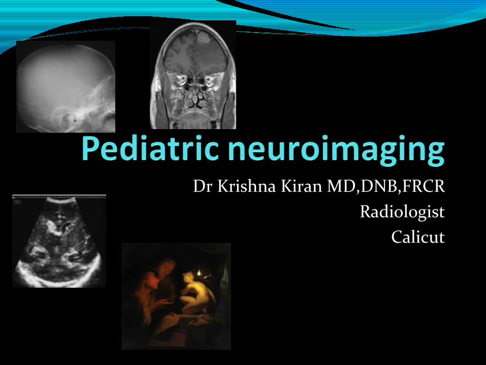
Dr Krishna Kiran MD,DNB,FRCRRadiologist
Calicut
Tools available.What is normal.?What to use and when..?

Available tools.Xray skull
NeurosonogramCT brainMRI brainPET CTDigital subtraction angiography.
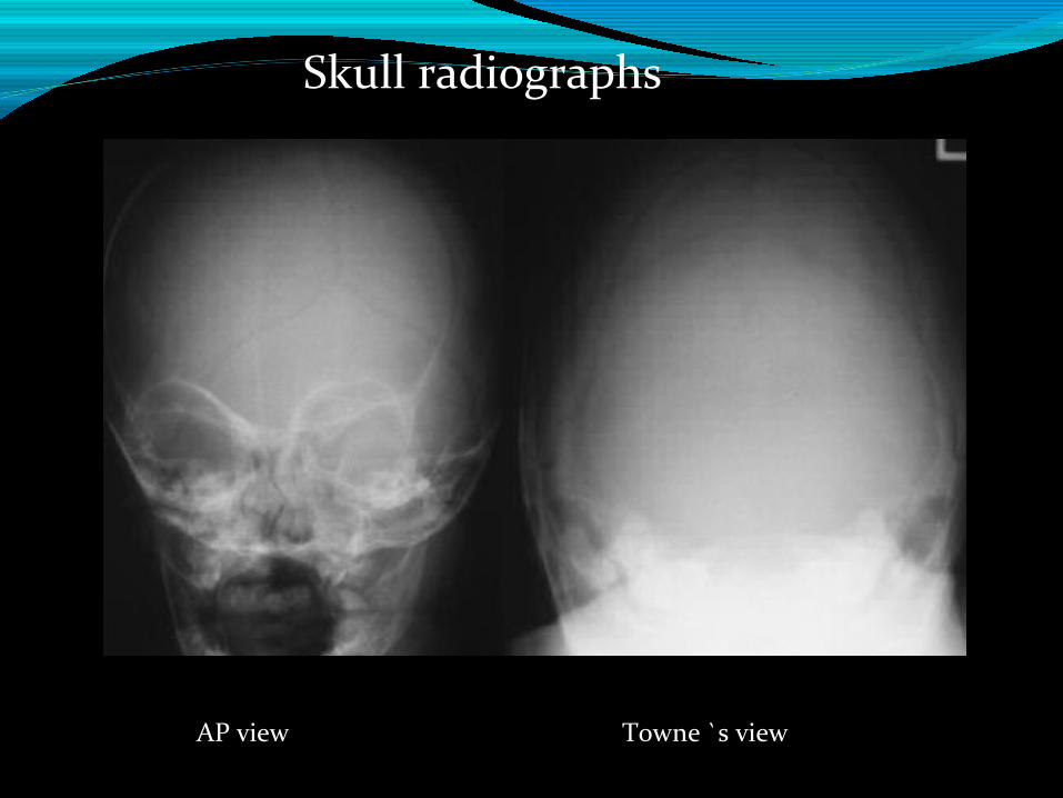
AP view Towne `s view
Skull radiographs
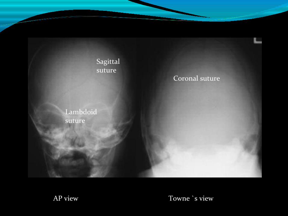
AP view Towne `s view
Sagittal suture
Lambdoid suture
Coronal suture
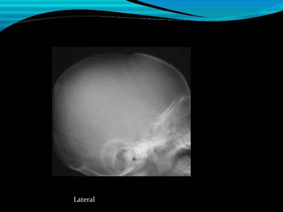
Lateral
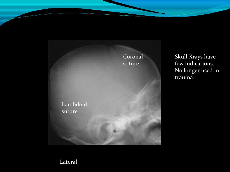
Lateral
Lambdoid suture
Coronal suture
Skull Xrays have few indications.No longer used in trauma.
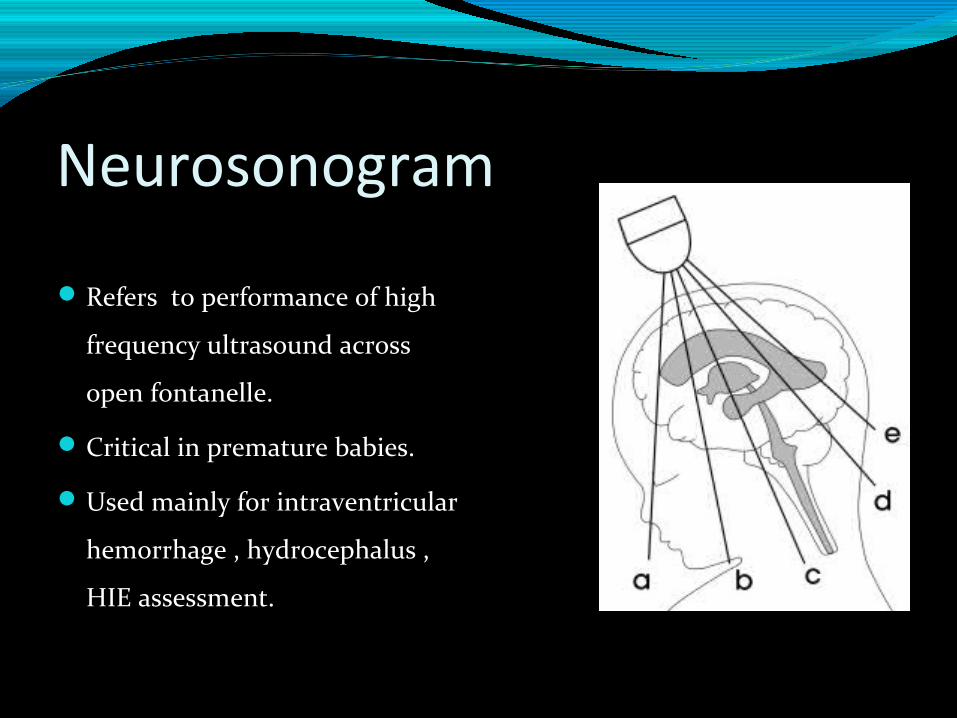
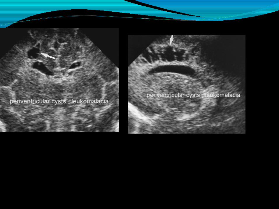
Neurosonogram
Refers to performance of high
frequency ultrasound across
open fontanelle.
Critical in premature babies.
Used mainly for intraventricular
hemorrhage , hydrocephalus ,
HIE assessment.
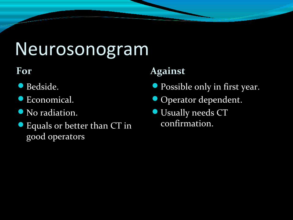
Neurosonogram For Against Bedside.Economical.No radiation.Equals or better than CT in
good operators
Possible only in first year.Operator dependent.Usually needs CT
confirmation.
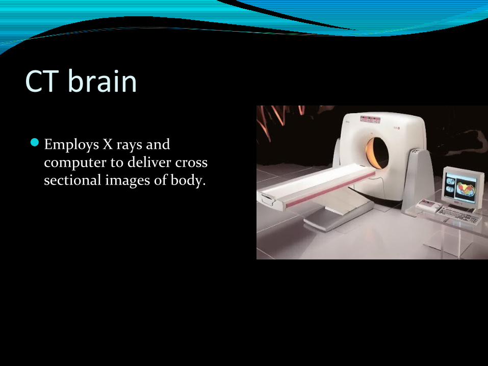
CT brain
Employs X rays and computer to deliver cross sectional images of body.
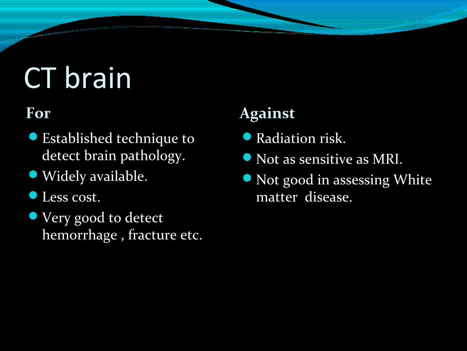
CT brainFor Against Established technique to
detect brain pathology.Widely available.Less cost.Very good to detect
hemorrhage , fracture etc.
Radiation risk.Not as sensitive as MRI.Not good in assessing White
matter disease.
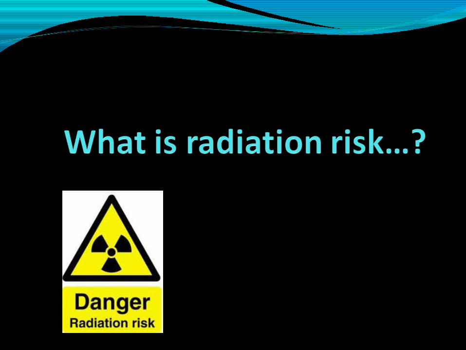
What is radiation risk..?For a cumulative dose of 50-60 mGy (milli Gray a unit
of radiation absorbed dose) there is 3 fold increase in
brain tumors when performing brain CT , 3 fold
increase in leukemia when red marrow is exposed.
What is radiation risk..?In a typical CT scanner , 2-3 head scans will give a
dose of 50-60 mGy to a child.
Berrington de González A, Mahesh M, Kim KP, Bhargavan M, Lewis R, Mettler F, Land C. Projected cancer risks from computed tomographic scans performed in the United States in 2007. Archives of Internal Medicine 2009; 169: 2071-7.
Why is radiation risk important to children..?Children live longer than adults , hence more time
for radiation effects to manifest. It takes 20-40 years
for cancer to manifest after exposure.
If not careful , children may get same dose as adults.
Children more sensitive to radiation.
Also increased risk of radiation induced cataract.
However remember that CT study can be lifesaving many times.
Hence use judiciously.
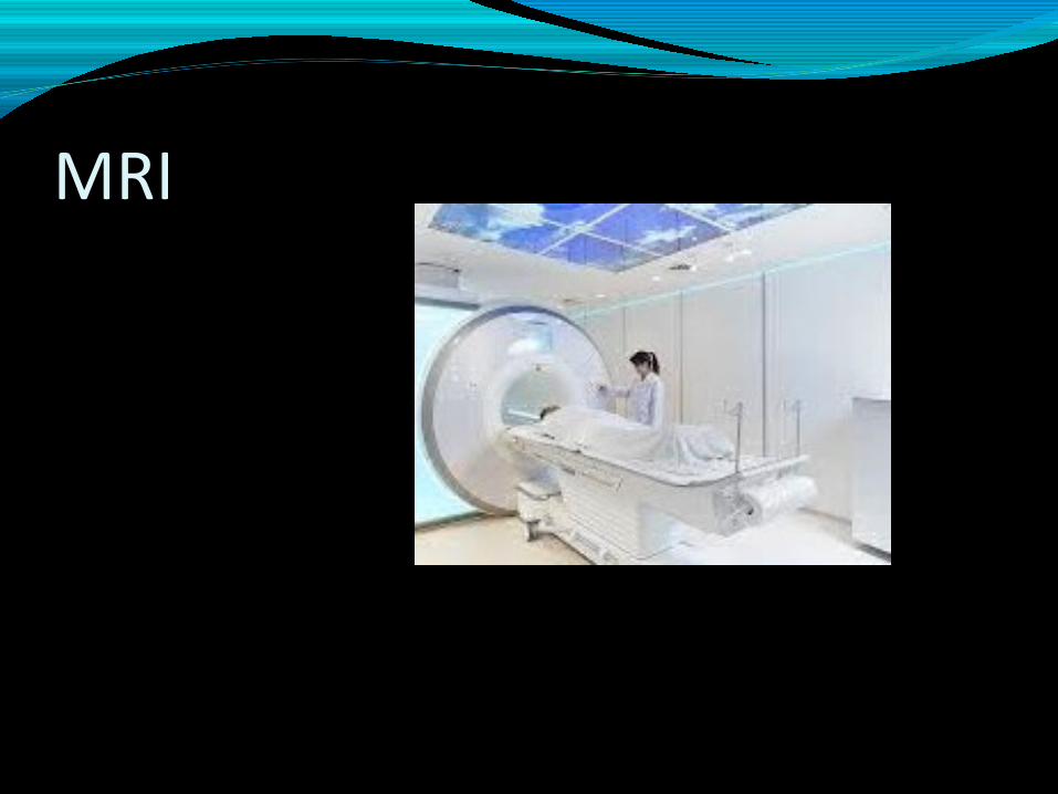
MRI
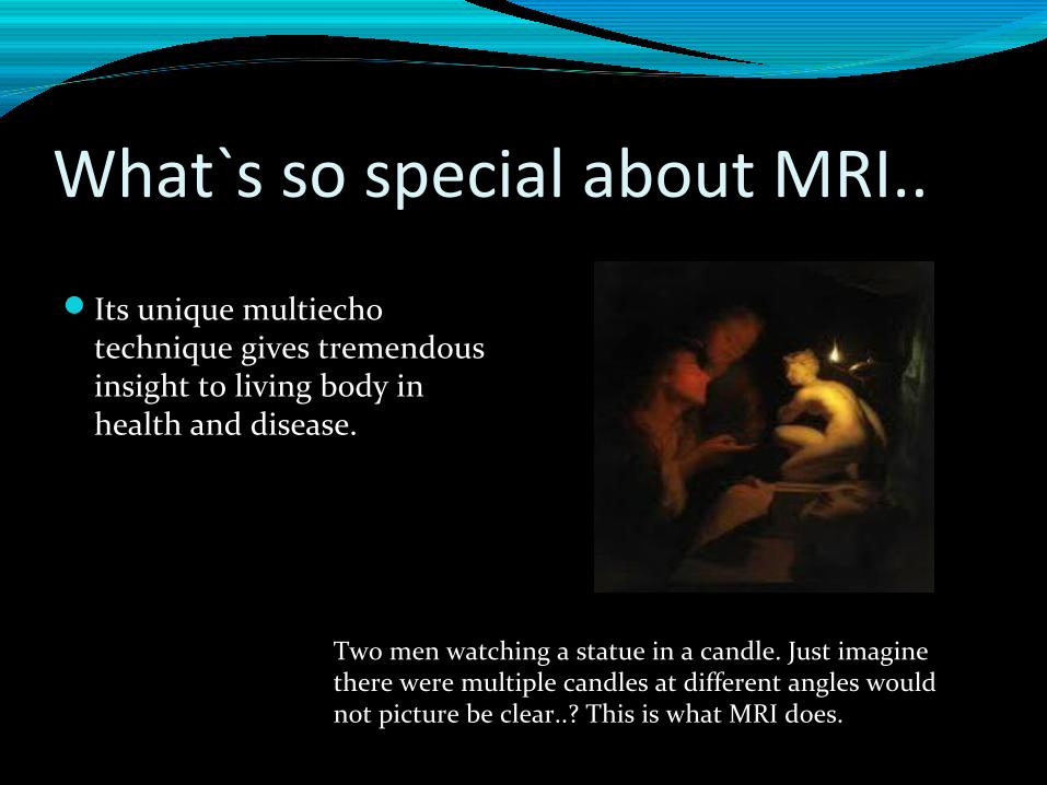
What`s so special about MRI..
Its unique multiecho technique gives tremendous insight to living body in health and disease.
Two men watching a statue in a candle. Just imagine there were multiple candles at different angles would not picture be clear..? This is what MRI does.
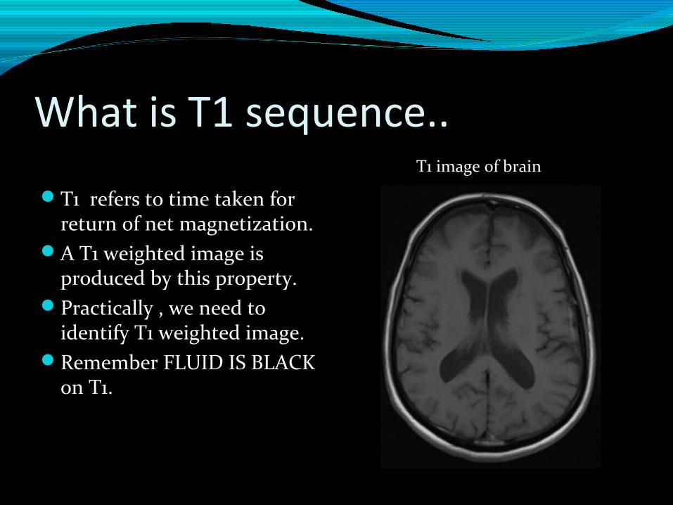
What is T1 sequence..
T1 refers to time taken for return of net magnetization.
A T1 weighted image is produced by this property.
Practically , we need to identify T1 weighted image.
Remember FLUID IS BLACK on T1.
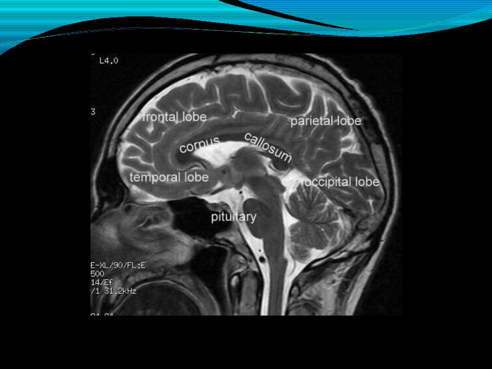
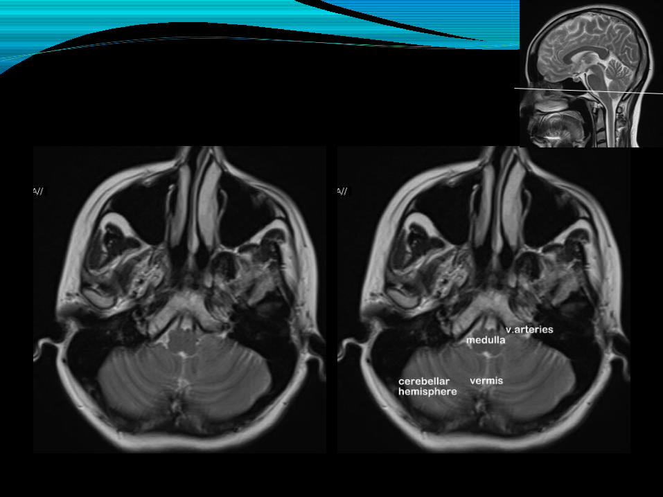
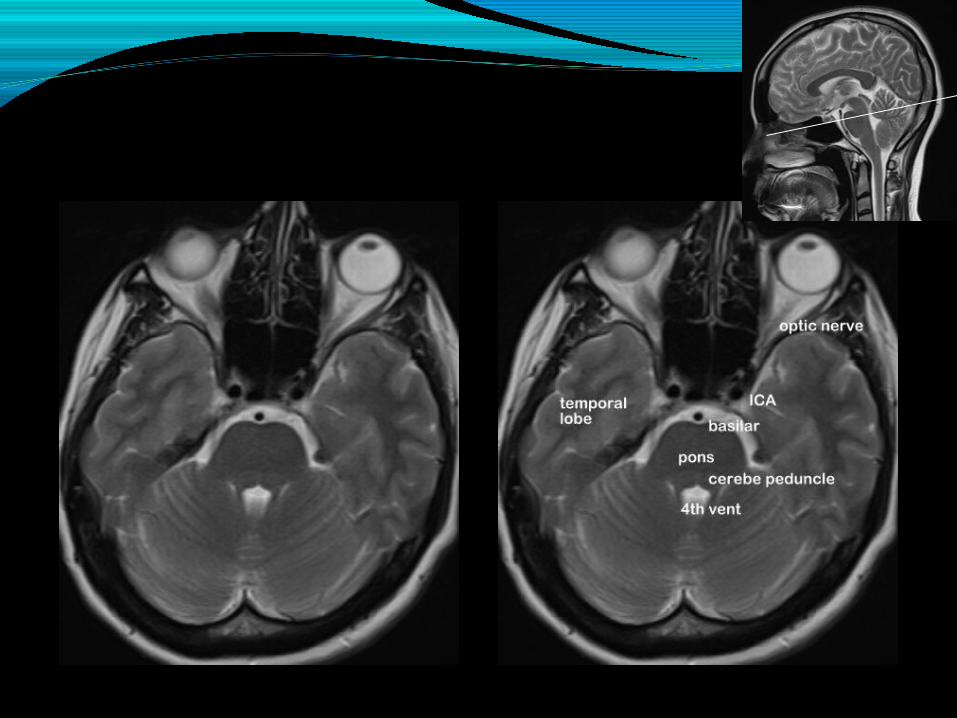
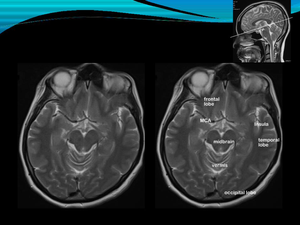
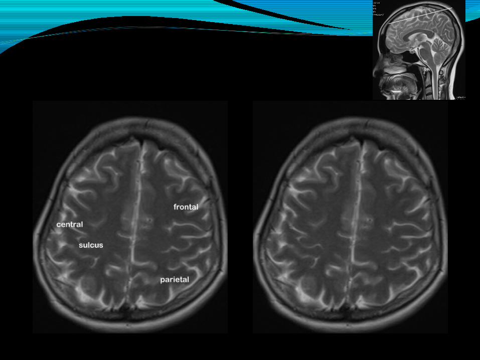
T1 image of brain
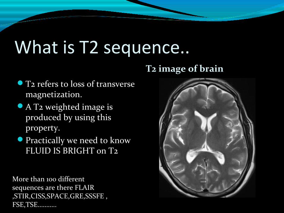
What is T2 sequence..T2 image of brain
T2 refers to loss of transverse magnetization.
A T2 weighted image is produced by using this property.
Practically we need to know FLUID IS BRIGHT on T2
More than 100 different sequences are there FLAIR ,STIR,CISS,SPACE,GRE,SSSFE , FSE,TSE………..
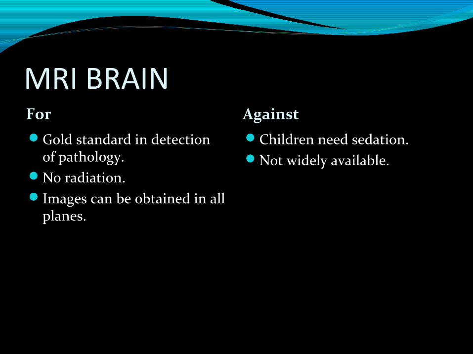
MRI BRAINFor Against Gold standard in detection
of pathology.No radiation.Images can be obtained in all
planes.
Children need sedation.Not widely available.
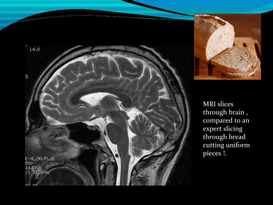
MRI slices through brain , compared to an expert slicing through bread cutting uniform pieces !.

Let us see some clinical situations…
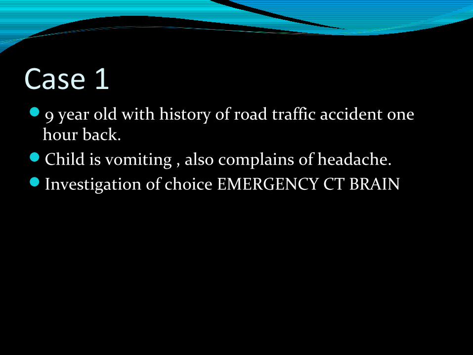
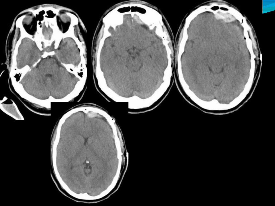
Case 1 9 year old with history of road traffic accident one
hour back.Child is vomiting , also complains of headache.Investigation of choice EMERGENCY CT BRAIN
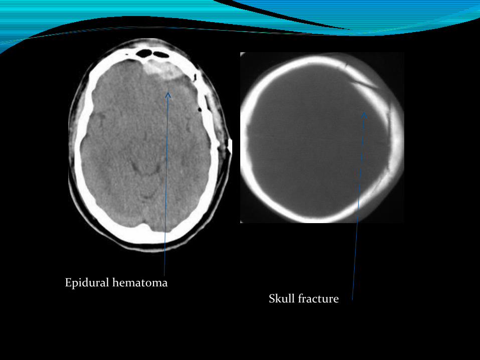
Epidural hematomaSkull fracture
Contd..Neurosurgeon decided not to operate.Child was managed conservatively , they also took 2
more CT brain scans.Child was discharged with request to do brain
imaging after 6 weeks.Which is the appropriate test..?MRI brain is the more appropriate test here.
CT brain is the first modality in trauma.It depicts hemorrhage and skull fractures well.MRI is second line investigation in head trauma.
Fractures may be missed on MRI.
Case 25 year old child with headache , early morning
projectile vomiting.You suspect an SOL.MRI with contrast is the ideal test.
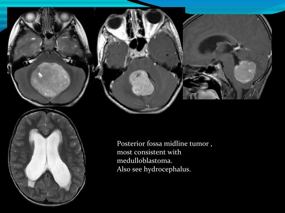
Posterior fossa midline tumor , most consistent with medulloblastoma. Also see hydrocephalus.
Case 37 year old boy with deterioration in school
performance , vision deterioration , reduced hearing.? Leukodystrophy.MRI brain is the investigation of choice.
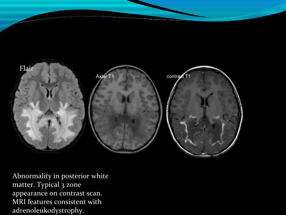
Flair
Abnormality in posterior white matter. Typical 3 zone appearance on contrast scan. MRI features consistent with adrenoleukodystrophy.
Case 410 year old child with chronic headache.Clinical examination- normal. Fundus- normal.Parents insist on SCAN..In this setting where clinical suspicion is low , both
MRI or CT may be used.
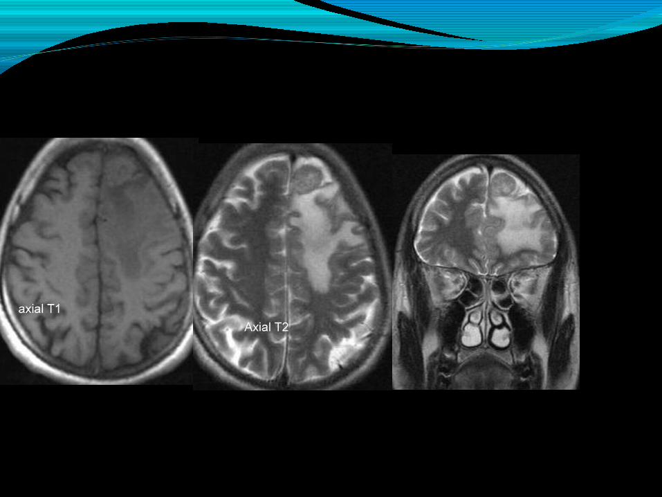
Case 5First episode of right focal seizures followed by
generalized tonic clonic seizures.Again MRI is the modality of choice
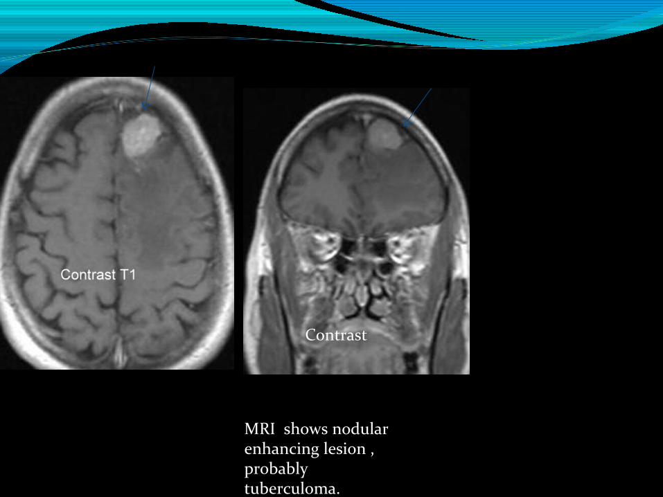
Contrast
MRI shows nodular enhancing lesion , probably tuberculoma.
How accurate is MRI ..?Depending upon signal , enhancement pattern MRI
gives approximate diagnosis.One needs to be watchful , plan for histopathology
confirmation when warranted.
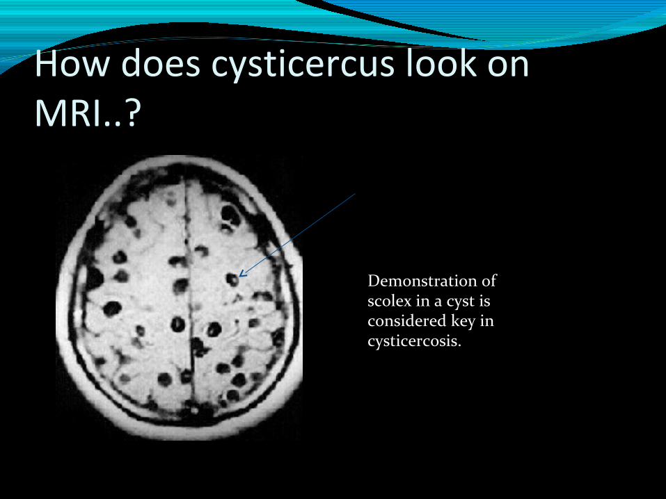
How does cysticercus look on MRI..?
Demonstration of scolex in a cyst is considered key in cysticercosis.
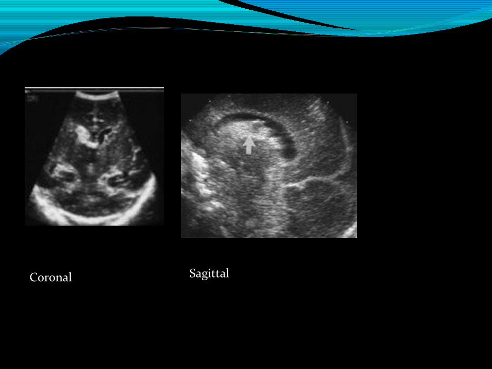
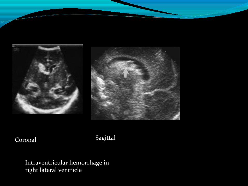
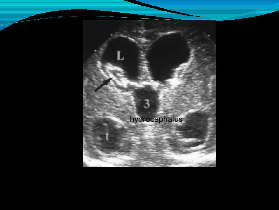
Case 6Premature baby in NICU , you sudden notice sudden
drop in hematocrit , bulging fontanelle.You are not inclined to shift baby out of NICU. Bedside NEUROSONOGRAM is the ideal test.
Coronal Sagittal
Coronal Sagittal
Intraventricular hemorrhage in right lateral ventricle
Coronal Sagittal
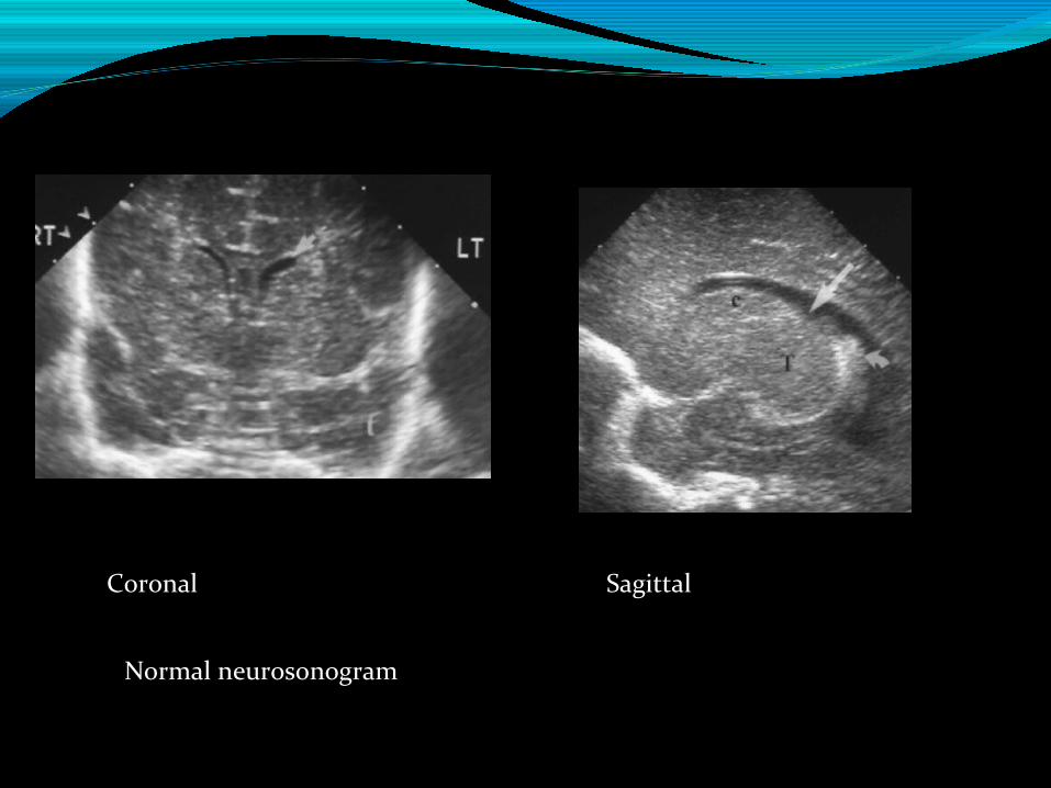
Normal neurosonogram
Conclusion Appropriate use of imaging is essential.
CT brain can be lifesaving., particularly in trauma. But
use it sparingly. Remember radiation effects.
MRI brain is modality of choice in most chronic
pediatric neuro conditions.
Less than 1 year think of neurosonogram.