pediatric pharmacology and pathology - kansas optometrickansasoptometric.org/pdfs/handouts for...
TRANSCRIPT

Pediatric Pharmacology and Pathology
Valerie M. Kattouf O.D., F.A.A.O.
Illinois College of Optometry Chief, Pediatric Binocular Vision Service
Associate Professor

In the next 2 hours…….
Ocular Medications and Children
Brief review of examination techniques/modifications for children
Common Presentations of Pediatric Pathology
Ocular Medications & Children
The rules:
– birth → 2 years old = 1/2 dose
– 2-3 years old = 2/3 dose
– > 3 years old = adult dose
If only 50 % is absorbed may be 10x maximum dosage

Ocular Medications & Children
Pediatric systems differ in:
– drug excretion
kidney is the main site of drug excretion
diminished 2 renal immaturity
– biotransformation
liver is organ for drug metabolism
Impaired 2 enzyme immaturity
Punctal Occlusion for 3-4 minutes ↓ systemic absorption by 40%
Ocular Medications & Children
Systemic absorption occurs through…..
– Mucous membrane of Nasolacrimal Duct 80% of each gtt passing through NLD system is available for rapid systemic
absorption by the nasal mucosa
– Conjunctiva
– Oropharynx
– Digestive system (if swallowed) Modified by variation in Gastric pH, delayed gastric emptying & intestinal
mobility
– Skin (2 overflow from comjunctival sac) Greatest in infants
Blood volume of neonate 1/20 adult Therefore absorbed meds are more concentrated at this age

Ocular Medications & Children
Ocular Meds with strongest potential for pediatric SE :
– 10 % Phenylephrine
– 2 % Epinephrine
– 1 % Atropine
– 2 % Cyclopentalate
– 1 % Prednisone

Ocular Medications & Children
Distribution to Site of Action in Pediatric Patients determined by :
– Size of body fluid compartment
– Muscle mass
– Fat storage
– Tissue blood flow
– Protein binding capabilities
Package inserts warn
”safety and efficacy has not been established in children”
FDA recognizes that accepted medical practice often includes prescribing medications for use in patient populations that are not included in approved labeling (PDR ophthalmology)

Ocular Medications & Children
Package inserts warn
”safety and efficacy has not been established in children”
FDA recognizes that accepted medical practice often includes prescribing medications for use in patient populations that are not included in approved labeling (PDR ophthalmology)

MODIFICATION OF AN EYE EXAMINATION FOR THE PEDIATRIC PATIENT

Pediatric Examination Procedures
Case History
Visual Acuity
EOM
Pupils
Refractive Error Assessment
Alignment / Posture
Anterior Segment Evaluation
Posterior Segment Evaluation

Case History
Perinatal History
• Full term?
• Complications during pregnancy / delivery?
• Birth weight / prematurity 5 lb 8 oz = normal Premature = < 37 weeks
• Oxygen exposure?
Case History
Medical History
• Medications ?
• Allergic to medications ?
• Allergies?
• Review of Systems
• Has the child ever been hospitalized?

Visual Acuity

OCULAR MOTILITY EXAMINATION
Rule OUT Amblyogenic Risk Factors
Goal of the pediatric eye examination……

NLDO Patient
7 month old male
– c/o tearing OU since 2 mo of age
– Hx of asthma / allergies (Albuterol / Claritin)
– Recurrent discharge OU
Anterior Segment Examination
– 2+-3+ Blepharitis
– (+) purulent discharge in both eyes
– 3+ tear prism OD, OS
Assessment / Plan
– NLDO with 2° Bacterial Conjunctivitis OD
– Rx Polytrim qid OD x 2 week
– Warm compress / Hydrostatic Massage qid
– RTC 1 week
NLDO Patient
2 week F/U, 8 month old male
– Excellent compliance with treatment, no more tearing – No discharge or staining – Cycloplegic Retinoscopy - +5.00 -0.50 x 180 OU
– Assessment / Plan Resolved NLDO d/c Polytrim, RTC if symptoms recur High Hyperopia – above age appropriate RTC 3 months to repeat cycloplegic refraction
Anterior / Posterior Segment
20D Lens
Hand Held Slit Lamp
Burton Lamp
BIO
Direct Ophthalmoscope
Anterior Segment Examination Guidelines
Lids / Lid Margins – Observe for :
Shape irregularity Discharge on lashes/lid margin
– Evert Lower lids to expose Bulbar/ Palpebral conjunctiva, observe for:
Follicles Papillae Discharge Edema
Cornea / Iris / Lens – Observe clarity / opacities/ irregularity
Anterior Segment Norms
Corneal Horizontal Diameter in Neonate
9-10 mm
Corneal Horizontal Diameter in a 1 year old
11 mm
Corneal Horizontal Diameter in Adult
11.5 –12.0 mm
Reached by 3-4 years

Anterior Segment Norms
Pupils Size Constricted - 1.2 - 2 mm Fully dilated - 7.5 – 8 mm Resting – 2.5 - 4 mm
In infancy pupillary rxn to light less than in childhood
Often absent in very premature infants (1st response at 28-32 wks)
IOP
8-15 mmHg
Increases by 1 mmHg/yr from birth to age 5

Posterior Pole Evaluation
Optic Nerve Head Color Size Symmetry
Macula Integrity / reflex
Vessels Tortuosity / attenuation
Minimal peripheral views

Ocular Pharmaceuticals
Side Effects of Diagnostic Pharmaceutical Agents in Children

Medication Class Example Ocular/
Local Side Effect Systemic Side
Effect
Adrenergic Agonist Phenylephrine Conjunctival Blanching
Hypertension, tachycardia, arrhythmias
Cholinergic Agents Cyclopentalate
Atropine
Tropicamide
Ocular irritation, follicular conjunctivitis, cutaneous hyperemia
Hyperactivity, restlessness, delirium, seizures, GI disturbance, temperature elevation, respiratory depression

Side Effects of Diagnostic Pharmaceutical Agents in Children
Phenylephrine risks – Hyperthyroidism and cardiovascular problems
may result is tachyarrhythmia
Premature / ROP patients = systemic side effects – Use Cyclomydril 0.2% cyclo / 1 % phenylephrine
no increased blood pressure
Cholinergic Agents – Education: parents may call with dilation concerns not expecting it to last into
next day
– Atropine toxicity more susceptible to Lightly pigmented Brain damage Down Syndrome

Treatment with Atropine
Proper patient selection… – Moderate-high hyperopia
– Moderate amblyopia (20/100 or better visual acuity)
Administration schedule
– 1 gtt 1.0% Atropine sulfate daily vs. weekend only frequency of installation can be reduced as acuity improvement is
observed
minimal installation is one drop of 1% Atropine sulfate two times per week
Side Effects / Emergency contact information

Atropine Instillation for Amblyopia
Our evaluation found that ________________________ has amblyopia of the right / left / both eyes.
Amblyopia is reduced vision in an eye that has not received adequate or appropriate use during early childhood, often known as “lazy eye”, and has many causes which have been explained by your doctor. If not treated, the amblyopic eye may never develop good vision and may even be functionally blind.
The treatment of amblyopia may require multiple therapy methods in which your doctor may prescribe occlusion therapy with patching or atropine instillation. Active amblyopia vision therapy may be recommended to enhance the effectiveness of occlusion therapy. Glasses are also prescribed in most cases.
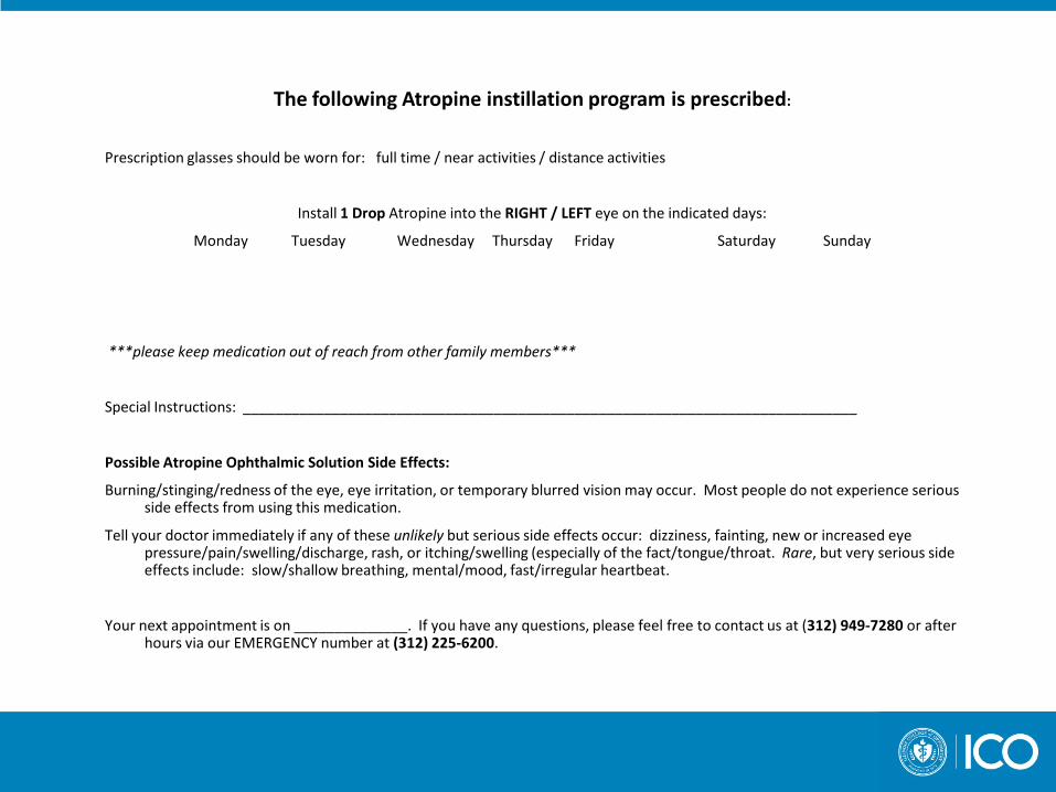
The following Atropine instillation program is prescribed:
Prescription glasses should be worn for: full time / near activities / distance activities
Install 1 Drop Atropine into the RIGHT / LEFT eye on the indicated days:
Monday Tuesday Wednesday Thursday Friday Saturday Sunday
***please keep medication out of reach from other family members***
Special Instructions: ____________________________________________________________________________
Possible Atropine Ophthalmic Solution Side Effects:
Burning/stinging/redness of the eye, eye irritation, or temporary blurred vision may occur. Most people do not experience serious side effects from using this medication.
Tell your doctor immediately if any of these unlikely but serious side effects occur: dizziness, fainting, new or increased eye pressure/pain/swelling/discharge, rash, or itching/swelling (especially of the fact/tongue/throat. Rare, but very serious side effects include: slow/shallow breathing, mental/mood, fast/irregular heartbeat.
Your next appointment is on ______________. If you have any questions, please feel free to contact us at (312) 949-7280 or after hours via our EMERGENCY number at (312) 225-6200.

Ocular Pharmaceuticals
Commonly Used Ocular Anti-Biotic Medications in Children

Administration of Ocular Meds Ointment vs. Drops
Ointment
– blurred vision
– ↑ contact dermatitis
Drops
– ↑ risk of systemic toxicity
– ↓ contact time with cornea (diluted by tears)
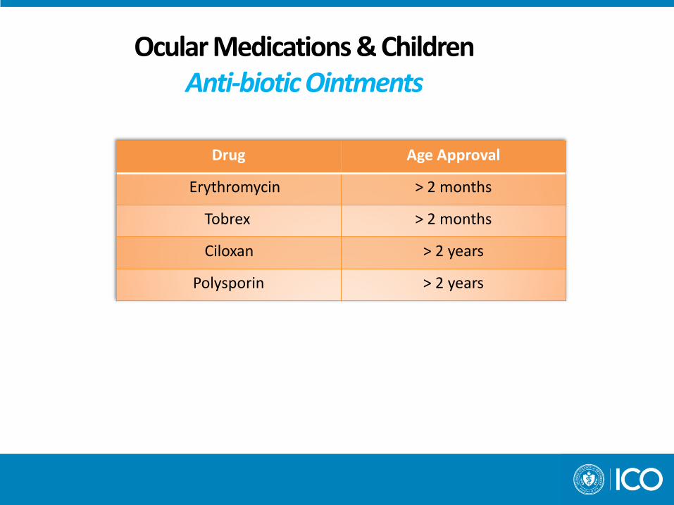
Ocular Medications & Children Anti-biotic Ointments
Drug Age Approval
Erythromycin > 2 months
Tobrex > 2 months
Ciloxan > 2 years
Polysporin > 2 years

Ocular Medications & Children Anti-biotic Drops
Drug Age Approval
Polytrim ≥ 2 months
Azasite ≥ 1 yr
Besivance ≥1 yr
Ciloxan ≥1 yr
Ocuflox ≥1 yr
Quixin ≥1 yr
Vigamox ≥1 yr
Zymar ≥1 yr
Zymaxid ≥1 yr
Iquix ≥6 yr
Gentamycin unknown
Sulfacetamide unknown

Ocular Medications & Children Anti-biotic Drops
• Polytrim – Broad spectrum, effective, inexpensive
• AzaSite – macrolide anti-biotic (Z-pack)
• Prolonged ½ life - ↓ dosing schedule • 1 gtt q 8-12 hrs (tid) x 2 days
• 1 gtt qd x 5 days
• Broad spectrum, effective, expensive

Ocular Medications & Children Anti-biotic Drops
• Fluoroquinolones (concentration dependent)
• Besivance (0.6%)– new, Advanced A-B Vehicle: DuraSite mucoadhesive – provides enhanced ocular surface
residency time Dosing = tid (q 8 hrs) Pediatric schedule: AM → after school → at bed time
• Ciloxan (0.3%) • Ocuflox (0.3%) • Quixin (0.5%) • Vigamox (0.5%) • Zymar (0.3%) • Zymaxid (0.5%) ↑ concentration may enhance clinical results • Iquix (1.5%)

Ocular Pharmaceuticals
Commonly Used Ocular Allergy Medications in Children

Ocular Medications & Children / Topical Allergy Drops Anti –histamine/ mast cell stabilizer
Rx
Drug Age approval
Pataday/Patanol ≥3 years old
Lastacaft ≥2 years old
Elestat ≥3 years old
Bepreve ≥2 years old
Optivar ≥3 years old
Anti –histamine/ mast cell stabilizer OTC
Drug Age approval
Zaditor ≥ 3 years old
Claritin Eye ≥3 years old
Refresh Eye Itch Relief
≥3 years old
Alaway ≥3 years old
Topical Corticosteroid Rx
Drug Age Approval
Alrex ≥3 years old
Ocular Pharmaceuticals
Commonly Used Ocular Medications in Children/ Additional Agents
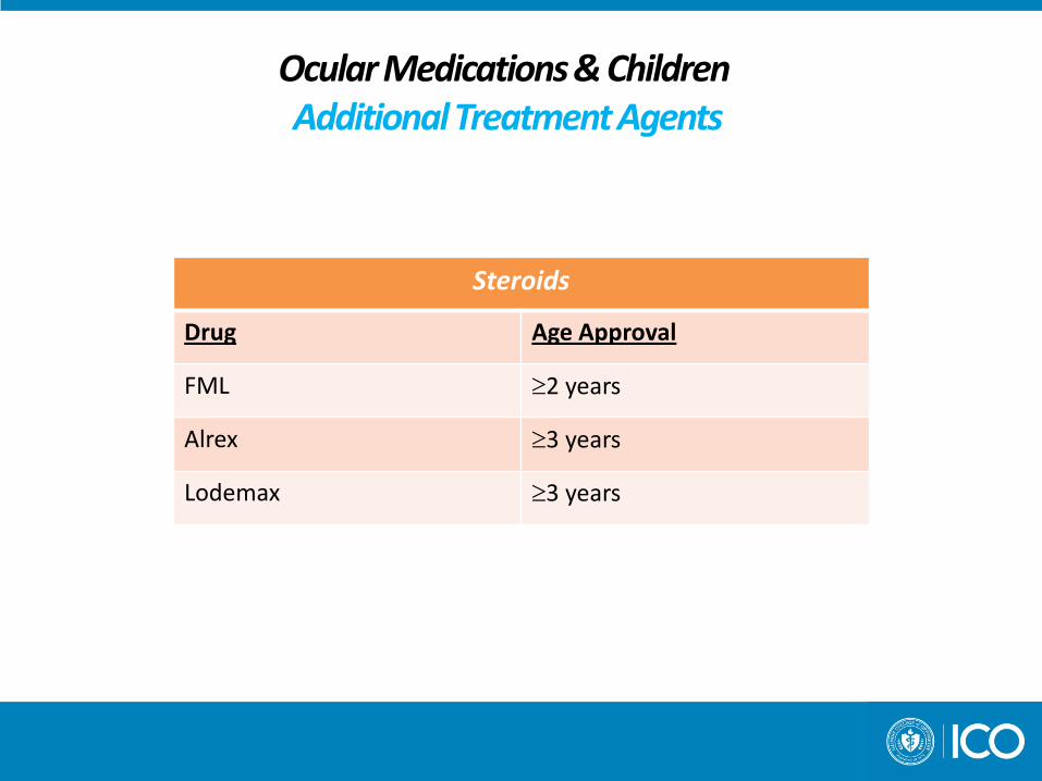
Ocular Medications & Children Additional Treatment Agents
Steroids
Drug Age Approval
FML ≥2 years
Alrex ≥3 years
Lodemax ≥3 years
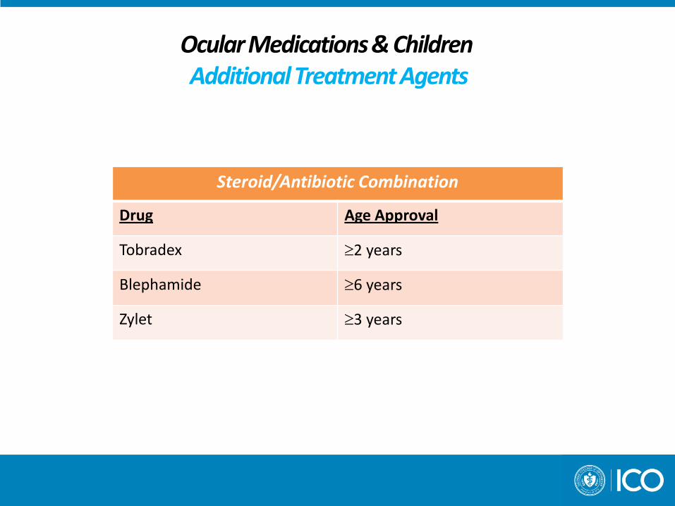
Ocular Medications & Children Additional Treatment Agents
Steroid/Antibiotic Combination
Drug Age Approval
Tobradex ≥2 years
Blephamide ≥6 years
Zylet ≥3 years

Ocular Medications & Children Topical Steroids
• Tobradex has dexamethasone as steroid agent known to increase IOP • Alrex • Zylet • Lotemax
• Have loteprednol as steroid agent = less likely to increase IOP

Ocular Medications & Children Topical Steroids
• Alrex (0.2% Lodeprednol)
• Approved for treatment of seasonal allergic conjunctivitis • Lubricant included/ increases comfort (viscous nature for soothing)
Lotemax (0.5% Lodeprednol) • Use for intraocular inflammation
Anterior uveitis Post-op Ocular allergy / GPC
• Zylet (0.5% Lodeprednol) • Treatment of inflammation + Ocular surface disease • Conjunctivitis/ blepharoconjunctivitis

Ocular Medications & Children Additional Treatment Agents
Antiviral Agents
Drug Age Approval
Viroptic ≥6 years
Zirgan ≥2 years
Ocular Medication Installation

Tips for ocular medication administration
Drop administration – Immobilize child
– Retract lower lid
– Single drop in cul de sac
Alternative method – Patient supine
– Drop in inner canthus while eyes are closed
Confirm that medication reaches tear film when eyes are open Wipe excess fluid from cheek When done properly ocular absorption is comparable to the conventional installation
method

Instructions for home use
Emergency contact number
Diagnosis
Name of medication – Dosage Instructions Which eye (s) How many times per day When to discontinue
Follow-up visit date(s)
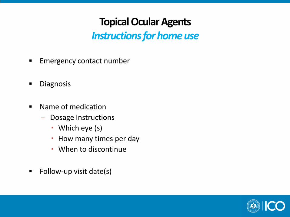
Topical Ocular Agents Instructions for home use
Emergency contact number
Diagnosis
Name of medication – Dosage Instructions Which eye (s) How many times per day When to discontinue
Follow-up visit date(s)

Prescribing Oral Medications to Children

Prescribing for Children: Guidelines & Helpful Hints
Children ≥12 years and older = dosed as adult Children ≤11 years
• Look up dosage – given in mg/kg/day
• Determine weight in kg – 1kg = 2.2lbs
• Mg x Kg = DAILY dose
• Divide daily dose to get desired doses per day
• Choose closest available dosage strength
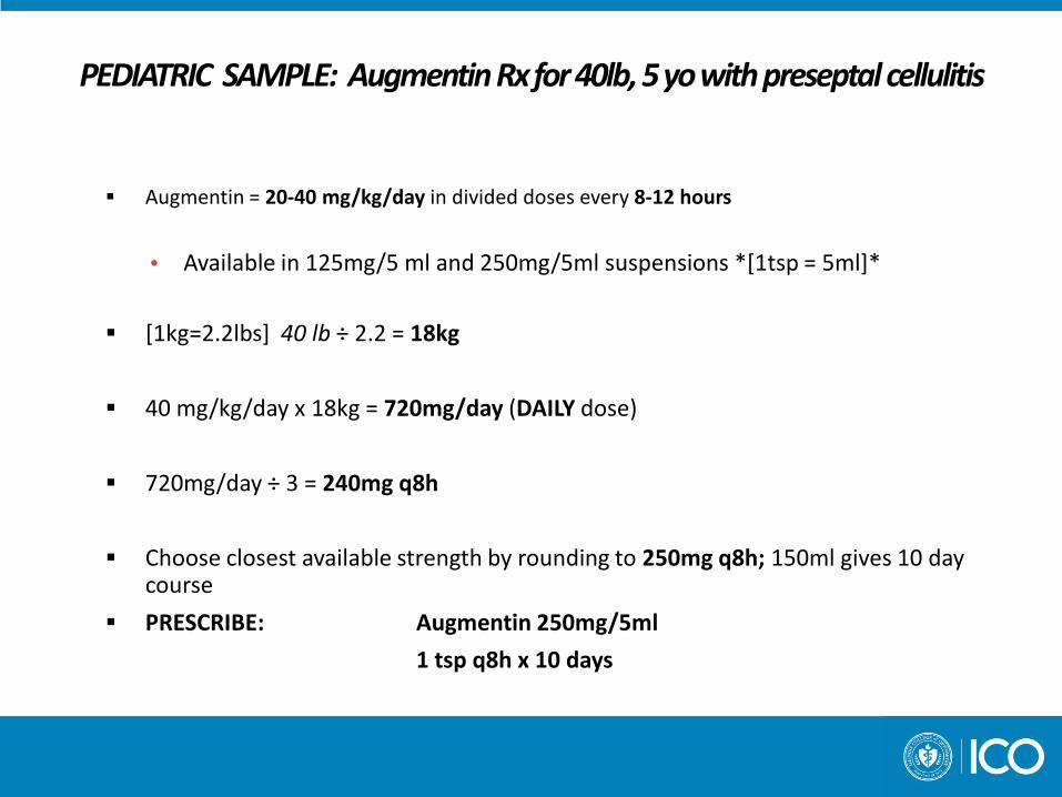
PEDIATRIC SAMPLE: Augmentin Rx for 40lb, 5 yo with preseptal cellulitis
Augmentin = 20-40 mg/kg/day in divided doses every 8-12 hours
• Available in 125mg/5 ml and 250mg/5ml suspensions *[1tsp = 5ml]*
[1kg=2.2lbs] 40 lb ÷ 2.2 = 18kg
40 mg/kg/day x 18kg = 720mg/day (DAILY dose)
720mg/day ÷ 3 = 240mg q8h
Choose closest available strength by rounding to 250mg q8h; 150ml gives 10 day course
PRESCRIBE: Augmentin 250mg/5ml
1 tsp q8h x 10 days

Oral Anti-Biotics
Children with no Penicillin Allergy
Penicillin V x 10 days
children < 30kg/65 lbs = 250mg bid
children > 30kg/65 lbs = 500mg bid
Amoxicillin x 10 days
children < 30kg/65 lbs = 40mg/kg/day
children > 30kg/65 lbs = 250mg tid

Oral Anti-Biotics
Children with Penicillin Allergy (for children < 60 lbs)
Eurythromycin x 10 days 40mg/kg/day tid
Azithromycin (Zithromax) x 5 days 12mg/kg qd
Cephalexin (Keflex) 25-30 mg/kg ≤ 4000 mg/day

Prescribing for Children: Guidelines & Helpful Hints
Consult pediatrician for children ≤ 5 years of age
In most cases prescribe the highest recommended mg/kg/day
Pharmacists are very helpful in dosing
For drug information:
Epocrates.com
Drugs.com

Anterior Segment Pathology Pediatric Red Eyes / Conjunctivitis

Differential Diagnosis The Pediatric Red Eye
Conjunctivitis
Bacterial
Viral
Allergic

Rules of Thumb
If itches, it’s allergic
If it burns, it’s dry eyes
If eye cannot open in the AM it is likely bacterial
If they have a cold it is viral

CONJUNCTIVITIS Bacterial vs. Viral
DIFFERENCES
Conjunctival Discharge
Conjunctival Response
Systemic Associations
Determination important because it drives decisions about treatment and school exclusion
SIMILARITIES
Bilateral
Eyelid Swelling
Conjunctival Erythema

Viral Conjunctivitis
Typically caused by adenovirus
Signs
• Watery discharge (typically bilateral??)
• Erythema
• Conjunctival response = follicular
• Often in presence of a viral URI (upper respiratory infection) • May have palpable pre-auricular node often on more affected side
Most common conjunctivitis seen in school aged children

Viral Conjunctivitis Tx
Treatment Options – Self limiting therefore supportive treatment
- Artificial Tears
- Cool Compress
- Instruction of proper hygiene / avoidance of family members
- Discuss daycare / School attendance issues
- Frequently asked questions…….
- How long does conjunctivitis/pink eye last?
- Signs and symptoms of conjunctivitis usually improve within three to seven days.
- When is it appropriate for a child to return to school or child care?
- When tearing and discharge are no longer present

Bacterial Conjunctivitis
Typically caused by haemophilus influenzae / streptococcus pneumoniae
Signs
• Purulent discharge
• Minimal erythema
• Conjunctival response = follicular + papillary
Bacterial Conjunctivitis Tx
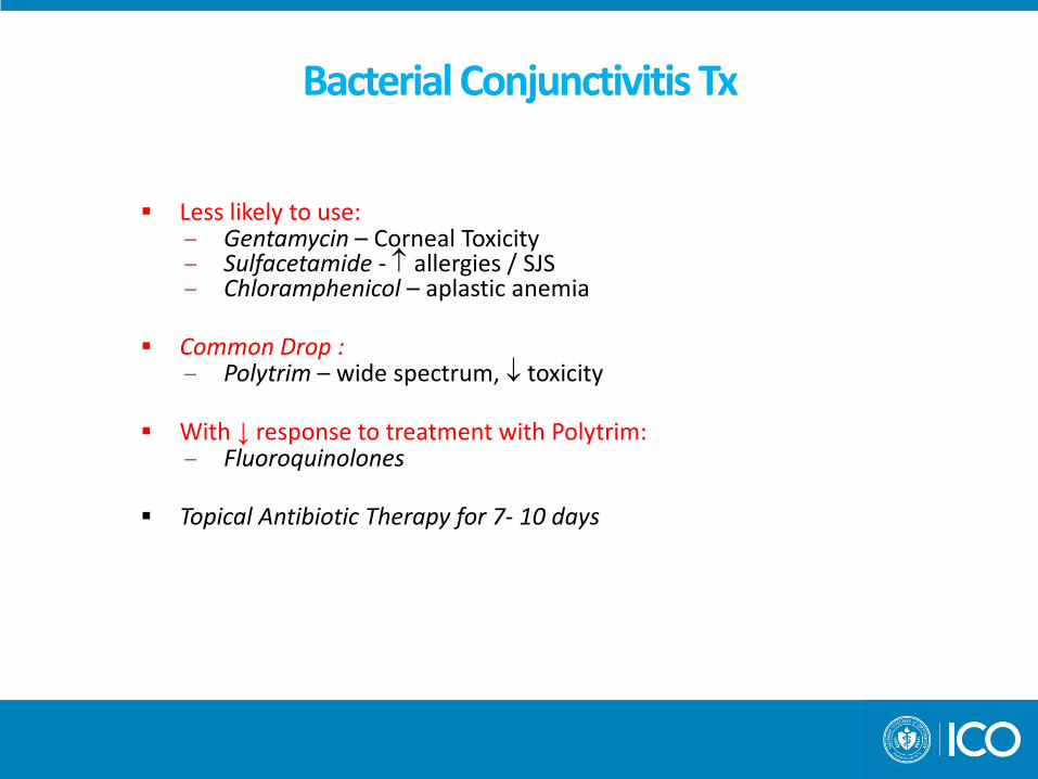
Less likely to use: – Gentamycin – Corneal Toxicity – Sulfacetamide - ↑ allergies / SJS – Chloramphenicol – aplastic anemia
Common Drop :
– Polytrim – wide spectrum, ↓ toxicity
With ↓ response to treatment with Polytrim: – Fluoroquinolones
Topical Antibiotic Therapy for 7- 10 days
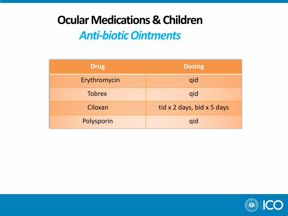
Ocular Medications & Children Anti-biotic Ointments
Drug Dosing
Erythromycin qid
Tobrex qid
Ciloxan tid x 2 days, bid x 5 days
Polysporin qid

Ocular Medications & Children Anti-biotic Drops
Drug Dosing
Polytrim qid
Azasite tid x 2days, qd x 5days
Besivance tid
Ciloxan 1gtt q 2hrs x 2days, qid x 5 days
Ocuflox 1gtt q 2hrs x 2days, qid x 5 days
Quixin 1gtt q 2hrs x 2days, qid x 5 days
Vigamox tid
Zymar 1gtt q 2hrs x 2days, qid x 5 days
Zymaxid 1gtt q 2hrs x 2days, qid x 5 days
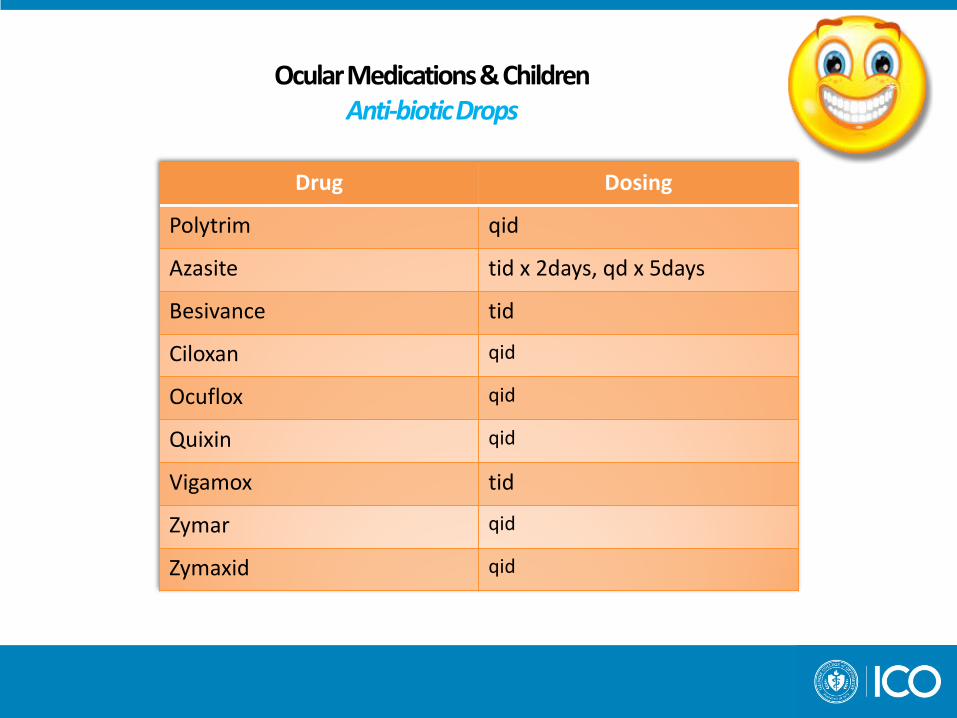
Ocular Medications & Children Anti-biotic Drops
Drug Dosing
Polytrim qid
Azasite tid x 2days, qd x 5days
Besivance tid
Ciloxan qid
Ocuflox qid
Quixin qid
Vigamox tid
Zymar qid
Zymaxid qid

Anterior Segment Pathology Chronic Blepharitis

Chronic Blepharitis
May result in:
• Chronic blepharoconjunctivitis
• Recurrent chalazia
• Loss of lashes / madarosis
• Thickening of lid margins
Treatment
• Warm compress/massage/lid scrubs
• Topical Anti-biotic (drop vs. ung)
• Oral Antibiotic
• Surgical Excision

Anterior Segment Pathology Nasolacrimal Duct Obstruction

Nasolacrimal Duct Obstruction
Clinical Characteristics – 5-6% of newborns – Constant tearing – Redness irritation of lids – **With secondary conjunctivitis discharge injection swelling over innermost aspect of lower lid pain fever

Nasolacrimal Duct Obstruction Etiology and Anatomy
Incomplete opening of lower end of the NLD along side of nose between inner canthus of eyelid and inferior turbinate of the nasal cavity

Nasolacrimal Duct Obstruction
TREATMENT OPTIONS
1) Warm compress / Hydrostatic Massage
2) Topical Antibiotic Drops
3) Probing
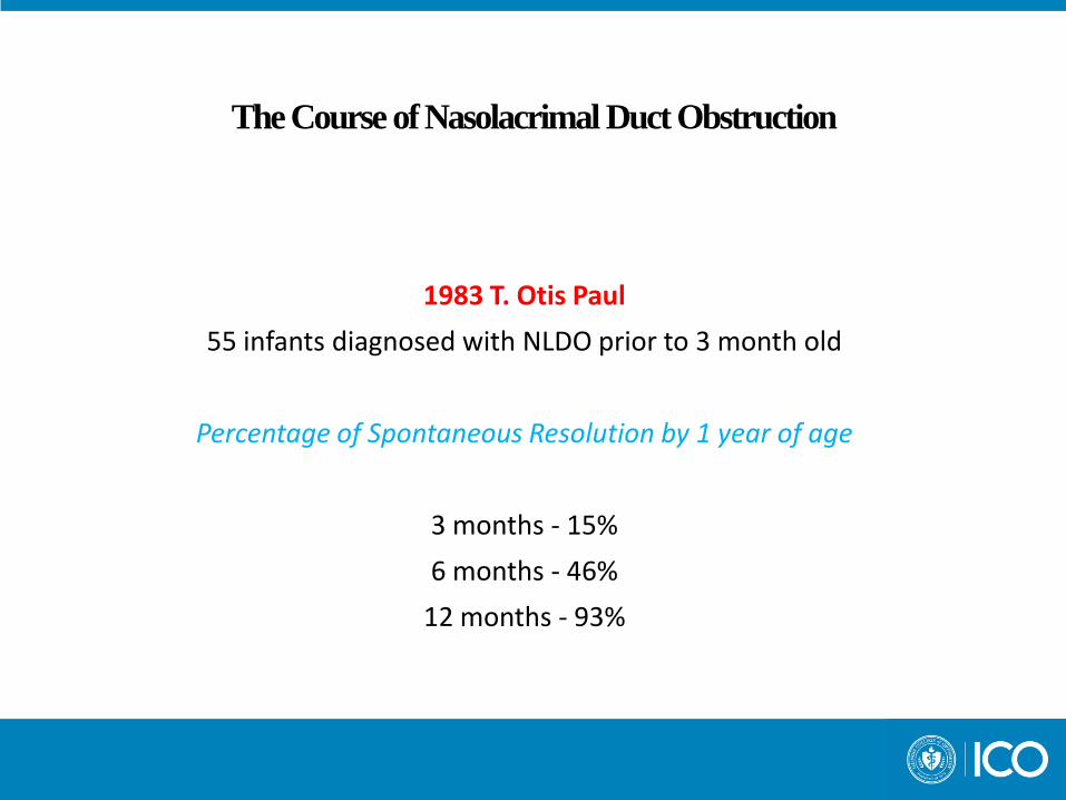
The Course of Nasolacrimal Duct Obstruction
1983 T. Otis Paul
55 infants diagnosed with NLDO prior to 3 month old
Percentage of Spontaneous Resolution by 1 year of age
3 months - 15%
6 months - 46%
12 months - 93%

NLDO Patient
4 week old AA male – Right eye tearing since birth – Red right eye with purulent discharge x 2 weeks
– Anterior Segment Evaluation
Erythema and Edema OD Yellow-green discharge OD (+) Tear lake OD >>>OS 3 = fluorecein disappearance test (-) corneal staining
– Assessment / Plan
NLDO with 2° Bacterial Conjunctivitis OD Rx Polytrim qid OD x 1 week Warm compress / Hydrostatic Massage qid RTC 1 week

NLDO Patient 5 weeks old
– ↓ Lid edema and tear lake – No NaFl stain – 2-3 = fluorecein disappearance test – (+)mucous in tear film
Continue with previous treatment regimen. RTC 2 weeks
8 weeks old – Mom notes tearing has decreased significantly – Minimal lid edema and injection – Minimal discharge – (+) tear lake OD – 2-3 = fluorecein disappearance test
↓ Polytrim bid ( to qid if conjunctivitis worsens) Continue with warm compress / massage Add lids scrubs with baby shampoo

NLDO Patient
12 weeks old – Mild lid edema – (-) discharge – ↓ tearing OD > OS – 2 = fluorecein disappearance test – d/c Polytrim, restart if conjunctivitis recurs
4 month old AA male
– Tearing significantly improved – 1 = fluorecein disappearance test – Partial vs. Resolved NLDO – Restart Polytrim / return to clinic if conjunctivitis returns

Instructions
Warm Compress
– 5- 10 minutes of continuous warmth
– Options
Lacrimal Sac Massage
– Use index finger wrapped in clean, thin, cloth
– Begin between infants eyebrow
– Drag finger down towards affected side, closing lid simultaneously
– Continue movement, pressing firmly into the canthus
– Continue onto cheek
– 10 strokes / tid

What are differentials for tearing in an infant?
NLDO
Conjunctivitis
Corneal Abrasion / Foreign Body
#1 = Congenital Glaucoma
Presentations of Pediatric Pathology
Congenital Glaucoma

Differential Dx of NLDO
Congenital Glaucoma Incidence
From 1/10,000-1/25,000 live birth presents during first year of life
Characteristics Unilateral / bilateral (2/3, usually asymmetric) 2/3 males Steamy cornea/edema Photophobia Tearing / Epiphora ↑ corneal diameter Axial elongation with myopia Elevated IOP

Differential Dx of NLDO
Characteristics
Unilateral / bilateral (2/3, usually asymmetric)
2/3 males Steamy cornea/edema Photophobia Tearing / Epiphora ↑ corneal diameter Axial elongation with myopia Elevated IOP

Congenital Glaucoma
Etiology – Membrane covering TM? – Anomalous high insertion of iris Infant angle not adult like Pale TM Indistinct Schwalbe’s line Flat peripheral iris Iris processes to TM
Open angle AC deeper in Cong Glaucoma than normal infant angle

Congenital Glaucoma
Examination
– IOP Norm = 8-15 mm Hg
– Corneal Horizontal Diameter Measurement
Norm = 9-10 mm infant, 11 mm by 1 year old
– Anterior Segment Evaluation
– A scan Axial length norms
Newborn - approximately 16 mm 18 months - 20.3 mm 2-5 yr. Old - increase avg. 1.1 mm 5-13 yr. old - increase of 1.3
– Rx determination
– DFE
Usually need EUA Check IOP under light sedation/ meds ↓ IOP

Nasal and Ocular Allergy
70-90% of allergic rhinitis patients have ocular allergy
Allergic rhinoconjunctivitis
25% of the population
80-90% of all allergic disorders

Allergic Cascade
IgE

Treatment of Allergy Patient
Identify / Remove antigen Reduce edema Reduce inflammation
Topical Ocular Agent
• Allergy medication
• Steroid
• Combination of the two
Oral medication Referral to allergist

Ocular Medications & Children / Topical Allergy Drops
Ocular Allergy Medications
Drug Dosing
Pataday qd
Patanol bid
Lastacaft qd
Elestat bid
Bepreve bid
Zaditor bid
Alrex qid

Anterior Segment Pathology Pediatric Red Eyes / Conjunctivitis
Vernal Keratoconjunctivitis
Vernal Keratoconjunctivitis
Vision Threatening
Chronic, bilateral conjunctival inflammatory disorder
Male > Female
Typically onset before 10 years of age, resolution by puberty
Seen most in warm, dry, climates???
Significant atopic history

Vernal Keratoconjunctivitis
Symptoms • Pain • Itching (severe) • Conjunctival injection • Ptosis • Mucous discharge
Clinical Signs:
• Large papillae • Conjunctival hyperemia with edema • Horner-Trantas dots = clumps of eosinophils with dead epithelial
cells

Summary of Pediatric Red Eyes
Conjunctivitis
– Bacterial
– Viral
– Allergic
More severe
– Preseptal cellulitis
– Orbital cellulitis

Anterior Segment Pathology Preseptal and Orbital Cellulitis

PRESEPTAL CELLULITIS
Definition - infection of soft tissues of the eyelid and periocular region anterior to the orbital septum
Clinical Characteristics
– eyelid edema
– erythema
– warmth of eyelid
– conjunctival chemosis / ocular discharge
– NOT PRESENT
proptosis
restriction of ocular motility
pain with eye movement

PRESEPTAL CELLULITIS
Possible Etiologies
– Chronic Blepharitis / conjunctivitis
– Internal Hordeolum
– Acute Dacryocystitis
– Penetrating Injury
– Bite Wounds
– Respiratory Infection
– Sinusitis
– Dermatitis
PRESEPTAL CELLULITIS
Differential Diagnosis
– Orbital Cellulitis ****
– Allergic Lid Edema
– Viral Conjunctivitis with Lid Edema

PRESEPTAL CELLULITIS
How to determine severity / treatment options:
– Is patient toxic?
– Is patient/parent non-compliant with treatment?
– Child < 5 years old
– No improvement within 3-4 days of administering oral anti-biotic

PRESEPTAL CELLULITIS
TREATMENT OPTIONS
Mild > 5 y.o.
ORAL ANTIBIOTICS
Moderate to Severe
Consult
Hospitalization
IV ANTIBIOTICS

ORBITAL CELLULITIS
Definition – infection of the soft tissues of the orbit posterior to the orbital septum
Clinical Characteristics
– unilateral orbital tenderness – Pain on eye movement – PARALYSIS of extraocular muscles – Proptosis – Papillodema – Blurred vision – fever / systemic illness
ORBITAL CELLULITIS
Differential Diagnosis
– Preseptal Cellulitis
– Differentiation made by :
Fever
Vision loss
Motility limitation
Proptosis

ORBITAL CELLULITIS
TREATMENT OPTIONS
CT Scan
IV ANTIBIOTICS

Cellulitis Case 5 yo AA female
– Left eye swelling x 5 days
– Given Augmentin (ER) – NI
– Symptoms worsening
– (+) injection, discharge and tenderness
– (-) hx of trauma or allergies
Examination Findings (ER)
– VA = 20/20 OD, OS
– 4+ lid edema OS
– area of tenderness left upper brow
– (+) injection and discharge
– (-) cell / flare in AC
– (-) proptosis
– (-) EOM restriction,
– (-) Pain on eye movement
Cellulitis Case
Assessment / Plan
– Likely Preseptal cellulitis – Ordered CT orbit / sinus (if abscess seen = admit) – Rx Zymar tid – Continue with Augmentin – RTC 1 day
– One week follow-up
– Mom notes decreased edema and injection – Possible hordeolum in left UL – (+) UL edema – (+) conjunctival injection – (-) discharge – Continue with – Zymar tid – Augmentin (10 day cycle) – RTC 1 week

Cellulitis Case
Two-week Follow-up
– Elicit history of styes and allergies as per Mom
– (+) papillae and mild conjunctival chemosis
– Minimal injection
– Minimal lid edema
– D/C anti-biotic medications
– Rx Patanol prn for ocular allergies
– Rx Lid scrubs and warm compresses bid to aid in decreasing development of chronic hordeola
Presentations of Pediatric Pathology
Leukokoria

Leukokoria
Must determine anatomic location of lesion
– Differential Diagnosis: Congenital Cataract
Retinoblastoma
Coat’s Disease
Retinopathy of Prematurity
Persistent Hyperplastic Primary Vitreous (PHPV)

Leukokoria
Congenital Cataracts

Leukokoria Differential Diagnosis
Congenital Cataracts
1/10,000, 400-500 infants per year
Risk of Image Degradation Amblyopia

Congenital Cataract
Treatment
– Cataract Extraction
IOL implant – Contact Lens Fit – Amblyopia Therapy
Contact Lens Fit
– May combine with spectacles – ↓ magnification 20-30 % with specs
8-12 % with contact lenses – Improves development, cosmesis

Leukokoria
Must determine anatomic location of lesion
– Differential Diagnosis: Congenital Cataract
Retinoblastoma
Coat’s Disease
Retinopathy of Prematurity
Persistent Hyperplastic Primary Vitreous (PHPV)

Leukokoria
Coat’s Disease

Coat’s Disease Epidemiology
Exudative retinitis, retinal telangiectasis
Increased permeability of these abnormal retinal vessels causes leakage of the serum into intraretinal and subretinal spaces
Inheritance pattern unknown
Very rare; young males (M:F, 3:1)
80% unilateral
Characterized by abnormal vessel development
Poor prognosis in advanced stages
Retinal detachment in advanced stages
Coats Disease Stages
I = abnormal dilation of retinal blood vessels
II= telangiectasia and exudation
III= exudative retinal detachment
IV= total retinal detachment
V = characterized by irreversible blindness

Coats Disease Diagnosis
CT Scan
the globe appears hyper dense compared to normal vitreous due to the exudate

Coats Disease Treatment
Ablation of causative lesions
Laser photocoagulation or cryotherapy
Steroid and anti-VEGF injections
Enucleation

Leukokoria
Retinopathy of Prematurity (ROP)

Leukokoria Differential Diagnosis
Retinopathy of Prematurity A premature infant is an infant born before 37 weeks gestation
Prematurity used to be defined as any infant weighing less than 5.5 lbs
ROP 40% of infants with birth weight ≥ 3 lbs 50%-80% of neonates under ≤ 2 lbs

Retinopathy of Prematurity
Retinal vascular disease secondary to premature birth, low birth weight, and use of supplemental oxygen
Clinical severity can range from mild with no visual defects to aggressive with neovascularization, retinal detachment, and blindness.

Retinopathy of Prematurity
Pathophysiology
– Retinal vasculature begins at 16 weeks gestation
– Proliferation of capillaries will form the mature retinal vessels
– The nasal portion of the retina becomes completely vascularized to the ora serrata by 32 weeks gestation
– The temporal portion is completed at 40-42 weeks gestation
Retinopathy of Prematurity
Defined in 5 stages
– Stage 1 – flat demarcation line between vascular and avascular tissue
– Stage 5 – total retinal detachment
Strabismus and high refractive error common

Retinopathy of Prematurity
Classification System
1) Location
Zones 1,2, and 3
2) Extent of Disease
Extent of disease based on clock hours
3) Staging of the Disease
Severity of Disease into stages from 0 to 5
Stage 5 being most severe

Zones
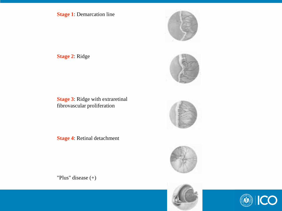
Stage 1: Demarcation line
Stage 2: Ridge
Stage 3: Ridge with extraretinal fibrovascular proliferation
Stage 4: Retinal detachment
"Plus" disease (+)

Leukokoria Differentials
Cataract
Retinoblastoma
Retinopathy of Prematurity
Posterior Segment Pathology
Optic Nerve Hypoplasia

Optic Nerve Hypoplasia
Under development of Optic Nerve during pregnancy
Causes: Unknown
Association with maternal DM, alcohol abuse, young maternal age, etc
May be isolated as Optic Nerve issue or associated with neurological and hormonal abnormalities
Wide spectrum of visual function and affect on VF
Optic Nerve Hypoplasia Associated Conditions
Midline anomalies of brain Septo optic dysplasia (absence of septum pellucidum and corpus callosum)
Anomalies of ventricles
Cerebral atrophy
Tumors (rare)
Hormonal insufficiencies Thyroid
Growth hormone
Pituitary
Adrenal
anti-diuretic hormone (ADH)

Brother – bilateral optic nerve hypoplasia OD -3.00 -1.00 x180 20/60 OS -1.00 -1.50 x 180 20/25

Sister - unilateral optic nerve hypoplasia pl -0.50 x 180 20/ 20 -5.25 sph 20/40

Common Presentations of Pediatric Pathology Ocular Trauma

Ocular Trauma
Most common cause of acquired blindness in children Boys 4x > vs girls Typically unilateral Cosmetic significance can be large Employment prospects are often reduced Birth Trauma 25% all births 50% difficult births Most common Chemical conjunctivitis (silver nitrate gtts) Conjunctival hemes
Ocular Trauma Types of Trauma
– Eyelid trauma
– Subconjunctival hemorrhages May mask underlying penetrating or perforating injury
– Corneal abrasions
Typically patch children/cycloplegics
– Eyewall Lacerations Monitor IOP May need ultrasound if anterior segment heme or cataract is present
– Non-accidental trauma

Ocular Trauma
Non-accidental trauma
– Referral/collaboration with pediatrician
– Full history/exam with photograph
– Involvement of social services
– Rule out other injury (X-ray, CT, MRI, etc)

Child Abuse
40 % of abused children show ocular signs Be suspicious of frequent history of ocular /physical injury
Types – Periorbital ecchymosis – Corneal abrasions – Lacerations – Hyphema – Angle recession – Cataracts – Dislocated lens – Retinal injuries


Ocular Trauma Case
11 year old AA male
– Battery assault / hit by fists – Patient lying flat with collar / backbrace – (+) Pain , photophobia and blurred vision
– VA
OD 20/100 OS 20/40
– EOM FROM OD, OS
– Tonometry
OD 36 OS 17
– Pupils: OD minimally reactive, (-) APD
Ocular Trauma Case
Anterior Segment Evaluation – (+) hematoma – Blood in AC / Hyphema
Posterior Segment Evaluation
– C/D 0.2 rd OD, OS
Treatment – Homatropine 1% bid OD – Timolol bid OD – Bed rest, elevated head to 30° - only restroom privileges – RTC 1 day

Ocular Trauma Case
11 year old AA male, One day follow-up
– (+) Pain , photophobia
– VA OD 20/40 OS 20/25
– (+) hematoma, right orbit, eyelid shut OD
– 3 + AC cells
– Hyphema
– Mild corneal haze
– 1+ injection of conjunctiva
– Tonometry OD 15 OS 11
– Traumatic Hyphema
Pred Forte qid, Homatropine bid, Timolol bid Bed rest, elevate head of bed 30 degrees, only restroom privileges RTC 1 day

Ocular Trauma Case
11 year old AA male, Two day follow-up
– VA
OD 20/60 PH 20/40 OS 20/25
– 3 + AC cells – Hyphema – 1+ injection of conjunctiva
– Tonometry
OD 15 OS 11
– C/D 0.2 OU, unremarkable DFE
– Resolving Traumatic Hyphema
Pred Forte q 2 hours, Homatropine bid, Timolol bid Strict bed rest RTC 1 day

Ocular Trauma Case
11 year old AA male, Three day follow-up (9/21/05)
– VA
OD 20/40 PH 20/40 OS 20/25
– 2 + AC cells – Hyphema resolving – 1+ injection of conjunctiva
– Tonometry
OD 10 OS 10
– Resolving Traumatic Hyphema
Pred Forte qid, Homatropine bid, d/c Timolol RTC 2 days
Ocular Trauma Case
11 year old AA male, Five days later (9/26/05)
– VA 20/20 OD, OS – AC deep and quiet – Hyphema resolved – Lid edema OD
– Tonometry OD 15 OS 14
– Resolving Traumatic Hyphema d/c meds RTC 1 week

Ocular Trauma
Hyphema in Childhood
– Rule out further intraocular damage – Rest!!!! – Watch the IOP – Avoid Aspirin / NSAID – Follow daily 1-2 wks (to ↓ risk of re-bleed) – Long Term must rule out: angle recession (and eventual development of glaucoma) dislocated lens posterior segment damage

Blow-Out Fracture of Orbit
Ethmoidal plate affected
Symptoms – Pain / pain on eye movement – Loss of sensation of cheek – Diplopia – Blurred vision
Signs
– Ecchymosis – Ptosis – Limitation of ocular in upgaze
Radiologic evaluation important

Summary
Ocular Meds in Children Pediatric Examination techniques NLDO Congenital Glaucoma Conjunctivitis Preseptal / Orbital Cellulitis Leukokoria Differentials Ocular Trauma
Use an “ocular emergency” visit for a child to educate the
parent on the importance of early and regular optometric visits
