pelvis and lower limb grant kennedy. objectives to cover this huge topic adequately in just over an...
TRANSCRIPT

PELVIS AND LOWER PELVIS AND LOWER LIMBLIMB
Grant KennedyGrant Kennedy

ObjectivesObjectives
To cover this huge topic adequately To cover this huge topic adequately in just over an hour.in just over an hour.
Special thanks to Tintinalli, UTDOL, Special thanks to Tintinalli, UTDOL, Dr. Buckley, Rob and Shawn’s Dr. Buckley, Rob and Shawn’s REMERGS web page.REMERGS web page.

Pelvic Fractures: Pelvic Fractures: EpidemiologyEpidemiology
Majority due to high impact blunt Majority due to high impact blunt trauma (MVA, pedestrian vs. vehicle trauma (MVA, pedestrian vs. vehicle etc.) but also secondary to falls in etc.) but also secondary to falls in frail elderly frail elderly
Mortality overall = 10%Mortality overall = 10% Mortality 50% if open #Mortality 50% if open #

Pelvic AnatomyPelvic Anatomy
PelvisPelvis = sacrum, coccyx + 2 = sacrum, coccyx + 2 innominate bonesinnominate bones
Innominate bonesInnominate bones = ilium, ischium, = ilium, ischium, pubispubis
Sacrum + innominate bones form a Sacrum + innominate bones form a ringring
Strength from ligamentous supports Strength from ligamentous supports (largely posterior aspect of ring)(largely posterior aspect of ring)

Pelvic AnatomyPelvic Anatomy
5 joints:5 joints: LumbosacralLumbosacral Sacroiliac (x2)Sacroiliac (x2) Sacrococcygeal Sacrococcygeal SymphysisSymphysis

Pelvic AnatomyPelvic Anatomy

Anterior Support:Anterior Support:– Symphysis pubisSymphysis pubis
Fibrocartilaginous Fibrocartilaginous joint covered by ant & joint covered by ant & post symphyseal post symphyseal ligamentsligaments
– Pubic ramiPubic rami Posterior Support:Posterior Support:
– ~majority of stability~majority of stability Iliolumbar ligamentsIliolumbar ligaments Sacroiliac ligamentsSacroiliac ligaments Sacrospinous Sacrospinous
ligamentligament Sacrotuberous Sacrotuberous
ligamentligament

Vascular AnatomyVascular Anatomy Vessels lie close Vessels lie close
to posterior to posterior pelvic wallspelvic walls
Venous bleeding Venous bleeding most common most common (sacral plexus)(sacral plexus)
Most commonly Most commonly injured arteries injured arteries are superior are superior gluteal and gluteal and internal internal pudendalpudendal
Pelvic AnatomyPelvic Anatomy
Nerve supply through the pelvis Nerve supply through the pelvis derived from lumbar and sacral derived from lumbar and sacral plexusesplexuses
Other structures: lower GI/GUOther structures: lower GI/GU

History & PhysicalHistory & Physical AMPLE HxAMPLE Hx Mechanism/Ambulating at SceneMechanism/Ambulating at Scene Numbness/Weakness/Bowel + Bladder DysfxnNumbness/Weakness/Bowel + Bladder Dysfxn
Inspect:Inspect: Destot’s sign: Hematoma above inguinal Destot’s sign: Hematoma above inguinal
ligament or over scrotumligament or over scrotum Blood at urethral meatus (urologic injury?)—if so, Blood at urethral meatus (urologic injury?)—if so,
ED cystourethrogram. Insert foley a small ED cystourethrogram. Insert foley a small amount (and lightly put up the balloon). Inject amount (and lightly put up the balloon). Inject 100-150 cc of dye into bladder and have x-ray 100-150 cc of dye into bladder and have x-ray taken at same time. taken at same time.
Flank ecchymosesFlank ecchymoses

History + PhysicalHistory + Physical
Examine pelvis only once!Examine pelvis only once! AP compression on ASISAP compression on ASIS AP compression on symphysisAP compression on symphysis Lateral compression on iliac crestsLateral compression on iliac crests
Distal neurovascular exam!Distal neurovascular exam!
BimanualBimanual should be performed on all women w/ pelvic #should be performed on all women w/ pelvic # If blood, do speculum to assess for vaginal laceration (open If blood, do speculum to assess for vaginal laceration (open
#)#)
DREDRE in everyone (High riding prostate? Lack of tone?) in everyone (High riding prostate? Lack of tone?) Earle’s sign:Earle’s sign:
– Presence of bony prominence, palpable hematoma, or tender # Presence of bony prominence, palpable hematoma, or tender # line on DREline on DRE

ImagingImaging Plain films are NOT necessary in stable Plain films are NOT necessary in stable
trauma patient with no lower abdo-pelvic trauma patient with no lower abdo-pelvic complaints, normal exam and GCS >13complaints, normal exam and GCS >13
X Rays:X Rays: AP AP Inlet/OutletInlet/Outlet JudetJudet CT Scan:CT Scan: Evaluates extent of posterior injuries and Evaluates extent of posterior injuries and
retroperitoneal bleeding, superior imaging retroperitoneal bleeding, superior imaging of sacrum and acetabulum, associated of sacrum and acetabulum, associated injuriesinjuries

ImagingImaging
AP VIEW:AP VIEW: Identifies most fracturesIdentifies most fractures Look for disruption in iliopubic and Look for disruption in iliopubic and
ilioischial lines, sacral foramina, ilioischial lines, sacral foramina, radiographic U, Shenton’s Linesradiographic U, Shenton’s Lines
Following are abnormal:Following are abnormal: Symphysis >5mm Symphysis >5mm Vertical offset left vs. right rami (>1-2mm) Vertical offset left vs. right rami (>1-2mm) SI joint > 5mmSI joint > 5mm


Inlet viewInlet view– X-ray beam at 60X-ray beam at 60oo to to
plate directed towards plate directed towards feetfeet
– Used to look for AP Used to look for AP displacement of ring displacement of ring fractures.fractures.
Outlet viewOutlet view– Beam aimed 30Beam aimed 30oo
towards headtowards head– Used to see Sup-Inf Used to see Sup-Inf
displacement.displacement.

ImagingImaging
Look for any evidence of damage to Look for any evidence of damage to the posterior pelvic structuresthe posterior pelvic structures– Clues on X-rays:Clues on X-rays:
L5 transverse process avulsion (iliolumbar L5 transverse process avulsion (iliolumbar ligament)ligament)
Ischial spine avulsion (sacrospinous ligament)Ischial spine avulsion (sacrospinous ligament) Unable to clearly make out sacral foraminaUnable to clearly make out sacral foramina Assymmetry of sacral foraminaAssymmetry of sacral foramina Avulsion at lower lip of lateral sacrum Avulsion at lower lip of lateral sacrum
(sacrotuberous ligament)(sacrotuberous ligament)

Pelvic Fracture Pelvic Fracture ComplicationsComplications
Hemorrhage: Hemorrhage: up to 6L of blood can up to 6L of blood can collect in retroperitoneal space!collect in retroperitoneal space!
Open #: Open #: high mortality if not high mortality if not recognized; communication to rectum, recognized; communication to rectum, vagina, skinvagina, skin
examine posterior skin carefully, do not examine posterior skin carefully, do not probe wounds, probe wounds,
perineal wounds = operative perineal wounds = operative debridement/irrigation, debridement/irrigation,
rectum = diverting colostomyrectum = diverting colostomy

Pelvic Fracture Pelvic Fracture ComplicationsComplications
Urologic Injury:Urologic Injury: (15%) # of symphysis (15%) # of symphysis have highest incidence of urologic have highest incidence of urologic injury, injury,
Microhematuria = no need for Microhematuria = no need for cystourethrogramcystourethrogram
Gross hematuria = cystourethrogram + Gross hematuria = cystourethrogram + CTCT
Neurologic Injury: Neurologic Injury: with sacral #, sx of with sacral #, sx of cauda equina, plexopathy, radiculopathycauda equina, plexopathy, radiculopathy

Pelvic Fracture Pelvic Fracture ComplicationsComplications
Gynecologic Injury:Gynecologic Injury: laceration, laceration, abruption, uterine perforationabruption, uterine perforation
Intra-abdominal Injury: Intra-abdominal Injury: rectum, rectum, colon, small bowelcolon, small bowel
Injuries by Association: Injuries by Association: due to due to high force mechanism… thoracic high force mechanism… thoracic aortic rupture, diaphragmatic ruptureaortic rupture, diaphragmatic rupture

Pelvic FracturesPelvic Fractures
5 General Categories:5 General Categories: 1. Pelvic Ring1. Pelvic Ring 2. Acetabular2. Acetabular 3. Sacral3. Sacral 4. Avulsion type4. Avulsion type 5. Single bone5. Single bone

Pelvic Ring FracturesPelvic Ring Fractures
Young Classification System:Young Classification System: Differentiates fracture patterns based Differentiates fracture patterns based
on mechanism of injury/direction of on mechanism of injury/direction of causative forcecausative force
3 major fracture patterns:3 major fracture patterns: 1. lateral compression (50%)1. lateral compression (50%) 2. antero-posterior compression (25%)2. antero-posterior compression (25%) 3. vertical shear (5%)3. vertical shear (5%)

Young Classification:Young Classification: Lateral Lateral
CompressionCompression– (50%) – transverse # (50%) – transverse #
of pubic rami, of pubic rami, ipsilateral or ipsilateral or contralateral to contralateral to posterior injuryposterior injury LC I – sacral LC I – sacral
compression on side of compression on side of impactimpact
LC II – iliac wing # on LC II – iliac wing # on side of impactside of impact
LC III – LC-I or LC-II on LC III – LC-I or LC-II on side of impact w/ side of impact w/ contralateral APC injurycontralateral APC injury

AP CompressionAP Compression (25%)(25%)– Symphyseal and / or Symphyseal and / or
Longitudinal Rami Longitudinal Rami FracturesFractures APC I –diastasis of the APC I –diastasis of the
pubic symphysis pubic symphysis and/or anterior SI jointand/or anterior SI joint
APC II – disrupted APC II – disrupted anterior SI joint, anterior SI joint, sacrotuberous, and sacrotuberous, and sacrospinous ligaments sacrospinous ligaments (intact post SI ligs)(intact post SI ligs)
APC III – complete SI APC III – complete SI joint disruption w/ joint disruption w/ lateral displacement lateral displacement and disruption of and disruption of sacrotuberous and sacrotuberous and sacrospinous ligamentssacrospinous ligaments

Tile B1 / Young APC II

Young Classification Young Classification System:System:
Vertical ShearVertical Shear (5%) (5%) – Symphyseal diastasis Symphyseal diastasis
or vertical or vertical displacement displacement anteriorly and anteriorly and posteriorly; usually posteriorly; usually through SI joint, through SI joint, occasionally through occasionally through iliac wingiliac wing

Tile C1/ Young VS

Pelvic Fracture Pelvic Fracture ManagementManagement
Stable vs. UnstableStable vs. Unstable
Young Classification:Young Classification: LC I, APC I = several days bedrest +/- LC I, APC I = several days bedrest +/-
external fixator, followed by external fixator, followed by progressive weight bearing as progressive weight bearing as toleratedtolerated
LC II and III, APC II and III, VS = surgeryLC II and III, APC II and III, VS = surgery

Pelvic Fracture Pelvic Fracture ManagementManagement
Buckley:Buckley: Full weight bearing for lateral Full weight bearing for lateral
compression #s that lack significant compression #s that lack significant deformity, isolated pubic rami fracturesdeformity, isolated pubic rami fractures
Indications for surgery:Indications for surgery: ongoing ongoing hemorrhage, displaced posterior pelvic hemorrhage, displaced posterior pelvic injury, symphysis diastasis >2.5 cminjury, symphysis diastasis >2.5 cm

Pelvic Fracture Pelvic Fracture Management of the Management of the
Unstable PatientUnstable Patient ABC’s & initial stabilizationABC’s & initial stabilization (IV access, (IV access,
crystalloid, blood products)crystalloid, blood products) Application of Application of Pelvic Sheet/Binder/External Pelvic Sheet/Binder/External
fixatorfixator (open-book with intact posterior (open-book with intact posterior ligaments has most potential for benefit)ligaments has most potential for benefit)
Adjuncts:Adjuncts: Foley (but not if blood at meatus) Foley (but not if blood at meatus) FASTFAST to assess for intraperitoneal injury (and to assess for intraperitoneal injury (and
help with disposition—laparotomy vs. angio)help with disposition—laparotomy vs. angio) AP pelvisAP pelvis ABX (ancef) and Tetanus if open.ABX (ancef) and Tetanus if open.

Pelvic Fracture Pelvic Fracture Management of the Management of the
Unstable PatientUnstable Patient
FAST +, Unstable =FAST +, Unstable = Laparotomy first Laparotomy first
FAST -, Unstable =FAST -, Unstable = Angio Angio
STABLE but with significant # =STABLE but with significant # = CT. If ‘brash’ on CT = ongoing bleed, CT. If ‘brash’ on CT = ongoing bleed, needs angioneeds angio

PELVIC BINDERPELVIC BINDER
Benefits:Benefits: Reduces pelvic volume (tamponade Reduces pelvic volume (tamponade
effect)effect) Stabilizes # fragmentsStabilizes # fragments Improves patient comfortImproves patient comfort

PELVIC BINDERPELVIC BINDER
Application:Application: Apply at level of greater trochantersApply at level of greater trochanters Avoid over-reduction (esp lateral Avoid over-reduction (esp lateral
compression #) as can increase compression #) as can increase internal rotation deformity, increase internal rotation deformity, increase bleedingbleeding
Aim for anatomical reduction (legs, Aim for anatomical reduction (legs, trochanters, patellae should be trochanters, patellae should be neutral)neutral)


AcetabulumAcetabulum
Forms the ‘socket’ for the femoral headForms the ‘socket’ for the femoral head
Fusion of 3 bones:Fusion of 3 bones: 1. iliac1. iliac (superior dome—chief weight- (superior dome—chief weight-
bearing surface)bearing surface) 2. pubis2. pubis (anterior-inferior—thin, easily (anterior-inferior—thin, easily
fractured)fractured) 3. ischium3. ischium (posterior-inferior-thick) (posterior-inferior-thick)

AcetabulumAcetabulum

AcetabulumAcetabulum
Also classically described as having 2 Also classically described as having 2 columns:columns:
1. Anterior column1. Anterior column (anterior iliac (anterior iliac wing, superior pubic ramus, anterior wing, superior pubic ramus, anterior wall of acetabulum)wall of acetabulum)
2. Posterior column2. Posterior column (ischium, ischial (ischium, ischial tuberosity, posterior wall of tuberosity, posterior wall of acetabulum)acetabulum)

Acetabular FracturesAcetabular Fractures Nearly all associated with hip dislocationsNearly all associated with hip dislocations Sciatic nerve injury commonSciatic nerve injury common MVA most common mechanismMVA most common mechanism
Imaging:Imaging: Judet views (AP, 45 degree iliac oblique, 45 Judet views (AP, 45 degree iliac oblique, 45
degree obturator oblique)degree obturator oblique) CT scan (x-ray negative but suspicious; clarifying CT scan (x-ray negative but suspicious; clarifying
operative or non-operative)operative or non-operative)
Judet-Letournel Classification System:Judet-Letournel Classification System: Simple (5 types) vs. Complex (combos)Simple (5 types) vs. Complex (combos)
Acetabular FracturesAcetabular Fractures
Judet ClassificationJudet Classification Simple Fractures:Simple Fractures: 1. Posterior Wall 1. Posterior Wall 2. Posterior Column 2. Posterior Column 3. Anterior Wall 3. Anterior Wall 4. Anterior Column4. Anterior Column 5. Transverse 5. Transverse


Acetabular Fracture Acetabular Fracture ManagementManagement
ABCsABCs Neurovascular examNeurovascular exam Reduction of hip dislocation Reduction of hip dislocation Ortho consultOrtho consult AdmissionAdmission
Buckley:Buckley: Non-Displaced = Non-Displaced = nonnon weight bearing x 6-8 weight bearing x 6-8
weeksweeks Displaced >2mm intra-articular = surgeryDisplaced >2mm intra-articular = surgery

Sacral FracturesSacral Fractures
Mechanism:Mechanism:– Direct trauma or forced flexionDirect trauma or forced flexion
Key distinction is Vertical (high Key distinction is Vertical (high energy/unstable) vs. Transverseenergy/unstable) vs. Transverse
O/E: O/E: pain on DREpain on DRE
Dx:Dx:– AP pelvis, CTAP pelvis, CT

Vertical Sacral FracturesVertical Sacral Fractures
Denis Classification:Denis Classification: Zone 1Zone 1—lateral to sacral neural —lateral to sacral neural
foramina (6% L5 root injury)foramina (6% L5 root injury) Zone 2Zone 2—through sacral neural —through sacral neural
foramina (28% sciatic injury)foramina (28% sciatic injury) Zone 3Zone 3—medial to sacral neural —medial to sacral neural
foramina (50% bowel/bladder, sexual foramina (50% bowel/bladder, sexual dysfunction)dysfunction)


Transverse Sacral Transverse Sacral FracturesFractures
Potential for neurologic injury depends Potential for neurologic injury depends on level of # lineon level of # line
Nerve root injury uncommon below S4Nerve root injury uncommon below S4
High incidence of neuro deficit if # line High incidence of neuro deficit if # line above S2above S2

Sacral FracturesSacral Fractures
Treatment of High-Energy Treatment of High-Energy Vertical: Vertical: ABCs etc. Surgical ABCs etc. Surgical stabilizationstabilization
Treatment of Transverse:Treatment of Transverse:
– Neuro deficitsNeuro deficits urgent spine consult urgent spine consult
– No neuro deficitsNo neuro deficits ice, bed rest, ice, bed rest, analgesia & ortho f/u in 1 weekanalgesia & ortho f/u in 1 week

Coccyx FracturesCoccyx Fractures Mechanism:Mechanism:
– Fall in seated positionFall in seated position Presentation:Presentation:
– Pain w/ sitting, standing, or defecatingPain w/ sitting, standing, or defecating– Local tendernessLocal tenderness
Dx:Dx:– Clinical. X-rays not needed! (pain on Clinical. X-rays not needed! (pain on
compression during DRE)compression during DRE) Tx:Tx:
--rest, ice, donut-ring cushion, stool softeners--rest, ice, donut-ring cushion, stool softeners– Coccygectomy if persistent chronic painCoccygectomy if persistent chronic pain

CASECASE
13 yo boy presents with pain in his 13 yo boy presents with pain in his hip after kicking a soccer ball…hip after kicking a soccer ball…


Avulsion FracturesAvulsion FracturesMechanism:Mechanism:
– Forced contraction of muscle avulsing bony Forced contraction of muscle avulsing bony fragment (soccer & gymnastics)fragment (soccer & gymnastics)
Most common types:Most common types:– Ischial tuberosity Ischial tuberosity hamstring hamstring– ASIS avulsion ASIS avulsion sartorius sartorius– AIIS AIIS rectus femoris rectus femoris
Tx:Tx:– RICE, crutches (for comfort), f/u w/ family MDRICE, crutches (for comfort), f/u w/ family MD– IT= hip ext; ASIS + AIIS = hip flexIT= hip ext; ASIS + AIIS = hip flex– >2 cm displacement = surgery >2 cm displacement = surgery

CASECASE
53 year old German female presents 53 year old German female presents with pain in her groin after having with pain in her groin after having fallen skiing.fallen skiing.
Mechanism: landed and fell back Mechanism: landed and fell back onto buttocks/’tail bone’. onto buttocks/’tail bone’.


Isolated Ramus FractureIsolated Ramus Fracture
Mechanism:Mechanism: Fall in elderly; stress # in young Fall in elderly; stress # in young
athleteathlete Presentation:Presentation: Inability to ambulate, local painInability to ambulate, local pain TX:TX: Ice, rest, analgesics, crutches with Ice, rest, analgesics, crutches with
progressive weight bearing.progressive weight bearing.

Sup and Inf Rami # Sup and Inf Rami # (unilateral)(unilateral)
Generally StableGenerally Stable Conservative managementConservative management Look for complicating associated Look for complicating associated
injuries: posterior pelvic impaction, SI injuries: posterior pelvic impaction, SI joint injury, acetabular # (may need joint injury, acetabular # (may need CT to identify these)CT to identify these)

Sup and Inf Rami # Sup and Inf Rami # (bilateral)(bilateral)
Straddle #Straddle # GU injuries common!GU injuries common! CT pelvis needed to plan surgical CT pelvis needed to plan surgical
mgmtmgmt Consult ORTHOConsult ORTHO Tx:Tx: SURGERYSURGERY

What is the nameWhat is the name
of this type of #??of this type of #??

Duverney (Iliac Wing) Duverney (Iliac Wing) FractureFracture
Mechanism:Mechanism:– Direct traumaDirect trauma
Presentation:Presentation:– Localized pain, swelling, tendernessLocalized pain, swelling, tenderness– abdominal tendernessabdominal tenderness– Associated acetabular #Associated acetabular #
Dx:Dx:– AP pelvisAP pelvis
Tx:Tx:– Minimally displaced Minimally displaced ortho f/u in 1 week, rest, ice, ortho f/u in 1 week, rest, ice,
strappingstrapping– Severely displaced Severely displaced ORIF ORIF– Concerning abdo examConcerning abdo exam CT abdo/pelvis CT abdo/pelvis


Hip DislocationsHip Dislocations
3 Types:3 Types:Posterior (80%)>>Anterior>>CentralPosterior (80%)>>Anterior>>Central
Associated injuries:Associated injuries:#-dislocation with femoral head or acetabulum#-dislocation with femoral head or acetabulumSciatic nerve (posterior); Femoral nerve/vessels Sciatic nerve (posterior); Femoral nerve/vessels
(anterior)(anterior)
Mechanism:Mechanism: Adults: MVA (high energy), polytrauma (assoc knee Adults: MVA (high energy), polytrauma (assoc knee
injuries) injuries) Elderly/Prosthetics/Kids: low energyElderly/Prosthetics/Kids: low energy

Hip Dislocations--Hip Dislocations--PresentationsPresentations
Anterior Dislocation:Anterior Dislocation:
extremity in abduction/external extremity in abduction/external rotation (similar to fem neck #)rotation (similar to fem neck #)
Posterior Dislocation:Posterior Dislocation:
extremity shortened, internally extremity shortened, internally rotated, adductedrotated, adducted
DX:DX: AP/Lateral Pelvis. AP/Lateral Pelvis.
Hip DislocationsHip Dislocations
Treatment:Treatment: Orthopedic Emergency!Orthopedic Emergency! ABCs/initial stabilizationABCs/initial stabilization R/O associated life threatening injuriesR/O associated life threatening injuries Risk of AVN increases in direct Risk of AVN increases in direct
proportion to delay in adequate proportion to delay in adequate reductionreduction
Simple (ie. no #) Ant/Post dislocations Simple (ie. no #) Ant/Post dislocations should be reduced urgently in ED using should be reduced urgently in ED using Allis, Stimson or Whistler maneuversAllis, Stimson or Whistler maneuvers

Post Reduction: Allis MethodPost Reduction: Allis Method

Post Reduction: Stimson Post Reduction: Stimson MethodMethod

Hip DislocationsHip Dislocations Call Ortho for irreducible dislocations Call Ortho for irreducible dislocations
(incarcerated tendon, intra-articular (incarcerated tendon, intra-articular osteochondral fragment)osteochondral fragment)
Post Reduction…Post Reduction… Obtain post reduction films (including CT if Obtain post reduction films (including CT if
associated acetabular # or other pelvic associated acetabular # or other pelvic injury)injury)
Check ROM to ensure stability of the hip, Check ROM to ensure stability of the hip, neurovascular statusneurovascular status
Simple dislocation w/out # = zimmer x Simple dislocation w/out # = zimmer x 1wk, crutches w/ weight bearing as 1wk, crutches w/ weight bearing as tolerated and ortho f/u tolerated and ortho f/u

Hip Dislocations-Special Hip Dislocations-Special CircumstancesCircumstances
Associated Femoral Head #:Associated Femoral Head #: More common w/ anteriorMore common w/ anterior Can still attempt closed reductionCan still attempt closed reduction Consult orthoConsult ortho
Hip Prosthesis:Hip Prosthesis: Consult orthoConsult ortho No time urgency as AVN not an issueNo time urgency as AVN not an issue

Injuries to the FemurInjuries to the Femur
Anatomy:Anatomy: Fem Head + Acetabulum = Ball and socket Fem Head + Acetabulum = Ball and socket
jointjoint Fibrous capsule extends from acetabulum to Fibrous capsule extends from acetabulum to
intertrochanteric lineintertrochanteric line Blood supply to femoral head from med and lat Blood supply to femoral head from med and lat
femoral circumflex arteries, branch of obturatorfemoral circumflex arteries, branch of obturator Vessels course beneath reflection of capsule Vessels course beneath reflection of capsule
and along ligamentum teres (less important)and along ligamentum teres (less important) Easily disrupted with # leading to AVNEasily disrupted with # leading to AVN


Injuries to the FemurInjuries to the Femur
History:History: AMPLEAMPLE Hx of Osteoporosis?Hx of Osteoporosis? Hx of Steroids? (RF for AVN)Hx of Steroids? (RF for AVN) Hx of Cancer, Radiation, Chemo? Hx of Cancer, Radiation, Chemo?
(pathologic #)(pathologic #) Medical causes for falls? (syncope Medical causes for falls? (syncope
etc.)etc.)

Injuries to the FemurInjuries to the Femur O/E:O/E: Inspect pelvis/hip/kneeInspect pelvis/hip/knee Neurovascular status (fem nerve/artery in Neurovascular status (fem nerve/artery in
subtrochanteric or shaft #; sciatic nerve in subtrochanteric or shaft #; sciatic nerve in hip # or dislocations)hip # or dislocations)
Assess for open #Assess for open #
Imaging:Imaging: APAP LateralLateral

Injuries to the FemurInjuries to the Femur
General Management:General Management: ABCs and initial stabilizationABCs and initial stabilization Type and Crossmatch (can lose 3L of Type and Crossmatch (can lose 3L of
blood w/ shaft #)blood w/ shaft #) Pre-hospital Hare or Sager traction Pre-hospital Hare or Sager traction
splints for shaft or subtrochanteric #splints for shaft or subtrochanteric # Contraindications to traction: open Contraindications to traction: open
#, nerve injury, femoral neck (may #, nerve injury, femoral neck (may further compromise blood flow)further compromise blood flow)
Injuries to the FemurInjuries to the Femur
Open Fractures:Open Fractures: Type I = < 1cm (Ancef)Type I = < 1cm (Ancef) Type II = > 1 – 10 cm (Ancef + Gent)Type II = > 1 – 10 cm (Ancef + Gent) Type III = > 10 cm (Ancef + Gent)Type III = > 10 cm (Ancef + Gent)
Irrigate and cover w/ saline guazeIrrigate and cover w/ saline guaze TetanusTetanus Splint + ConsultSplint + Consult

Injuries to the FemurInjuries to the Femur
Classification of Hip Fractures:Classification of Hip Fractures: 1. Intracapsular:1. Intracapsular: Femoral headFemoral head Femoral neckFemoral neck 2. Extracapsular:2. Extracapsular: Greater or Lesser TrochanterGreater or Lesser Trochanter IntertrochantericIntertrochanteric SubtrochantericSubtrochanteric

Injuries to the FemurInjuries to the Femur

Femoral Head FracturesFemoral Head Fractures
Infrequently in isolationInfrequently in isolation Usually in conjunction w/ dislocationUsually in conjunction w/ dislocation Types: capital, depression, shearTypes: capital, depression, shear Consult OrthoConsult Ortho Treatment:Treatment: If associated dislocation—attempt If associated dislocation—attempt
reduction in EDreduction in ED ORIF if failure to reduceORIF if failure to reduce

Femoral Head FracturesFemoral Head Fractures
Treatment: (Buckley)Treatment: (Buckley) Non-displaced, stable # = limited Non-displaced, stable # = limited
weight bearing with crutches for 6 weight bearing with crutches for 6 weeksweeks
Displaced (>2mm) head fragment, or Displaced (>2mm) head fragment, or associated femoral neck or associated femoral neck or acetabular # = ORIFacetabular # = ORIF


Femoral Neck FracturesFemoral Neck Fractures
Garden ClassificationGarden Classification
Types:Types: Subcapital vs. TranscervicalSubcapital vs. Transcervical
All are intracapsular (precarious All are intracapsular (precarious blood supply)blood supply)


Femoral Neck FracturesFemoral Neck Fractures
Mechanism: Mechanism: minor trauma in elderly minor trauma in elderly (osteoporosis); high energy in young(osteoporosis); high energy in young
Presentation:Presentation: ranges from limp and ranges from limp and mild groin pain (non-displaced #) to mild groin pain (non-displaced #) to unable to weight bear w/ externally unable to weight bear w/ externally rotated, abducted and shortened limbrotated, abducted and shortened limb

Femoral Neck FracturesFemoral Neck Fractures Dx:Dx: AP/Lateral—look for disruption of Shenton’s AP/Lateral—look for disruption of Shenton’s
Line, Trabecular network, Normal and Reverse SLine, Trabecular network, Normal and Reverse S
Significant hip pain w/ weight bearing and normal Significant hip pain w/ weight bearing and normal radiographs = possible occult fem neck #, may radiographs = possible occult fem neck #, may need CT or MR to diagnoseneed CT or MR to diagnose
Treatment: Analgesia in ED, ORIFTreatment: Analgesia in ED, ORIF
Complications: Complications: AVN, non-union, osteomyelitis, AVN, non-union, osteomyelitis, emboliemboli

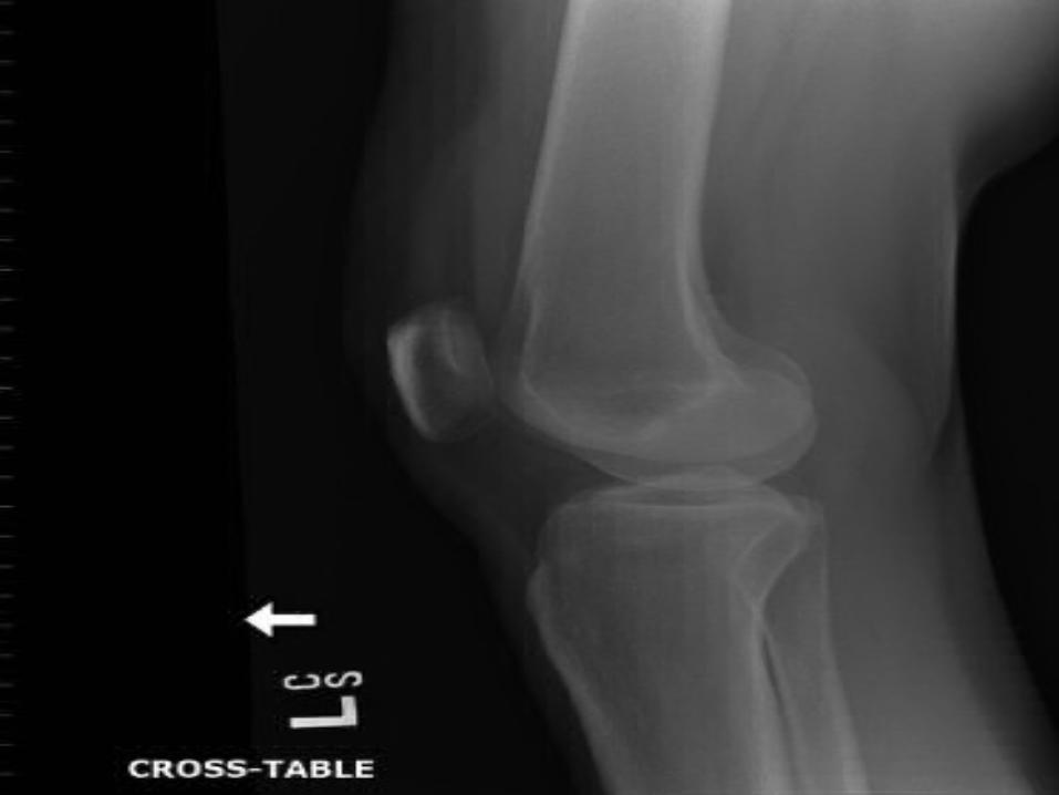
What typeWhat type of # isof # is this?this?
Donk SignDonk Sign
Trochanteric FracturesTrochanteric Fractures
Greater Trochanter:Greater Trochanter: Direct trauma vs. avulsion of gluteus Direct trauma vs. avulsion of gluteus
mediusmedius Pain with abduction/extensionPain with abduction/extension Tender to palp over greater trochTender to palp over greater troch TX: TX: Conservative, gradual weight-bearing until Conservative, gradual weight-bearing until
asymptomaticasymptomatic >1cm displaced: ortho consult for fixation>1cm displaced: ortho consult for fixation

Trochanteric FracturesTrochanteric Fractures
Lesser Trochanter:Lesser Trochanter: Avulsion of iliopsoasAvulsion of iliopsoas Pain w/ flexion/internal rotationPain w/ flexion/internal rotation
TX:TX: Conservative, gradual weight bearingConservative, gradual weight bearing >2cm displaced: ortho for screw >2cm displaced: ortho for screw
fixationfixation

WhatWhat type oftype of # is this?# is this?

Intertrochanteric Intertrochanteric FracturesFractures
Extracapsular, thus less risk of AVNExtracapsular, thus less risk of AVN Fall in elderlyFall in elderly High energy force in youngHigh energy force in young TX:TX: ABCs; analgesiaABCs; analgesia Exclude other life threatening injuriesExclude other life threatening injuries ORTHO for Dynamic Hip Screw fixationORTHO for Dynamic Hip Screw fixation Complications: non-union, infection, blood Complications: non-union, infection, blood
lossloss

TypeType
OfOf
#?#?

Subtrochanteric Subtrochanteric FracturesFractures
Occur b/w the lesser trochanter and Occur b/w the lesser trochanter and proximal 5 cm of femoral shaftproximal 5 cm of femoral shaft
Elderly: fall in osteoporotic bone, Elderly: fall in osteoporotic bone, pathological #spathological #s
Young: high energy traumaYoung: high energy trauma Comminution and deformity commonComminution and deformity common TX:TX: ABCs, Ortho for ORIF ABCs, Ortho for ORIF Complications: hemodynamic Complications: hemodynamic
instability, fat embolus, non-unioninstability, fat embolus, non-union


Femoral Shaft FracturesFemoral Shaft Fractures Young w/ high energy trauma (falls, MVAs, Young w/ high energy trauma (falls, MVAs,
gunshot etc.)gunshot etc.) Classification: transverse, oblique, spiral, Classification: transverse, oblique, spiral,
wedge, comminutedwedge, comminuted 50% have assoc. ligamentous damage to knee50% have assoc. ligamentous damage to knee TX: TX: ABCs (significant hemorrhage can occur) ABCs (significant hemorrhage can occur) Look for other life threatening injuriesLook for other life threatening injuries Traction splints in pre-hospital settingTraction splints in pre-hospital setting Ortho for ORIF (IM rod) vs. plating for Ortho for ORIF (IM rod) vs. plating for
comminuted (union rates approach 100%)comminuted (union rates approach 100%)

CaseCase
68y male injured in 68y male injured in MVCMVC
c/o left leg painc/o left leg pain

Case continuedCase continued
Type of #?Type of #?

Distal Femur FracturesDistal Femur Fractures
Supracondylar, Intracondylar (intra-Supracondylar, Intracondylar (intra-articular), Condylar (intra-articular)articular), Condylar (intra-articular)
Isolated, T or Y patternIsolated, T or Y pattern

Distal Femur FracturesDistal Femur Fractures

Distal Femur FracturesDistal Femur Fractures
Tx:Tx: ABCs ABCs Check neurovascular exam. Check neurovascular exam. (# in close proximity to femoral and (# in close proximity to femoral and
popliteal arteries!—may need angio if popliteal arteries!—may need angio if in question)in question)
Splint and consult OrthoSplint and consult Ortho All require ORIF (per Buckley)All require ORIF (per Buckley)

Distal Femur FracturesDistal Femur Fractures
Complications:Complications:– thrombophlebitisthrombophlebitis– fat embolus syndromefat embolus syndrome– delayed union or malunion if reduction is delayed union or malunion if reduction is
incomplete or not maintainedincomplete or not maintained– intraarticular or quadriceps adhesions if intraarticular or quadriceps adhesions if
the fracture is intraarticularthe fracture is intraarticular– angulation deformitiesangulation deformities– osteoarthritisosteoarthritis

Knee InjuriesKnee Injuries
Fractures:Fractures: 1. distal femur (covered already)1. distal femur (covered already) 2. patellar2. patellar 3. proximal tibia3. proximal tibia 4. proximal fibula4. proximal fibulaSoft Tissue Injuries:Soft Tissue Injuries: Dislocations (patellar, tib-fem), Dislocations (patellar, tib-fem),
Ligamentous and Meniscal injuries Ligamentous and Meniscal injuries

AnatomyAnatomy
Main joints:Main joints:– PatellofemoralPatellofemoral– TibiofemoralTibiofemoral
Main bones:Main bones:– Distal FemurDistal Femur– PatellaPatella– Proximal tibiaProximal tibia– (fibula head)(fibula head)

Knee AnatomyKnee Anatomy
Medial Stabilizers of the Knee:Medial Stabilizers of the Knee: MCL, joint capsule, MCL, joint capsule,
semimembranosus, pes anserinussemimembranosus, pes anserinus
Lateral Stabilizers of the Knee:Lateral Stabilizers of the Knee: LCL, joint capsule, IT band, biceps LCL, joint capsule, IT band, biceps
tendon, popliteal arcuate complextendon, popliteal arcuate complex

Knee InjuriesKnee Injuries DDX of Anterior Knee Pain:DDX of Anterior Knee Pain: Plateau/Patellar #Plateau/Patellar # Pre-patellar BursitisPre-patellar Bursitis Quads/Patellar TendonitisQuads/Patellar Tendonitis Patellofemoral Pain SyndromePatellofemoral Pain Syndrome Chondromalacia PatellaeChondromalacia Patellae Osgood SchlattersOsgood Schlatters PlicaPlica Meniscal injuryMeniscal injury Ligamentous injuryLigamentous injury Osteochondritis DessicansOsteochondritis Dessicans Synovial ChondrinosisSynovial Chondrinosis

Knee InjuriesKnee Injuries
DDX of Hemarthrosis:DDX of Hemarthrosis: ACLACL PCLPCL Meniscal tearMeniscal tear Osteochondral #Osteochondral # Capsular tearCapsular tear
BUT NOT MCL nor LCL!BUT NOT MCL nor LCL!

Knee Injuries--HistoryKnee Injuries--History
AMPLEAMPLE Mechanism particularly importantMechanism particularly important Hx of prior knee injuries, surgeriesHx of prior knee injuries, surgeries Inability to weight bearInability to weight bear Locking (meniscus vs. intra-articular Locking (meniscus vs. intra-articular
body)body) Giving Way (ligamentous vs. meniscus)Giving Way (ligamentous vs. meniscus) ““Pop”! (ACL)Pop”! (ACL)

Knee Injuries--Knee Injuries--ExaminationExamination
COMPARE TO HEALTHY KNEECOMPARE TO HEALTHY KNEE Inspection (swelling, bruising, Inspection (swelling, bruising,
deformity)deformity) Palpation: (joint line tenderness? Palpation: (joint line tenderness?
effusion? point tenderness?)effusion? point tenderness?) ROM ROM Ligamentous/Meniscal Stress TestingLigamentous/Meniscal Stress Testing

Ligament/Meniscal Stress Ligament/Meniscal Stress TestingTesting
Anterior Drawer (ACL):Anterior Drawer (ACL): not reliable. not reliable. FN = effusion, hamstring spasm, techniqueFN = effusion, hamstring spasm, technique FP = PCL injuryFP = PCL injury
Lachman’s Test (ACL):Lachman’s Test (ACL): reliable, even in reliable, even in acute.acute.
Posterior Drawer (PCL)Posterior Drawer (PCL)
McMurray’s TestMcMurray’s Test (Meniscal): (Meniscal): int rotation int rotation stresses lateral meniscus, ext rotation stresses stresses lateral meniscus, ext rotation stresses medial meniscusmedial meniscus
Collateral Ligament Stress (MCL, LCL): Collateral Ligament Stress (MCL, LCL):

Knee Injuries--ImagingKnee Injuries--Imaging
Standard XR Views:Standard XR Views: APAP Lateral (fat fluid level = lipohemarthrosis = intra-Lateral (fat fluid level = lipohemarthrosis = intra-
articular #)articular #) Oblique (tibial plateau)Oblique (tibial plateau)Special XR Views:Special XR Views: Tunnel (intercondylar region, tibial spines)Tunnel (intercondylar region, tibial spines) Skyline (patellar)Skyline (patellar)CT:CT: helps fully delineate extent of tib plateau # helps fully delineate extent of tib plateau #MR:MR: meniscal, ligamentous meniscal, ligamentousU/S:U/S: popliteal cysts, popliteal aneurysms popliteal cysts, popliteal aneurysms


CASECASE
28 year old MOBHOB (Huffman, 28 year old MOBHOB (Huffman, 2007)2007)
Beaten about legs by some jerk Beaten about legs by some jerk yielding a bat.yielding a bat.
Tender in several places.Tender in several places.
X-ray shows…X-ray shows…


Fractures of the PatellaFractures of the Patella
Mech:Mech: direct blow vs. avulsion (forceful direct blow vs. avulsion (forceful contraction of quads)contraction of quads)
Classification:Classification: transverse (most transverse (most common), vertical, comminuted, common), vertical, comminuted, avulsion-typeavulsion-type
O/EO/E-focal tenderness, swelling. NEED to -focal tenderness, swelling. NEED to check extensor mechanism via straight-check extensor mechanism via straight-legleg
XR-XR- watch for normal variants (bipartite) watch for normal variants (bipartite)

Fractures of the PatellaFractures of the Patella
TX:TX: extra-articular, non-displaced, in-tact extra-articular, non-displaced, in-tact extensor mechanism = Zimmer splint extensor mechanism = Zimmer splint (vs. long-leg cast) x 4 wks, progressive (vs. long-leg cast) x 4 wks, progressive wt bearing, isometric exercises, passive wt bearing, isometric exercises, passive ROM ROM
displaced >3mm and involving articular displaced >3mm and involving articular surface, inadequate extensor surface, inadequate extensor mechanism, comminuted = ORIF (tension mechanism, comminuted = ORIF (tension band wire w/ suturing of retinaculum)band wire w/ suturing of retinaculum)

CASECASE
65 year old female from Japan 65 year old female from Japan presents post fall skiing.presents post fall skiing.
Had collided with a snowboarder.Had collided with a snowboarder. Knee had twisted (external rotation Knee had twisted (external rotation
of leg)of leg) Felt ‘pop’.Felt ‘pop’.


Fractures of the Tibial Fractures of the Tibial SpinesSpines
Tib spine = intercondylar eminence = Tib spine = intercondylar eminence = consists of medial and lateral tubercleconsists of medial and lateral tubercle
Anteriorly: ACL, ant horns of menisciAnteriorly: ACL, ant horns of menisci Posteriorly: PCL, post horns of menisciPosteriorly: PCL, post horns of menisci Anterior injury 10x more common than Anterior injury 10x more common than
posteriorposterior Results in cruciate ligament instability/tearResults in cruciate ligament instability/tear Mech:Mech: AP force against the proximal tibia AP force against the proximal tibia
while in flexion (MVA, sports), twisting, while in flexion (MVA, sports), twisting, hyperflexion, hyperextensionhyperflexion, hyperextension

Fractures of the Tibial Fractures of the Tibial SpinesSpines
Type I--incomplete Type I--incomplete avulsion, no avulsion, no displacedisplace
Type II--incomplete Type II--incomplete avulsion, displace avulsion, displace of anterior but not of anterior but not postpost
Type III--complete Type III--complete displacement (+/- displacement (+/- rotation)rotation)

Fractures of Tibial Fractures of Tibial SpinesSpines
O/E:O/E: hemarthrosis, inability to extend hemarthrosis, inability to extend fullyfully
Lachman + if anterior spineLachman + if anterior spine XR:XR: AP/Lateral/may need tunnel view AP/Lateral/may need tunnel view TX:TX: incomplete or non-displaced = incomplete or non-displaced =
immobilize in full extension (competitor), immobilize in full extension (competitor), protected weight bearing, ortho f/uprotected weight bearing, ortho f/u
Complete, displaced = ortho consult for Complete, displaced = ortho consult for ORIF vs. arthroscopic to restore normal ORIF vs. arthroscopic to restore normal ACL function ACL function

CASECASE
35 yo woman presents with pain in 35 yo woman presents with pain in her knee, unable to weight bear after her knee, unable to weight bear after having gone off a jump skiing, landed having gone off a jump skiing, landed on flat surface…on flat surface…
XR shows the following…XR shows the following…


Fractures of the Tibial Fractures of the Tibial PlateauPlateau
Mech:Mech: valgus/varus force combined with axial valgus/varus force combined with axial load, driving femoral condyles into articulating load, driving femoral condyles into articulating surface of tibia VS. direct blowsurface of tibia VS. direct blow
Lateral plateau > medial plateauLateral plateau > medial plateau May have assoc. ligamentous injuryMay have assoc. ligamentous injury O/E:O/E: pain, swelling, decrease ROM, assess pain, swelling, decrease ROM, assess
neurovascular (high incidence of popliteal a. neurovascular (high incidence of popliteal a. inj)inj)
XR:XR: often # is difficult to detect, may only often # is difficult to detect, may only show lipohemarthrosis on lateral, CT if neededshow lipohemarthrosis on lateral, CT if needed

Fracture of the Tibial Fracture of the Tibial PlateausPlateaus
Segond fracture:Segond fracture: Bony avulsion off the lateral tib Bony avulsion off the lateral tib
plateau (lateral capsular sign)plateau (lateral capsular sign) Strong association w/ ACL disruptionStrong association w/ ACL disruption

Fractures of the Tibial Fractures of the Tibial PlateauPlateau
TX:TX: Non-displaced, no depression of Non-displaced, no depression of
articular surface = knee immobilizer, articular surface = knee immobilizer, elevation x 24-48 hrs, ortho f/u, non-elevation x 24-48 hrs, ortho f/u, non-weight bearing x 6-8 weeksweight bearing x 6-8 weeks
Displaced >2mm, depressed Displaced >2mm, depressed articular surface = surgeryarticular surface = surgery

Ligamentous Injuries of the Ligamentous Injuries of the KneeKnee
Grading of Ligamentous Sprains:Grading of Ligamentous Sprains: Grade I: Pain but no laxityGrade I: Pain but no laxity Grade II: Laxity w/ firm end pointGrade II: Laxity w/ firm end point Grade III: Laxity w/out firm end pointGrade III: Laxity w/out firm end point
Cruciate ligament injuries often Cruciate ligament injuries often accompany collateral ligament accompany collateral ligament injuries!injuries!

Ligamentous Injuries of the Ligamentous Injuries of the KneeKnee
Medial Collateral Ligament (MCL):Medial Collateral Ligament (MCL): Mech:Mech: valgus force valgus force Dx:Dx: pain or laxity w/ valgus stress pain or laxity w/ valgus stress TX:TX: non-operative, knee immobilizer non-operative, knee immobilizer
(2 wks) then hinge brace (8 wks), (2 wks) then hinge brace (8 wks), weight bearing as tolerated (will likely weight bearing as tolerated (will likely need crutches early on), RICEneed crutches early on), RICE
Ultimately physio/quad strengthening Ultimately physio/quad strengthening

Ligamentous Injuries of the Ligamentous Injuries of the KneeKnee
Lateral Collateral Ligament Lateral Collateral Ligament (LCL):(LCL):
Mech:Mech: hyperextension + varus force hyperextension + varus force DX:DX: pain or laxity w/ varus stress pain or laxity w/ varus stress TX:TX: conservative as per MCL conservative as per MCL

Ligamentous Injuries of the Ligamentous Injuries of the KneeKnee
Anterior Cruciate Ligament (ACL):Anterior Cruciate Ligament (ACL): Mech:Mech: pivoting, rotation w/ valgus stress, pivoting, rotation w/ valgus stress,
hyperextensionhyperextension DX: DX: + Lachman; hemarthrosis in 70%; + Lachman; hemarthrosis in 70%;
‘pop’ in 70%; watch for assoc. injuries ‘pop’ in 70%; watch for assoc. injuries (50% have meniscal tears); Segond # (50% have meniscal tears); Segond #
TX: TX: Initially conservative; ROM limiting Initially conservative; ROM limiting brace; weight bearing as tolerated; long-brace; weight bearing as tolerated; long-term = hamstring strenghtening/brace vs. term = hamstring strenghtening/brace vs. reconstructionreconstruction

Ligamentous Injuries of the Ligamentous Injuries of the KneeKnee
Posterior Cruciate Ligament (PCL):Posterior Cruciate Ligament (PCL): Mech: Mech: dashboard (MVA) w/ direct blow to dashboard (MVA) w/ direct blow to
anterior tibia; hyperflexion; anterior tibia; hyperflexion; hyperextensionhyperextension
DX: DX: + posterior drawer, posterior sag+ posterior drawer, posterior sag TX: TX: non-operative unless persistent non-operative unless persistent
instability post rehab/quads strengthening instability post rehab/quads strengthening or other associated injuries (meniscal tear, or other associated injuries (meniscal tear, combined ligamentous injury etc.) combined ligamentous injury etc.)

Meniscal InjuriesMeniscal Injuries
Medial 2x more common (and Medial 2x more common (and posterior peripheral aspect)posterior peripheral aspect)
Damage associated with early OADamage associated with early OA Avascular except peripheral 1/3Avascular except peripheral 1/3 MECH:MECH: twisting on weight-bearing twisting on weight-bearing
kneeknee Associated with MCL/ACL (Terrible Associated with MCL/ACL (Terrible
Triad!)Triad!)
Meniscal InjuriesMeniscal Injuries
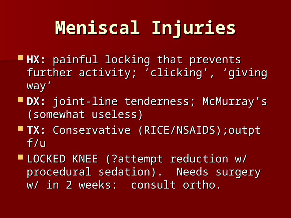
HX:HX: painful locking that prevents painful locking that prevents further activity; ‘clicking’, ‘giving way’further activity; ‘clicking’, ‘giving way’
DX:DX: joint-line tenderness; McMurray’s joint-line tenderness; McMurray’s (somewhat useless)(somewhat useless)
TX:TX: Conservative (RICE/NSAIDS);outpt Conservative (RICE/NSAIDS);outpt f/uf/u
LOCKED KNEE (?attempt reduction w/ LOCKED KNEE (?attempt reduction w/ procedural sedation). Needs surgery w/ procedural sedation). Needs surgery w/ in 2 weeks: consult ortho.in 2 weeks: consult ortho.

CASECASE
40 year old obese male skier…40 year old obese male skier… Fell and had immediate pain in his Fell and had immediate pain in his
knee.knee. Unable to weight bear.Unable to weight bear.


Tibial-Femoral Knee Tibial-Femoral Knee DislocationDislocation
Types:Types: Anterior, Posterior, Medial, Anterior, Posterior, Medial, LateralLateral
MECH:MECH: sporting accidents, falls sporting accidents, falls High incidence of popliteal artery injury, High incidence of popliteal artery injury,
peroneal nerve injury, compartmentperoneal nerve injury, compartment Normal pulses do not r/o vascular injuryNormal pulses do not r/o vascular injury
TX:TX: Immediate reduction (longitudinal Immediate reduction (longitudinal traction), Zimmer splint and Ortho traction), Zimmer splint and Ortho consult for surgical stabilizationconsult for surgical stabilization

Tibial-Femoral Knee Tibial-Femoral Knee DislocationDislocation
Check neurovascular pre- and Check neurovascular pre- and post:post:
Absent pulse (post) = Immediate Absent pulse (post) = Immediate Vascular Surgery Consult + Vascular Surgery Consult + reposition/relocatereposition/relocate
Decreased or absent pulse pre w/ Decreased or absent pulse pre w/ return post = Angioreturn post = Angio
Pulse present pre and post = serial Pulse present pre and post = serial exams vs. ANGIO ALL (per Betzner)exams vs. ANGIO ALL (per Betzner)

Patella DislocationPatella Dislocation
Patella displaced laterally over lateral Patella displaced laterally over lateral condyle (most common)condyle (most common)
Mech:Mech: twisting on extended knee; twisting on extended knee; TX:TX: Reduction in ER (+/- under Reduction in ER (+/- under
sedation)sedation) XR post reduction to r/o #XR post reduction to r/o # Zimmer x 1 wk with crutches. Then Zimmer x 1 wk with crutches. Then
knee sleeve x 3 weeks with knee sleeve x 3 weeks with progressive weight bearing, gentle progressive weight bearing, gentle ROM and isometric quad strengtheningROM and isometric quad strengthening

Soft Tissue InjuriesSoft Tissue Injuries
Patellar TendonitisPatellar Tendonitis—overuse-pain to palp over —overuse-pain to palp over inferior pole--tx conservativeinferior pole--tx conservative
Osteochondritis DissecansOsteochondritis Dissecans—idiopathic--—idiopathic--articular cartilage and subchondral bone articular cartilage and subchondral bone dislodged—tx epiphyses open = protective dislodged—tx epiphyses open = protective weight bearing. epiphyses closed = weight bearing. epiphyses closed = arthroscopyarthroscopy
Quads/Patellar Tendon RuptureQuads/Patellar Tendon Rupture—violent —violent contraction of quads—tx surgical repaircontraction of quads—tx surgical repair
Baker’s CystBaker’s Cyst—aspiration, surgical, vs. —aspiration, surgical, vs. resolutionresolution

Soft Tissue InjuriesSoft Tissue Injuries Chondromalacia PatellaeChondromalacia Patellae—softening of —softening of
articular cartilage secondary to articular cartilage secondary to patellofemoral malalignment/abrnormal patellofemoral malalignment/abrnormal tracking of patella. Tx= tracking of patella. Tx= Rest/NSAIDS/quads+hip strengthening/braceRest/NSAIDS/quads+hip strengthening/brace
PlicaPlica—redundant folds of synovium that —redundant folds of synovium that become inflammed. Leads to pain/stiffness. become inflammed. Leads to pain/stiffness.
Dx: clinical/exclusion Tx: conservativeDx: clinical/exclusion Tx: conservative OsteonecrosisOsteonecrosis—bony infarction. —bony infarction.
Spontaneous vs. secondary causes (steroids, Spontaneous vs. secondary causes (steroids, SLE, EtOH, Sickle etc). Dx-MRI (XR normal). SLE, EtOH, Sickle etc). Dx-MRI (XR normal). Tx-Early=protected weight bearing/NSAIDS. Tx-Early=protected weight bearing/NSAIDS. Advanced=debridement/bone graft/TKAAdvanced=debridement/bone graft/TKA

Leg InjuriesLeg Injuries

Leg Injuries-AnatomyLeg Injuries-Anatomy Bones: Bones: Tibia/Fibula Tibia/Fibula 4 compartments:4 compartments: 1. Anterior1. Anterior--ant tib artery, deep peroneal nerve --ant tib artery, deep peroneal nerve
(dorsiflexion; sensory = web space of 1(dorsiflexion; sensory = web space of 1stst and 2 and 2ndnd toes)toes)
2. Lateral2. Lateral—superficial peroneal nerve (foot —superficial peroneal nerve (foot eversion; sensory = lateral dorsal foot)eversion; sensory = lateral dorsal foot)
3. Superficial Posterior3. Superficial Posterior—ankle plantar flexors —ankle plantar flexors (gastroc, soleus), sural nerve = lateral heel (gastroc, soleus), sural nerve = lateral heel sensationsensation
4. Deep Posterior4. Deep Posterior—post tibial artery; tibial nerve —post tibial artery; tibial nerve = toe plantar flexors, sensation to sole of foot= toe plantar flexors, sensation to sole of foot


Fibular FracturesFibular Fractures
Proximally = attachment for LCL, biceps Proximally = attachment for LCL, biceps femoris tendonfemoris tendon
Common peroneal wraps around fibular Common peroneal wraps around fibular headhead
Usually in setting of # to TibiaUsually in setting of # to Tibia Mech:Mech: direct trauma vs. twisting on direct trauma vs. twisting on
planted foot, inversion or eversion of planted foot, inversion or eversion of ankleankle
Only bears 15% of body weight, thus pts Only bears 15% of body weight, thus pts can often ambulate with isolated #can often ambulate with isolated #

Fibular FracturesFibular Fractures
ED Tx:ED Tx: ABCs; neurovascular; assess ABCs; neurovascular; assess for knee/ankle injuries; stirrup splint for knee/ankle injuries; stirrup splint to prevent varus/valgus stress x 3-4 to prevent varus/valgus stress x 3-4 wks; RICE; crutches if needed for pain; wks; RICE; crutches if needed for pain;
Consult Ortho for:Consult Ortho for: lateral lateral compartment syndrome/peroneal compartment syndrome/peroneal nerve injury; comminuted #, nerve injury; comminuted #, associated tibial #, badly displaced #, associated tibial #, badly displaced #, assoc knee/ankle joint injuriesassoc knee/ankle joint injuries


Tibial Shaft FracturesTibial Shaft Fractures
Major weight bearing bone!Major weight bearing bone!
Open #s common due to superficial Open #s common due to superficial locationlocation
Watch for compartment syndromeWatch for compartment syndrome

Tibial Shaft FracturesTibial Shaft Fractures
ED TX: ED TX: ABCs, neurovascular exam; close ABCs, neurovascular exam; close inspection to r/o open #; analgesics; long-inspection to r/o open #; analgesics; long-leg posterior splint and consult Ortholeg posterior splint and consult Ortho
Definitive Tx:Definitive Tx: ORIF/IM rod VS. ORIF/IM rod VS. Consider long-leg cast (metatarsal heads Consider long-leg cast (metatarsal heads
to upper thigh) and non-weight bearing to upper thigh) and non-weight bearing IFIF displaced <5mm, rotated <10 degrees, displaced <5mm, rotated <10 degrees, angulated <10 degrees and not angulated <10 degrees and not shortenedshortened

Ankle InjuriesAnkle Injuries
Anatomy of an Ankle:Anatomy of an Ankle: 3 Primary Joints:3 Primary Joints:
Medial malleolus w/medial talusMedial malleolus w/medial talus Tibial plafond w/ talar domeTibial plafond w/ talar dome Lat malleolus w/ lat talusLat malleolus w/ lat talus
3 Bones:3 Bones: Tibia, Fibula and TalusTibia, Fibula and Talus
3 sets of Ligaments:3 sets of Ligaments:– Lateral collaterals (ATFL, CFL, PTFL)Lateral collaterals (ATFL, CFL, PTFL)– Syndesmotic LigamentsSyndesmotic Ligaments– Medial collaterals (Deltoid)Medial collaterals (Deltoid)


Ankle InjuriesAnkle Injuries
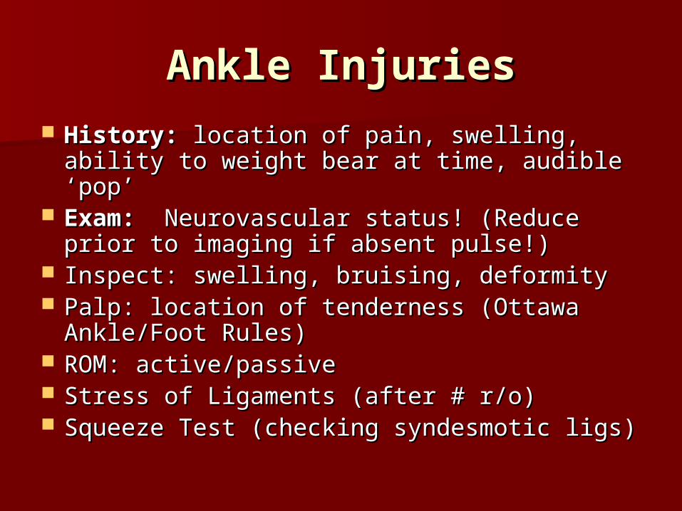
History:History: location of pain, swelling, ability location of pain, swelling, ability to weight bear at time, audible ‘pop’to weight bear at time, audible ‘pop’
Exam:Exam: Neurovascular status! (Reduce Neurovascular status! (Reduce prior to imaging if absent pulse!)prior to imaging if absent pulse!)
Inspect: swelling, bruising, deformityInspect: swelling, bruising, deformity Palp: location of tenderness (Ottawa Palp: location of tenderness (Ottawa
Ankle/Foot Rules)Ankle/Foot Rules) ROM: active/passiveROM: active/passive Stress of Ligaments (after # r/o)Stress of Ligaments (after # r/o) Squeeze Test (checking syndesmotic ligs)Squeeze Test (checking syndesmotic ligs)

Ankle InjuriesAnkle Injuries
OTTAWA ANKLE RULES:OTTAWA ANKLE RULES: X-ray if…X-ray if… Pain in malleolar zone and 1 of…Pain in malleolar zone and 1 of… Inability to weight bear 4 steps both Inability to weight bear 4 steps both
immediately and at time of evaluationimmediately and at time of evaluation Bony tenderness at post edge of Bony tenderness at post edge of
distal 6 cm of either the lateral or distal 6 cm of either the lateral or medial malleolusmedial malleolus

Approach to AnkleApproach to Ankle Go through complete approach (ABC’s)Go through complete approach (ABC’s) 3 views- AP, lat, Mortise (15-203 views- AP, lat, Mortise (15-20° int rot) ° int rot)
ankle, ankle, Direct evidence of injury: assess bonesDirect evidence of injury: assess bones Indirect evidence of injuries: are all ankle Indirect evidence of injuries: are all ankle
measurements normal? Joint effusion?measurements normal? Joint effusion?

Ankle FracturesAnkle Fractures
What are stable fractures?What are stable fractures? Ankle forms a ringAnkle forms a ring
– Disruption of only 1 structure Disruption of only 1 structure
is stableis stable– Disruption of Disruption of > 1 is unstable> 1 is unstable
Assymetry in gap betweenAssymetry in gap between
talar dome and malleolitalar dome and malleoli
on mortis view= unstableon mortis view= unstable

Ankle FracturesAnkle Fractures Management of Stable Fractures:Management of Stable Fractures:
Chip/Avulsion #s <3mm = Tx as Sprain (ie. Chip/Avulsion #s <3mm = Tx as Sprain (ie. WBAT, RICE, NSAIDS, Early ROM/physio)WBAT, RICE, NSAIDS, Early ROM/physio)
Chip/Avulsion#s >3mm = splint and f/u Chip/Avulsion#s >3mm = splint and f/u with Orthowith Ortho
Non-displaced, non-intra-articular, stable Non-displaced, non-intra-articular, stable #s: = 2 wks NWB cast, 3-5 wks WB cast. #s: = 2 wks NWB cast, 3-5 wks WB cast. Ortho f/u in 1 wks to ensure # hasn’t Ortho f/u in 1 wks to ensure # hasn’t slippedslipped

Ankle FracturesAnkle Fractures
Indications for Immediate Indications for Immediate Reduction Prior to X Ray:Reduction Prior to X Ray:
Neurovascular compromiseNeurovascular compromise Gross DeformityGross Deformity Skin TentingSkin Tenting

Ankle FracturesAnkle Fractures Ortho Consultation for the Following:Ortho Consultation for the Following: Open #Open # Pilon #Pilon # Bimalleolar/Trimalleolar #Bimalleolar/Trimalleolar # Lateral Malleolar (Weber B and C)Lateral Malleolar (Weber B and C) Lateral Malleolar Weber A2, A3 (some will Lateral Malleolar Weber A2, A3 (some will
fix/some will cast)fix/some will cast) Isolated Medial Malleolar with significant Isolated Medial Malleolar with significant
displacementdisplacement Isolated Posterior Malleolar with significant Isolated Posterior Malleolar with significant
displacementdisplacement

Diagnosis?Classification?Diagnosis?Classification?Treatment? Treatment?
Does it change you mgmt if they Does it change you mgmt if they have a tender deltoid ligament?have a tender deltoid ligament?

Lateral Malleolar Lateral Malleolar FracturesFractures
Stability depends on location of # to Stability depends on location of # to tib-talar tib-talar
Danis-Weber Classification (A,B,C):Danis-Weber Classification (A,B,C):
A:A: # below tibiotalar joint # below tibiotalar joint A1: no deltoid (medial) tenderness, no post A1: no deltoid (medial) tenderness, no post
malleolar #malleolar # A2: w/ deltoid (medial) tendernessA2: w/ deltoid (medial) tenderness A3: w/ post malleolar #A3: w/ post malleolar #
B:B: # at the level of tibiotalar joint # at the level of tibiotalar joint
C:C: # above the tibiotalar joint # above the tibiotalar joint

Lateral Malleolar Lateral Malleolar FracturesFractures
Treatment:Treatment: Weber A1Weber A1 (stable): NWB x 2 wks (below (stable): NWB x 2 wks (below
knee plaster, fiberglass, or air cast) then knee plaster, fiberglass, or air cast) then WBAT w/ air cast x 3 wks; f/u with Ortho in WBAT w/ air cast x 3 wks; f/u with Ortho in 1 wk1 wk
Weber A2:Weber A2: Consult Ortho (some will fix Consult Ortho (some will fix surgically, some will cast). Do stress view surgically, some will cast). Do stress view to see if mortis opens up.to see if mortis opens up.
Weber A3:Weber A3: Bimalleolar = Ortho for Bimalleolar = Ortho for surgerysurgery

Lateral Malleolar Lateral Malleolar FracturesFractures
Treatment:Treatment: Weber B: Weber B: consult Ortho; 50% have consult Ortho; 50% have
injury to syndesmosis and widening injury to syndesmosis and widening of medial joint spaceof medial joint space
Weber C:Weber C: consult Ortho; frequent consult Ortho; frequent injury to syndesmosisinjury to syndesmosis

Type of Fracture?Type of Fracture?

Medial Malleolar Medial Malleolar FracturesFractures
Commonly associated with lateral or Commonly associated with lateral or posterior malleolar disruption = Orthoposterior malleolar disruption = Ortho
Significant displacement = OrthoSignificant displacement = Ortho R/O R/O Maisonneuve’s #Maisonneuve’s # = Ortho = Ortho
Minimally displaced = NWB (below Minimally displaced = NWB (below knee cast) x 2 wks; WBAT w/ walking knee cast) x 2 wks; WBAT w/ walking boot x 3-5 wks; f/u w/ Ortho at 1 wkboot x 3-5 wks; f/u w/ Ortho at 1 wk

TRIVIA TIMETRIVIA TIME
Name of the rare variation of a Name of the rare variation of a Maisonneuve Fracture in which Maisonneuve Fracture in which the proximal fibula gets trapped the proximal fibula gets trapped behind the tibia?behind the tibia?

TRIVIA TIMETRIVIA TIME
Name of the rare variation of a Name of the rare variation of a Maisonneuve Fracture in which Maisonneuve Fracture in which the proximal fibula gets trapped the proximal fibula gets trapped behind the tibia?behind the tibia?
The Bosworth Fracture!The Bosworth Fracture!

Posterior Malleolar Posterior Malleolar FracturesFractures
Rarely in isolationRarely in isolation
Isolated, non-displaced, <25% of Isolated, non-displaced, <25% of joint surface = cast + NWB x 2 wks; joint surface = cast + NWB x 2 wks; WBAT x 3-5 wks with air cast. Ortho WBAT x 3-5 wks with air cast. Ortho f/u at 1 wkf/u at 1 wk
Otherwise consult OrthoOtherwise consult Ortho

Diagnosis? Stable or unstable?Diagnosis? Stable or unstable?
Bi or Tri-Malleolar #sBi or Tri-Malleolar #s
All unstable because of disruption of All unstable because of disruption of two or more elements of the ankle two or more elements of the ankle ringring
Syndesmosis injury is commonSyndesmosis injury is common
All require Ortho consultationAll require Ortho consultation

Name of this type of fracture? Other Name of this type of fracture? Other associated #s?associated #s?

Pilon FracturesPilon Fractures
Fall from heightFall from height Talus driven into Tibial PlafondTalus driven into Tibial Plafond Distal Tibial Metaphysis #s (+ Fibula)Distal Tibial Metaphysis #s (+ Fibula) 50% are open #s!50% are open #s! Associated #s are common Associated #s are common
(calcaneus, tib-plateau, pelvis, C,T,L (calcaneus, tib-plateau, pelvis, C,T,L spine)spine)
ORTHO!ORTHO!

The Foot (last section!)The Foot (last section!)

HINDFOOT
MIDFOOT
FOREFOOT
calcaneustalus
Medialnavicular
cuneiforms
cuboid
metatarsals
phalanges
sesamoids

Choparts
Lisfrancs
MTP
IP

Type of #Do you need to speak to Ortho??ottawa ankle rules

Talar #sTalar #s
Osteochondral # of Talar DomeOsteochondral # of Talar Dome
X-rays commonly normalX-rays commonly normal Ottawa Ankle Rules may miss theseOttawa Ankle Rules may miss these TX= Cast or Splint and refer to Ortho TX= Cast or Splint and refer to Ortho
as outpatientas outpatient

Describe #?Describe #?
At risk for??At risk for??

Talar #sTalar #s
At risk for AVN due to tenuous blood supplyAt risk for AVN due to tenuous blood supply All talar fractures require Ortho f/uAll talar fractures require Ortho f/u Minor (chip/avulsion of head,neck,body or Minor (chip/avulsion of head,neck,body or
osteochondral # of talor dome) = as osteochondral # of talor dome) = as outpatient after splinting (non-weight outpatient after splinting (non-weight bearing –Rigby)bearing –Rigby)
Major (the rest) = in EDMajor (the rest) = in ED Per Buckley: ORIF for any displaced #, Per Buckley: ORIF for any displaced #,
fractures w/ >2mm gapping, loose fractures w/ >2mm gapping, loose osteochondral bodyosteochondral body


10°

apex ofanterior process
apex of posterior facet
Posteriortuberosity

Calcaneal #sCalcaneal #s
Intra-articular vs. Extra-articularIntra-articular vs. Extra-articular Calcaneus # ManagementCalcaneus # Management Order Harris (axial view), may need CTOrder Harris (axial view), may need CT Probably should speak to Ortho for all Probably should speak to Ortho for all
since x-rays under-estimate extent of since x-rays under-estimate extent of injury and tx varies considerablyinjury and tx varies considerably
But…non-displaced, extra-articular – But…non-displaced, extra-articular – NWB cast x 6 wksNWB cast x 6 wks
Intra-articular, displaced ? ORIFIntra-articular, displaced ? ORIF

Sub-talar DislocationSub-talar Dislocation
Tibio-talar joint remains in tactTibio-talar joint remains in tact Disruption of talonavicular and Disruption of talonavicular and
calcaneotalar jointscalcaneotalar joints Attempt reduction in ER and consult Attempt reduction in ER and consult
OrthoOrtho If successful, f/u x-rays (+/- CT), short If successful, f/u x-rays (+/- CT), short
leg splint; ortho f/uleg splint; ortho f/u ORIF for irreducible dislocation, ORIF for irreducible dislocation,
significant debris in joint spacesignificant debris in joint space

Navicular FracturesNavicular Fractures
RareRare Risk of AVNRisk of AVN Tx:Tx: Dorsal avulsion, tuberosity # with Dorsal avulsion, tuberosity # with
minimal articular surface involvement minimal articular surface involvement = walking cast x 6 wks; ortho f/u= walking cast x 6 wks; ortho f/u
Body #, displaced, > 20% of articular Body #, displaced, > 20% of articular surface = ORIFsurface = ORIF

Describe injury.Describe injury. Name this injury.Name this injury. Management?Management?

Describe injury.Describe injury. Name this injuryName this injury
– LisfrancLisfranc Management?Management?
– OR for any OR for any displacement of displacement of 1,2,3 metartarsal 1,2,3 metartarsal basesbases
– Fracture of the base Fracture of the base of the 2of the 2ndnd metatarsal metatarsal is pathognomonicis pathognomonic

Metatarsal Base #sMetatarsal Base #s Metatarsal Base of Great Toe:Metatarsal Base of Great Toe: Consult OrthoConsult Ortho
Metatarsal Base #s 2-4:Metatarsal Base #s 2-4: R/O Lisfranc injury.R/O Lisfranc injury.
Recall 2Recall 2ndnd metatarsal base is pathognomonic for metatarsal base is pathognomonic for Lisfranc.Lisfranc.
Non-displaced = Below Knee Cast and f/u with Non-displaced = Below Knee Cast and f/u with OrthoOrtho
Displaced = Attempt reduction and consult OrthoDisplaced = Attempt reduction and consult Ortho

What type of #?What type of #?
Treatment?Treatment?
JONES #:JONES #:
NWB cast (classic teaching) vs. NWB cast (classic teaching) vs. weight bearing (Buckley)weight bearing (Buckley)

Describe.Management
Walking cast x 2-3 weeksAvulsion type #

Metatarsal Shaft # Metatarsal Shaft # Treatment:Treatment:
Metatarsal Shaft #s 2-5Metatarsal Shaft #s 2-5 Nondisplaced or min displaced = Nondisplaced or min displaced =
Treatments vary!: stiff shoe, walking cast Treatments vary!: stiff shoe, walking cast w/ WBAT, or cast w/ NWB x 4 wks.w/ WBAT, or cast w/ NWB x 4 wks.
Displaced (>3mm) or angulated >10 Displaced (>3mm) or angulated >10 degrees = closed reduction w/ toe traps; degrees = closed reduction w/ toe traps; cast and NWB x 4-6 wks. Consult ortho in cast and NWB x 4-6 wks. Consult ortho in EDED

Metatarsal #sMetatarsal #s
Great toe metatarsal shaftGreat toe metatarsal shaft Non-displaced = NWB castNon-displaced = NWB cast (it’s a (it’s a
major WB surface!) x 6 wks; f/u with major WB surface!) x 6 wks; f/u with OrthoOrtho
Displaced = Attempt closed Displaced = Attempt closed reduction and consult Ortho in ED reduction and consult Ortho in ED (will likely pin)(will likely pin)

Metatarsal Head and Metatarsal Head and Neck #sNeck #s
Non-displaced = walking cast 4-6 Non-displaced = walking cast 4-6 wkswks
Displaced (common) = consult ortho Displaced (common) = consult ortho re ? ORIF, as even if reduction re ? ORIF, as even if reduction achieved with toe traps they often achieved with toe traps they often slipslip

Phalangeal #’sPhalangeal #’s
Indication for surgery: open #, Indication for surgery: open #, displaced intra-articular # of Great displaced intra-articular # of Great ToeToe
Otherwise: reduce, buddy taping, Otherwise: reduce, buddy taping, protective orthosis, weight bearing protective orthosis, weight bearing as toleratedas tolerated