peptoid residues make diverse, hyperstable collagen triple
TRANSCRIPT

doi.org/10.26434/chemrxiv.12921995.v1
Peptoid Residues Make Diverse, Hyperstable Collagen Triple HelicesJulian Kessler, Grace Kang, Zhao Qin, Helen Kang, Frank Whitby, Thomas Cheatham, Christopher Hill, YangLi, S. Michael Yu
Submitted date: 05/09/2020 • Posted date: 08/09/2020Licence: CC BY-NC-ND 4.0Citation information: Kessler, Julian; Kang, Grace; Qin, Zhao; Kang, Helen; Whitby, Frank; Cheatham,Thomas; et al. (2020): Peptoid Residues Make Diverse, Hyperstable Collagen Triple Helices. ChemRxiv.Preprint. https://doi.org/10.26434/chemrxiv.12921995.v1
The triple-helical structure of collagen, responsible for collagen’s remarkable biological and mechanicalproperties, has inspired both basic and applied research in synthetic peptide mimetics for decades. Sincenon-proline amino acids weaken the triple helix, the cyclic structure of proline has been considered necessary,and functional collagen mimetic peptides (CMPs) with diverse sidechains have been difficult to produce. Herewe show that N-substituted glycines (N-glys), also known as peptoid residues, exhibit a general triple-helicalpropensity similar to or greater than proline, allowing synthesis of thermally stable triple-helical CMPs withunprecedented sidechain diversity. We found that the N-glys stabilize the triple helix by sterically promotingthe preorganization of individual CMP chains into the polyproline-II helix conformation. Our findings weresupported by the crystal structures of two atomic-resolution N-gly-containing CMPs, as well as experimentaland computational studies spanning more than 30 N-gly-containing peptides. We demonstrated that N-glysidechains with diverse exotic moieties including a ‘click’-able alkyne and a photo-sensitive sidechain can beincorporated into stable triple helices, enabling functional applications such spatio-temporal control of celladhesion and migration on a gelatin matrix. The folding principles discovered in this study open upopportunities for a new generation of collagen mimetic therapeutics and materials with extraordinaryproperties.
File list (2)
download fileview on ChemRxivChemRxiv PEPTOID-CMP_manuscript 20200905_submitt... (1.52 MiB)
download fileview on ChemRxivChemRxiv-SI-Peptoid-CMP_submitted (20200905).pdf (4.82 MiB)

1
Peptoid Residues Make Diverse, Hyperstable Collagen Triple Helices
Julian L. Kessler1, Grace Kang1, Zhao Qin2, Helen Kang1, Frank G. Whitby3, Thomas E.
Cheatham III4, Christopher P. Hill3, Yang Li1,*, and S. Michael Yu1,5
1Department of Biomedical Engineering, University of Utah, Salt Lake City, Utah 84112, USA
2Department of Civil & Environmental Engineering, Collagen of Engineering & Computer
Science, Syracuse University, Syracuse, New York 13244, USA
3Department of Biochemistry, University of Utah School of Medicine, Salt Lake City, UT 84112,
USA
4Department of Medicinal Chemistry, College of Pharmacy, L. S. Skaggs Pharmacy Research
Institute, University of Utah, Salt Lake City, Utah 84112, USA
5Department of Pharmaceutics and Pharmaceutical Chemistry, University of Utah, Salt Lake
City, Utah 84112, USA
*Corresponding Author: Yang Li ([email protected])
Abstract
The triple-helical structure of collagen, responsible for collagen’s remarkable biological and
mechanical properties, has inspired both basic and applied research in synthetic peptide
mimetics for decades. Since non-proline amino acids weaken the triple helix, the cyclic structure
of proline has been considered necessary, and functional collagen mimetic peptides (CMPs)
with diverse sidechains have been difficult to produce. Here we show that N-substituted
glycines (N-glys), also known as peptoid residues, exhibit a general triple-helical propensity
similar to or greater than proline, allowing synthesis of thermally stable triple-helical CMPs
with unprecedented sidechain diversity. We found that the N-glys stabilize the triple helix by
sterically promoting the preorganization of individual CMP chains into the polyproline-II helix
conformation. Our findings were supported by the crystal structures of two atomic-resolution
N-gly-containing CMPs, as well as experimental and computational studies spanning more
than 30 N-gly-containing peptides. We demonstrated that N-gly sidechains with diverse exotic
moieties including a ‘click’-able alkyne and a photo-sensitive sidechain can be incorporated into
stable triple helices, enabling functional applications such spatio-temporal control of cell
adhesion and migration on a gelatin matrix. The folding principles discovered in this study
open up opportunities for a new generation of collagen mimetic therapeutics and materials with
extraordinary properties.

2
Introduction
Among the twenty canonical amino acids, proline (Pro), formally an imino acid, stands out
as the single residue that features an N-substitution. Pro’s unique cyclic sidechain mandates
restricted backbone dihedral angles and, when incorporated into a protein chain, its tertiary
amide group lacks the N-hydrogen atom to donate a hydrogen bond. As a result, Pro disrupts
the regular secondary conformations favored by most amino acids, such as α-helices and β-
sheets, and promotes distinct folding patterns, such as β-turns and polyproline helices. From a
chemical perspective, there are numerous N-substituted α-amino acids as possible protein
building blocks1, of which evolution has sampled only one candidate for ribosomal protein
expression: Pro. The rules governing the folding of N-substituted amino acids and their
relationship to Pro’s unique conformation are intriguing topics for both fundamental science
and practical applications of de novo protein designs2.
Collagen, the most abundant protein in vertebrates, best exemplifies a natural protein
structure dictated by Pro’s folding. The collagen chain comprises a repetitive sequence of Gly-X-
Y triplets, where the X and Y positions are often occupied by Pro and its post-translational
hydroxylation product, 4(R)-hydroxyproline (Hyp)3. As many as 22% of all residues in human
collagen are either Pro or Hyp4 and, as a result, the defining structural motif of collagen is the
intertwining of three polyproline II-type helices5,6. This unique triple helix motif is responsible
for collagen’s remarkable properties, including higher-order assembly7, mechanical strength8,
resistance to proteases, and binding with numerous cell receptors9. The triple helix has also
inspired many synthetic designs aimed at recapitulating nature’s supramolecular chemistry10,11.
Because collagens are large, insoluble proteins that are difficult to study holistically, many
research groups have turned to synthetic collagen mimetic peptides (CMPs)3,6,12. CMPs featuring
GlyProPro or GlyProHyp triplets have been synthesized as models of the collagen triple helix
since the late 1960s13. Studies of CMPs revealed that all natural amino acids destabilize the triple
helix when replacing Pro in the GlyProHyp triplet14, while hydroxylation of Pro at the Y
position stabilizes the structure3. Throughout the 1990s12 and more recenlty15,16, a substantial
portion of CMP studies have focused on uncovering and exploiting the stabilizing effects of the

3
post-translational modification of Pro on the triple helix by leveraging synthetic Pro
derivatives3,15-19.
Despite more than five decades of vigorous research, there have been few attempts to
produce a stable collagen triple helix without Pro, and the structural forces imposed on the
triple-helical conformation by unnatural N-substituted amino acids remain almost entirely
unexplored. As probably the only precedent, Goodman and coworkers showed that replacing
the central Pro with N-isobutylglycine (Nleu) within the GlyProPro triplet of a CMP results in a
more stable triple helix20. They suggested that the stabilization results from interchain
interactions between the hydrophobic sidechains of Nleu and the adjacent Pro21,22; however this
hypothesis was not tested further. Meanwhile, peptoids, which are synthetic oligomers of N-
substituted glycines (the simplest subset of N-substituted α-amino acids, e.g., Nleu), have been
studied for decades as a promising class of synthetic peptidomimetics in biomedicine and
material science23,24. Repetitive peptoid sequences with various sidechains have been shown to
form robust conformations resembling polyproline helices25-29, as well as a multitude of
secondary and higher order structures including helical bundles30, superhelices31, and two-
dimensional sheets32,33. Yet, so far, the structural influences of peptoid residues upon collagen
triple helicity remain unclear.
Here we present a systematic study on the triple-helical folding propensity of N-substituted
glycines (N-glys, Fig. 1a), and demonstrate their potential for making stable collagen triple
helices with extraordinarily diverse sidechain structures. Using host-guest peptide systems for
both collagen-mimetic and polyproline peptides, we examined more than 30 different peptides
featuring N-glys with a spectrum of sidechain chemistries, for their triple-helical and
polyproline-II-helix (PPII) propensities. We also acquired atomic-resolution crystal structures of
two N-gly containing CMPs and performed molecular dynamics (MD) simulations, which
together provide insights into the molecular structure of peptoid residues within collagen triple
helices and their folding mechanism. Furthermore, to demonstrate untility, we present stable
triple helices featuring exotic N-gly sidechains including light sensitive ones, which enabled
spatiotemporal control of cell adhesion and migration on a collagen substrate. So far,
construction of synthetic collagen mimetics has critically relied on Pro and its derivatives; the

4
design principles uncovered in this study drastically expand the library of residues with high
triple-helical propensity, which has immense implications for a new generation of collagen-
mimetic therapeutics and materials.
Results and Discussion
Triple-helical stability of CMPs with N-glys. To investigate the triple-helical folding
propensity of peptoids residues, we inserted a series of N-gly guests into the central X position
of a conventional CMP host peptide with the sequence: Ac-(GlyProHyp)3-Gly-X-Hyp-
(GlyProHyp)3-NH2 (designated as X-CMP, Fig. 1b, and supplementary Materials and
Methods)34. We measured the X-CMPs’ triple-helical stability via thermal unfolding experiments
monitored under circular dichroism (CD, Fig. 1c, Supplementary Section 3 and Table S1), first
in a group of N-glys with sidechains selected from the canonical amino acids (Fig. 1d). It is
known that Pro is the most stabilizing amino acid at position X, and substitution from Pro to
another canonical amino acid clearly reduces Tm by 4-17 °C (Fig. 1d, column AA)14.
Surprisingly, we found that many of the N-glys with canonical sidechains were at least as stable
as Pro, and almost all N-gly residues were more stable than their amino acid counterparts (Fig.
1d, exception: Nval), with the biggest Tm difference seen between Nphe- and Phe-CMP (Fig.
1c). These results demonstrate that, in the X position of the CMP, shifting the sidechain from the
Cα carbon to the nitrogen (i.e., transforming a canonical amino acid to its peptoid analogue)
may improve triple-helical stability.
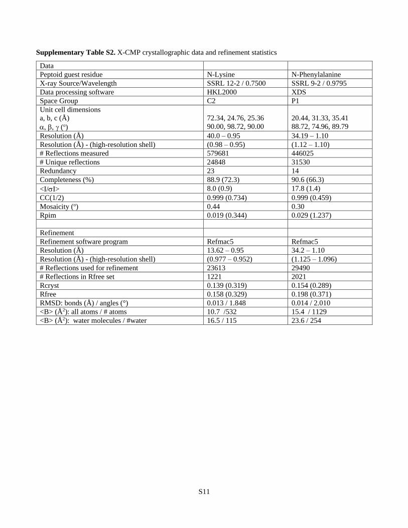
Crystal structures of N-gly-CMPs. To confirm the triple-helical folding of N-gly-CMPs and
to decipher the precise molecular structure of the N-glys within the peptide conformation, we
determined the X-ray crystal structures of two compounds. We chose Nlys-CMP and Nphe-
CMP for crystallographic analysis because of their high stabilities and differing N-gly sidechain
moieties, and determined their structures at resolutions of 0.95 Å and 1.10 Å , respectively(Fig.
2a, 2b; Supplementary Table S2, Fig. S1, S2). The overall conformation in both crystal
structures was consistent with those reported for conventional CMPs found in the Protein Data
Bank (PDB, SI Table S3), with the average (φ, ψ) angles and standard deviations of all non-

5
terminal Pro, Hyp, and Gly residues being (-70 ± 6°, 162 ± 6°), (-58 ± 5°, 147 ± 7°), and (-69 ± 3°,
174 ± 5°), respectively (Fig. 2c, SI Fig. S3). The dihedral angles of the two N-glys [Nlys: (-86 ± 3°,
176 ± 3°), Nphe: (-82 ± 3°, 177 ± 3°)] were also within the range populated by the X-position
residues of collagen peptides in the PDB (Fig. 2d), though slightly shifted from the PDB’s
average angles (Δφ = -13°, Δψ = +21°). This deviation did not significantly alter the backbone
structure and there was neither kinking nor bending of the triple helix near the N-gly in either
crystal structure. Furthermore, the inter-strand hydrogen bonding pattern, formed along the
length of the triple helix, agreed well with that seen in other collagen peptides in the PDB (Fig.
2e), although the hydrogen bonds formed with N-glys appeared to adopt a slightly less ideal
geometry (Fig. 2f). This suggests that the triple-helical stability of N-gly-CMPs does not
originate from improved hydrogen bonding35 of N-glys.
Probing the structural factors for N-glys’ triple-helical stability. Next, we introduced a series
of guest N-glys into the X-CMPs to understand how sidechain structures affect triple-helical
stability (Fig. 3). We first focused on hydrophobic sidechains, which previous reports suggested
to be the main stabilizing factor in N-gly-CMPs21,22. We found that the aliphatic Nchx as well as
the aromatic Nphe produced triple helices with stability notably higher than Pro (Fig. 3a). In
addition, Nphe derivatives having an electron-rich (Ntyr) or poor (Nnbz) aromatic ring
generated triple helices with similar stability, suggesting that neither the aromaticity nor its
electron density is crucial to triple helix stabilization. Meanwhile, although structurally similar,
the hydrophilic Ndxn and the deprotonated Ntyr (pH 12) were both more stabilizing than Pro
and only slightly less stabilizing than their hydrophobic analogs in the group (Fig. 3a). These
results suggest that the hydrophobicity of the peptoid residue might not be the main driving
force for the triple helix stabilization. Furthermore, we synthesized a pair of CMPs featuring
central guest triplets of GlyNpheHyp and GlyNpheIle to examine the hydrophobic interactions
between adjacent residues (SI Table S4). Introduction of Ile did not provide any additional
stabilizing effect, further de-emphasizing hydrophobicity in triple helix stabilization.
Next, we investigated the charged and hydrophilic peptoid sidechains (Fig. 3b). Although
charged Nasp and Nlys destabilized the triple helices modestly through electrostatic repulsions,

6
when we altered the pH so their sidechains were uncharged, both X-CMPs were as stable as
Pro-CMP. Also, we noted that peptoid residues with similar sidechain configurations conferred
highly similar Tm values (Fig. 3b: Nasp, Nasn, and Nleu; Fig. 3a: Nchx, Nphe, Nnbz, Ntyr,
Ndxn), while within each group, Tm values of the hydrophobic residues were only higher by 2-
4 °C.
Lastly, we found that progressively increasing the size of the aliphatic side chains produced a
concomitant increase in Tm of the corresponding X-CMP (Fig. 3c). Knowing that the
hydrophobicity has modest stabilizing effects for triple helices, we reasoned that the large
increase in stability from Gly-CMP (Tm, 37 °C) to Nchx-CMP (Tm, 63 °C) in this series cannot be
explained by the hydrophobicity alone, and were prompted to look into other mechanisms by
which peptoid residues stabilize the collagen triple helix.
Polyproline-II-helix (PPII) propensity of N-glys. It is generally accepted that Pro and Hyp
residues stabilize the collagen triple helix by pre-organizing individual collagen strands into the
PPII conformation36. Therefore, we hypothesized that N-glys, with their Pro-like tertiary amide
structure, could induce similar pre-organizational effects in N-gly-CMPs. To test this
hypothesis, we first surveyed a large set of small molecule crystal structures containing N-glys
from the Cambridge Structural Database (SI Table S5, S6). We found that the dihedral angles of
these peptoid residues largely fall within the most probable range for Pro, with the highest
density present in the area corresponding to a PPII helix (Fig. 4a). This suggests that similar to
Pro, N-glys have a high natural propensity for the PPII conformation.
To systematically study the N-glys’ propensity to adopt the PPII conformation, we employed
another host-guest peptide sequence: Ac-GlyProPro-X-ProProGlyTyr-NH2, designated as X-
PP5, where X represents the N-gly or amino acid residue replacing the central Pro within the
(Pro)5 sequence. This short, proline-rich host peptide was previously utilized to measure the
PPII propensity of the twenty canonical amino acids based on the CD intensity near 228 nm37,38.
Except for Sar-PP5, all N-gly-CMPs showed PPII CD intensity higher than Pro-PP5 (Fig. 4b) and
their amino acid counterparts37. Notably, the PPII propensities of Nphe, Nlys, and Nleu were
higher than that of Hyp, which is known for having the highest PPII propensity among all
7
natural amino acids39. This systematic investigation supports that N-glys promote the intrinsic
PPII folding of individual peptide strands, which can contribute to triple-helical
stabilization. Energy-wise, the free energy bonus arising from the PPII preorganization by just a
few bulky N-glys in a triple helix (Fig. 4b, Δ ΔG) can add up to be roughly an extra amide-
amide hydrogen bond (−2.0 kcal/mol)40.
Previous studies have shown that peptoids with specific N-substitutions can form stable
polyproline helices25-29,41. So far, the frontier of peptoid research has largely focused on control
over the cis-trans isomerization of peptoid amide bonds (i.e., adoptions of PPI or PPII
conformation) through specific steric or stereoelectronic effects induced by N-aryl or N-Cα-
chiral sidechains25-29,42. In this work, we systematically quantitated the general PPII propensity
of peptoid residues. We learned that although Pro has the highest PPII propensity among all
canonical amino acids, it is on the low side among the peptoid residues (Fig. 4b). The strong
PPII propensity of peptoid residues as compared to the amino acids implies that Pro may be the
an imperfect, biosynthetic surrogate for N-glys43 where PPII folding is needed, but its moderate
folding propensity can be improved via post-translational hydroxylation (e.g., in collagen).
MD simulation and steric effects of the N-substitution. We conducted MD simulations for
triple-helical X-CMPs incorporating guest residues Gly, Sar, Nleu, Nchx, or Pro (Fig. 4c). We
based our simulations on the crystal structures of Nphe- and Nlys-CMPs (Fig. 2) and chose N-
glys with simple aliphatic side chains that lack potential for electrostatic and hydrogen bonding
interactions. The simulation results demonstrated that the Nleu and Nchx residues displayed
energy landscapes very similar to Pro, with a low-energy valley confined to the regions (φ, ψ
angles) for PPII and the triple helix. In comparison, Sar which has a smaller sidechain, and to a
greater extent Gly, showed more conformational flexibility (Fig. 4c). These simulation results
are in agreement with our CD experiments, which showed that an N-gly’s PPII propensity (i.e.,
Gly < Sar < Nleu) and the triple-helical stability of its corresponding X-CMP correlate with the
size of its N-substitution (Fig. 3c & 4c). As a plausible explanation for this steric effect, a bulky
sidechain can impose a steric restraint to the N-gly’s backbone carbonyl, thereby hindering the
rotation around its nitrogen-carbon bond and constraining the φ angle to values appropriate for
8
triple-helical folding (Fig. 4d). This is evident in the most flexible guest residue Gly, where
addition of a single N-methyl group from Sar increases the Tm by 13 °C (Fig. 3c). Additionally,
we believe that an N-gly’s ψ angle is less sterically influenced by the N-substitution because it is
located further from the sidechain, and that it is more likely to assume a value close to ± 180°
because the α-carbon has no sidechain.
The pyrrolidine ring of Pro has been assumed to be a structural requisite for collagen
mimetics for decades3,44. In an attempt to replace Pro in CMPs, Raines and coworkers found
that converting Pro to N-methyl-L-alanine (NMe-Ala), which removes only the γ carbon and
eliminates the ring structure, substantially destabilizes the triple helix45 (SI Fig. S4). From this
observation, they concluded that the conformational restrictions imposed by the Pro ring are
more important for triple-helical stability than the presence of an N-substitution45. In contrast,
we discovered that N-glys with bulky sidechains can form well-folded collagen triple helices
without Pro’s cyclic sidechain. We believe that the low triple helicity of NMe-Ala45 is due to its
poor PPII propensity (Fig. 4b). Without the ring, steric repulsion between the two adjacent
methyl groups of NMe-Ala precludes formation of the dihedral angles necessary to form the
polyproline-II helix, thereby nullifying any stabilizing effect from the N-methyl group (SI Fig.
S4).
N-Cα chiral branching. To understand why Nval, the only peptoid residue with an N-Cα-
branched sidechain in our library, has unexpected, low triple-helical stability (Tm: 36 °C, Fig.
1d), we created a group of X-CMPs featuring guest residues NEt, Nval, Nphe, Nspe and Nrpe
(Fig. 5). The Tm differences (Fig. 5a) indicated that adding an N-Cα methyl group to an N-gly
destabilizes the triple helix, and that the triple-helical folding was greatly affected by the
chirality of the N-Cα-branching (Fig. 5a, Nspe versus Nrpe). For each chirality, 3D modeling
based on the Nphe-CMP crystal structure revealed a specific location of possible steric clash
between the branching methyl group and an adjacent peptide strand (Fig. 5b,c), which provides
an explanation for Nval’s low triple-helicity despite its higher-than-Pro PPII propensity (Fig.
4b). Although additional confirmatory studies are needed, this is the first time that a

9
steroselective steric interaction at the N-α-carbon is presented in the context of collagen triple
helix.
Sidechain functionalization. After a series of mechanistic investigations into triple-helical
folding, we turned to leveraging the diverse pool of peptoid residues to produce functionalized
collagen mimetics for potential applications. As one example, we demonstrated facile sidechain
functionalization through ‘click’ chemistry, utilizing an alkyne-bearing residue (Nakn, Fig. 6a,
SI Methods and SI Discussions). In another example, to demonstrate precise control over the
structure and function of CMP-based molecules and materials, we synthesized Nnbz-CMP, an
X-CMP featuring a UV-cleavable N-o-nitrobenzyl (nbz) sidechain46. We hypothesized that
Nnbz-CMP would form a robust triple helix, but UV irradiation would convert the stabilizing
Nnbz residue into Gly, thereby dramatically weakening the triple helix (Fig. 6b). CD
measurements confirmed this hypothesis (Fig. 6c). Moreover, characterizations of CMPs
incorporating more than one Nnbz residues (e.g., Nnbz2,3, SI Table S7) indicated that (i) the
stabilizing effect of N-glys may be additive, and that (ii) the increased number of Pro → Nnbz
substitutions resulted in even wider Tm differences before and after UV irradiation (Fig. 6c).
Next, we examined the Nnbz-substituted CMP’s capacity to hybridize to denatured collagen
and tested their release afterwards by UV irradiation. Previously, we reported that single-strand
CMPs [e.g., (GlyProHyp)9] can form hybrid triple helices with collagens that are denatured by
heat, proteases, or mechanical damage9,69. Using Nnbz2-CMP labeled with fluorescein (F-
Nnbz2-CMP), We found that Nnbz2-CMP displayed a drastically higher level of binding to
gelatin (heat-denatured collagen) than the host peptide Pro-CMP, consistent with its stronger
triple-helical propensity (Fig. 6d). Furthermore, upon brief UV irradiation, over 80% of the
bound F-Nnbz2-CMP was released from the gelatin matrix (Fig. 6d).
Spatio-temporal modification of cell culture substrates. To further demonstrate X-CMP’s
applications in biomaterials, we conjugated Nnbz2-CMP to a multi-arm poly(ethylene glycol)
(MA-PEG) polymer (SI Fig. S5a,b). The resulting conjugate (MA-PEG-CMP) readily formed a
hydrogel that was physically crosslinked via its Nnbz2-CMP triple helices; this stable hydrogel

10
showed the ability to dissolve and release its bound contents on demand as the CMP crosslinks
unfold upon UV irradiation (SI Fig. S5). Next, taking advantage of PEG’s ability to repel cell
adhesion and Nnbz2-CMP’s UV-triggered unbinding from gelatin (Fig. 6d), we demonstrated
that a crosslinked gelatin film can be coated with MA-PEG-CMP to accurately photo-pattern cell
attachment in selected areas (SI Fig. S6), or to trigger cell migration into defined regions at a
designated time (Fig. 6e,f). These proof-of-concept experiments showcase the practical use of X-
CMPs for creating complex cell culture substrates with spatio-temporal controls.
Billions of stable, Pro-free triple helices. Lastly, we synthesized X7-CMP, a (Gly-X-Hyp)7
sequence where each X position was polulated with a different peptoid residue (Fig. 6g). With a
Tm 1 °C higher than (GlyProHyp)7, X7-CMP represents a hyperstable, synthetic collagen triple-
helix with the greatest sidechain-diversity to date. With hundreds of amines commercially
available for efficient solid-phase peptoid synthesis, this work expands the amino-acid library
for stable CMPs by more than an order of magnitude. Even with a modest library of 20 N-glys
for X-substitution, for a short sequence of (Gly-X-Hyp)7, over 1.2 billion (207) triple-helical
peptides can be generated. The design principles discovered in this study open the door for not
only deciphering new fundamental molecular interactions (e.g., n→π*)47 in polyproline and
triple-helical folding, but also developing a new class of collagen-mimetic therapeutics and
biomaterials with remarkable functionalities. With the unprecedented structural diversity
offered by X-CMPs, we anticipate the long-awaited new era of de novo design of functional
collagen peptidomimetics.

11
Fig. 1 | A single N-substituted glycine (N-gly), also known as a peptoid residue, in the
central Pro position within (GlyProHyp)7 shows high triple-helical propensity. a, General
structures of a canonical amino acid, Pro, and an N-gly. b, Chemical structure of the X-CMP, a
host-guest collagen mimetic peptide with the sequence of a (GlyProHyp)7, where the Pro
residue in the central X position is substituted, for example, with an N-gly residue. c, An X-
CMP triple helix dissociates into single strands under gradual heating; monitored under CD,
the middle point of this two-state transition is defined as the X-CMP’s melting temperature
(Tm). The Nphe sidechain at the X position resulted in a CMP triple helix considerably more
stable than Pro (ΔTm: +7 °C). In comparison, the amino acid with the same sidechain was highly
destabilizing (ΔTm: -17 °C). d, The triple helical stabilities of X-CMPs featuring N-glys with
canonical sidechains, quantitated in ΔTm (in comparison to the host peptide: Pro-CMP) . Almost
universally, peptoid residues (right column) have higher stability than their amino acid
counterparts (left column) except for Nval, and many have stabilities similar to or better than
Pro. *ΔTm for amino acid residues taken from reference21. **Nhcy (homocysteine) is a similar but
not exact equivalent to cysteine.

12
Fig. 2 | CMPs containing peptoid residues adopt a triple-helical conformation nearly
identical to the native collagen. a,b Crystal structures of Nlys-CMP (a) and Nphe-CMP (b);
each shows a triple helix with the N-gly residue uniquely colored (Nlys in orange and Nphe in
green). c, A Ramachandran plot showing the φ and ψ angles of all amino acid residues in the
crystal structure of Nlys-CMP (orange) and Nphe-CMP (green), overlaid with the φ and ψ
angles from the reported CMP crystal structure in the PDB (grey, see SI for structure IDs used
for comparison). d, A Ramachandran plot showing the φ and ψ angles for only the N-gly
residues (Nlys in orange, Nphe in green), overlaid with the φ and ψ angles (in grey) from the X
position of Gly-X-Y repeats of reported CMP crystal structures. The peptoid residues adopt a
conformation common to the X position in previously reported Gly-X-Y collagen sequences, yet
with slightly shifted average angles (Δφ: -13°, Δψ: +21°). e, A plot of the N-H-O angle versus the
N-O distance of all the interstrand amide-amide H-bonds in the crystal structures of Nlys-CMP
(orange) and Nphe-CMP (green) overlaid with that (in grey) of all the H-bonds in the reported
crystal structure of CMPs. f, A plot, same as e but showing H-bonds associated only with N-gly
residues in the crystal structures (Nlys in orange, and Nphe in green). The H-bonds parameters
for both residues are consistent with previously reported crystal structures of collagen peptides.

13
Fig. 3 | The structure of the peptoid sidechain affects triple-helical propensity. a, Tm values
for a series of X-CMPs with N-Cα-substituted 6-membered rings in the sidechains. There is little
difference in triple-helical stability among the aliphatic, aromatic, and hydrophilic rings, and
every peptide had a Tm higher than the host: Pro-CMP. b, Although charged groups
destabilized the triple helix at physiological pH, when the sidechains were uncharged, both
Nlys and Nasp, as well as the hydrophilic Nasn, were as stabilizing as Pro. c, A series of X-
CMPs with aliphatic sidechains of increasing size showing concomitantly increasing Tm values,
implying an robust steric effect. Although the hydrophobicity in this series increases as the
aliphatic sidechains increase in size, according to series a and b, hydrophobicity has only a
minor effect on stability. The results suggest that the tertiary amide structure N-gly (which
hinders the α helix and β sheet conformations) and the size of the N-substituted sidechain play
the most critical roles in stabilization of N-gly-CMP triple helices.

14
Fig. 4 | N-gly residues have a strong polyproline-II helix (PPII) propensity which improve
the triple-helical stability of X-CMPs. a, A survey of φ and ψ angles of N-glys from high
resolution crystal structures of small molecules (from Cambridge Structure Database). N-glys
have a high natural propensity to fold into either the left- or right-handed polyproline
conformation (φ ± 90°, ψ 180°). Watermarks show the regions highly populated by Pro residues.
b, Structure of the PPII host-guest peptide X-PP5 and the comparative CD spectra of Gly-PP5,
Pro-PP5, and Nleu-PP5. The PPII propensity of each X residue within the X-PP5 model was
assessed by the ellipticity value of the characteristic CD signal near 228 nm. All N-glys, with the
exception of Sar, had a θmax higher than Pro indicating peptoids’ high intrinsic PPII propensity.
*No peak was observed near 228 nm for NMe-Ala (Supplementary data). c, Metadynamics
calculations of the free energy landscape of the guest residues in Gly-, Sar-, Nleu-, Nchx-, and
Pro-CMP. Each model yielded a native state consistent with the expected triple helix and PPII
conformations (-70°, 170°); however, peptides with small guest residues (Gly and Sar) showed a
secondary non-PPII energy well. d, Newman-like projections of N-glys with the grey dashed
line indicating the line of sight for the projection. N-glys with large side chains can experience
major steric clashes with its carbonyl group. Such reduced conformational flexibility of the N-
gly may help preorganize the individual chains into a PPII conformation, thereby stabilizing the
triple-helical assembly.

15
Fig. 5 | N-Cα branching of peptoid residues affects triple-helical folding. a, With a N-Cα
branch, Nval- and Nspe-CMPs, had Tm values 14-16 °C lower than their unbranched analogs,
NEt and Nphe (ΔTm: -14 ~ -16 °C). Moreover, while residue Nspe conferred a triple helix with a
Tm of 48 °C, its enantiomer, Nrpe completely abolished the triple-helical folding. b,c, Molecular
modeling based on solved crystal structure of Nphe-CMP showing potential steric clashes
within the triple helix introduced by the N-Cα methyl branch of Nspe and Nrpe. The R-branch
of Nrpe (cyan), pointing directly toward the inner core of the triple helix, may clash with the
backbone carbonyl of the cross-chain Gly and interfere with proper backbone assembling; in
contrast, the S-branch (magenta) in Nspe may clash with the hydroxyl group of a cross-chain
Hyp, which may be less destabilizing since the steric repulsion may be alleviated by changing
the Hyp’s ring pucker (shown in half-transparency in b).

16
Fig. 6 | X-CMPs enable new functional design strategies. a, Facile modification of the alkyne-
sidechain through alkyne-azide ‘click’ chemistry. Nakn-CMP retained strong triple-helical
propensity after the conjugation, indicating that the N-substitution can well tolerate the
spatially demanding triazole units within the triple helix (Supplementary Discussions). b,
Reaction scheme of photo-conversion of the Nnbz residue to Gly by UV light. c, CD melting
curves showing UV-induced triple helix destabilization of Nnbz-CMPs. A single Pro → Nnbz
substitution (Nnbz-CMP) in the central triad stabilized the triple helix (Tm: 55 °C → 62 °C),
while photo-cleavage of the N-o-nitrobenzyl group destabilized the triple helix (Tm: 62 °C →
37 °C). Additional substitutions of Pro with Nnbz within the CMP (Nnbz2- and Nnbz3-CMPs,
SI Table S7) further increased the ΔTm of the peptide before and after UV irradiation. d,
Comparative fluorescence intensity of gelatin films treated with fluorescently labeled X-CMPs.
F-Nnbz2-CMP demonstrated higher affinity to gelatin than F-Pro-CMP, as well as triple-helical
unfolding and releasing after UV irradiation. e, Schematics of UV-patterning on a gelatin
substrate mediated by Nnbz2-CMP: MA-PEG-CMPs bound onto the crosslinked gelatin film
prevented cell attachment; however, UV irradiation released the PEG-CMP conjugate, exposing
gelatin that supports cell adhesion. f, Light micrographs of MDA-MB-231 cells grown to a
confluency only in regions devoid of MA-PEG-CMP on a gelatin film (top). After exposing this

17
cell culture to UV light through a photomask (e), which unbound the MA-PEG-CMPs in
selected areas, the cells began to infiltrate the patterned areas with exposed gelatin (bottom,
taken 1 d after UV treatment). g, Pro-free X7-CMP hosting seven peptoid residues with diverse
sidechain structures. Its high Tm of 56 °C demonstrates a hyper-stable triple-helical collagen
peptide with the greatest sidechain diversity reported to date.
References
1. Horne, W.S. & Grossmann, T.N. Proteomimetics as protein-inspired scaffolds with defined tertiary
folding patterns. Nat. Chem. 12, 331-337 (2020).
2. Huang, P., Boyken, S.E. & Baker, D. The coming of age of de novo protein design. Nature 537, 320-
327 (2016).
3. Shoulders, M.D. & Raines, R.T. Collagen structure and stability. Annu. Rev. Biochem. 78, 929-958
(2009).
4. Ramshaw, J.A.M., Shah, N.K. & Brodsky, B. Gly-X-Y tripeptide frequencies in collagen: A context for
host–guest triple-helical peptides. J. Struct. Biol. 122, 86-91 (1998).
5. Ramachandran, G.N. & Kartha, G. Structure of collagen. Nature 176, 593-595 (1955).
6. Bella, J., Eaton, M., Brodsky, B. & Berman, H.M. Crystal and molecular structure of a collagen-like
peptide at 1.9 A resolution. Science 266, 75 (1994).
7. Orgel, J.P.R.O., Irving, T.C., Miller, A. & Wess, T.J. Microfibrillar structure of type I collagen in situ.
Proc. Natl. Acad. Sci. U. S. A. 103, 9001 (2006).
8. Zitnay, J.L. et al. Molecular level detection and localization of mechanical damage in collagen
enabled by collagen hybridizing peptides. Nature Communications 8, 14913 (2017).
9. Leitinger, B. Transmembrane collagen receptors. Annu. Rev. Cell. Dev. Biol. 27, 265-290 (2011).
10. O'Leary, L.E.R., Fallas, J.A., Bakota, E.L., Kang, M.K. & Hartgerink, J.D. Multi-hierarchical self-
assembly of a collagen mimetic peptide from triple helix to nanofibre and hydrogel. Nat. Chem. 3, 821
(2011).
11. Tanrikulu, I.C., Forticaux, A., Jin, S. & Raines, R.T. Peptide tessellation yields micrometre-scale
collagen triple helices. Nat. Chem. 8, 1008 (2016).
12. Holmgren, S.K., Taylor, K.M., Bretscher, L.E. & Raines, R.T. Code for collagen's stability deciphered.
Nature 392, 666-667 (1998).
13. Sakakibara, S., Kishida, Y., Kikuchi, Y., Sakai, R. & Kakiuchi, K. Synthesis of poly-(L-prolyl-L-
prolylglycyl) of defined molecular weights. Bull. Chem. Soc. Jpn. 41, 1273-1273 (1968).
14. Persikov, A.V., Ramshaw, J.A.M., Kirkpatrick, A. & Brodsky, B. Amino acid propensities for the
collagen triple-helix. Biochemistry 39, 14960-14967 (2000).
15. Maaßen, A. et al. Triple-helix-stabilizing effects in collagen model peptides containing PPII-helix-
preorganized diproline modules. Angew. Chem. Int. Ed. 59, 5747-5755 (2020).
16. Aronoff, M.R., Egli, J., Schmitt, A. & Wennemers, H. Alkylation of γ-Azaproline Creates
Conformationally Adaptable Proline Derivatives for pH-Responsive Collagen Triple Helices.
Chemistry – A European Journal 26, 5070-5074 (2020).
17. Erdmann, R.S. & Wennemers, H. Functionalizable collagen model peptides. J. Am. Chem. Soc. 132,
13957-13959 (2010).

18
18. Siebler, C., Erdmann, R.S. & Wennemers, H. Switchable proline derivatives: Tuning the
conformational stability of the collagen triple helix by pH changes. Angew. Chem. Int. Ed. 53, 10340-
10344 (2014).
19. Hentzen, N.B., Smeenk, L.E.J., Witek, J., Riniker, S. & Wennemers, H. Cross-linked collagen triple
helices by oxime ligation. J. Am. Chem. Soc. 139, 12815-12820 (2017).
20. Goodman, M., Melacini, G. & Feng, Y. Collagen-like triple helices incorporating peptoid residues. J.
Am. Chem. Soc. 118, 10928-10929 (1996).
21. Feng, Y., Melacini, G. & Goodman, M. Collagen-based structures containing the peptoid residue N-
Isobutylglycine (Nleu): synthesis and biophysical studies of Gly-Nleu-Pro sequences by circular
dichroism and optical rotation. Biochemistry 36, 8716-8724 (1997).
22. Melacini, G., Feng, Y. & Goodman, M. Collagen-based structures containing the peptoid residue N-
Isobutylglycine (Nleu): conformational analysis of gly-nleu-pro sequences by 1H-NMR and
molecular modeling. Biochemistry 36, 8725-8732 (1997).
23. Simon, R.J. et al. Peptoids: a modular approach to drug discovery. Proceedings of the National Academy
of Sciences 89, 9367 (1992).
24. Robertson, E.J. et al. Design, synthesis, assembly, and engineering of peptoid nanosheets. Acc. Chem.
Res. 49, 379-389 (2016).
25. Wu, C.W., Sanborn, T.J., Huang, K., Zuckermann, R.N. & Barron, A.E. Peptoid oligomers with α-
chiral, aromatic side chains: sequence requirements for the formation of stable peptoid helices. J.
Am. Chem. Soc. 123, 6778-6784 (2001).
26. Wu, C.W. et al. Structural and spectroscopic studies of peptoid oligomers with α-chiral aliphatic side
chains. J. Am. Chem. Soc. 125, 13525-13530 (2003).
27. Stringer, J.R., Crapster, J.A., Guzei, I.A. & Blackwell, H.E. Extraordinarily robust polyproline type I
peptoid helices generated via the incorporation of α-chiral aromatic N-1-Naphthylethyl side chains.
J. Am. Chem. Soc. 133, 15559-15567 (2011).
28. Roy, O. et al. Homogeneous and robust polyproline type I helices from peptoids with nonaromatic
α-chiral side chains. J. Am. Chem. Soc. 139, 13533-13540 (2017).
29. Shah, N.H. et al. Oligo(N-aryl glycines): a new twist on structured peptoids. J. Am. Chem. Soc. 130,
16622-16632 (2008).
30. Lee, B.-C., Zuckermann, R.N. & Dill, K.A. Folding a nonbiological polymer into a compact
multihelical structure. J. Am. Chem. Soc. 127, 10999-11009 (2005).
31. Murnen, H.K., Rosales, A.M., Jaworski, J.N., Segalman, R.A. & Zuckermann, R.N. Hierarchical self-
assembly of a biomimetic diblock copolypeptoid into homochiral superhelices. J. Am. Chem. Soc. 132,
16112-16119 (2010).
32. Mannige, R.V. et al. Peptoid nanosheets exhibit a new secondary-structure motif. Nature 526, 415-420
(2015).
33. Nam, K.T. et al. Free-floating ultrathin two-dimensional crystals from sequence-specific peptoid
polymers. Nature Materials 9, 454-460 (2010).
34. Zuckermann, R.N., Kerr, J.M., Kent, S.B.H. & Moos, W.H. Efficient method for the preparation of
peptoids [oligo(N-substituted glycines)] by submonomer solid-phase synthesis. J. Am. Chem. Soc.
114, 10646-10647 (1992).
35. Zhang, Y., Malamakal, R.M. & Chenoweth, D.M. Aza-glycine induces collagen hyperstability. J. Am.
Chem. Soc. 137, 12422-12425 (2015).
36. Adzhubei, A.A., Sternberg, M.J.E. & Makarov, A.A. Polyproline-II helix in proteins: Structure and
function. J. Mol. Biol. 425, 2100-2132 (2013).
37. Brown, A.M. & Zondlo, N.J. A propensity scale for type II polyproline helices (PPII): aromatic amino
acids in proline-rich sequences strongly disfavor PPII due to proline–aromatic interactions.
Biochemistry 51, 5041-5051 (2012).

19
38. Pandey, A.K., Thomas, K.M., Forbes, C.R. & Zondlo, N.J. Tunable control of polyproline helix (PPII)
structure via aromatic electronic effects: an electronic switch of polyproline helix. Biochemistry 53,
5307-5314 (2014).
39. Horng, J.-C. & Raines, R.T. Stereoelectronic effects on polyproline conformation. Protein Sci. 15, 74-83
(2006).
40. Jenkins, C.L., Vasbinder, M.M., Miller, S.J. & Raines, R.T. Peptide bond isosteres: Ester or (E)-alkene
in the backbone of the collagen triple helix. Org. Lett. 7, 2619-2622 (2005).
41. Butterfoss, G.L., Renfrew, P.D., Kuhlman, B., Kirshenbaum, K. & Bonneau, R. A preliminary survey
of the peptoid folding landscape. J. Am. Chem. Soc. 131, 16798-16807 (2009).
42. Crapster, J.A., Guzei, I.A. & Blackwell, H.E. A peptoid ribbon secondary structure. Angewandte
Chemie (International ed. in English) 52, 5079-5084 (2013).
43. Delauney, A.J. & Verma, D.P.S. Proline biosynthesis and osmoregulation in plants. The Plant Journal
4, 215-223 (1993).
44. Egli, J., Schnitzer, T., Dietschreit, J.C.B., Ochsenfeld, C. & Wennemers, H. Why proline? Influence of
ring-size on the collagen triple helix. Org. Lett. 22, 348-351 (2020).
45. Cram, D.J. The design of molecular hosts, guests, and their complexes. Science 240, 760 (1988).
46. Li, Y. et al. Targeting collagen strands by photo-triggered triple-helix hybridization. Proc. Natl. Acad.
Sci. U. S. A. 109, 14767-14772 (2012).
47. Gorske, B.C., Stringer, J.R., Bastian, B.L., Fowler, S.A. & Blackwell, H.E. New strategies for the design
of folded peptoids revealed by a survey of noncovalent interactions in model systems. J. Am. Chem.
Soc. 131, 16555-16567 (2009).
Acknowledgements
The authors thank Rodrigo Galindo-Murillo and Xiaolei Zhu for consultation on the simulations of X-
CMP structures and Hendra Wahyudi for assistance in the synthesis of Fmoc-Nnbz-OH. This research
was funded by grants from the National Institutes of Health (R01AR071358, R21EY029430, and
R21OD026618) awarded to SMY.
Author contributions
Y.L. and J.L.K. designed the studies. J.L.K., Y.L., G.K., and H.K. performed the syntheses. J.L.K and Y.L.
performed the experiments. J.L.K. and F.G.W. solved the crystal structures. C.P.H. oversaw the
crystallography. Z.Q. and T.E.C. performed the simulations. J.L.K., S.M.Y., and Y.L. wrote the paper. All
authors were involved in the completion of this work.
Competing interests
The authors declare no competing interests.

download fileview on ChemRxivChemRxiv PEPTOID-CMP_manuscript 20200905_submitt... (1.52 MiB)

S1
Peptoid Residues Make Diverse, Hyperstable Collagen Triple Helices
Julian L. Kessler1, Grace Kang1, Zhao Qin2, Helen Kang1, Frank G. Whitby3, Thomas E. Cheatham III4,
Christopher P. Hill3, Yang Li1,* and S. Michael Yu1,5
1Department of Biomedical Engineering, University of Utah, Salt Lake City, Utah 84112, USA 2Department of Civil & Environmental Engineering, Collagen of Engineering & Computer Science, Syracuse
University, Syracuse, New York 13244, USA 3Department of Biochemistry, University of Utah School of Medicine, Salt Lake City, UT 84112, USA 4Department of Medicinal Chemistry, College of Pharmacy; University of Utah, Salt Lake City, 84112, USA 5Department of Pharmaceutics and Pharmaceutical Chemistry, University of Utah, Salt Lake City, Utah 84112,
USA
*Correspondence to: Yang Li ([email protected])
Table of Contents
Supplementary Section 1: Materials and Methods S2
Materials S2
Solid phase synthesis S3
On-resin functionalization via “click” chemistry S5
Cleavage protocol S5
Purification and mass spectrometry S5
Circular dichroism spectroscopy S6
X-ray Crystallography S6
MD Simulations S7
Gelatin binding assays S7
UV triggered hydrogel dissolution and spatial control of cell attachment S8
Supplementary Section 2: Figures and Tables S10
Supplementary Table S1. Chemical structures of all guest residues and their Tm. S10
Supplementary Table S2. X-ray crystallography parameters. S11
Supplementary Table S3. Existing PDB entries of collagen model peptides. S12
Supplementary Table S4. Primary structures and Tm of X-CMP with double guests. S13
Supplementary Table S5. Existing CSD entries of peptoid-containing small molecules S14
Supplementary Table S6. Examples of crystal structures of peptoids in CSD S15
Supplementary Table S7. Primary structures and Tm of Nnbz-, Nnbz2-, and Nnbz3-
CMP.
S16
Supplementary Fig. S1. Crystal structure of Nlys-CMP. S17
Supplementary Fig. S2. Crystal structure of Nphe-CMP. S18
Supplementary Fig. S3. Ramachandran plots for Nlys- and Nphe-CMP. S19
Supplementary Fig. S4. Newman-like projection of NMe-Ala. S20
Supplementary Fig. S5. MA-PEG-CMP and UV-induced unfolding. S21
Supplementary Fig. S6. Spatial control of cell adhesion. S22
Supplementary Section 3: HPLC, MALDI and CD of all peptides S23
Analyses of X-CMP peptides (in alphabetical order) S23
Analyses of X-PP5 peptides (in alphabetical order) S52
Supplementary Section 4: Supplementary Discussion S58
References S59
S2
Supplementary Section 1: Materials and methods
Materials
All commercial chemicals and solvents were used as received. The solvents, resin, Fmoc amino acids, and reagents
used in the synthesis and purification of the peptides and peptoids were purchased from the suppliers listed in the
table below.
General reagents Supplier Catalog number
N-Methylpyrrolidinone (NMP) Fisher Scientific BP1172
N,N-Dimethylformamide (DMF) Fisher Scientific BP1160
Dimethyl sulfoxide (DMSO) Sigma-Aldrich 276855
Methylene Chloride (DCM) Fisher Scientific D37
Acetonitrile Fisher Scientific A998
TentaGel R RAM resin Peptides International RTS-9995-PI
Fmoc-Gly-OH EMD Millipore 852001
Fmoc-Pro-OH EMD Millipore 852017
Fmoc-Hyp(tBu)-OH EMD Millipore 852036
Fmoc-Lys(Boc)-OH AAPPTec AFK105
Fmoc-Phe-OH AAPPTec AFF101
Fmoc-Tyr(tBu)-OH EMD Millipore 852020
Fmoc-Ile-OH Advanced ChemTech FI2326
Piperidine Sigma-Aldrich 104094
Bromoacetic acid Sigma-Aldrich 17000
N,N’-Diisopropylcarbodiimide (DIC) Sigma-Aldrich D125407
O-(7-Azabenzotriazol-1-yl)-N,N,N',N'-
tetramethyluronium hexafluorophosphate (HATU) Chem-Impex 12881
1-Hydroxy-7-azabenzotriazole (HOAT) AAPPTec CXZ012
(7-Azabenzotriazol-1-
yloxy)trispyrrolidinophosphonium
hexafluorophosphate (PyAOP)
AAPPTec CXZ070
N-Ethyldiisopropylamine (DIEA) EMD Millipore 845017
Acetic anhydride Alfa Aesar L04295
Trifluoroacetic acid (TFA) Fisher Scientific BP618
Triisopropylsilane (TIS) Sigma-Aldrich 233781
1,2-Ethanedithiol (EDT) Sigma-Aldrich 02390
DL-Dithiothreitol Sigma-Aldrich D0632
Anisole Sigma-Aldrich 123226
Ethyl ether Fisher Scientific E138

S3
Solid phase synthesis
All sequences were prepared on TentaGel R RAM resin (substitution level: 0.2 mmol/g) via solid phase synthesis.
Typically, 50 mg of resin (containing 10 µmol of reaction sites) was used to prepare one host-guest peptide/peptoid
sequence. Prior to the first coupling, resin was swelled in 0.4 mL of NMP for 30 min. Each amino acid residue was
coupled by agitating the resin with a solution of Fmoc protected residue (50 µmol), HATU (50 µmol, 19 mg),
HOAT (50 µmol, 6.8 mg) and DIEA (75 µmol, 13 µL) in 0.4 mL of NMP for over 3 hr. The Fmoc protective group
was removed by treating the resin with piperidine (20% by volume in NMP) for 20 min. Following each reaction,
the resin was drained and washed with NMP (3×10 mL). All coupling and deprotection reactions were monitored
by the standard Kaiser (for primary amines) and chloranil (for secondary amines) tests.
Except residues Sar, NMe-Ala, and Nnbz, which were coupled by using the respective Fmoc-protected compounds
and the above-mentioned HATU chemistry, all other peptoid residues were incorporated on-resin using the two-
step sub-monomer method reported by Zuckermann and others1.
(1) Acylation reactions were performed by addition of a solution of bromoacetic acid (100 µmol, 13.9 mg) and DIC
(98 µmol, 15.2 µL) in DMF (0.5 mL) to 10 µmol of resin-bound amine. Reaction mixtures were agitated at room
temperature for 30 min. Each acylation was repeated once before the resin was drained and washed with NMP
(3×10 mL). (2) Each displacement reaction was performed by addition of a primary amine (200 µmol) in 0.5 mL
of NMP, followed by agitation overnight at room temperature. Following the displacement, the resin was drained
and washed with DMF (3×10 mL). The reaction was monitored by the chloranil test. Typically, resin beads turned
strongly green in a chloranil test following a displacement reaction of 2 hr, suggesting successful coupling of a
peptoid; we chose to run the reactions overnight to ensure completion. In the table below, the primary amine used
to create each peptoid residue is listed, and special reaction conditions and observations are noted. In general, the
next amino acid following a peptoid residue (e.g., Fmoc-Gly-OH or Fmoc-Pro-OH) was coupled with full
completion using the above-mentioned 5 eq HATU protocol.
NH
O
HN
RR-NH2
NH
O
BrBr
OH
O
DICH2N
(1) (2)

S4
Residue R-NH2 Reagent Supplier Catalog
number Note
Sar Fmoc-Sar-OH EMD Millipore 852055 Coupled by Fmoc & HATU chemistry
NMe-Ala Fmoc-N-methyl-L-alanine Chem-Impex 2650 Coupled by Fmoc & HATU chemistry
Nnbz Fmoc(N-o-nitrobenzyl)Gly-
OH Synthesized in-house2,3 Coupled by Fmoc & HATU chemistry
Nakn Propargylamine Sigma-Aldrich P50900
: in Kaiser
test, resin beads and the solution do not
turn dark blue despite the presence of
primary amine, perhaps because the
Nakn side chain affects the ninhydrin
reactions.
Nasn Glycinamide Combi-Blocks QA-8748
Glycinamide is not soluble in NMP.
The displacement reaction was carried
out in 0.5 mL DMSO.
Nasp Glycine tert-butyl ester Alfa Aesar L16258
Nchx Cyclohexanemethylamine Sigma-Aldrich 101842
Ncys 2-(tert-butylsulfanyl)ethan-1-
amine Enamine
EN300-
110537
Ndxn C-[1,4]Dioxan-2-yl-
methylamine Matrix Scientific 008403
NEt Ethylamine solution (2.0 M in
THF) Sigma-Aldrich 395072
Nleu Isobutylamine Sigma-Aldrich I14150
Nlys N-Boc-1,4-butanediamine Chem-Impex 31317
Nmet 2-(Methylthio)ethylamine Sigma-Aldrich 632929
Nphe Benzylamine Sigma-Aldrich 185701
Nrpe (R)-(+)-α-Methylbenzylamine Sigma-Aldrich 115541 The Gly residue following Nrpe/Nspe
was coupled by agitating the resin (10
µmol) in a solution of Fmoc-Gly-OH
(100 µmol) and DIC (100 µmol) in 0.5
mL of DMF. Nspe (S)-(−)-α-Methylbenzylamine Sigma-Aldrich 115568
Ntyr (4-tert-
butoxyphenyl)methanamine Enamine
EN300-
55516
Nval Isopropylamine Sigma-Aldrich 471291
The Gly residue following Nval was
coupled by agitating the resin (10
µmol) in a solution of Fmoc-Gly-OH
(100 µmol) and DIC (100 µmol) in 0.5
mL of DMF.
Ndota
1,4,7,10-
Tetraazacyclododecane-1,4,7-
tris(t-butyl acetate)-10-(4-
aminobutyl)acetamide
Macrocyclics B-279
The displacement reaction was
completed by agitating the resin (10
µmol) in 0.5 mL of NMP containing
the primary amine (63 µmol) and DIEA
(115 µmol) overnight.
Nman
2-Aminoethyl 2,3,4,6-tetra-O-
acetyl-α-D-mannopyranoside
hydrochloride
Synthose AM876
DIEA (250 µmol, 43.5 µL) was added
to the reaction mixture in the
displacement reaction.

S5
On-resin functionalization via “click” chemistry
The azido compound (60 µmol), such as 2-Azidoethanol (Carbosynth, FA07084) or 2-Azidoethyl α-D-
mannopyranoside (Synthose, AM482), was dissolved in 0.5 mL of DMF along with [Cu(MeCN)4]PF6 (10 µmol,
3.7 mg, Sigma-Aldrich, 346276) and TBTA (20 µmol, 10.6 mg, Sigma-Aldrich, 678937). This solution was added
to the resin-bound alkyne-containing peptide Nakn-CMP (5 µmol), and the mixture was agitated at room
temperature overnight. The resin was drained and washed with DMF (5×10 mL) before the “click”-functionalized
product was cleaved.
Cleavage protocol
Following the coupling and Fmoc-deprotection of the last Gly residue, the N-terminal amine of each sequence was
acetylated by mixing the resin (5 µmol) with a solution of acetic anhydride (500 mM), HOAT (14.7 mM), and
DIEA (129 mM) in 0.5 mL of NMP at room temperature for 30 min. The resin was drained, washed with NMP
(3×10 mL) and DCM (5×10 mL). Unless otherwise mentioned below, the resin (5 µmol) was treated with 1 mL
mixture of TFA / TIS / water (95:2.5:2.5) for over 3 hr with stirring. Subsequently, the resin was washed with 0.5
mL of TFA twice. After the resin was filtered, the TFA cleavage solution was collected and evaporated under a
stream of nitrogen down to approximately 0.5 mL. The crude peptide/peptoid products were precipitated by adding
5 mL of cold ethyl ether to the TFA solution on ice.
Sequence Special cleavage protocol
Nmet Treat the resin with a mixture of TFA/EDT/water/TIS (94:2.5:2.5:1) for over 3 h.
Nhcy
Step 1: for 5 µmol of crude product, treat the resin with a mixture (1 mL) of TFA/TIS/water
(95:2.5:2.5) for over 2 hr, and collect the crude product.
Step 2: Remove the tBu protective group from the Ncys residue by stirring the crude product in a
mixture (3 mL) of TFA/DMSO/anisole (97.9:2:0.1) at room temperature for 1 hr, precipitate and
collect the product again.
Step 3: dry the crude product and dissolve it in 2 mL of water containing 15 mg of dithiothreitol to
reduce any oxidized thiol groups before purification.
Purification and mass spectrometry
All peptides were purified by reverse-phase high performance liquid chromatography (HPLC) on an Agilent Zorbax
SB C-18 column, using a mixture of water (A) and acetonitrile (B) (linear gradient: 5% to 35% acetonitrile over 30
min). Both eluents A and B contain 0.1% TFA. The column oven was heated to 70 °C to prevent triple helix
formation. After semi-preparative purifications, the collected fractions containing the target peptides were analyzed
by analytical HPLC again using the following gradient parameters.
Time (min) 0 2 4 25 25.5 30
B% 5% 5% 10% 40% 5% 5%

S6
The flow rates used for semi-preparative and analytical HPLC were 4 and 1.5 mL/min, respectively. The pure
fractions were lyophilized. The lyophilized products were reconstituted in pure water as stock solutions to be used
for structural characterizations. Peptide concentrations in the stock solutions were determined by measuring the
ultraviolet (UV) absorbance of the solutions at 214 nm (for the collagen mimetic host-guest sequences, extinction
coefficient: 2200 M-1cm-1 per peptide bond) or 280 nm (for the Tyr-containing polyproline host-guest sequences,
extinction coefficient: 1280 M-1cm-1 per tyrosine residue) on a SpectraMax M2e microplate reader (Molecular
Devices) using a quartz cell with 1 cm cell path length.
All purified peptides/peptoids were verified by mass spectrometry. Mass spectra were obtained at the University of
Utah Mass Spectrometry and Proteomics core facility on a Bruker UltrafleXtreme matrix-assisted laser
desorption/ionization time-of-flight (MALDI/TOF) mass spectrometer.
Circular dichroism (CD) spectroscopy
CD measurements of the peptide solutions were recorded in quartz cells with a path length of 0.1 cm, on a JASCO
J-1500 CD spectrophotometer. Prior to CD spectra or melting experiments, peptide stock solutions were diluted to
150 µM in 1×PBS (pH: 7.4 for triple helical peptides) or in 5 mM phosphate buffer (pH: 7.0 for polyproline host-
guest peptides) and heated at 80 °C for 5 min followed by incubation at 4 °C for at least 48 hr. Sequences containing
guest residues with acidic or basic side chains were also diluted to 150 µM in NaOH (10 or 35 mM) or HCl (15
mM) solutions.
CD spectra were scanned at 4 °C (for triple helical peptides) or 25 °C (for polyproline host-guest peptides) using
the following parameters: bandwidth, 5 nm; digital integration time, 16 s; scanning speed, 20 nm/min; data pitch,
0.1 nm. For the polyproline host-guest series, all reported spectra were the average of three independent scans, and
were corrected from the blank buffer background.
Thermal melting curves were obtained by monitoring the ellipticity at 225 nm from 4 °C to 90 °C at a heating rate
of 0.5 °C/min. The mean residue ellipticity (MRE, [θ]) was calculated using the equation [θ]=(θ×m)/(c×l×n), where
θ is measured ellipticity (mdeg), m is molecular weight (g/mol), c is concentration (mg/mL), l is path length of the
cuvette (mm), and n is the number of amino acid residues in the peptide. The derivative of a melting curve was
generated using the JASCO Spectra Manager software (Version 2.10.05), and the temperature at the minimum of
the derivative curve was defined as the melting temperature (Tm). Each Tm value reported in this study was averaged
from two CD thermal unfolding experiments, in which the difference between the two measured Tm values was less
than 1 °C for all triple helical host-guest peptides.
X-ray crystallography
Purified, lyophilized Nlys-CMP [Ac-(GlyProHyp)3-GlyNlysHyp-(GlyProHyp)3] was dissolved in water to a final
concentration of 5.0 mg/mL. Crystals were grown by vapor diffusion in sitting drops of 0.3 μL of the peptide

S7
solution and 0.3 μL of 2.1 M DL-malic acid (pH 7.0) corresponding to well F8 in the commercially-available crystal
screen JCSG-plus (Molecular Dimensions) at 21 °C. Crystals appeared within 10 days. Purified, lyophilized Nphe-
CMP [Ac-(GlyProHyp)3-GlyNpheHyp-(GlyProHyp)3] was dissolved in water to a final concentration of 10.0
mg/mL. Crystals were grown by vapor diffusion in sitting drops of 0.3 μL of the peptide solution and 0.3 μL of 2.4
M sodium malonate dibasic monohydrate (pH 7.0) corresponding to well F9 in the commercially available crystal
screen JCSG-plus (Molecular Dimensions) at 4 °C. Crystals appeared within 1 week.
In preparation for data collection, crystals were suspended in a small rayon loop attached to a mounting pin,
immersed in 20 μL crystallization buffer with 25% added glycerol, then cryocooled by plunging into liquid nitrogen.
Data were collected from crystals maintained at 100 K. Diffraction data were collected on beam lines 12-2 (Nlys-
CMP) and 9-2 (Nphe-CMP) at the Stanford Synchrotron Radiation Lightsource (SSRL). The resulting data were
integrated and scaled using HKL20004. Phases were determined by molecular replacement with Phaser-MR5 using
1G9W as a search model. Models were built with COOT6 and refined with Refmac57. UCSF Chimera version 1.14
was used to render molecular structure figures. Data and refinement statistics are indicated in Supplementary Table
S3.
Molecular Dynamics (MD) Simulations
We built the molecular structure and atomic interaction of different peptoids by combining DFT calculation with
CHARMM General Force field (CHARMM GenFF)8. The standard CHARMM27 force field9 was updated to
enable the modeling and simulation of peptide-peptoid hybrid system. To be more specific, the DFT calculation for
each peptoid was performed by using HyperChem (Hypercube, Inc. Gainesville, FL, USA), which uses the 3-21G
basis set10 to represent the electronic wave function. Geometric optimization with the conjugate gradient method
was used to find the minimized coordinates of all the atoms. These optimized structures were used to make
automatic analogy with the CHARMM GenFF to define the charge distribution within the molecule, and parameters
of bond, angle and dihedral angle and vdw parameters. All the parameters had penalty score generally lower than
10, indicating the analogy was fair8. The intrinsic coordinate of each peptoid molecule was also defined according
to the optimized structure, ensuring the initial geometry had optimum energy.
Gelatin binding assays
Fluorescently labeled peptides F-Pro-CMP and F-Nnbz2-CMP [sequences: F-GlyGlyGly-(GlyProHyp)7, F-Ahx-
(GlyProHyp)2(GlyNnbzHyp)2(GlyProHyp)3; GlyGlyGly: spacer, Ahx: aminohexanoic acid, spacer] were prepared
by reacting the N-terminal amines of the sequences on-resin with 6 molar equivalents of 5(6)-carboxyfluorescein
(designated as F-, Sigma-Aldrich, 21877) activated by 6 molar equivalents of PyAOP with 12 molar equivalents of
DIEA in NMP for over 24 hr. The labeled peptides were cleaved from the resin, purified by HPLC with protection

S8
from ambient light, and analyzed by MALDI [F-Pro-CMP MALDI predicted mass: 2417, observed mass: 2440
(+Na); F-Nnbz2-CMP predicted mass: 2549, observed mass: 2572 (+Na)]. The pure fluorescent peptides were
lyophilized and dissolved in 1×PBS.
Wells of a 96-well plate were coated with approximately 6 µL of 85 °C gelatin (Sigma-Aldrich, G2500) solution in
1×PBS (10% w/v) and incubated at 4 °C for 15 min to allow gelation. The thin gelatin hydrogel films were
crosslinked with a MES buffered solution (pH 4.7, 100 µL/well) containing 2 mM of NHS and 10 mM EDC with
shaking overnight at room temperature. The crosslinked gelatin films were washed with 200 µL of 25 °C 1×PBS
10 times. PBS solution (50 µL) containing 10 µM of F-Pro- or F-Nnbz2-CMP was heated at 85 °C for 15 min to
dissociate the triple helices before being immediately added to each well and allowed to bind for 1 hr at room
temperature. Following binding, the plate was washed three times with 1×PBS at room temperature before the
fluorescence (ex: 489 nm, em: 533 nm) was measured on a SpectraMax M2e microplate reader. Subsequently, half
the wells treated with F-Nnbz2-CMP were exposed to UV light for 15 min (mercury arc lamp, 365 nm, 15.5
mW/cm2). Afterwards, the gelatin hydrogels were washed with 1×PBS at room temperature (3×30 sec, 4×15 min)
before their fluorescence was measured again. The binding experiment for each peptide was run three times with
protection from ambient light.
UV-triggered hydrogel dissolution and spatial control of cell attachment
To prepare multi-arm (MA) PEG-CMPs, 8-arm PEG-Mal-40000 (Jenkem) was dissolved in 1×PBS to produce an
8% solution and added directly to CysGlyGlyGly-(GlyProHyp)2-(GlyNnbzHyp)2-(GlyProHyp)3 [MALDI predicted
mass: 2352.3 Da, observed mass: 2353.1 Da] (8 eq, dry powder). Gelation was observed almost immediately.
Conjugation was confirmed on HPLC (70% X-CMP conjugated to polymer, Supplementary Fig. S5). Fluorescently
labeled gels were prepared by dissolving the dry MA-PEG-CMP in a solution containing 650 μM F-Nnbz2-CMP
and 1×PBS. To observe UV-induced unfolding and dissolution of the hydrogel, the Nnbz2X-CMP containing MA-
PEG-CMP hydrogel was melted by heating to 80 °C and 50 μL of the melted gel was added to the bottom of a 1.7
mL conical tube and allowed to gel at 4 °C for 15 min. After gelation, 250 μL of 1×PBS (25 °C) was added atop
the green colored gel. No diffusion of fluorescence into the PBS was observed. The gel was then exposed to UV
light (Mercury arc lamp, 365 nm, 33 mW/cm2) for 30 min until the gel was no longer visible at the bottom of the
tube. After UV irradiation, the green color fully permeated the solution indicating release of the Nnbz2X-CMP and
dissolution of the gel (Supplementary Fig. S5).
To utilize the MA-PEG-CMP to spatially control cell attachment and migration, we prepared a thin gelatin film on
the bottom of a 6-well plate (size: ~1 cm2, thickness: ~50 μm) and crosslinked it with EDC/NHS. MA-PEG-CMP
was melted by heating to 80 °C and diluted in hot 1×PBS to a final concentration of 31.25 μM. The MA-PEG-CMP
solution (85 μL, 80 °C) was applied to the film surface and allowed to dry completely. Wells patterned before cell
addition (Supplementary Fig. S6) were exposed to UV light through a transparency mask placed directly onto the

S9
film surface for 10 min (mercury arc lamp, 365 nm, 15.5 mW/cm2). Patterns were produced using CorelDraw and
printed onto transparences by CAD/Art Services (Brandon, OR). Films were then washed with 1×PBS and sterilized
with 70% ethanol by immersion in water for 1 hr and washed with sterile 1×PBS. MDA-MB-231 cells (1.4 × 10⁴
cells/mL) in 3 mL of Dulbecco’s modified eagle medium (supplemented with 10% fetal bovine serum and 1% mix
of penicillin-streptomycin) were added to each well and were incubated at 37 °C in a 5% CO2 atmosphere. Growth
media was changed every 2 days. Cell attachment was monitored with an EVOS light microscope. Once cells grew
to 80% confluency (2 days), unpatterned wells were drained of growth media and exposed to UV light (Mercury
arc lamp, 365 nm, 33 mW/cm2) through a photomask elevated by thin spacers (0.9 mm, to prevent contact with the
film surface) for 8 min. After patterning, wells were briefly washed with 1×PBS, growth media was replaced, and
cells were incubated for an additional 24 hr.

S10
Supplementary Section 2: Supplementary Tables and Figures
Supplementary Table S1. Name, structure, and Tm of the host-guest CMPs used in this study.
Name Structure Tm (°C)
N-gly residues
Nakn
52 Nhcy
54 Nrpe
< 4
Nasn
55 Nleu
58 Nspe
48
Nasp
50 (PBS) Nlys
55 (PBS) Ntyr
61 (PBS) 56 (HCl) 54 (NaOH) 59 (NaOH)
Nchx
63 Nmet
55 Sar
50
Ndxn
59 Nnbz
62
NEt
52 Nphe
62
Amino Acids
Gly
37 Lys
47 (PBS) Phe
38 46 (NaOH)
Pro
55
HN N
O O
N
OH
O
NH2HN N
O O
N
OH
O
HN N
OH
OO
3 3
X
O
N
O
N
O
HS
N
O
N
O
O
NH2
N
O
N
O
N
O
O
OH
N
O
NH2
N
O
OH
N
O
N
O
S
N
O
N
O
O
O
N
O
O2N
N
O
N
O
HN
OHN
O
NH2
HN
O
N
O
S11
Supplementary Table S2. X-CMP crystallographic data and refinement statistics
Data
Peptoid guest residue N-Lysine N-Phenylalanine
X-ray Source/Wavelength SSRL 12-2 / 0.7500 SSRL 9-2 / 0.9795
Data processing software HKL2000 XDS
Space Group C2 P1
Unit cell dimensions
a, b, c (Å)
(o)
72.34, 24.76, 25.36
90.00, 98.72, 90.00
20.44, 31.33, 35.41
88.72, 74.96, 89.79
Resolution (Å) 40.0 – 0.95 34.19 – 1.10
Resolution (Å) - (high-resolution shell) (0.98 – 0.95) (1.12 – 1.10)
# Reflections measured 579681 446025
# Unique reflections 24848 31530
Redundancy 23 14
Completeness (%) 88.9 (72.3) 90.6 (66.3)
<I/I> 8.0 (0.9) 17.8 (1.4)
CC(1/2) 0.999 (0.734) 0.999 (0.459)
Mosaicity (o) 0.44 0.30
Rpim 0.019 (0.344) 0.029 (1.237)
Refinement
Refinement software program Refmac5 Refmac5
Resolution (Å) 13.62 – 0.95 34.2 – 1.10
Resolution (Å) - (high-resolution shell) (0.977 – 0.952) (1.125 – 1.096)
# Reflections used for refinement 23613 29490
# Reflections in Rfree set 1221 2021
Rcryst 0.139 (0.319) 0.154 (0.289)
Rfree 0.158 (0.329) 0.198 (0.371)
RMSD: bonds (Å) / angles (°) 0.013 / 1.848 0.014 / 2.010
<B> (Å2): all atoms / # atoms 10.7 /532 15.4 / 1129
<B> (Å2): water molecules / #water 16.5 / 115 23.6 / 254

S12
Supplementary Table S3. Protein Data Bank (PDB) IDs for collagen crystal structures used in backbone
analysis.
1BKV 1V6Q 3WN8
1CAG 2D3H 4AXY
1CGD 2V53 4DMT
1EI8 2V53 4LOR
1G9W 3A1H 4Z1R
1V4F 3P46

S13
Supplementary Table S4. X-CMP sequences with X and Y substitutions at the central repeat designed to
investigate the effect of hydrophobic neighbors on triple helical stability. For these constructs, we selected Nphe
and Ile to replace Pro and Hyp. Following Goodman’s hypothesis that hydrophobic effects are the critical factor
for inducing triple helix formation, we expected that the destabilization resulting from the Y position Hyp → Ile
substitution would be attenuated when X = Nphe, since hydrophobic interactions between the Nphe-Ile pair are
stronger than Pro-Ile. Ile → Hyp increased Tm by 10 or 11 °C and Pro → Nphe increased Tm by 6 or 7 °C. Since
individual substitutions resulted in changes that are nearly identical regardless of neighboring residues, we believe
that adjacent hydrophobic interactions do not contribute significantly to the triple helix stability. These results
further support the notion that hydrophobicity of peptoid is not the most important factor in stabilization the
collagen triple helix.
Name Structure Tm (°C) Name Structure Tm (°C)
Nphe-Ile
51 Pro-Ile
45
Nphe-Hyp
62 Pro-Hyp
55
HN N
O O
N
OH
O
NH2HN N
O O
N
OH
O
HN
YO
3 3
X
O
N
O
HN
O
N
O
HN
O
N
O
N
O
OH
N
O
N
O
OH

S14
Supplementary Table S5. Reference codes for peptoid structures analyzed from the Cambridge Molecular
Database (CSD).
ACEVEM CUCPAR GIKBEJ KALWAX QECXEE XEDKOH
ACEVIQ CUCPEV GLSARM KEPNAU QETQEO YIYJUN
ACTDGU10 CUKGIA HAXMOI KEXZAQ QETQIS YOCWUK
ACUMOC CUQRUB HAXMOI10 LEBDEB QICFEP YOJNUH
ACUMUI CUQSAI HEYZAN LIPDOD QODTIO YUHDIQ
ALASAR CYDSAR HEYZER LOSLEL ROLDUR YUHDOW
ASUXOB CYGSGS HUCVIK LOWGOT TBXSAG YUHDUC
BEHTEN CYTSAR HUCVOQ MIJVEH UKUTUP YUHFAK
BIPWAY DEDQAU HXSARM NAFJEJ UMAYEM YUHGIT
BIPWAY10 DEKSAN ICYSPA NAMPIA UWUFUO YUHGOZ
BIPWEC10 DETWUU IGEJUB NIZHIO UXOYEN ZADREB
BRAXGU FIFSOE IJAQIV NUWNEY UXOYOX ZADRIF
CALSAR FOLMUQ IJAQIV10 OGAZOM VACLOD ZAJDUJ
CAZSEB GABHEX IPUQAN OGAZUS VACLUJ ZAJZAO
CBBLPB10 GABXEN IPUQER PAGTIA VEJGEY ZATJEL
CHPSAR GEDYOG JOKMAX PAGTOG VEJGIC ZEWREA
COSARC10 GEDYUM KADFOL PAGTUM VEJGUO ZEWRIE
CPSAYL10 GIDNUC KALVIE PIJPAZ WEBZEI CTSARC GIJZUW KALVOK POHMUU WEXPOE CUCNUJ GIKBAF KALVUQ POXPAT WEXPUK

S15
Supplementary Table S6. Examples of peptoid-containing small molecule structures from CSD.
CSD-
REFCODE Chemical structure Crystal structure
POXPAT
ACUMUI
HXSARM
PAGTIA
YUHGIT

S16
Supplementary Table S7. UV-responsive X-CMPs. Additional Nnbz groups increase Tm which indicates that
peptoid stabilization by N-gly is additive. When nitrobenzyl side chain is removed by exposure to UV light, the
triple helix becomes less stable as the Nnbz residue is converted to glycine.
Name Primary structure Tm
(°C) Tm (°C) post UV
Nnbz-CMP
62 37
Nnbz2-CMP
73 18
Nnbz3-CMP
79 < 4
HN N
O O
N
OH
O
NH2HN N
O O
N
OH
O
HN N
O O
N
OH
OO
3 3
O2N
HN N
O O
N
OH
O
HN N
O O
N
OH
OO
2
O2N
HN N
O O
N
OH
O
NH2HN N
O O
N
OH
O
3
O2N
HN N
O O
N
OH
O
HN N
O O
N
OH
OO
2
O2N
HN N
O O
N
OH
O
O2N
HN N
O O
N
OH
O
NH2HN N
O O
N
OH
O
2
O2N
S17
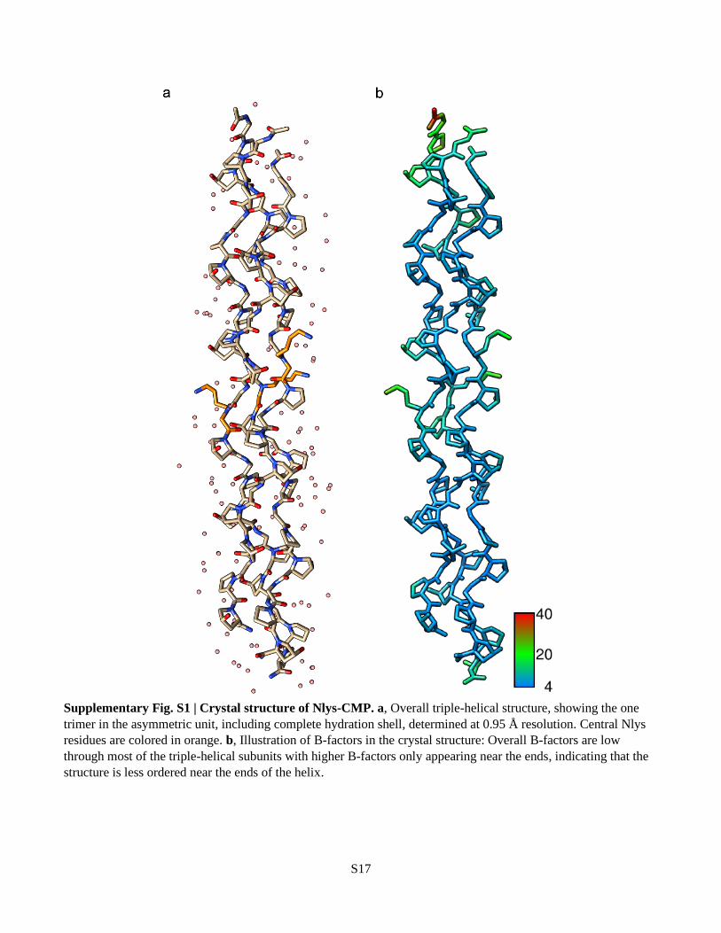
Supplementary Fig. S1 | Crystal structure of Nlys-CMP. a, Overall triple-helical structure, showing the one
trimer in the asymmetric unit, including complete hydration shell, determined at 0.95 Å resolution. Central Nlys
residues are colored in orange. b, Illustration of B-factors in the crystal structure: Overall B-factors are low
through most of the triple-helical subunits with higher B-factors only appearing near the ends, indicating that the
structure is less ordered near the ends of the helix.

S18
Supplementary Fig. S2 | Crystal structure of Nphe-CMP. a, Overall triple-helical structure, showing the two
trimers in the asymmetric unit, including the complete hydration shell, determined at 1.10 Å resolution. Central
Nphe residues are colored in green. b, Illustration of B-factors in the crystal structure: Overall B-factors are low
through most of the triple helical subunits with higher B-factors only appearing near the ends, indicating that the
structure is less ordered near the ends of the helix.

S19
Supplementary Fig. S3a | Distribution of glycine φ and ψ angles in the solved crystal structures for Nlys- (red)
and Nphe-CMPs (blue) vs glycine residues in collagen structures from literature (grey). Average dihedral angles
(φ, ψ) for all non-terminal glycine residues are almost identical to previously reported structures: (-69 ± 3°, 174 ±
5°). We removed the terminal glycines because they have very high B-factors. The collagen structures used for
comparison are listed in Supplementary Table S4.
Supplementary Fig. S3b | Distribution of proline φ and ψ angles in the solved crystal structures for Nlys- (red)
and Nphe-CMPs (blue) vs X position amino acids in collagen structures from literature (grey). Average dihedral
angles for all proline residues are (-70 ± 6°, 162 ± 6°). The collagen structures used for comparison are listed in
Supplementary Table S4.
Supplementary Fig. S3c | Distribution of hydroxyproline φ and ψ angles in the solved crystal structures for Nlys-
(red) and Nphe-CMPs (blue) vs Y position amino acids in collagen structures from literature (grey). Average
dihedral angles for all proline residues are (-58 ± 5°, 147 ± 7°). The collagen structures used for comparison are
listed in Supplementary Table S4.
-180
-150
-120
-90 -60 -30
(
)
()
120
150
180
-90 -60 -30
(
)
()
-180
-90
0
90
180
-180 -90 0 90 180
(
)
()
-180
-90
0
90
180
-180 -90 0 90 180
(
)
()
120
150
180
-90 -60 -30
(
)
()
-180
-90
0
90
180
-180 -90 0 90 180
(
)
()
120
150
180
-90 -60 -30
(
)
()

S20
Supplementary Fig. S4 | Newman-like Projection of NMe-Ala showing the steric clash of adjacent methyl
groups in the polyproline backbone conformation.

S21
Supplementary Fig. S5 | MA-PEG-CMPs produce a stable hydrogel capable of UV-induced dissolution. a,
Schematic of hydrogel formation by reacting an 8-arm PEG hydrogel with X-CMPs. Maleimide groups at the
termini of the 8-arm PEG react with the thiol-containing X-CMPs to form a triple helix crosslinked hydrogel. b,
HPLC of MA-PEG-CMP showing 70% of the X-CMP conjugated to the 8-arm PEG. c, MA-PEG-CMP mixed
with F-Nnbz2-CMP forms a green hydrogel that traps the green colored F-Nnbz2-CMP via triple helical
crosslinks. Upon UV exposure, the triple helical crosslinks unfold, releasing the bound F-Nnbz2X-CMP into
solution.

S22
Supplementary Fig. S6 | Light micrographs showing spatial control of cell attachment by MA-PEG-CMP.
MDA-MB-231 cells seeded onto a gelatin substrate coated with the MA-PEG-CMP hydrogel (a). The coated
hydrogel had previously been exposed to UV light through a photomask (b) and cells bound only to areas which
had been exposed. The results indicate that the MA-PEG-CMP binds to gelatin preventing cell attachment to the
gelatin substrate and can be photo-removed to recover cell adherence.
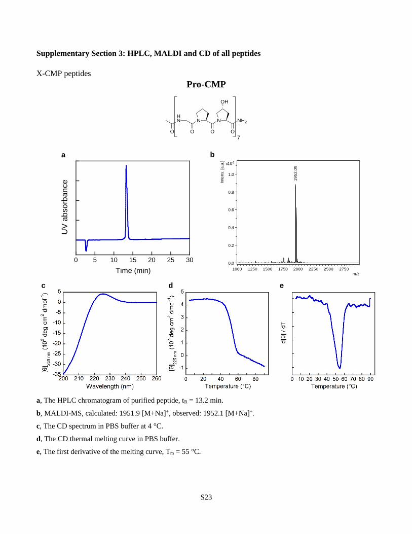
S23
Supplementary Section 3: HPLC, MALDI and CD of all peptides
X-CMP peptides
Pro-CMP
a, The HPLC chromatogram of purified peptide, tR = 13.2 min.
b, MALDI-MS, calculated: 1951.9 [M+Na]+, observed: 1952.1 [M+Na]+.
c, The CD spectrum in PBS buffer at 4 °C.
d, The CD thermal melting curve in PBS buffer.
e, The first derivative of the melting curve, Tm = 55 °C.
HN N
O O
N
OH
O
NH2
O7
0 5 10 15 20 25 30
UV
ab
so
rban
ce
Time (min)
19
52
.09
0.0
0.2
0.4
0.6
0.8
1.0
4x10
Inte
ns. [a
.u.]
1000 1250 1500 1750 2000 2250 2500 2750m/z
c d e
a b

S24
Pro-Ile-CMP
a, The HPLC chromatogram of purified peptide, tR = 15.7 min.
b, MALDI-MS, calculated: 1951.9 [M+Na]+, observed: 1951.5 [M+Na]+, 1967.5 [M+K]+.
c, The CD spectrum in PBS buffer at 4 °C.
d, The CD thermal melting curve in PBS buffer.
e, The first derivative of the melting curve, Tm = 45.5 °C.
HN N
O O
N
OH
O
NH2
HN N
O O
N
OH
O
HN N
O O
HN
OO
3 3
0 5 10 15 20 25 30
UV
ab
so
rban
ce
Time (min)
19
51
.47
0
1000
2000
3000
4000Inte
ns. [a
.u.]
1000 1250 1500 1750 2000 2250 2500 2750m/z
c d e
a b

S25
Gly-CMP
a, The HPLC chromatogram of purified peptide, tR = 13.3 min.
b, MALDI-MS, calculated: 1911.9 [M+Na]+, observed: 1911.4 [M+Na]+.
c, The CD spectrum in PBS buffer at 4 °C.
d, The CD thermal melting curve in PBS buffer.
e, The first derivative of the melting curve, Tm = 37 °C.
HN N
O O
N
OH
O
NH2
HN N
O O
N
OH
O
HN
HN
O O
N
OH
OO
3 3
0 5 10 15 20 25 30
UV
ab
so
rban
ce
Time (min)
19
11
.38
0
1000
2000
3000
4000Inte
ns. [a
.u.]
1000 1250 1500 1750 2000 2250 2500 2750m/z
c d e
a b

S26
Lys-CMP
a, The HPLC chromatogram of purified peptide, tR = 12.5 min.
b, MALDI-MS, calculated: 1960.9 [M+H]+, observed: 1960.7 [M+H]+, 1982.7 [M+Na]+, 1998.7 [M+K]+.
c, The CD spectrum in PBS buffer at 4 °C.
d, The CD thermal melting curves in PBS buffer (blue) and 32 mM NaOH solution (pH 12.51, red).
e, The first derivatives of the melting curves, Tm = 47 °C (PBS, blue) or 46 °C (NaOH, red).
HN N
O O
N
OH
O
NH2
HN N
O O
N
OH
O
HN
HN
O O
N
OH
OO
3 3
NH2
0 5 10 15 20 25 30
UV
ab
so
rban
ce
Time (min)
19
60
.67
0.0
0.2
0.4
0.6
0.8
4x10
Inte
ns. [a
.u.]
1000 1250 1500 1750 2000 2250 2500 2750m/z
c d e
a b

S27
Phe-CMP
a, The HPLC chromatogram of purified peptide, tR = 16.4 min.
b, MALDI-MS, calculated: 2001.9 [M+Na]+, observed: 2001.9 [M+Na]+.
c, The CD spectrum in PBS buffer at 4 °C.
d, The CD thermal melting curve in PBS buffer.
e, The first derivative of the melting curve, Tm = 38 °C.
HN N
O O
N
OH
O
NH2
HN N
O O
N
OH
O
HN
HN
O O
N
OH
OO
3 3
0 5 10 15 20 25 30
UV
ab
so
rban
ce
Time (min)
20
01
.85
0
1000
2000
3000
4000
5000
Inte
ns. [a
.u.]
1250 1500 1750 2000 2250 2500 2750m/z
c d e
a b

S28
Sar-CMP
a, The HPLC chromatogram of purified peptide, tR = 12.8 min.
b, MALDI-MS, calculated: 1925.9 [M+Na]+, observed: 1926.8 [M+Na]+.
c, The CD spectrum in PBS buffer at 4 °C.
d, The CD thermal melting curve in PBS buffer.
e, The first derivative of the melting curve, Tm = 50 °C.
HN N
O O
N
OH
O
NH2
HN N
O O
N
OH
O
HN N
O O
N
OH
OO
3 3
0 5 10 15 20 25 30
UV
ab
so
rban
ce
Time (min)
19
26
.78
0
2000
4000
6000
8000
Inte
ns. [a
.u.]
1250 1500 1750 2000 2250 2500 2750m/z
c d e
a b

S29
Nakn-CMP
a, The HPLC chromatogram of purified peptide, tR = 15.7 min.
b, MALDI-MS, calculated: 1949.9 [M+Na]+, observed: 1951.1[M+Na]+.
c, The CD spectrum in PBS buffer at 4 °C.
d, The CD thermal melting curve in PBS buffer.
e, The first derivative of the melting curve, Tm = 52 °C.
HN N
O O
N
OH
O
NH2
HN N
O O
N
OH
O
HN N
O O
N
OH
OO
3 3
0 5 10 15 20 25 30
UV
ab
so
rban
ce
Time (min)
c d e
a b
19
51
.09
0
500
1000
1500
Inte
ns.
[a.u
.]
1000 1250 1500 1750 2000 2250 2500 2750m/z

S30
Nakn-OH-CMP
a, The HPLC chromatogram of purified peptide, tR = 16.3 min.
b, MALDI-MS, calculated: 2036.9 [M+Na]+, observed: 2037.6 [M+Na]+.
c, The CD spectrum in PBS buffer at 4 °C.
d, The CD thermal melting curve in PBS buffer.
e, The first derivative of the melting curve, Tm = 55 °C.
HN N
O O
N
OH
O
NH2
HN N
O O
N
OH
O
HN N
O O
N
OH
OO
3 3
NN
N
OH
c d e
a b
20
37
.62
0
500
1000
1500
2000
2500
Inte
ns. [a
.u.]
1000 1250 1500 1750 2000 2250 2500 2750m/z
-35
-30
-25
-20
-15
-10
-5
0
5
200 210 220 230 240 250 260
[] 2
25
nm
(10
3 d
eg
cm
2 d
mo
l-1)
Wavelength (nm)
0 5 10 15 20 25 30
UV
ab
so
rban
ce
Time (min)
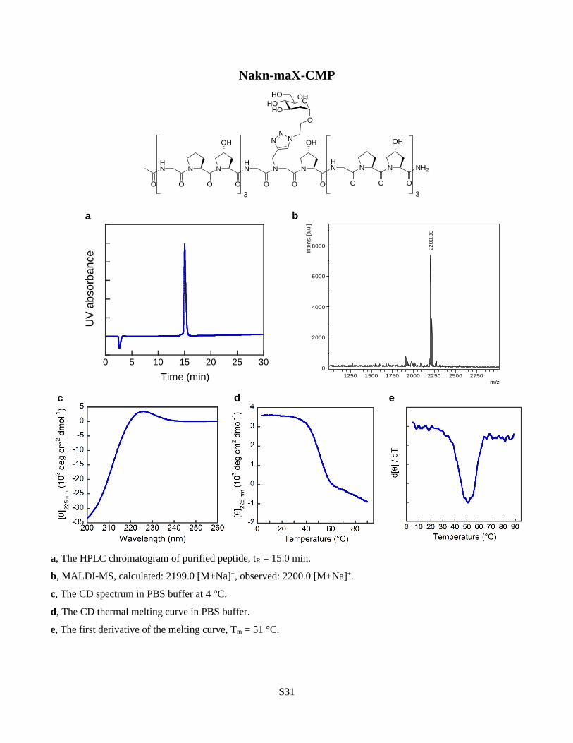
S31
Nakn-maX-CMP
a, The HPLC chromatogram of purified peptide, tR = 15.0 min.
b, MALDI-MS, calculated: 2199.0 [M+Na]+, observed: 2200.0 [M+Na]+.
c, The CD spectrum in PBS buffer at 4 °C.
d, The CD thermal melting curve in PBS buffer.
e, The first derivative of the melting curve, Tm = 51 °C.
HN N
O O
N
OH
O
NH2
HN N
O O
N
OH
O
HN N
O O
N
OH
OO
3 3
NN
N
O
OHOHO
HO OH
0 5 10 15 20 25 30
UV
ab
so
rban
ce
Time (min)
c d e
a b
22
00
.00
0
2000
4000
6000
8000
Inte
ns.
[a.u
.]
1250 1500 1750 2000 2250 2500 2750m/z

S32
Nasn-CMP
a, The HPLC chromatogram of purified peptide, tR = 11.6 min.
b, MALDI-MS, calculated: 1968.9 [M+Na]+, observed: 1968.5 [M+Na]+.
c, The CD spectrum in PBS buffer at 4 °C.
d, The CD thermal melting curve in PBS buffer.
e, The first derivative of the melting curve, Tm = 55 °C.
HN N
O O
N
OH
O
NH2
HN N
O O
N
OH
O
HN N
O O
N
OH
OO
3 3
O
NH2
19
68
.48
0
1000
2000
3000
4000
5000
Inte
ns. [a
.u.]
1000 1250 1500 1750 2000 2250 2500 2750m/z
0 5 10 15 20 25 30
UV
ab
so
rban
ce
Time (min)
c d e
a b

S33
Nasp-CMP
a, The HPLC chromatogram of purified peptide, tR = 12.7 min.
b, MALDI-MS, calculated: 1945.9 [M-H]-, observed: 1945.5 [M-H]-.
c, The CD spectrum in PBS buffer at 4 °C.
d, The CD thermal melting curves in PBS buffer (blue) and 15 mM HCl solution (pH 1.82, red).
e, The first derivatives of the melting curves, Tm = 50 °C (PBS, blue) or 56 °C (HCl, red).
HN N
O O
N
OH
O
NH2
HN N
O O
N
OH
O
HN N
O O
N
OH
OO
3 3
O
OH
0 5 10 15 20 25 30
UV
ab
so
rban
ce
Time (min)
19
45
.51
0
2000
4000
6000
Inte
ns. [a
.u.]
1000 1250 1500 1750 2000 2250 2500 2750m/z
c d e
a b
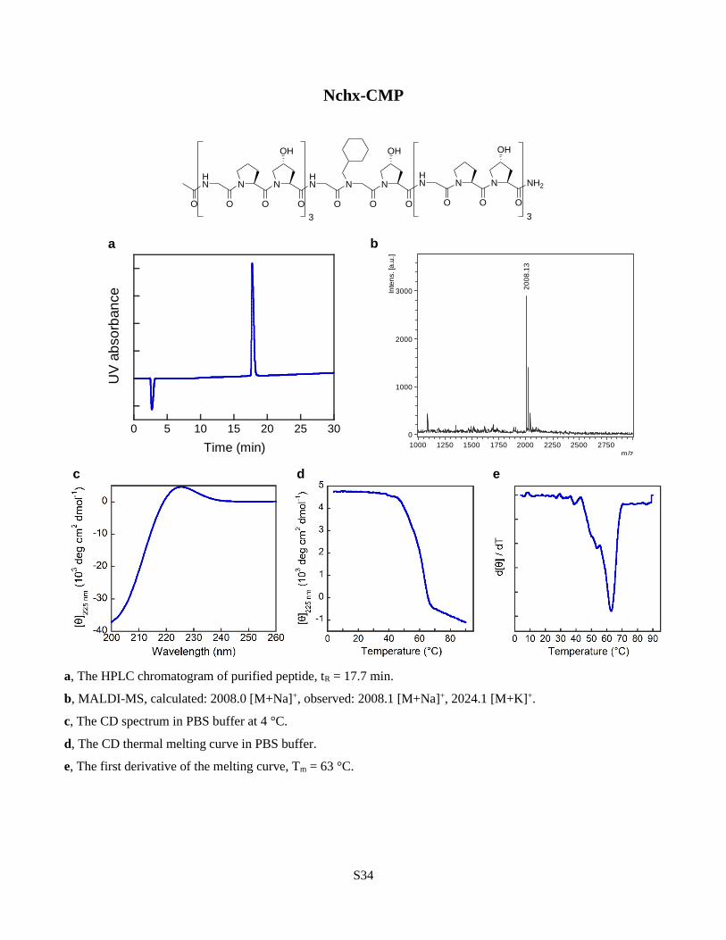
S34
Nchx-CMP
a, The HPLC chromatogram of purified peptide, tR = 17.7 min.
b, MALDI-MS, calculated: 2008.0 [M+Na]+, observed: 2008.1 [M+Na]+, 2024.1 [M+K]+.
c, The CD spectrum in PBS buffer at 4 °C.
d, The CD thermal melting curve in PBS buffer.
e, The first derivative of the melting curve, Tm = 63 °C.
HN N
O O
N
OH
O
NH2
HN N
O O
N
OH
O
HN N
O O
N
OH
OO
3 3
0 5 10 15 20 25 30
UV
ab
so
rban
ce
Time (min)
20
08
.13
0
1000
2000
3000Inte
ns. [a
.u.]
1000 1250 1500 1750 2000 2250 2500 2750m/z
c d e
a b

S35
Ndxn-CMP
a, The HPLC chromatogram of purified peptide, tR = 14.2 min.
b, MALDI-MS, calculated: 2011.9 [M+Na]+, observed: 2011.5 [M+Na]+, 2027.5 [M+K]+.
c, The CD spectrum in PBS buffer at 4 °C.
d, The CD thermal melting curve in PBS buffer.
e, The first derivative of the melting curve, Tm = 59 °C.
HN N
O O
N
OH
O
NH2
HN N
O O
N
OH
O
HN N
O O
N
OH
OO
3 3
O
O
0 5 10 15 20 25 30
UV
ab
so
rban
ce
Time (min)
20
11
.54
0.0
0.2
0.4
0.6
0.8
1.0
4x10
Inte
ns. [a
.u.]
1000 1250 1500 1750 2000 2250 2500 2750m/z
c d e
a b

S36
NEt-CMP
a, The HPLC chromatogram of purified peptide, tR = 15.7 min.
b, MALDI-MS, calculated: 1939.9 [M+Na]+, observed: 1939.5 [M+Na]+.
c, The CD spectrum in PBS buffer at 4 °C.
d, The CD thermal melting curve in PBS buffer.
e, The first derivative of the melting curve, Tm = 52 °C.
HN N
O O
N
OH
O
NH2
HN N
O O
N
OH
O
HN N
O O
N
OH
OO
3 3
0 5 10 15 20 25 30
UV
ab
so
rban
ce
Time (min)
c d e
a b
19
39
.50
0
1000
2000
3000
4000
Inte
ns.
[a.u
.]
1000 1250 1500 1750 2000 2250 2500 2750m/z

S37
Nhcy-CMP
a, The HPLC chromatogram of purified peptide, tR = 16.2 min.
b, MALDI-MS, calculated: 1971.9 [M+Na]+, observed: 1972.1 [M+Na]+.
c, The CD spectrum in PBS buffer at 4 °C.
d, The CD thermal melting curve in PBS buffer.
e, The first derivative of the melting curve, Tm = 54 °C.
HN N
O O
N
OH
O
NH2
HN N
O O
N
OH
O
HN N
O O
N
OH
OO
3 3
HS
0 5 10 15 20 25 30
UV
ab
so
rban
ce
Time (min)
c d e
a b
19
72
.10
0
500
1000
1500
2000
2500
3000
Inte
ns.
[a.u
.]
1000 1250 1500 1750 2000 2250 2500 2750m/z

S38
Nleu-CMP
a, The HPLC chromatogram of purified peptide, tR = 15.2 min.
b, MALDI-MS, calculated: 1967.9 [M+Na]+, observed: 1967.9 [M+Na]+, 1983.9 [M+K]+.
c, The CD spectrum in PBS buffer at 4 °C.
d, The CD thermal melting curve in PBS buffer.
e, The first derivative of the melting curve, Tm = 58 °C.
HN N
O O
N
OH
O
NH2
HN N
O O
N
OH
O
HN N
O O
N
OH
OO
3 3
0 5 10 15 20 25 30
UV
ab
so
rban
ce
Time (min)
19
67
.92
0
2000
4000
6000
8000
Inte
ns. [a
.u.]
1000 1250 1500 1750 2000 2250 2500 2750m/z
c d e
a b

S39
Nlys-CMP
a, The HPLC chromatogram of purified peptide, tR = 12.9 min.
b, MALDI-MS, calculated: 1960.9 [M+H]+, observed: 1961.0 [M+H]+, 1983.0 [M+Na]+, 1999.0 [M+K]+.
c, The CD spectrum in PBS buffer at 4 °C.
d, The CD thermal melting curves in PBS buffer (blue) and 35 mM NaOH solution (pH 12.54, red).
e, The first derivatives of the melting curves, Tm = 55 °C (PBS, blue) or 56 °C (NaOH, red).
HN N
O O
N
OH
O
NH2
HN N
O O
N
OH
O
HN N
O O
N
OH
OO
3 3
H2N
0 5 10 15 20 25 30
UV
ab
so
rban
ce
Time (min)
19
60
.97
19
98
.97
0
500
1000
1500
2000
2500
3000
Inte
ns. [a
.u.]
1000 1250 1500 1750 2000 2250 2500 2750m/z
c d e
a b

S40
NMe-Ala-CMP
a, The HPLC chromatogram of purified peptide, tR = 13.1 min.
b, MALDI-MS, calculated: 1939.9 [M+Na]+, observed: 1940.3 [M+Na]+.
c, The CD spectrum in PBS buffer at 4 °C.
d, The CD thermal melting curve of in PBS buffer.
e, The first derivative of the melting curve, Tm = 40 °C.
HN N
O O
N
OH
O
NH2
HN N
O O
N
OH
O
HN N
O O
N
OH
OO
3 3
0 5 10 15 20 25 30
UV
ab
so
rban
ce
Time (min)
19
40
.33
0
1000
2000
3000
4000
5000
6000
Inte
ns. [a
.u.]
1000 1250 1500 1750 2000 2250 2500 2750m/z
c d e
a b

S41
Nmet-CMP
a, The HPLC chromatogram of purified peptide, tR = 16.7 min.
b, MALDI-MS, calculated: 1986.9 [M+Na]+, observed: 1987.1 [M+Na]+.
c, The CD spectrum in PBS buffer at 4 °C.
d, The CD thermal melting curve in PBS buffer.
e, The first derivative of the melting curve, Tm = 55 °C.
HN N
O O
N
OH
O
NH2
HN N
O O
N
OH
O
HN N
O O
N
OH
OO
3 3
S
c d e
a b
19
87
.08
0
1000
2000
3000
4000
Inte
ns.
[a.u
.]
1000 1250 1500 1750 2000 2250 2500 2750m/z
0 5 10 15 20 25 30
UV
ab
so
rban
ce
Time (min)

S42
Nnbz-CMP
a, The HPLC chromatogram of purified peptide, tR = 15.9 min.
b, MALDI-MS, calculated: 2046.9 [M+Na]+, observed: 2046.9 [M+Na]+.
c, The CD spectrum in PBS buffer at 4 °C.
d, The CD thermal melting curve in PBS buffer.
e, The first derivative of the melting curve, Tm = 62 °C.
HN N
O O
N
OH
O
NH2
HN N
O O
N
OH
O
HN N
O O
N
OH
OO
3 3
O2N
0 5 10 15 20 25 30
UV
ab
so
rban
ce
Time (min)
20
46
.86
0
2000
4000
6000
8000Inte
ns. [a
.u.]
1000 1250 1500 1750 2000 2250 2500 2750m/z
c d e
a b

S43
Nnbz2-CMP
a, The HPLC chromatogram of purified peptide, tR = 18.3 min.
b, MALDI-MS, calculated: 2141.9 [M+Na]+, observed: 2141.9 [M+Na]+.
c, The CD spectrum in PBS buffer at 4 °C.
d, The CD thermal melting curve in PBS buffer.
e, The first derivative of the melting curve, Tm = 73 °C.
HN N
O O
N
OH
O
HN N
O O
N
OH
OO
2
O2N
HN N
O O
N
OH
O
NH2
HN N
O O
N
OH
O
3
O2N
0 5 10 15 20 25 30
UV
ab
so
rban
ce
Time (min)
21
41
.91
0.0
0.5
1.0
1.5
4x10
Inte
ns. [a
.u.]
1000 1250 1500 1750 2000 2250 2500 2750m/z
c d e
a b

S44
Nnbz3-CMP
a, The HPLC chromatogram of purified peptide, tR = 20.4 min.
b, MALDI-MS, calculated: 2236.9 [M+Na]+, observed: 2236.9 [M+Na]+.
c, The CD spectrum in PBS buffer at 4 °C.
d, The CD thermal melting curve in PBS buffer.
e, The first derivative of the melting curve, Tm = 79 °C.
HN N
O O
N
OH
O
HN N
O O
N
OH
OO
2
O2N
HN N
O O
N
OH
O
O2N
HN N
O O
N
OH
O
NH2
HN N
O O
N
OH
O
2
O2N
0 5 10 15 20 25 30
UV
ab
so
rban
ce
Time (min)
22
36
.92
0.00
0.25
0.50
0.75
1.00
1.25
4x10
Inte
ns. [a
.u.]
1000 1250 1500 1750 2000 2250 2500 2750m/z
c d e
a b

S45
Nphe-CMP
a, The HPLC chromatogram of purified peptide, tR = 15.6 min.
b, MALDI-MS, calculated: 2001.9 [M+Na]+, observed: 2001.8 [M+Na]+, 2017.8 [M+K]+.
c, The CD spectrum in PBS buffer at 4 °C.
d, The CD thermal melting curve in PBS buffer.
e, The first derivative of the melting curve, Tm = 62 °C.
HN N
O O
N
OH
O
NH2
HN N
O O
N
OH
O
HN N
O O
N
OH
OO
3 3
0 5 10 15 20 25 30
UV
ab
so
rban
ce
Time (min)
20
01
.83
0
2000
4000
6000
8000
Inte
ns. [a
.u.]
1000 1250 1500 1750 2000 2250 2500 2750m/z
c d e
a b

S46
Nphe-Ile-CMP
a, The HPLC chromatogram of purified peptide, tR = 19.2 min.
b, MALDI-MS, calculated: 2001.9 [M+Na]+, observed: 2002.6 [M+Na]+.
c, The CD spectrum -Ile in PBS buffer at 4 °C.
d, The CD thermal melting curve in PBS buffer.
e, The first derivative of the melting curve, Tm = 51 °C.
HN N
O O
N
OH
O
NH2
HN N
O O
N
OH
O
HN N
O O
HN
OO
3 3
0 5 10 15 20 25 30
UV
ab
so
rban
ce
Time (min)
20
02
.57
0
2000
4000
6000
Inte
ns. [a
.u.]
1000 1250 1500 1750 2000 2250 2500 2750m/z
c d e
a b

S47
Nrpe-CMP
a, The HPLC chromatogram of purified peptide, tR = 18.6 min.
b, MALDI-MS, calculated: 2015.9 [M+Na]+, observed: 2016.6 [M+Na]+.
c, The CD spectrum in PBS buffer at 4 °C.
d, The CD thermal melting curve in PBS buffer.
e, The first derivative of the melting curve.
HN N
O O
N
OH
O
NH2
HN N
O O
N
OH
O
HN N
O O
N
OH
OO
3 3
0 5 10 15 20 25 30
UV
ab
so
rban
ce
Time (min)
c d e
a b
20
16
.64
0
1000
2000
3000
4000
5000
Inte
ns.
[a.u
.]
1000 1250 1500 1750 2000 2250 2500 2750m/z

S48
Nspe-CMP
a, The HPLC chromatogram of purified peptide, tR = 18.7 min.
b, MALDI-MS, calculated: 2015.9 [M+Na]+, observed: 2015.7 [M+Na]+.
c, The CD spectrum in PBS buffer at 4 °C.
d, The CD thermal melting curve in PBS buffer.
e, The first derivative of the melting curve, Tm = 48 °C.
HN N
O O
N
OH
O
NH2
HN N
O O
N
OH
O
HN N
O O
N
OH
OO
3 3
c d e
a b
20
15
.68
0
2000
4000
6000
8000
Inte
ns.
[a.u
.]
1000 1250 1500 1750 2000 2250 2500 2750m/z
0 5 10 15 20 25 30
UV
ab
so
rban
ce
Time (min)
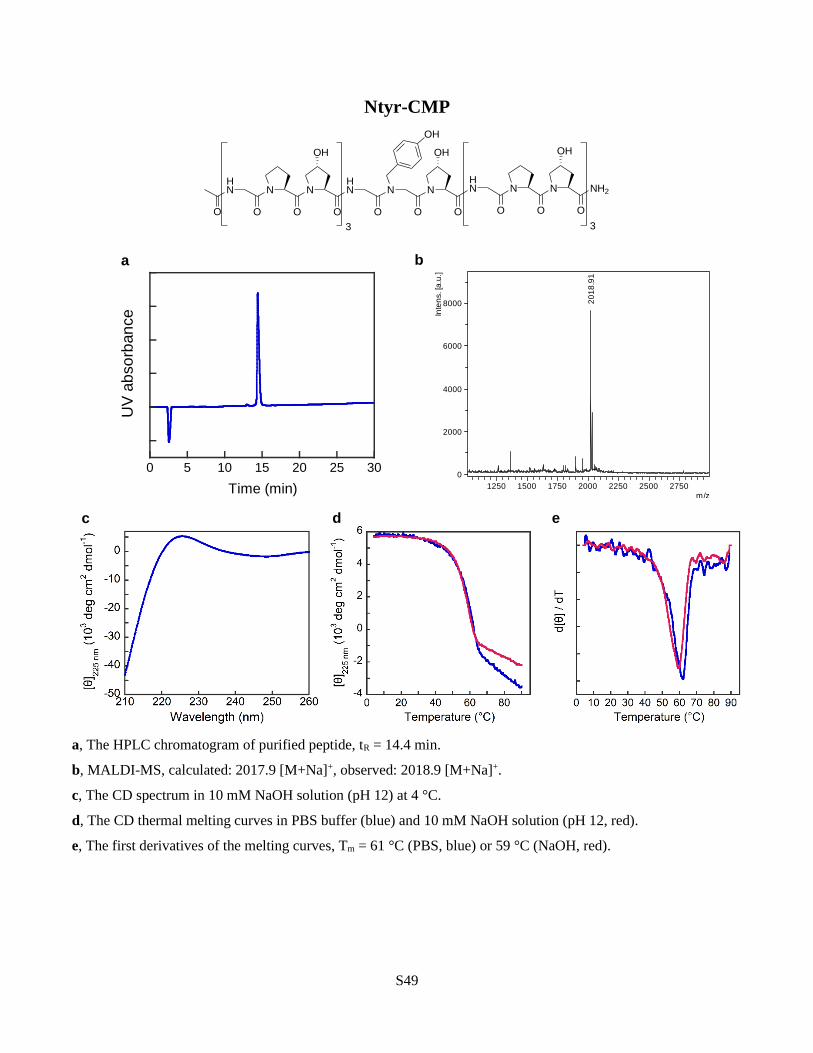
S49
Ntyr-CMP
a, The HPLC chromatogram of purified peptide, tR = 14.4 min.
b, MALDI-MS, calculated: 2017.9 [M+Na]+, observed: 2018.9 [M+Na]+.
c, The CD spectrum in 10 mM NaOH solution (pH 12) at 4 °C.
d, The CD thermal melting curves in PBS buffer (blue) and 10 mM NaOH solution (pH 12, red).
e, The first derivatives of the melting curves, Tm = 61 °C (PBS, blue) or 59 °C (NaOH, red).
HN N
O O
N
OH
O
NH2
HN N
O O
N
OH
O
HN N
O O
N
OH
OO
3 3
OH
0 5 10 15 20 25 30
UV
ab
so
rban
ce
Time (min)
20
18
.91
0
2000
4000
6000
8000
Inte
ns. [a
.u.]
1250 1500 1750 2000 2250 2500 2750m/z
c d e
a b

S50
Nval-CMP
a, The HPLC chromatogram of purified peptide, tR = 16.3 min.
b, MALDI-MS, calculated: 1953.9 [M+Na]+, observed: 1955.0 [M+Na]+.
c, The CD spectrum in PBS buffer at 4 °C.
d, The CD thermal melting curve in PBS buffer.
e, The first derivative of the melting curve, Tm = 36 °C.
HN N
O O
N
OH
O
NH2
HN N
O O
N
OH
O
HN N
O O
N
OH
OO
3 3
c d e
a b
19
55
.00
0.00
0.25
0.50
0.75
1.00
1.25
4x10
Inte
ns.
[a.u
.]
1250 1500 1750 2000 2250 2500 2750m/z
0 5 10 15 20 25 30
UV
ab
so
rban
ce
Time (min)

S51
X7-CMP
a, The HPLC chromatogram of purified peptide, tR = 19.4 min.
b, MALDI-MS, calculated: 2516.2 [M+H]+, observed: 2517.3 [M+H]+.
c, The CD spectrum in PBS buffer at 4 °C.
d, The CD thermal melting curve in PBS buffer.
e, The first derivative of the melting curve, Tm = 56 °C.
HN N
O O
N
OH
O
HN N
O O
N
OH
O
HN N
O O
N
OH
O
HN N
O O
N
OH
O
HN N
O O
N
OH
O
NH2HN N
O O
N
OH
O
HN N
O O
N
OH
O
H2NHN O
N N
NN
OH
O
OH
O
HO
O
O
HO
O
c d e
a b
0 5 10 15 20 25 30
UV
ab
so
rban
ce
Time (min)
25
17
.32
0
2000
4000
6000
8000Inte
ns. [a
.u.]
1500 1750 2000 2250 2500 2750 3000 3250m/z
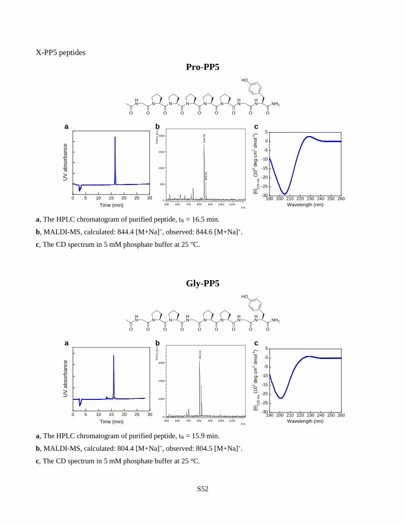
S52
X-PP5 peptides
Pro-PP5
a, The HPLC chromatogram of purified peptide, tR = 16.5 min.
b, MALDI-MS, calculated: 844.4 [M+Na]+, observed: 844.6 [M+Na]+.
c, The CD spectrum in 5 mM phosphate buffer at 25 °C.
Gly-PP5
a, The HPLC chromatogram of purified peptide, tR = 15.9 min.
b, MALDI-MS, calculated: 804.4 [M+Na]+, observed: 804.5 [M+Na]+.
c, The CD spectrum in 5 mM phosphate buffer at 25 °C.
HN N
O O
N
O
N N
O O
N
O
HN
O O
HN NH2
O
HO
HN N
O O
N
O
HN N
O O
N
O
HN
O O
HN NH2
O
HO
80
4.5
3
0
1000
2000
3000
Inte
ns. [a
.u.]
500 600 700 800 900 1000 1100m/z
a b c
0 5 10 15 20 25 30
UV
ab
so
rban
ce
Time (min)
-30
-25
-20
-15
-10
-5
0
5
190 200 210 220 230 240 250 260
[] 2
25
nm
(10
3 d
eg
cm
2 d
mo
l-1)
Wavelength (nm)
84
4.5
88
60
.56
0
500
1000
1500
2000
Inte
ns. [a
.u.]
500 600 700 800 900 1000 1100m/z
a b c
0 5 10 15 20 25 30
UV
ab
so
rban
ce
Time (min)
-30
-25
-20
-15
-10
-5
0
5
190 200 210 220 230 240 250 260
[] 2
25
nm
(10
3 d
eg
cm
2 d
mo
l-1)
Wavelength (nm)

S53
Hyp-PP5
a, The HPLC chromatogram of purified peptide, tR = 15.9 min.
b, MALDI-MS, calculated: 860.4 [M+Na]+, observed: 860.4 [M+Na]+.
c, The CD spectrum in 5 mM phosphate buffer at 25 °C.
Sar-PP5
a, The HPLC chromatogram of purified peptide, tR = 16.6 min.
b, MALDI-MS, calculated: 818.4 [M+Na]+, observed: 818.5 [M+Na]+, 834.5 [M+K]+.
c, The CD spectrum in 5 mM phosphate buffer at 25 °C.
HN N
O O
N
O
N N
O O
N
O
HN
O O
HN NH2
O
HO
OH
HN N
O O
N
O
N N
O O
N
O
HN
O O
HN NH2
O
HO
a b c
81
8.5
38
34
.53
0
500
1000
1500
2000
2500
Inte
ns. [a
.u.]
500 600 700 800 900 1000 1100m/z
0 5 10 15 20 25 30
UV
ab
so
rban
ce
Time (min)
-30
-25
-20
-15
-10
-5
0
5
190 200 210 220 230 240 250 260
[] 2
25
nm
(10
3 d
eg
cm
2 d
mo
l-1)
Wavelength (nm)
86
0.4
4
0
1000
2000
3000
4000
Inte
ns. [a
.u.]
500 600 700 800 900 1000 1100m/z
a b c
0 5 10 15 20 25 30
UV
ab
so
rban
ce
Time (min)
-30
-25
-20
-15
-10
-5
0
5
190 200 210 220 230 240 250 260
[] 2
25
nm
(10
3 d
eg
cm
2 d
mo
l-1)
Wavelength (nm)

S54
Nasn-PP5
a, The HPLC chromatogram of purified peptide, tR = 18.9 min.
b, MALDI-MS, calculated: 861.4 [M+Na]+, observed: 861.5 [M+Na]+, 877.5 [M+K]+.
c, The CD spectrum in 5 mM phosphate buffer at 25 °C.
Nasp-PP5
a, The HPLC chromatogram of purified peptide, tR = 16.2 min.
b, MALDI-MS, calculated: 838.4 [M-H]-, observed: 836.9 [M-H]-.
c, The CD spectrum in 5 mM phosphate buffer at 25 °C.
HN N
O O
N
O
N N
O O
N
O
HN
O O
HN NH2
O
HO
O
NH2
HN N
O O
N
O
N N
O O
N
O
HN
O O
HN NH2
O
HO
O
OH
83
6.8
8
0
1000
2000
3000
Inte
ns. [a
.u.]
500 600 700 800 900 1000 1100m/z
a b c
0 5 10 15 20 25 30
UV
ab
so
rban
ce
Time (min)
-30
-25
-20
-15
-10
-5
0
5
190 200 210 220 230 240 250 260
[] 2
25
nm
(10
3 d
eg
cm
2 d
mo
l-1)
Wavelength (nm)
a b c
86
1.5
18
77
.48
0
1000
2000
3000
Inte
ns. [a
.u.]
500 600 700 800 900 1000 1100m/z
0 5 10 15 20 25 30
UV
ab
so
rban
ce
Time (min)
-30
-25
-20
-15
-10
-5
0
5
190 200 210 220 230 240 250 260
[] 2
25
nm
(10
3 d
eg
cm
2 d
mo
l-1)
Wavelength (nm)

S55
Nleu-PP5
a, The HPLC chromatogram of purified peptide, tR = 19.9 min.
b, MALDI-MS, calculated: 860.4 [M+Na]+, observed: 860.6 [M+Na]+.
c, The CD spectrum in 5 mM phosphate buffer at 25 °C.
Nlys-PP5
a, The HPLC chromatogram of purified peptide, tR = 15.9 min.
b, MALDI-MS, calculated: 853.4 [M+H]+, observed: 853.6 [M+H]+, 875.4 [M+Na]+.
c, The CD spectrum in 5 mM phosphate buffer at 25 °C.
HN N
O O
N
O
N N
O O
N
O
HN
O O
HN NH2
O
HO
HN N
O O
N
O
N N
O O
N
O
HN
O O
HN NH2
O
HOH2N
85
3.6
2
0
500
1000
1500
2000
2500
3000
Inte
ns. [a
.u.]
500 600 700 800 900 1000 1100m/z
a b c
0 5 10 15 20 25 30
UV
ab
so
rban
ce
Time (min)
-30
-25
-20
-15
-10
-5
0
5
190 200 210 220 230 240 250 260
[] 2
25
nm
(10
3 d
eg
cm
2 d
mo
l-1)
Wavelength (nm)
86
0.5
9
0
2000
4000
6000
8000
Inte
ns. [a
.u.]
500 600 700 800 900 1000 1100m/z
a b c
0 5 10 15 20 25 30
UV
ab
so
rban
ce
Time (min)
-30
-25
-20
-15
-10
-5
0
5
190 200 210 220 230 240 250 260
[] 2
25
nm
(10
3 d
eg
cm
2 d
mo
l-1)
Wavelength (nm)

S56
NMe-Ala-PP5
a, The HPLC chromatogram of purified peptide, tR = 17.1 min.
b, MALDI-MS, calculated: 832.4 [M+Na]+, observed: 832.3 [M+Na]+, 848.2 [M+K]+.
c, The CD spectrum in 5 mM phosphate buffer at 25 °C.
Nphe-PP5
a, The HPLC chromatogram of purified peptide, tR = 21.1 min.
b, MALDI-MS, calculated: 894.4 [M+Na]+, observed: 894.6 [M+Na]+.
c, The CD spectrum in 5 mM phosphate buffer at 25 °C.
HN N
O O
N
O
N N
O O
N
O
HN
O O
HN NH2
O
HO
HN N
O O
N
O
N N
O O
N
O
HN
O O
HN NH2
O
HO
89
4.5
69
10
.53
0
1000
2000
3000
4000Inte
ns. [a
.u.]
500 600 700 800 900 1000 1100m/z
a b c
-30
-25
-20
-15
-10
-5
0
5
190 200 210 220 230 240 250 260
[] 2
25
nm
(10
3 d
eg
cm
2 d
mo
l-1)
Wavelength (nm)
0 5 10 15 20 25 30
UV
ab
so
rban
ce
Time (min)
83
2.2
58
48
.22
0
1000
2000
3000
4000
5000
Inte
ns. [a
.u.]
500 600 700 800 900 1000 1100m/z
a b c
0 5 10 15 20 25 30
UV
ab
so
rban
ce
Time (min)
-30
-25
-20
-15
-10
-5
0
5
190 200 210 220 230 240 250 260
[] 2
25
nm
(10
3 d
eg
cm
2 d
mo
l-1)
Wavelength (nm)
1
1

S57
Nval-PP5
a, The HPLC chromatogram of purified peptide, tR = 21.0 min.
b, MALDI-MS, calculated: 846.4 [M+Na]+, observed: 846.5 [M+Na]+.
c, The CD spectrum in 5 mM phosphate buffer at 25 °C.
HN N
O O
N
O
N N
O O
N
O
HN
O O
HN NH2
O
HO
84
6.5
10
1000
2000
3000
4000
Inte
ns. [a
.u.]
500 600 700 800 900 1000 1100m/z
a b c
-30
-25
-20
-15
-10
-5
0
5
190 200 210 220 230 240 250 260
[] 2
25
nm
(1
03 d
eg
cm
2 d
mo
l-1)
Wavelength (nm)
0 5 10 15 20 25 30
UV
ab
so
rban
ce
Time (min)

S58
Supplementary Section 4: Supplementary Discussion
X-CMP functionalization through click chemistry. One simple way to introduce functionality into a
CMP is to install an alkyne or azido group, which allows facile derivatization through ‘click’ chemistry.
To prepare an X-CMP capable of undergoing such reactions, we prepared Nakn-CMP which features a
central N-gly residue bearing an alkyne moiety. Nakn’s unbranched aliphatic sidechain is similar to
that of NEt (Fig. 6a) and the two residues produced almost identical CD melting temperatures. Click
reactions between the sidechain alkyne and azides bearing unprotected hydroxyl or mannose moieties
proceeded efficiently on resin in the presence of Cu(I)/TBTA (Fig. 6a and SI Methods). Following click
conjugation with 2-azidoethanol, the Tm of the Nakn triple helix increased by 3 °C, presumably due to
the increased size of the N-Cα-triazole ring (Fig. 6a). Conjugation with 2-azidoethylmannose had
almost no effect on Tm (Fig. 6a). Previously, click reactions were used to functionalize CMP via
(4R)azidoproline; however the formation of the triazole-proline units resulted in Tm drop of at least
7 °C11. Our results demonstrate that the N-appended sidechain can better tolerate spatially demanding
triazole units within the triple helix than Cγ‐substituted proline derivatives11 and showcase a superior
approach for click-functionalization of CMPs.

S59
References
1. Zuckermann, R. N., Kerr, J. M., Moosf, W. H. & Kent, S. B. H. Efficient Method for the Preparation of
Peptoids [Oligo(N-substituted glycines)] by Submonomer Solid-Phase Synthesis. J. Am. Chem. Soc. 114,
10646–10647 (1992).
2. Li, Y. et al. Targeting collagen strands by photo-triggered triple-helix hybridization. Proc. Natl. Acad. Sci.
109, 14767–14772 (2012).
3. Li, Y. et al. Non-Covalent Photo-Patterning of Gelatin Matrices Using Caged Collagen Mimetic Peptides.
Macromol. Biosci. 15, 52–62 (2015).
4. Otwinowski, Z. & Minor, W. Processing of X-Ray Diffraction Data Collected in Oscillation Mode. in
Methods in Enzymology (eds. Carter Jr., C. W. & Sweet, R. M.) vol. 276 307–326 (Academic Press, 1997).
5. McCoy, A. J. et al. Phaser crystallographic software. J. Appl. Crystallogr. 40, 658–674 (2007).
6. Emsley, P., Lohkamp, B., Scott, W. G. & Cowtan, K. Features and development of Coot. Acta Crystallogr.
Sect. D Biol. Crystallogr. 66, 486–501 (2010).
7. Murshudov, G. N. et al. REFMAC 5 for the refinement of macromolecular crystal structures research
papers. Acta Crystallogr. Sect. D Biol. Crystallogr. D67, 355–367 (2011).
8. Vanommeslaeghe, K. et al. CHARMM General Force Field: A Force Field for Drug-Like Molecules
Compatible with the CHARMM All-Atom Additive Biological Force Fields. J. Comput. Chem. 31, 671–
690 (2010).
9. MacKerell, A. D. et al. All-atom empirical potential for molecular modeling and dynamics studies of
proteins. J. Phys. Chem. B 102, 3586–3616 (1998).
10. Binkley, J. S., Pople, J. A. & Hehre, W. J. Self-consistent molecular orbital methods. 21. Small split-
valence basis sets for first-row elements. J. Am. Chem. Soc. 102, 939–947 (1980).
11. Erdmann, R. S. & Wennemers, H. Functionalizable collagen model peptides. J. Am. Chem. Soc. 132,
13957–13959 (2010).

download fileview on ChemRxivChemRxiv-SI-Peptoid-CMP_submitted (20200905).pdf (4.82 MiB)