periappendageal lichen nitidus: report of a case
TRANSCRIPT

J Cutan Pathol 2002: 29: 125–128 Copyright C Blackwell Munksgaard 2002Blackwell Munksgaard . Printed in Denmark
Journal ofCutaneous Pathology
ISSN 0303-6987
Periappendageal lichen nitidus: report ofa caseBackground: The histology of lichen nitidus has been described Scott Sanders1,previously but a follicular variant has not been emphasized. De Anne H. Collier1,Method: We report a case of lichen nitidus with periappendageal Rachelle Scott1,inflammation resulting in histologic similarities to lichen striatus. Hong Wu2 andResults: This case extends the spectrum of histologic findings in N. Scott McNutt2
lichen nitidus and shows overlap in the distribution of the inflammatory 1Department of Dermatology and 2Division ofinfiltrate in lichen nitidus and lichen striatus. Dermatopathology, Department of Pathology,
Cornell University Weill Medical Center,New York, New York, USASanders S, Collier DAH, Scott R, Wu H, McNutt NS.
Periappendageal lichen nitidus: report of a case.Scott Sanders, Department of Dermatology, CornellJ Cutan Pathol 2002; 29: 125–128. C Blackwell Munksgaard 2002. University Weill Medical Center, 525 East 68th Street,New York, New York 10021, USATel: π1 212 772 0128e-mail: skinsand/aol.com
Accepted 9 July, 2001
Lichen nitidus is an idiopathic inflammatory derma-tosis first described nearly a century ago by FelixPinkus.1,2 Its etiology and relationship to lichenplanus have been the subject of continuing specu-lation.3,4 The clinical and histopathologic features oflichen nitidus are considered to be fairly standard,without much variation,5 although unusual presen-tations have been described previously.6–25 We reportherein a case of lichen nitidus with periappendagealinflammation. Literature review reveals a similar caseof lichen nitidus with perifollicular granulomas.26 Al-though periappendageal inflammation is common inlichen striatus,27 three cases of lichen striatus combin-ing periappendageal inflammation with superficialfeatures of lichen nitidus have been reported.28,29
These three cases of lichen striatus thus have an histo-pathology identical to our case. Our case extends thehistopathologic features of lichen nitidus and maysuggest a morphologic spectrum between lichennitidus and lichen striatus.
Case reportAn 8-year-old African-American boy presented withan asymptomatic rash on his face, legs and armswhich had been present for 11 months. The child hadbeen born with a tracheoesophageal fistula which was
125
repaired with three surgeries, the first at age 1 yearand the last at age 5. Despite a gastric feeding tube,he had experienced multiple aspiration pneunomiasand was ultimately diagnosed with gastroesophagealreflux disease and asthma. Serial tuberculin tests werenegative. At the time of presentation he was takingcimetidine for 18 months and albuterol inhalers forseveral years.
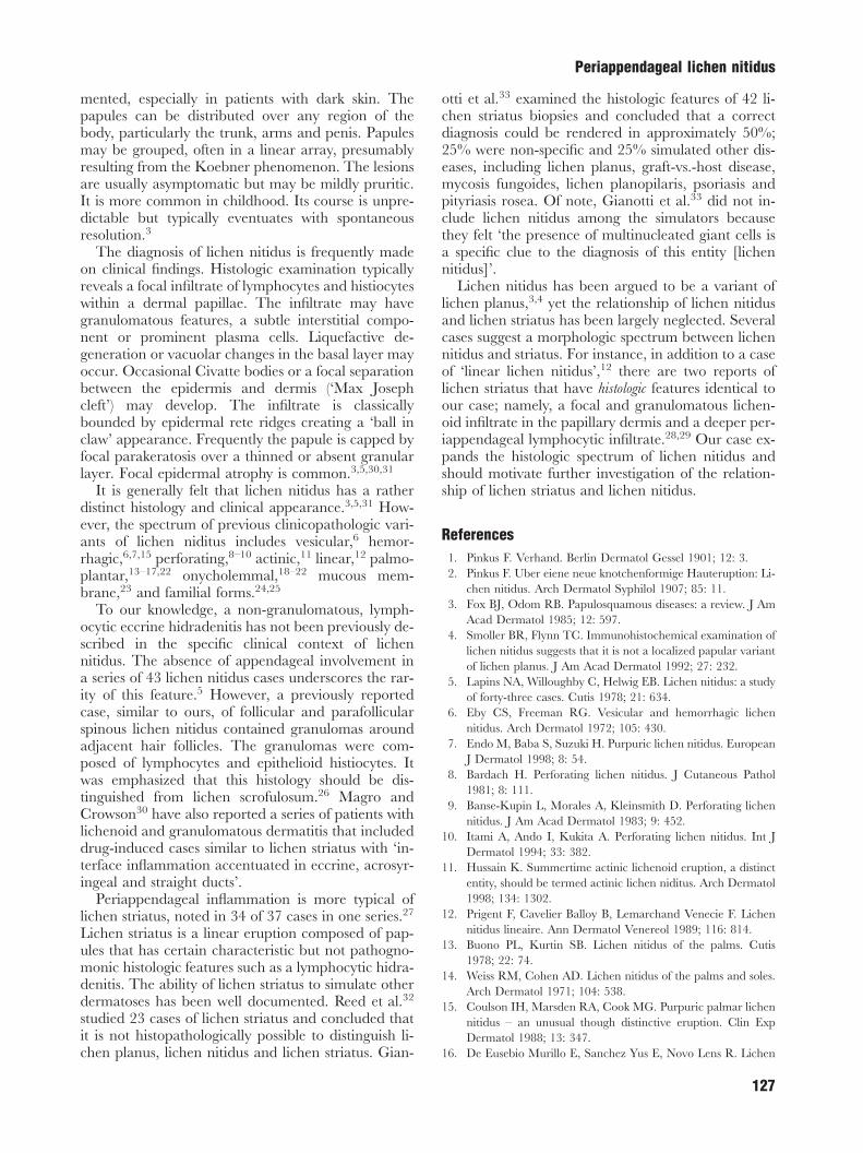
The skin lesions consisted of 1- to 2-mm flat-topped, slightly hypopigmented papules widely scat-tered over the trunk and extremities. In areas, thepapules were aligned in a linear fashion, suggesting aKoebner phenomenon. (Fig. 1) The patient noted thatthe eruption began on the left arm and became gen-eralized over the course of a year. No mucosal ornail lesions were noted. A clinical diagnosis of lichennitidus was made. Because of the child’s extensiveprior health problems and concern of the family, a 3-mm punch biopsy was done within an area of pruriticpapules on the left arm.
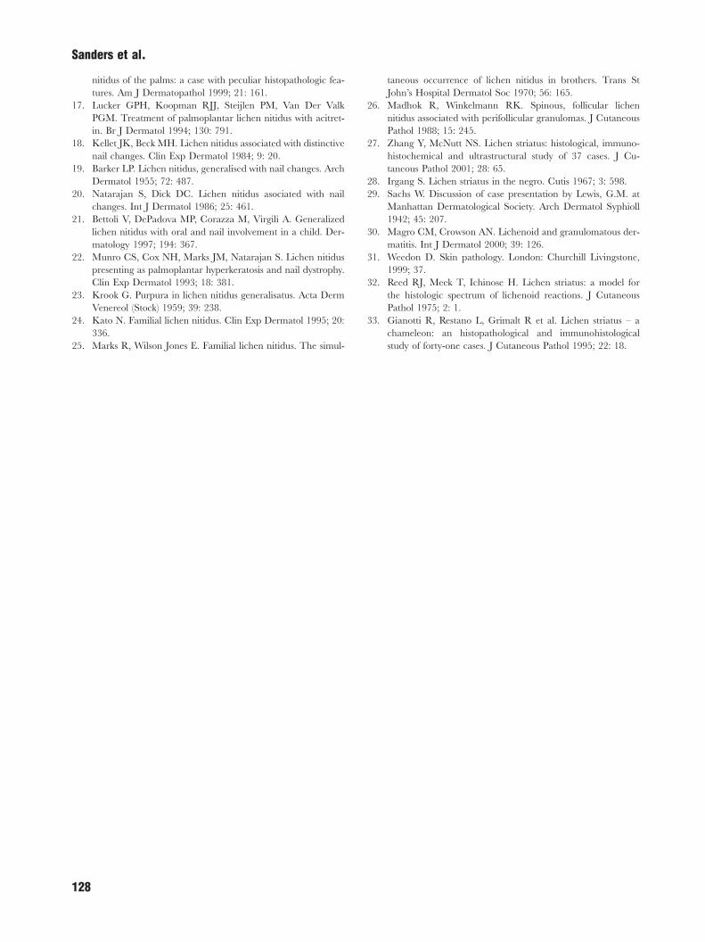
PathologyHematoxylin and eosin (H&E)-stained sections re-vealed a lymphocytic infiltrate in the papillary dermiswith admixed macrophages and rare multinucleatedcells. Eosinophils and plasma cells were not seen. The

Sanders et al.
infiltrate was in intimate apposition to the dermal–epidermal junction. Vacuolization of the basal kera-tinocytes and focal parakeratosis were noted. The in-flammatory infiltrate extended into the reticular der-mis along hair follicles and eccrine glands. (Figs. 2–5)
CommentLichen nitidus is an uncommon, idiopathic, chronicinflammatory dermatosis composed of numerous dis-
Fig. 1. Flat-topped, hypopigmented 1- to 2-mm papules distributedover the left upper arm.
Fig. 2. Low-power photomicrograph of hematoxylin and eosin-stained tissue profile demonstrating a mixed granulomatous infil-trate at the dermal–epidermal junction and underlying periappend-ageal, predominantly lymphocytic, inflammation.
126
crete, flat-topped and shiny papules. The papules aretypically no more than 3mm in diameter and oc-casionally may be dome-shaped or umbilicated. Al-though often flesh-colored, lesions may be hypopig-
Fig. 3. Granulomatous and lymphocytic lichenoid infiltrate withinthe papillary dermis. Focal parakeratosis surmounts areas of intensedermal inflammation.
Fig. 4. Lymphocytes surround a dermal granuloma.
Fig. 5. Lymphocytic eccrine hidradenitis.
Periappendageal lichen nitidus
mented, especially in patients with dark skin. Thepapules can be distributed over any region of thebody, particularly the trunk, arms and penis. Papulesmay be grouped, often in a linear array, presumablyresulting from the Koebner phenomenon. The lesionsare usually asymptomatic but may be mildly pruritic.It is more common in childhood. Its course is unpre-dictable but typically eventuates with spontaneousresolution.3
The diagnosis of lichen nitidus is frequently madeon clinical findings. Histologic examination typicallyreveals a focal infiltrate of lymphocytes and histiocyteswithin a dermal papillae. The infiltrate may havegranulomatous features, a subtle interstitial compo-nent or prominent plasma cells. Liquefactive de-generation or vacuolar changes in the basal layer mayoccur. Occasional Civatte bodies or a focal separationbetween the epidermis and dermis (‘Max Josephcleft’) may develop. The infiltrate is classicallybounded by epidermal rete ridges creating a ‘ball inclaw’ appearance. Frequently the papule is capped byfocal parakeratosis over a thinned or absent granularlayer. Focal epidermal atrophy is common.3,5,30,31
It is generally felt that lichen nitidus has a ratherdistinct histology and clinical appearance.3,5,31 How-ever, the spectrum of previous clinicopathologic vari-ants of lichen niditus includes vesicular,6 hemor-rhagic,6,7,15 perforating,8–10 actinic,11 linear,12 palmo-plantar,13–17,22 onycholemmal,18–22 mucous mem-brane,23 and familial forms.24,25
To our knowledge, a non-granulomatous, lymph-ocytic eccrine hidradenitis has not been previously de-scribed in the specific clinical context of lichennitidus. The absence of appendageal involvement ina series of 43 lichen nitidus cases underscores the rar-ity of this feature.5 However, a previously reportedcase, similar to ours, of follicular and parafollicularspinous lichen nitidus contained granulomas aroundadjacent hair follicles. The granulomas were com-posed of lymphocytes and epithelioid histiocytes. Itwas emphasized that this histology should be dis-tinguished from lichen scrofulosum.26 Magro andCrowson30 have also reported a series of patients withlichenoid and granulomatous dermatitis that includeddrug-induced cases similar to lichen striatus with ‘in-terface inflammation accentuated in eccrine, acrosyr-ingeal and straight ducts’.
Periappendageal inflammation is more typical oflichen striatus, noted in 34 of 37 cases in one series.27
Lichen striatus is a linear eruption composed of pap-ules that has certain characteristic but not pathogno-monic histologic features such as a lymphocytic hidra-denitis. The ability of lichen striatus to simulate otherdermatoses has been well documented. Reed et al.32
studied 23 cases of lichen striatus and concluded thatit is not histopathologically possible to distinguish li-chen planus, lichen nitidus and lichen striatus. Gian-
127
otti et al.33 examined the histologic features of 42 li-chen striatus biopsies and concluded that a correctdiagnosis could be rendered in approximately 50%;25% were non-specific and 25% simulated other dis-eases, including lichen planus, graft-vs.-host disease,mycosis fungoides, lichen planopilaris, psoriasis andpityriasis rosea. Of note, Gianotti et al.33 did not in-clude lichen nitidus among the simulators becausethey felt ‘the presence of multinucleated giant cells isa specific clue to the diagnosis of this entity [lichennitidus]’.
Lichen nitidus has been argued to be a variant oflichen planus,3,4 yet the relationship of lichen nitidusand lichen striatus has been largely neglected. Severalcases suggest a morphologic spectrum between lichennitidus and striatus. For instance, in addition to a caseof ‘linear lichen nitidus’,12 there are two reports oflichen striatus that have histologic features identical toour case; namely, a focal and granulomatous lichen-oid infiltrate in the papillary dermis and a deeper per-iappendageal lymphocytic infiltrate.28,29 Our case ex-pands the histologic spectrum of lichen nitidus andshould motivate further investigation of the relation-ship of lichen striatus and lichen nitidus.
References1. Pinkus F. Verhand. Berlin Dermatol Gessel 1901; 12: 3.2. Pinkus F. Uber eiene neue knotchenformige Hauteruption: Li-
chen nitidus. Arch Dermatol Syphilol 1907; 85: 11.3. Fox BJ, Odom RB. Papulosquamous diseases: a review. J Am
Acad Dermatol 1985; 12: 597.4. Smoller BR, Flynn TC. Immunohistochemical examination of
lichen nitidus suggests that it is not a localized papular variantof lichen planus. J Am Acad Dermatol 1992; 27: 232.
5. Lapins NA, Willoughby C, Helwig EB. Lichen nitidus: a studyof forty-three cases. Cutis 1978; 21: 634.
6. Eby CS, Freeman RG. Vesicular and hemorrhagic lichennitidus. Arch Dermatol 1972; 105: 430.
7. Endo M, Baba S, Suzuki H. Purpuric lichen nitidus. EuropeanJ Dermatol 1998; 8: 54.
8. Bardach H. Perforating lichen nitidus. J Cutaneous Pathol1981; 8: 111.
9. Banse-Kupin L, Morales A, Kleinsmith D. Perforating lichennitidus. J Am Acad Dermatol 1983; 9: 452.
10. Itami A, Ando I, Kukita A. Perforating lichen nitidus. Int JDermatol 1994; 33: 382.
11. Hussain K. Summertime actinic lichenoid eruption, a distinctentity, should be termed actinic lichen niditus. Arch Dermatol1998; 134: 1302.
12. Prigent F, Cavelier Balloy B, Lemarchand Venecie F. Lichennitidus lineaire. Ann Dermatol Venereol 1989; 116: 814.
13. Buono PL, Kurtin SB. Lichen nitidus of the palms. Cutis1978; 22: 74.
14. Weiss RM, Cohen AD. Lichen nitidus of the palms and soles.Arch Dermatol 1971; 104: 538.
15. Coulson IH, Marsden RA, Cook MG. Purpuric palmar lichennitidus – an unusual though distinctive eruption. Clin ExpDermatol 1988; 13: 347.
16. De Eusebio Murillo E, Sanchez Yus E, Novo Lens R. Lichen
Sanders et al.
nitidus of the palms: a case with peculiar histopathologic fea-tures. Am J Dermatopathol 1999; 21: 161.
17. Lucker GPH, Koopman RJJ, Steijlen PM, Van Der ValkPGM. Treatment of palmoplantar lichen nitidus with acitret-in. Br J Dermatol 1994; 130: 791.
18. Kellet JK, Beck MH. Lichen nitidus associated with distinctivenail changes. Clin Exp Dermatol 1984; 9: 20.
19. Barker LP. Lichen nitidus, generalised with nail changes. ArchDermatol 1955; 72: 487.
20. Natarajan S, Dick DC. Lichen nitidus asociated with nailchanges. Int J Dermatol 1986; 25: 461.
21. Bettoli V, DePadova MP, Corazza M, Virgili A. Generalizedlichen nitidus with oral and nail involvement in a child. Der-matology 1997; 194: 367.
22. Munro CS, Cox NH, Marks JM, Natarajan S. Lichen nitiduspresenting as palmoplantar hyperkeratosis and nail dystrophy.Clin Exp Dermatol 1993; 18: 381.
23. Krook G. Purpura in lichen nitidus generalisatus. Acta DermVenereol (Stock) 1959; 39: 238.
24. Kato N. Familial lichen nitidus. Clin Exp Dermatol 1995; 20:336.
25. Marks R, Wilson Jones E. Familial lichen nitidus. The simul-
128
taneous occurrence of lichen nitidus in brothers. Trans StJohn’s Hospital Dermatol Soc 1970; 56: 165.
26. Madhok R, Winkelmann RK. Spinous, follicular lichennitidus associated with perifollicular granulomas. J CutaneousPathol 1988; 15: 245.
27. Zhang Y, McNutt NS. Lichen striatus: histological, immuno-histochemical and ultrastructural study of 37 cases. J Cu-taneous Pathol 2001; 28: 65.
28. Irgang S. Lichen striatus in the negro. Cutis 1967; 3: 598.29. Sachs W. Discussion of case presentation by Lewis, G.M. at
Manhattan Dermatological Society. Arch Dermatol Syphioll1942; 45: 207.
30. Magro CM, Crowson AN. Lichenoid and granulomatous der-matitis. Int J Dermatol 2000; 39: 126.
31. Weedon D. Skin pathology. London: Churchill Livingstone,1999; 37.
32. Reed RJ, Meek T, Ichinose H. Lichen striatus: a model forthe histologic spectrum of lichenoid reactions. J CutaneousPathol 1975; 2: 1.
33. Gianotti R, Restano L, Grimalt R et al. Lichen striatus – achameleon: an histopathological and immunohistologicalstudy of forty-one cases. J Cutaneous Pathol 1995; 22: 18.