periodontal tissue changes after retention or relapse ... · molar intrusion, treatment result with...
TRANSCRIPT

Periodontal tissue changes after retention or
relapse following intrusive forces in rat molars
Yoon Jeong Choi
The Graduate School
Yonsei University
Department of Dental Science

Periodontal tissue changes after retention or
relapse following intrusive forces in rat molars
A Dissertation Thesis
Submitted to the Department of Dental Science
and the Graduate School of Yonsei University
in partial fulfillment of the
requirements for the degree of
Doctor of Philosophy of Dental Science
Yoon Jeong Choi
December 2008


감사의 글
치과대학을 졸업하고 교정학을 공부하기 시작한 것이 엊그제만 같습니다. 여전히 부
족한 저에게 ‘박사’라는 타이틀은 어색하고 부끄러울 따름입니다. 교정학을 공부할 수
있는 기회를 주시고 지금 이 순간까지 지도해주신 박영철 선생님께 진심으로 감사드
립니다. 교정학에 대한 것은 물론이거니와, 스승님으로서 너무나 존경하는 선생님 밑
에서 지낼 수 있었던 것은 저에게 더할 나위 없는 행복이었습니다. 바쁘신 와중에도
지도해주시고 임상적으로도 많은 가르침을 주시는 김형곤 선생님의 따뜻한 격려 덕에
논문이 완성될 수 있었습니다. 꼼꼼한 지도로 논문의 완성도를 높여주신 김진 선생님,
논문을 쓰면서 너무 많은 것들을 배웠습니다. 헛점 투성이였던 논문을 세심하게 지도
해주신 황충주 선생님께서 보내주신 격려와 응원 덕에 제가 여기까지 올 수 있었습니
다. 고민하던 저에게 훌륭한 아이디어를 주셨던 김경호 선생님께서는 실험의 처음부터
끝까지, 때로는 스승님으로 때로는 친구처럼 저의 고민을 들어주시고 해결책을 주셨습
니다. 지도해주신 다섯 분의 선생님께 감사하다는 말씀은 너무나 부족할 뿐입니다.
언제나 제 맘속에 큰 스승님으로 남아계신 유영규 선생님, 손병화 선생님, 만나뵐
때마다 논문에 관심을 보여주시고 용기를 주셨던 백형선 선생님, 유형석 선생님께도
감사의 말씀을 드립니다. 선생님들의 가르치심 덕에 오늘의 제가 있을 수 있었습니다.
논문의 큰 줄기를 잡아주시고 세심하고 예리하게 지적해주시고 도와주신 이기준 선생
님, 제가 선생님께 감사드릴 일이 너무나 많습니다. 매 단계마다 귀찮음을 마다않고
친절하고 세심하게 도움을 주셨던 정주령 선생님, 차정열 선생님께도 진심으로 감사드
립니다. 조직 사진마다 꼼꼼하게 설명해주시고 저보다 더 큰 열의로 가르쳐주신 문익
상 선생님, 여러 번의 귀찮은 질문에 대해 질문보다 항상 더 많이 가르쳐주신 김현실
선생님께도 가슴 깊이 감사드립니다. 힘들고 귀찮은 내색 없이 본인의 일들처럼 열심
히 도와주었던 조용민 선생님, 김진욱 선생님, 윤혜림 선생님 덕에 실험이 진행되고
마무리될 수 있었습니다. 매번 표현하지 못했지만, 지면을 빌어서나마 감사의 인사를
전합니다. 어렵고 힘들었던 수련 생활부터 지금까지 늘 힘이 되어주는 동기들–김경석,
김영재, 전재민, 주억-과 실험 내내 든든한 지원군이 되어주었던 의국 후배들-김인실,
이지연, 장정은, 황순신, 김진호-에게도 감사드립니다.
학부 때부터 어려운 결정의 순간마다 중요한 조언을 주신 육종인 선생님, 논문 뿐
아니라 일상의 수많은 고민들에 해결책을 주시는 스승이시면서 동시에 소중한 친구가
되어주시는 김의성 선생님, 늦은 시간까지 연구에 매진하시면서 논문에 매달리는 저에
게 뜨거운 격려를 주셨던 허종기 선생님, 박정원 선생님, 김선재 선생님, 신수정 선생
님, 교정에 대해서는 물론이거니와 다른 분야에서도 많은 조언을 주시는 이종석 선생
님께도 깊이 감사드립니다.
언제나 희로애락을 함께 해주는 소중한 친구들-주연, 혜자, 희진-과, 저에게 가족
과도 같은 언니들-난심, 미성, 민경, 은경, 가영, 승은-에게 감사의 마음을 전합니다.
마지막으로 저에게 무한한 사랑을 주시고 든든한 지원군이 되어주시는 사랑하는 가족
들께 이 논문을 바칩니다.
2008년 12월
저자 씀

v
Table of Contents
Abstract (English) ∙∙∙∙∙∙∙∙∙∙∙∙∙∙∙∙∙∙∙∙∙∙∙∙∙∙∙∙∙∙∙∙∙∙∙∙∙∙∙∙∙∙∙∙∙∙∙∙∙∙∙∙∙∙∙∙∙∙∙∙∙∙∙∙∙∙∙∙∙∙∙∙∙∙∙∙∙∙∙∙∙∙∙∙∙∙∙∙∙∙∙∙∙∙∙∙∙∙∙∙ ix
I. Introduction ∙∙∙∙∙∙∙∙∙∙∙∙∙∙∙∙∙∙∙∙∙∙∙∙∙∙∙∙∙∙∙∙∙∙∙∙∙∙∙∙∙∙∙∙∙∙∙∙∙∙∙∙∙∙∙∙∙∙∙∙∙∙∙∙∙∙∙∙∙∙∙∙∙∙∙∙∙∙∙∙∙∙∙∙∙∙∙∙∙∙∙∙∙∙∙∙∙∙∙∙∙∙∙∙∙∙∙∙∙∙ 1
II. Materials and Methods ∙∙∙∙∙∙∙∙∙∙∙∙∙∙∙∙∙∙∙∙∙∙∙∙∙∙∙∙∙∙∙∙∙∙∙∙∙∙∙∙∙∙∙∙∙∙∙∙∙∙∙∙∙∙∙∙∙∙∙∙∙∙∙∙∙∙∙∙∙∙∙∙∙∙∙∙∙∙∙∙∙∙∙∙∙∙∙∙∙ 4
1. Materials ∙∙∙∙∙∙∙∙∙∙∙∙∙∙∙∙∙∙∙∙∙∙∙∙∙∙∙∙∙∙∙∙∙∙∙∙∙∙∙∙∙∙∙∙∙∙∙∙∙∙∙∙∙∙∙∙∙∙∙∙∙∙∙∙∙∙∙∙∙∙∙∙∙∙∙∙∙∙∙∙∙∙∙∙∙∙∙∙∙∙∙∙∙∙∙∙∙∙∙∙∙∙∙∙∙∙∙∙∙∙∙∙ 4
2. Experimental procedures ∙∙∙∙∙∙∙∙∙∙∙∙∙∙∙∙∙∙∙∙∙∙∙∙∙∙∙∙∙∙∙∙∙∙∙∙∙∙∙∙∙∙∙∙∙∙∙∙∙∙∙∙∙∙∙∙∙∙∙∙∙∙∙∙∙∙∙∙∙∙∙∙∙∙∙∙∙∙∙∙∙∙∙ 5
3. Measurements ∙∙∙∙∙∙∙∙∙∙∙∙∙∙∙∙∙∙∙∙∙∙∙∙∙∙∙∙∙∙∙∙∙∙∙∙∙∙∙∙∙∙∙∙∙∙∙∙∙∙∙∙∙∙∙∙∙∙∙∙∙∙∙∙∙∙∙∙∙∙∙∙∙∙∙∙∙∙∙∙∙∙∙∙∙∙∙∙∙∙∙∙∙∙∙∙∙∙∙∙∙∙ 9
A. Measurements on periapical films ∙∙∙∙∙∙∙∙∙∙∙∙∙∙∙∙∙∙∙∙∙∙∙∙∙∙∙∙∙∙∙∙∙∙∙∙∙∙∙∙∙∙∙∙∙∙∙∙∙∙∙∙∙∙∙∙∙∙∙∙∙∙∙ 9
1) The changes of the vertical position of molar caused by cementum
apposition on root apex with aging ∙∙∙∙∙∙∙∙∙∙∙∙∙∙∙∙∙∙∙∙∙∙∙∙∙∙∙∙∙∙∙∙∙∙∙∙∙∙∙∙∙∙∙∙∙∙∙∙∙∙∙∙∙∙∙∙∙∙∙ 9
2) The changes following molar intrusion and
after retention or relapse following molar intrusion ∙∙∙∙∙∙∙∙∙∙∙∙∙∙∙∙∙∙∙∙∙∙∙∙∙∙ 10
B. Histomorphometric analysis ∙∙∙∙∙∙∙∙∙∙∙∙∙∙∙∙∙∙∙∙∙∙∙∙∙∙∙∙∙∙∙∙∙∙∙∙∙∙∙∙∙∙∙∙∙∙∙∙∙∙∙∙∙∙∙∙∙∙∙∙∙∙∙∙∙∙∙∙∙∙∙∙∙ 11
1) Depth from free gingival margin to the apical end of epithelium
in the mesial side of maxillary 1st molar ∙∙∙∙∙∙∙∙∙∙∙∙∙∙∙∙∙∙∙∙∙∙∙∙∙∙∙∙∙∙∙∙∙∙∙∙∙∙∙∙∙∙∙∙∙∙∙ 11
2) The number of osteoclasts on alveolar bone surface ∙∙∙∙∙∙∙∙∙∙∙∙∙∙∙∙∙∙∙∙∙∙∙∙∙ 12
3) Root resorption area ∙∙∙∙∙∙∙∙∙∙∙∙∙∙∙∙∙∙∙∙∙∙∙∙∙∙∙∙∙∙∙∙∙∙∙∙∙∙∙∙∙∙∙∙∙∙∙∙∙∙∙∙∙∙∙∙∙∙∙∙∙∙∙∙∙∙∙∙∙∙∙∙∙∙∙∙∙∙∙∙∙∙∙∙ 12
4. Statistical analysis ∙∙∙∙∙∙∙∙∙∙∙∙∙∙∙∙∙∙∙∙∙∙∙∙∙∙∙∙∙∙∙∙∙∙∙∙∙∙∙∙∙∙∙∙∙∙∙∙∙∙∙∙∙∙∙∙∙∙∙∙∙∙∙∙∙∙∙∙∙∙∙∙∙∙∙∙∙∙∙∙∙∙∙∙∙∙∙∙∙∙∙∙∙ 12
III. Results ∙∙∙∙∙∙∙∙∙∙∙∙∙∙∙∙∙∙∙∙∙∙∙∙∙∙∙∙∙∙∙∙∙∙∙∙∙∙∙∙∙∙∙∙∙∙∙∙∙∙∙∙∙∙∙∙∙∙∙∙∙∙∙∙∙∙∙∙∙∙∙∙∙∙∙∙∙∙∙∙∙∙∙∙∙∙∙∙∙∙∙∙∙∙∙∙∙∙∙∙∙∙∙∙∙∙∙∙∙∙∙∙∙∙ 13
1. The changes of body weight ∙∙∙∙∙∙∙∙∙∙∙∙∙∙∙∙∙∙∙∙∙∙∙∙∙∙∙∙∙∙∙∙∙∙∙∙∙∙∙∙∙∙∙∙∙∙∙∙∙∙∙∙∙∙∙∙∙∙∙∙∙∙∙∙∙∙∙∙∙∙∙∙∙∙∙∙ 13
2. Measurements on periapical films ∙∙∙∙∙∙∙∙∙∙∙∙∙∙∙∙∙∙∙∙∙∙∙∙∙∙∙∙∙∙∙∙∙∙∙∙∙∙∙∙∙∙∙∙∙∙∙∙∙∙∙∙∙∙∙∙∙∙∙∙∙∙∙∙∙∙ 13
A. The changes of the vertical position of molar caused by cementum
apposition on root apex with aging ∙∙∙∙∙∙∙∙∙∙∙∙∙∙∙∙∙∙∙∙∙∙∙∙∙∙∙∙∙∙∙∙∙∙∙∙∙∙∙∙∙∙∙∙∙∙∙∙∙∙∙∙∙∙∙∙∙ 13
B. The changes following molar intrusion and after retention or
relapse following molar intrusion ∙∙∙∙∙∙∙∙∙∙∙∙∙∙∙∙∙∙∙∙∙∙∙∙∙∙∙∙∙∙∙∙∙∙∙∙∙∙∙∙∙∙∙∙∙∙∙∙∙∙∙∙∙∙∙∙∙∙∙ 13
1) The changes following molar intrusion ∙∙∙∙∙∙∙∙∙∙∙∙∙∙∙∙∙∙∙∙∙∙∙∙∙∙∙∙∙∙∙∙∙∙∙∙∙∙∙∙∙∙∙∙∙∙∙∙∙∙∙ 13
2) The changes after retention following molar intrusion ∙∙∙∙∙∙∙∙∙∙∙∙∙∙∙∙∙∙∙∙∙∙ 14
3) The changes after relapse following molar intrusion ∙∙∙∙∙∙∙∙∙∙∙∙∙∙∙∙∙∙∙∙∙∙∙∙∙ 16
vi
4) The differences between the retention and the relapse groups
according to duration after molar intrusion ∙∙∙∙∙∙∙∙∙∙∙∙∙∙∙∙∙∙∙∙∙∙∙∙∙∙∙∙∙∙∙∙∙∙∙∙∙∙∙∙ 18
3. Micro computer tomographic findings ∙∙∙∙∙∙∙∙∙∙∙∙∙∙∙∙∙∙∙∙∙∙∙∙∙∙∙∙∙∙∙∙∙∙∙∙∙∙∙∙∙∙∙∙∙∙∙∙∙∙∙∙∙∙∙∙∙ 20
4. Histologic findings ∙∙∙∙∙∙∙∙∙∙∙∙∙∙∙∙∙∙∙∙∙∙∙∙∙∙∙∙∙∙∙∙∙∙∙∙∙∙∙∙∙∙∙∙∙∙∙∙∙∙∙∙∙∙∙∙∙∙∙∙∙∙∙∙∙∙∙∙∙∙∙∙∙∙∙∙∙∙∙∙∙∙∙∙∙∙∙∙∙∙∙∙ 24
A. Depth from free gingival margin to the apical end of epithelium
in the mesial side of maxillary 1st molar ∙∙∙∙∙∙∙∙∙∙∙∙∙∙∙∙∙∙∙∙∙∙∙∙∙∙∙∙∙∙∙∙∙∙∙∙∙∙∙∙∙∙∙∙ 24
B. Osteoclasts on the alveolar bone surface adjacent to root ∙∙∙∙∙∙∙∙∙∙∙∙∙∙∙∙∙∙ 27
1) Interradicular area ∙∙∙∙∙∙∙∙∙∙∙∙∙∙∙∙∙∙∙∙∙∙∙∙∙∙∙∙∙∙∙∙∙∙∙∙∙∙∙∙∙∙∙∙∙∙∙∙∙∙∙∙∙∙∙∙∙∙∙∙∙∙∙∙∙∙∙∙∙∙∙∙∙∙∙∙∙∙∙∙∙∙∙∙∙∙∙ 27
2) Interdental area between maxillary 2nd
and 3rd
molar ∙∙∙∙∙∙∙∙∙∙∙∙∙∙∙∙∙∙∙∙∙∙∙∙ 30
C. Root resorption area ∙∙∙∙∙∙∙∙∙∙∙∙∙∙∙∙∙∙∙∙∙∙∙∙∙∙∙∙∙∙∙∙∙∙∙∙∙∙∙∙∙∙∙∙∙∙∙∙∙∙∙∙∙∙∙∙∙∙∙∙∙∙∙∙∙∙∙∙∙∙∙∙∙∙∙∙∙∙∙∙∙∙∙∙∙∙ 32
D. Periodontal ligament ∙∙∙∙∙∙∙∙∙∙∙∙∙∙∙∙∙∙∙∙∙∙∙∙∙∙∙∙∙∙∙∙∙∙∙∙∙∙∙∙∙∙∙∙∙∙∙∙∙∙∙∙∙∙∙∙∙∙∙∙∙∙∙∙∙∙∙∙∙∙∙∙∙∙∙∙∙∙∙∙∙∙∙∙∙∙ 32
IV. Discussion ∙∙∙∙∙∙∙∙∙∙∙∙∙∙∙∙∙∙∙∙∙∙∙∙∙∙∙∙∙∙∙∙∙∙∙∙∙∙∙∙∙∙∙∙∙∙∙∙∙∙∙∙∙∙∙∙∙∙∙∙∙∙∙∙∙∙∙∙∙∙∙∙∙∙∙∙∙∙∙∙∙∙∙∙∙∙∙∙∙∙∙∙∙∙∙∙∙∙∙∙∙∙∙∙∙∙∙ 34
V. Conclusion ∙∙∙∙∙∙∙∙∙∙∙∙∙∙∙∙∙∙∙∙∙∙∙∙∙∙∙∙∙∙∙∙∙∙∙∙∙∙∙∙∙∙∙∙∙∙∙∙∙∙∙∙∙∙∙∙∙∙∙∙∙∙∙∙∙∙∙∙∙∙∙∙∙∙∙∙∙∙∙∙∙∙∙∙∙∙∙∙∙∙∙∙∙∙∙∙∙∙∙∙∙∙∙∙∙∙∙∙ 42
VI. References ∙∙∙∙∙∙∙∙∙∙∙∙∙∙∙∙∙∙∙∙∙∙∙∙∙∙∙∙∙∙∙∙∙∙∙∙∙∙∙∙∙∙∙∙∙∙∙∙∙∙∙∙∙∙∙∙∙∙∙∙∙∙∙∙∙∙∙∙∙∙∙∙∙∙∙∙∙∙∙∙∙∙∙∙∙∙∙∙∙∙∙∙∙∙∙∙∙∙∙∙∙∙∙∙∙∙ 44
Abstract (Korean) ∙∙∙∙∙∙∙∙∙∙∙∙∙∙∙∙∙∙∙∙∙∙∙∙∙∙∙∙∙∙∙∙∙∙∙∙∙∙∙∙∙∙∙∙∙∙∙∙∙∙∙∙∙∙∙∙∙∙∙∙∙∙∙∙∙∙∙∙∙∙∙∙∙∙∙∙∙∙∙∙∙∙∙∙∙∙∙∙∙∙∙∙∙∙∙∙∙∙∙ 49
vii
List of Figures
Figure 1. Intruding appliance for molar intrusion in rats ∙∙∙∙∙∙∙∙∙∙∙∙∙∙∙∙∙∙∙∙∙∙∙∙∙∙∙∙∙∙∙∙∙∙ 7
Figure 2. Rats which were sacrificed after retention or relapse following
two weeks of molar intrusion ∙∙∙∙∙∙∙∙∙∙∙∙∙∙∙∙∙∙∙∙∙∙∙∙∙∙∙∙∙∙∙∙∙∙∙∙∙∙∙∙∙∙∙∙∙∙∙∙∙∙∙∙∙∙∙∙∙∙∙∙∙∙∙ 7
Figure 3. Measurements on periapical film ∙∙∙∙∙∙∙∙∙∙∙∙∙∙∙∙∙∙∙∙∙∙∙∙∙∙∙∙∙∙∙∙∙∙∙∙∙∙∙∙∙∙∙∙∙∙∙∙∙∙∙∙∙∙∙∙ 10
Figure 4. Micro-CT 3D images of teeth (Buccal view) ∙∙∙∙∙∙∙∙∙∙∙∙∙∙∙∙∙∙∙∙∙∙∙∙∙∙∙∙∙∙∙∙∙ 21
Figure 5. Micro-CT 3D images of alveolar socket (Occlusal view) ∙∙∙∙∙∙∙∙∙∙∙ 23
Figure 6. Histomorphometric analyses of the control and the experimental
groups ∙∙∙∙∙∙∙∙∙∙∙∙∙∙∙∙∙∙∙∙∙∙∙∙∙∙∙∙∙∙∙∙∙∙∙∙∙∙∙∙∙∙∙∙∙∙∙∙∙∙∙∙∙∙∙∙∙∙∙∙∙∙∙∙∙∙∙∙∙∙∙∙∙∙∙∙∙∙∙∙∙∙∙∙∙∙∙∙∙∙∙∙∙∙∙∙∙∙∙∙∙∙∙ 25
Figure 7. Changes of gingival sulcus and junctional epithelium in the mesial
side of maxillary 1st molar ∙∙∙∙∙∙∙∙∙∙∙∙∙∙∙∙∙∙∙∙∙∙∙∙∙∙∙∙∙∙∙∙∙∙∙∙∙∙∙∙∙∙∙∙∙∙∙∙∙∙∙∙∙∙∙∙∙∙∙∙∙∙∙∙∙∙∙ 26
Figure 8. Interradicular alveolar bone and furcation area of the intruded
teeth ∙∙∙∙∙∙∙∙∙∙∙∙∙∙∙∙∙∙∙∙∙∙∙∙∙∙∙∙∙∙∙∙∙∙∙∙∙∙∙∙∙∙∙∙∙∙∙∙∙∙∙∙∙∙∙∙∙∙∙∙∙∙∙∙∙∙∙∙∙∙∙∙∙∙∙∙∙∙∙∙∙∙∙∙∙∙∙∙∙∙∙∙∙∙∙∙∙∙∙∙∙∙∙∙∙∙ 28
Figure 9. Osteoclasts in the interradicular area ∙∙∙∙∙∙∙∙∙∙∙∙∙∙∙∙∙∙∙∙∙∙∙∙∙∙∙∙∙∙∙∙∙∙∙∙∙∙∙∙∙∙∙∙∙∙∙ 29
Figure 10. Interdental area between intruded (M2) and non-intruded
(M3) teeth ∙∙∙∙∙∙∙∙∙∙∙∙∙∙∙∙∙∙∙∙∙∙∙∙∙∙∙∙∙∙∙∙∙∙∙∙∙∙∙∙∙∙∙∙∙∙∙∙∙∙∙∙∙∙∙∙∙∙∙∙∙∙∙∙∙∙∙∙∙∙∙∙∙∙∙∙∙∙∙∙∙∙∙∙∙∙∙∙∙∙∙∙∙∙∙ 31
Figure 11. Masson trichrome stained sections of root apex of maxillary
2nd
molar ∙∙∙∙∙∙∙∙∙∙∙∙∙∙∙∙∙∙∙∙∙∙∙∙∙∙∙∙∙∙∙∙∙∙∙∙∙∙∙∙∙∙∙∙∙∙∙∙∙∙∙∙∙∙∙∙∙∙∙∙∙∙∙∙∙∙∙∙∙∙∙∙∙∙∙∙∙∙∙∙∙∙∙∙∙∙∙∙∙∙∙∙∙∙∙∙ 33

viii
List of Tables
Table 1. Control and experimental groups ∙∙∙∙∙∙∙∙∙∙∙∙∙∙∙∙∙∙∙∙∙∙∙∙∙∙∙∙∙∙∙∙∙∙∙∙∙∙∙∙∙∙∙∙∙∙∙∙∙∙∙∙∙∙∙∙ 5
Table 2. The relative vertical position of molar caused by cementum
apposition on root apex in 13, 14 and 15 weeks old rats ∙∙∙∙∙∙∙∙∙∙∙∙∙∙∙ 14
Table 3. The relative vertical position of the intrusion (2wk-Intrusion
group) and the control groups ∙∙∙∙∙∙∙∙∙∙∙∙∙∙∙∙∙∙∙∙∙∙∙∙∙∙∙∙∙∙∙∙∙∙∙∙∙∙∙∙∙∙∙∙∙∙∙∙∙∙∙∙∙∙∙∙∙∙∙∙∙∙∙ 15
Table 4. The relative vertical position in the intrusion and the retention
groups ∙∙∙∙∙∙∙∙∙∙∙∙∙∙∙∙∙∙∙∙∙∙∙∙∙∙∙∙∙∙∙∙∙∙∙∙∙∙∙∙∙∙∙∙∙∙∙∙∙∙∙∙∙∙∙∙∙∙∙∙∙∙∙∙∙∙∙∙∙∙∙∙∙∙∙∙∙∙∙∙∙∙∙∙∙∙∙∙∙∙∙∙∙∙∙∙∙∙∙∙∙∙∙∙∙∙ 16
Table 5. The relative vertical position in the intrusion and the relapse groups
∙∙∙∙∙∙∙∙∙∙∙∙∙∙∙∙∙∙∙∙∙∙∙∙∙∙∙∙∙∙∙∙∙∙∙∙∙∙∙∙∙∙∙∙∙∙∙∙∙∙∙∙∙∙∙∙∙∙∙∙∙∙∙∙∙∙∙∙∙∙∙∙∙∙∙∙∙∙∙∙∙∙∙∙∙∙∙∙∙∙∙∙∙∙∙∙∙∙∙∙∙∙∙∙∙∙∙∙∙∙∙∙∙∙∙∙∙∙∙∙∙ 17
Table 6. The comparison of the retained and the relapsed teeth according to
duration after molar intrusion ∙∙∙∙∙∙∙∙∙∙∙∙∙∙∙∙∙∙∙∙∙∙∙∙∙∙∙∙∙∙∙∙∙∙∙∙∙∙∙∙∙∙∙∙∙∙∙∙∙∙∙∙∙∙∙∙∙∙∙∙∙∙∙∙ 19

ix
ABSTRACT
Periodontal tissue changes after retention or
relapse following intrusive forces in rat molars
Orthodontic miniscrew implants are commonly used in current practice to
intrude molars when correcting anterior openbite. However, there have been
few reports on the changes of the intruded teeth and periodontal tissue
during retention period following molar intrusion. The aim of this study was
to observe periodontal tissue changes after intrusion of posterior teeth in
rats using miniscrew implant and its features of retention or relapse.
Orthodontic miniscrew implant was placed behind maxillary left incisor in
ten week old rat and 50 gm of intrusion force was applied to the maxillary
left 1st and 2
nd molars for two weeks with Japanese NiTi wire. Periodontal
tissue changes after two weeks of molar intrusion and after a period of one
to two weeks of retention or relapse following molar intrusion were observed.
With molar intrusion, mild surface root resorption occurred and it was
repaired with cementum after retention or relapse following intrusion. Active
bone modeling and remodeling were seen in the alveolar bone adjacent to the
intruded teeth, and it was the most evident in the interradicular area. The
height of alveolar crest was decreased but there was no statistically
significant difference after intrusion with the control group. After retention,

x
however, the alveolar crest between intruded teeth was moved apically (p <
0.05). The apical end of epithelium moved with cement-enamel junction
causing long junctional epithelium formation after molar intrusion (p < 0.05).
Periodontal ligament was stretched in the tension side and compressed in the
pressure side. The free gingival margin receded and periodontal ligament
was remodeled after retention, resulting in normal and healthy periodontium.
Most teeth movements of relapse occurred in the early phase, therefore
initial retention is important for the stability of treatment.
Key Words : molar intrusion, retention, relapse, anterior openbite, absolute
anchorage, orthodontic miniscrew implant

1
Periodontal tissue changes after retention or
relapse following intrusive forces in rat molars
Yoon Jeong Choi, D.D.S., M.S.D.
Department of Dental Science, Graduate School, Yonsei University
(Directed by Prof. Young Chel Park, D.D.S., M.S.D., PhD)
I. Introduction
Skeletal anchorage simplified orthodontic treatment by counteracting
reactive forces and expanded the ranges to be corrected by orthodontic
treatment only. Recently, onplant1, miniplate
2-7 and orthodontic miniscrew
implant8-13
have been used for skeletal anchorage. They are divided
according to insertion site, technique and design, but they provide the same
function as absolute anchorage for tooth movement. Miniscrew implant has

2
the advantages of immediate loading, various insertion sites, uncomplicated
placement and removal procedures, and minimal expense for patients. The
success rate of miniscrew implant was reported to be over 90%14,15
, and
immediate loading was known not to affect the stability of miniscrew
implant16,17
. Antero-posterior tooth movement is not interfered by miniscrew
implant if it is placed with appropriate angulation to bone surface in
interproximal area15
.
The correction of anterior openbite with molar intrusion using miniscrew
implant8,10
is an example that the range of orthodontic treatment has been
expanded. Miniscrew implant helps to reduce the potential risk of surgery,
the troublesome of wearing extraoral appliance and the dependence upon
patient compliance during treatment.
In treatment of anterior openbite using miniscrew implant as an absolute
anchorage, the occlusal plane was rotated counterclockwise to close anterior
openbite as molars were intruded. Since facial soft tissue was changed with
molar intrusion, treatment result with molar intrusion is similar to that by
orthognathic surgery in respect to facial profile changes. Kuroda et al2 stated
that molar intrusion with skeletal anchorage is simpler and more useful than
two-jaw surgery in the treatment of patients with severe anterior openbite.
However, since there have been few reports on the changes of the intruded
teeth and periodontal tissue during retention period following molar intrusion,
relapse rates are controversial and even the effectiveness of anterior
openbite correction with molar intrusion has been questioned.

3
There are some reports of the changes after molar intrusion. It was stated
that clinically the changes in alveolar bone height with marginal bone
remodeling during molar intrusion were observed but any increase in gingival
pocket depth after posterior segmental molar intrusion was not seen,
although the temporary formation of a gingival pocket (pseudo-pocket) was
observed4. The tissues in the marginal alveolar crest showed a resorption
and remodeling of alveolar bone and in the interradicular and apical regions
had the typical characteristics of pressure zone; periodontal ligament
compression and cellfree zone18
.
For the long term stability of treatment it is essential to understand the
periodontal tissue changes following molar intrusion and during retention
period after molar intrusion, but it has scarcely been reported. Therefore,
the aim of this study was to observe periodontal tissue changes after
intrusion of posterior teeth in rats using miniscrew implant and its features
of retention or relapse.

4
II. Materials and Methods
1. Materials
29 ten weeks old female Sprague-Dawley rats, averaging 220 - 250 gm in
weight, were used. All animals were kept in stainless-steel cages in air-
conditioning and subjected to standard 12-hour light / dark cycle. They were
fed with a pellet diet (8811M0001, Extrusion, Superfeed Co. Ltd., Gangwon-
do, Korea) and tap water ad libitum. They were checked everyday in regard
to their health status.
The animals were divided into control and five experimental groups to
evaluate periodontal tissue changes following molar intrusion and after
retention or relapse following molar intrusion (Table 1). In the 2wk-
Intrusion group (n=5), molars were intruded for two weeks. In the 1wk-
Retention (n=5) and 2wk-Retention (n=5) groups, intruded molars were
maintained for one and two weeks, respectively, after two weeks of molar
intrusion. In the 1wk-Relapse (n=5) and 2wk-Relapse (n=5) groups,
intruding appliances were disengaged and periodontal tissue changes were
observed after one and two weeks, respectively, following two weeks of
molar intrusion. In the control group (n=4), the experimental condition was
the same as the 2wk-Intrusion group except the intrusion force.

5
Table 1. Control and experimental groups
Number
of
samples
Duration (unit : week)
0 1 2 3 4
Control 4 Implantation
of miniscrew
Occlusal
bonding material
2wk-
Intrusion 5
Implantation
of miniscrew
1wk-
Retention 5
Implantation
of miniscrew
2wk-
Retention 5
Implantation
of miniscrew
1wk-
Relapse 5
Implantation
of miniscrew
2wk-
Relapse 5
Implantation
of miniscrew
2wk-Intrusion, the experimental group in which molars were intruded for two weeks;
1wk-Retention and 2wk-Retention, the experimental groups in which intruded
molars were maintained for one and two weeks, respectively, after two weeks of
molar intrusion; 1wk-Relapse and 2wk-Relapse, the experimental groups in which
intruding appliances were disengaged and periodontal tissue changes were observed
after one and two weeks, respectively, following two weeks of molar intrusion.
2. Experimental procedures
The animals were immobilized with ether inhalation and anaesthetized with
intraperitoneal injection of Zoletil (Tiletamine 125ml, Zolazepam 125ml;
0.04ml Virbac, 060516 carros, France) and Rompun (Xylazine
hydroxychloride 23.32 mg/ml; 0.01 ml Bayer AG, 51368 Leverkusen,
German).
Orthodontic miniscrew implant (1.2 mm diameter, 7.0 mm length,
Intrusion
Intrusion
Intrusion
Intrusion
Intrusion
Retention
Retention
Relapse
Relapse

6
BioMaterials Korea Inc., Seoul, Korea) was inserted into the alveolar crest
behind maxillary left incisor, impression of upper arch was taken with poly-
vinysiloxane (Aquasil Ultra, Dentsply, York, PA, USA) and a dental cast
(New Plastone, GC Corp., Tokyo, Japan) was fabricated. With reference to
the fabricated cast, the improved superelastic nickel-titanium alloy wire
(L&H Titan, Tomy, Tokyo, Japan), 0.016 x 0.022 inches, was bent to
transfer an intrusive force of 50 gm18
parallel to the long axis of tooth by
using the direct electric resistance heat treatment method with a heat
bender19
(Fig. 1).
One week after miniscrew implantation, the fabricated wire was attached to
miniscrew implant and the occlusal tables of maxillary 1st and 2
nd molars on
left side. The space between the wire and the neck of miniscrew implant was
filled with flowable resin (Esthet-X®
Flow, Dentsply, York, PA, USA). GI
cement (Ultra Band-LokTM
, Reliance Orthod Prod. Itasca, IL, USA) was used
to bond wire to molars. Occlusal bonding material, GI cement, was also
bonded to maxillary left 3rd
molar and three maxillary right molars to prevent
unwanted extrusion of teeth(Fig. 1).
After two weeks of molar intrusion, the intruding appliance was disengaged,
impression of upper arch was taken and a dental cast was fabricated. In the
retention groups, three maxillary molars on each side were splinted by
occlusal bonding material again (Fig 2, A). In the relapse groups, all
appliances except miniscrew implant were removed (Fig. 2, B).

7
Figure 1. Intruding appliance for molar intrusion in rats. A, Schematic
drawing of the appliance; B, Superelastic NiTi wire transformed by heat
bender (top, occlusal view; bottom, lateral view); C, Rat which was sacrificed
after two weeks of molar intrusion. M1, M2 and M3 - maxillary 1st, 2
nd and
3rd
molar, respectively.
Figure 2. Rats which were sacrificed after retention or relapse following two
weeks of molar intrusion. A, Retention group; B, Relapse group.
NiTi wire
A
S
passive form activated form
Bite block
B C
B A
S

8
At the end of each experimental period the animals were sacrificed by
cervical dislocation under ether inhalation. Intrusion or retentive appliance
was disengaged, impression was taken and a dental cast was fabricated. The
appliance was checked everyday under ether inhalation and the body weight
of animals was measured every week.
The maxilla was dissected free and divided with a midline, sagittal cut.
Each half-cut maxilla was placed on the intraoral periapical film (Insight,
Kodak, Rochester, NY, USA). X-ray was exposed at the distance of 30 cm
from the film using intraoral radiographic apparatus (AnyRay, E-Woo
Technology Co. Ltd., Gyeonggi-do, Korea). The radiographic films were
processed in manual method. Micro computer tomography (SkyScan micro
CT 1076, Skyscan, Aartselaar, Belgium) was taken on one sample of each
five experimental groups (2wk-Intrusion, 1wk-Retention, 2wk-Retention,
1wk-Relapse and 2wk-Relapse groups).
Immediately after sacrifice, the maxilla, including the teeth, was fixed for
24 hours in 4% paraformaldehyde in 0.1M phosphate buffer (pH 7.4),
dehydrated in ethyl alcohol, decalcified in EDTA/HCl (Calci-Clear Rapid®,
National Diagnosics Inc., Atlanta, GA, USA), embedded in paraffin and cut
into 4 ㎛ thick sections in a sagittal direction. The sections were prepared
and stained with Hematoxylin-Eosin and Masson trichrome.

9
3. Measurements
A. Measurements on periapical films
With a digital camera (DFC300FX, Leica Microsystems Ltd., Wetzlar,
Germany) connected to a microscope (Leica MZ75, Leica Microsystems Ltd.,
Wetzlar, Germany), the processed periapical films were converted into digital
images which were magnified 6.7 times with measuring tool.
On the digital image a horizontal reference plane was created as a line
tangent to cranium below frontal-squamosal intersection at temporal crest20
,
and the following categories were measured with image measuring program
(Image J, Wayne Rasband, National Institutes of Health, USA).
1) The changes of the vertical position of molar caused by cementum
apposition on root apex with aging
In order to evaluate whether cementum apposition, a characteristic in rat
molars, would cause the vertical displacement of teeth according to the age
of rats, the vertical positions of crown and root were compared. The right
maxillary 3rd
molar (M3) in the 2wk-Intrusion, 1wk-Retention and 2wk-
Retention groups was used for comparison because bonding material had
been attached during the whole experimental period but intrusion force had
not been applied. The perpendicular distances from the below points to the
horizontal reference plane were measured (Fig. 3): (a) frontal-squamosal
intersection at the temporal crest, (b) middle cusp tip of M3, (c) distal root

10
apex of M3. To compensate for variance of individual skull size, the ratios of
measurements for frontal-squamosal intersection at the temporal crest,
which were constant during the experiment, were calculated: b/a and c/a, the
relative vertical positions of M3 crown and root, respectively.
Figure 3. Measurements on periapical film. a, Frontal-squamosal intersection
at the temporal crest; b, middle cusp tip of maxillary 3rd
molar (M3) - total
height; c, M3 distal root apex; d, distal cusp tip of maxillary 2nd
molar (M2);
e, alveolar crest between M2 and M3; f, alveolar crest between maxillary 1st
molar (M1) and M2; g, alveolar crest in M1 mesial side; h, M1 distal root
apex; i, M1 mesial root apex.
2) The changes following molar intrusion and after retention or relapse
following molar intrusion
To evaluate the amount of molar intrusion and root resorption and the

11
changes of alveolar bone, the perpendicular distance from the below points to
the horizontal reference plane were measured (Fig. 3): (b) total height – M3
middle cusp tip, (d) M2 distal cusp tip, (e) alveolar crest between M2 and
M3, (f) alveolar crest between M1 and M2, (g) alveolar crest in M1 mesial
side, (h) M1 distal root apex, (i) M1 mesial root apex.
Total height (b) was given a value of 1, as a reference, and other
measurements were converted accordingly.
B. Histomorphometric analysis
In order to observe periodontal tissue changes immediately after molar
intrusion and after retention or relapse, histomorphometric analyses were
performed with image measuring program (Image-Pro PLUSTM
, ver 3.0,
Media Cybernetics, Inc., MD, USA) in all the experimental and the control
groups. The mean value of measurements from three serial sections was
used to decrease the variation of block slicing.
1) Depth from free gingival margin to the apical end of epithelium in the
mesial side of maxillary 1st molar
Depth from free gingival margin to the apical end of epithelium in the
mesial side of maxillary 1st molar was measured to observe the changes of
gingival sulcus and junctional epithelium immediately after intrusion and after
retention or relapse period following intrusion. The perpendicular distance
from free gingival margin to the bottom line, which was made perpendicular

12
to root surface at the apical end of epithelium, was measured.
2) The number of osteoclasts on alveolar bone surface
The length of the alveolar surface adjacent to root of maxillary 1st and 2
nd
molars was measured and the number of osteoclasts on the alveolar bone
surface was counted. The number of osteoclasts was divided by the alveolar
bone length, thus the number of osteoclast per unit length was calculated.
3) Root resorption area
The root length of maxillary 1st and 2
nd molars and the size of crater, where
the continuity of cementum was lost representing the root length were
measured, thus the root resorption area per unit length was calculated.
4. Statistical analysis
The statistical analysis was carried out on SPSS ver 12.0 (SPSS Inc.,
Chicago, IL, USA). Data were presented as means ± standard deviations.
For significance of differences, the data were evaluated by independent t-
test in comparison between the intrusion (2wk-Intrusion) and control groups,
and between the retention and the relapse groups. To compare other
variables, one-way analysis of variance (ANOVA) and the post-hoc
Duncan’s multiple range test were performed. A p value less than .05 was
considered statistically significant.
13
III. Results
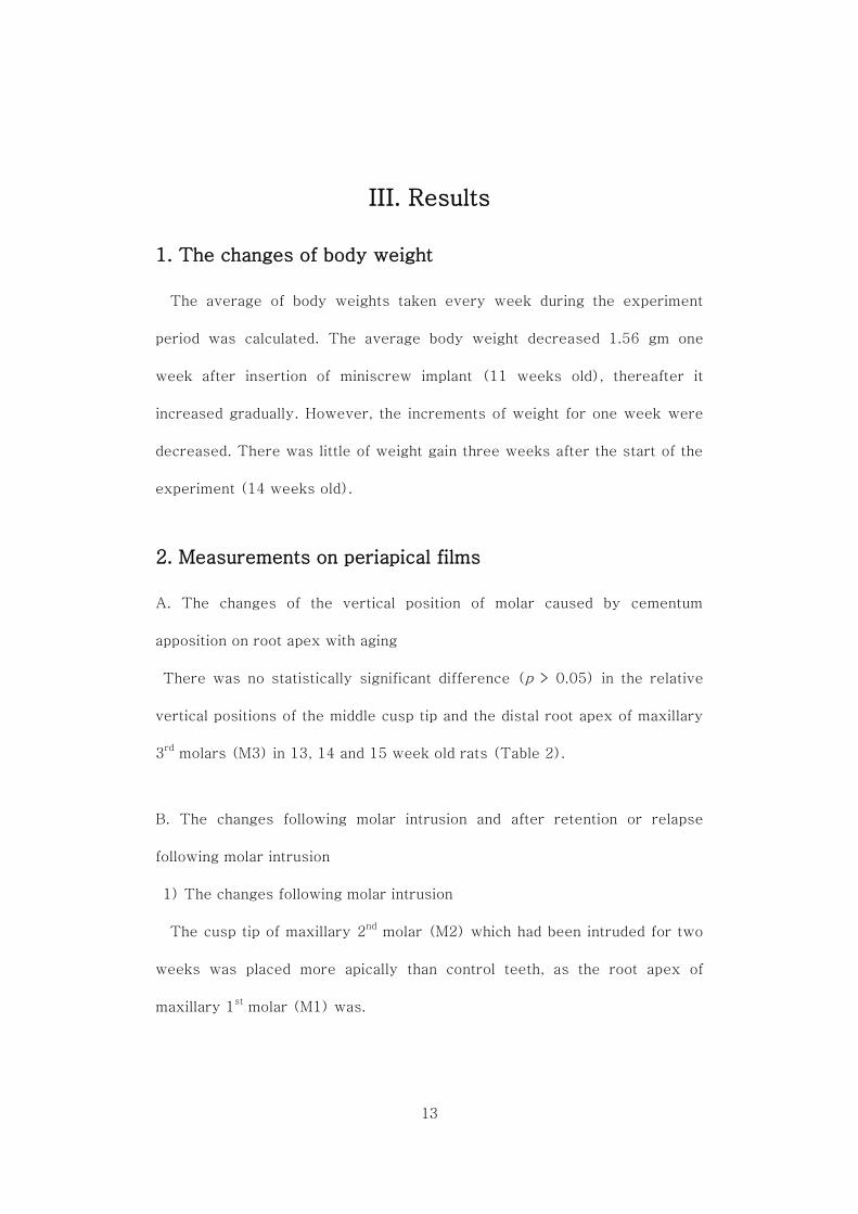
1. The changes of body weight
The average of body weights taken every week during the experiment
period was calculated. The average body weight decreased 1.56 gm one
week after insertion of miniscrew implant (11 weeks old), thereafter it
increased gradually. However, the increments of weight for one week were
decreased. There was little of weight gain three weeks after the start of the
experiment (14 weeks old).
2. Measurements on periapical films
A. The changes of the vertical position of molar caused by cementum
apposition on root apex with aging
There was no statistically significant difference (p > 0.05) in the relative
vertical positions of the middle cusp tip and the distal root apex of maxillary
3rd
molars (M3) in 13, 14 and 15 week old rats (Table 2).
B. The changes following molar intrusion and after retention or relapse
following molar intrusion
1) The changes following molar intrusion
The cusp tip of maxillary 2nd
molar (M2) which had been intruded for two
weeks was placed more apically than control teeth, as the root apex of
maxillary 1st molar (M1) was.

14
Table 2. The relative vertical position of molar caused by cementum
apposition on root apex in 13, 14 and 15 weeks old rats (p = 0.05)
13-week (n=5) 14-week (n=5) 15-week (n=5) Sig.
b/a 8.50 ± 0.31 8.48 ± 0.23 8.46 ± 0.16 NS
c/a 6.76 ± 0.15 6.76 ± 0.96 6.75 ± 0.13 NS
a : the perpendicular distance from the most superior point of frontal-squamosal
intersection at the temporal crest to the horizontal reference plane
b : the perpendicular distance from middle cusp tip of maxillary 3rd
molar (M3) to the
horizontal reference plane
c : the perpendicular distance from M3 distal root apex to the horizontal reference
plane
b/a : the relative vertical position of M3 middle cust tip
c/a : the relative vertical position of M3 distal root apex
NS : not significant
However, the differences of the relative height of root apex between the
intrusion and the control groups were less than those of cusp tip. The height
of the alveolar crest in the intrusion group was lower than the control group,
but there was no statistically significant difference in the mesial side of M1
and in the alveolar crest between M2 and M3, and also M1 and M2 (Table 3).
2) The changes after retention following molar intrusion
The relative vertical position of the intruded teeth for two weeks (2wk-
Intrusion group) and the maintained teeth after intrusion (1wk-Retention
and 2wk-Retention groups) showed no statistically significant difference.

15
Table 3. The relative vertical position of the intrusion (2wk-Intrusion
group) and the control groups
Intrusion Control Sig.
M2 distal cusp 0.964 ± 0.003 0.987 ± 0.002 **
Alveolar crest between
M2 and M3 0.892 ± 0.006 0.900 ± 0.003 NS
Alveolar crest between
M1 and M2 0.878 ± 0.011 0.882 ± 0.003 NS
Alveolar crest
in M1 mesial side 0.868 ± 0.009 0.870 ± 0.007 NS
M1 distal root apex 0.710 ± 0.004 0.725 ± 0.002 **
M1 mesial root apex 0.707 ± 0.012 0.725 ± 0.006 *
M1, M2 and M3; maxillary 1st, 2
nd and 3
rd molar, respectively.
* : p < 0.05
** : p < 0.01
NS : not significant
After one or two weeks of retention following intrusion, the relative height
of the alveolar crest in the M1 mesial side and between intruded M2 and
non-intruded M3 showed no statistically significant difference between after
intrusion and after retention. The alveolar crest between intruded M1 and M2
during retention was positioned more apically than immediately after two
weeks of molar intrusion.
The relative vertical position of the root apex of M1 was statistically
significantly higher after retention following intrusion than immediately after
intrusion (Table 4).

16
Table 4. The relative vertical position in the intrusion and the retention
groups
2wk-
Intrusion
1wk-
Retention
2wk-
Retention Sig.
M2 distal cusp 0.965 ± 0.003 0.965 ± 0.006 0.968 ± 0.008 NS
Alveolar crest
between M2 and M3 0.892 ± 0.006 0.897 ± 0.002 0.896 ± 0.003 NS
Alveolar crest
between M1 and M2
0.878 ± 0.011 0.865 ± 0.007 0.866 ± 0.003 *
A B B
Alveolar crest
in M1 mesial side 0.868 ± 0.009 0.867 ± 0.011 0.863 ± 0.002 NS
M1 distal root apex 0.710 ± 0.004 0.727 ± 0.009 0.729 ± 0.007
** A B B
M1 mesial root apex 0.707 ± 0.012 0.721 ± 0.006 0.720 ± 0.007
** A B B
M1, M2 and M3 - maxillary 1st, 2
nd and 3
rd molar, respectively; 2wk-Intrusion, two
weeks of molar intrusion; 1wk-Retention, one week of retention after two weeks of
molar intrusion; 2wk-Retention, two weeks of retention after two weeks of molar
intrusion.
* : p < 0.05
** : p < 0.01
NS : not significant
3) The changes after relapse following molar intrusion
The differences of the relative vertical position of M2 cusp tip between the
2wk-Intrusion and the 2wk-Relapse groups were compared with between
the intrusion and the control groups. In the 2wk-Intrusion group, M2 cusp tip
was positioned 0.023 apically than the control group, and M2 cusp tip of the
2wk-Relapse group 0.015 occlusally than the 2wk-Intrusion group.
Therefore, the relapse rate was calculated 41.67%.

17
Most of occlusal movements, called as relapse, occurred during the first
week of relapse. The alveolar crest between intruded M2 and non-intruded
M3 moved occlusally as teeth relapsed, but in the other alveolar crests there
was no statistically significant difference with the 2wk-Intrusion group. The
root apices of M1 were moved occlusally as the cusp tip of M2 did, but the
amount of the root apices movement was bigger than that of the cusp tip
(Table 5).
Table 5. The relative vertical position in the intrusion and the relapse groups
2wk-Intrusion 1wk-Relapse 2wk-Relapse Sig.
M2 distal cusp 0.964 ± 0.003 0.979 ± 0.006 0.979 ± 0.002
** A B B
Alveolar crest
between M2 and M3
0.892 ± 0.006 0.899 ± 0.004 0.901 ± 0.002 *
A B B
Alveolar crest
between M1 and M2 0.878 ± 0.011 0.878 ± 0.004 0.879 ± 0.008 NS
Alveolar crest
in M1 mesial side 0.868 ± 0.009 0.868 ± 0.009 0.866 ± 0.008 NS
M1 distal root apex 0.710 ± 0.004 0.730 ± 0.004 0.728 ± 0.008
** A B B
M1 mesial root apex 0.707 ± 0.012 0.726 ± 0.009 0.724 ± 0.007
* A B B
M1, M2 and M3 - maxillary 1st, 2
nd and 3
rd molar, respectively; 2wk-Intrusion, two
weeks of molar intrusion; 1wk-Relapse, one week of relapse after two weeks of
molar intrusion; 2wk-Relapse, two weeks of relapse after two weeks of molar
intrusion.
* : p < 0.05
** : p < 0.01
NS : not significant

18
4) The differences between the retention and the relapse groups according
to duration after molar intrusion
The relative vertical position of M2 cusp tip in the retention and the relapse
groups showed a distinct difference. The alveolar crest between intruded M2
and non-intruded M3 moved occlusally in both groups, but there was no
statistically significant difference between one week of retention and relapse
following intrusion. However, after two weeks of retention or relapse it was
placed more apically in the retention groups than the relapse groups. The
alveolar crest between M1 and M2 that were intruded for two weeks was
positioned more apically in the retention groups than the relapse groups. In
the alveolar crest of M1 mesial side, there was no statistically significant
difference between with and without retention. It was the same in the root
apices of M1 (Table 6).

19
Table 6. The comparison between the retained and the relapsed teeth
according to duration after molar intrusion
1wk-
Retention
1wk-
Relapse Sig.
2wk-
Retention
2wk-
Relapse Sig.
M2 distal cusp 0.965 ±
0.006
0.979 ±
0.006 *
0.968 ±
0.008
0.979 ±
0.002 *
Alveolar crest
between
M2 and M3
0.897 ±
0.002
0.899 ±
0.004 NS
0.896 ±
0.003
0.901 ±
0.002 *
Alveolar crest
between
M1 and M2
0.865 ±
0.007
0.878 ±
0.004 *
0.866 ±
0.003
0.879 ±
0.008 *
Alveolar crest
in M1 mesial side
0.867 ±
0.011
.0868 ±
0.009 NS
0.863 ±
0.002
0.866 ±
0.008 NS
M1 distal
root apex
0.727 ±
0.009
0.730 ±
0.004 NS
0.729 ±
0.007
0.728 ±
0.008 NS
M1 mesial
root apex
0.721 ±
0.006
0.726 ±
0.009 NS
0.720 ±
0.007
0.724 ±
0.007 NS
M1, M2 and M3 - maxillary 1st, 2
nd and 3
rd molar, respectively; 1wk-Retention, one
week of retention after two weeks of molar intrusion; 1wk-Relapse, one week of
relapse after two weeks of molar intrusion; 2wk-Retention, two weeks of retention
after two weeks of molar intrusion; 2wk-Relapse, two weeks of relapse after two
weeks of molar intrusion.
* : p < 0.05
NS : not significant

20
3. Micro computer tomographic findings
Intruded M1 and M2 were placed more apically than control teeth and they
were maintained well during retention period. In the relapse groups where
the intruding appliance was disengaged without retainer, intruded M1 and M2
showed a similar vertical position to M3 that was not intruded. In the relapse
groups, the difference of the vertical position of teeth according to the
duration of relapse after intrusion was not evident (Fig. 4).
Rough root surface with multiple small craters was observed in the 2wk-
Intrusion group, but it disappeared after retention or relapse. In M1 and M2
of the 2wk-Intrusion group, the mesial surface of roots, from which the
intrusion force was originated, showed root resorptions in apical 1/3 area
(Fig. 4, G). In the retention and the relapse groups root resorptions were
also observed, especially in short and small roots. There was no distinct
difference of roots in furcation area between the experimental and the
control groups.

21
Figure 4. Micro-CT 3D images of teeth (Buccal view). A, 2wk-Intrusion
group; B, Opposite side of 2wk-Intrusion group (control side); C, 1wk-
Retention group; D, 1wk-Relapse group; E, 2wk-Retention group; F, 2wk-
Relapse group; G, Bottom view of 2wk-Intrusion group. M1, M2 and M3 -
maxillary 1st, 2
nd and 3
rd molar, respectively.
A
C
M3
D
E F
M2 M1
M1 M2
M3
M2 M1
M3
M2
M2 M1 M3
M1
M3
B
M1 M2 M3
G M1 M2
M3

22
In the alveolar socket adjacent to furcation area of the 2wk-Intrusion and
the retention groups, the interradicular alveolar bone showed similar
configurations to the control group. Otherwise, in the relapse groups the
smooth texture of the interradicular alveolar bone was not observed and the
surface was rougher than in other groups. But the roughness was decreased
according to the duration of relapse (Fig. 5).
It was shown that the root apices of the intruded molars penetrated into the
nasal cavity in the 2wk-Intrusion group. Even though the perforated hole of
the apical alveolar bones in the control group was apparent, its number and
size were much smaller in the control group than the 2wk-Intrusion group.
The perforated wholes were also seen in the retention groups, but the size
was decreased in the 2wk-Retention group compared to the 1wk-Retention
group. In the relapse groups, the perforated area was notably diminished,
therefore the differences between the 2wk-Relapse and the control groups
were not evident (Fig. 5).

23
Figure 5. Micro-CT 3D images of alveolar socket (Occlusal view). A, 2wk-
Intrusion group; B, Opposite side of 2wk-Intrusion group (control side); C,
1wk-Retention group; D, 1wk-Relapse group; E, 2wk-Retention group; F,
2wk-Relapse group. M1 and M2 - maxillary 1st and 2
nd molar, respectively.
(Top, lingual; Bottom, buccal side). Arrowheads, the perforated hole of the
apical alveolar bone (the holes in M1 mesial apex were excluded because
they were seen in all groups); circles, interradicular alveolar bone adjacent
to furcation area.
C DD
E F
M2 M1 M1 M2
M2 M1
M2 M1
B
M1 M2
A Buccal
Lingual
M1 M2

24
4. Histologic findings
A. Depth from free gingival margin to the apical end of epithelium in the
mesial side of maxillary 1st molar
Depth from free gingival margin to the apical end of epithelium after two
weeks of molar intrusion was increased statistically significantly compared to
the control group and decreased again after two weeks of retention following
intrusion. Even though it had been decreased after retention or relapse, there
was no statistically significant difference between the 2wk-Intrusion group
and other experimental groups except the 2wk-Retention group. In the
retention and the relapse groups it was measured more than the control
group, but there was no statistically significant difference (Fig. 6, A).
The epithelial attachment reached the cement-enamel junction in both the
control and the experimental specimens. The junctional epithelium was
lengthened with molar intrusion, consequently the depth from free gingival
margin to the apical end of epithelium was increased. However, there was no
sign of edema or swelling of the gingiva. The thickness of junctional and
sulcular epitheliums was increased in the retention and the relapse groups
and it was more evident in the relapse groups. (Fig. 7).

25
Figure 6. Histomorphometric analyses of the control and the experimental
groups. A, Depth from free gingival margin to the apical end of epithelium; B,
Number of osteoclasts; C, Root resorption area.
* : p < 0.05
** : p < 0.01
(unit : ㎜)
A. Depth from free gingival
margin to the apical end
of epithelium Control 2wk-Intrusion
1wk-Retention
2wk-Retention
1wk-Relapse
2wk- Relapse
* *
Control 2wk-
Intrusion 1wk-
Retention 2wk-
Retention 1wk-
Relapse 2wk-
Relapse
(unit : number/㎜)
B. Number of osteoclasts
** **
Control 2wk-Intrusion
1wk-Retention
2wk-Retention
1wk-Relapse
2wk-Relapse
(unit : ㎜2/㎜)
C. Root resorption area *

26
Figure 7. Changes of gingival sulcus and junctional epithelium in the mesial
side of maxillary 1st molar. A, 2wk-Intrusion group; B, Control group; C,
1wk-Retention group; D, 1wk-Relapse group; E, 2wk-Retention group; F,
2wk-Relapse group. Arrow indicates CEJ (cement-enamel junction); D,
dentin; E, enamel cavity. (Magnification X 100)
D C
B
E
A
F
D D
D D
D
D
E E
E
E
E E

27
B. Osteoclasts on the alveolar bone surface adjacent to root
The number of osteoclasts per unit length of alveolar bone surface after two
weeks of molar intrusion was increased statistically significantly compared to
the control group and it was decreased again after retention or relapse
following molar intrusion (Fig. 6, B).
1) Interradicular area
In the experimental groups, active modeling and remodeling of alveolar
bone was seen compared to the control group. The incidence of blood
vessels in the apical and marginal alveolar bones as well as the interradicular
alveolar bone was high in the retention and the relapse groups, especially the
1wk-Retention group (Fig. 8).
After two weeks of molar intrusion many osteoclasts were seen, but the
number of osteoclasts was decreased after retention or relapse following
molar intrusion. Osteoclasts on the alveolar bone surface were observed
more in the retention groups than the relapse groups (Fig. 9).

28
Figure 8. Interradicular alveolar bone and furcation area of the intruded teeth.
A, 2wk-Intrusion group; B, Control group; C, 1wk-Retention group; D,
1wk-Relapse group; E, 2wk-Retention group; F, 2wk-Relapse group.
Arrowheads indicate root resorption area; AB, alveolar bone; P, pulp; R, root
dentin. (Magnification X 100)
D
E
B
F
A
C
P
p P
R
AB
AB
R
AB
AB
AB AB
R
R
R R
P P
P P

29
Figure 9. Osteoclasts in the interradicular area. A and B, 2wk-Intrusion
groups; C, 1wk-Retention group; D, 1wk-Relapse group; E, 2wk-Retention
group; F, 2wk-Relapse group. Arrowheads, osteoclasts; arrows, alveolar
bone remnants; AB, alveolar bone; P, pulp; R, root dentin. (Magnification X
200)
D
B A
C
AB AB
R R R
P
R R
AB
AB
AB AB
R R
E F

30
2) Interdental area between maxillary 2nd
and 3rd
molar
The apical end of epithelium of maxillary 2nd
and 3rd
molars was located at
CEJ in both the experimental and the control groups. The transseptal fibers
ran straight across the interdental septum in the control group, while they
were stretched toward intruded tooth after two weeks of intrusion. The
orientation was changed after retention or relapse. The transseptal fiber
bundles were imbedded approximately perpendicular to the cementum and
ran parallel to the imaginary line between CEJs of two adjacent teeth.
Direct and undermining bone resorptions on the marginal alveolar bone
adjacent to intruded teeth were observed in the 2wk-intrusion group. In
other experimental and the control groups, however, osteoclasts adjacent to
alveolar bone surface were hardly observed and osteoid tissue bordered by
osteoblasts was seen on the alveolar crest and into the resorptive lacunas
(Fig. 10).

31
Figure 10. Interdental area between intruded (M2) and non-intruded (M3)
teeth. A, 2wk-Intrusion group; B, Control group; C, 1wk-Retention group; D,
1wk-Relapse group; E, 2wk-Retention group; F, 2wk-Relapse group.
Arrowheads, osteoclasts; arrows, osteoid tissue bordered by osteoblasts; M2,
maxillary 2nd
molar; M3, maxillary 3rd
molar; AB, alveolar bone.
(Magnification X 100)
B A
F
M2 M2 M3
M3
AB
M2
M2 M3 M2
D AB
M2
M3
AB
E
M3
C
M3
AB
AB AB

32
C. Root resorption area
Surface root resorptions were observed in all experimental groups. The
area of root resorption in all experimental groups was statistically
significantly larger than the control group (Fig. 6, C). Root resorption area
after retention or relapse following molar intrusion was decreased compared
to the 2wk-Intrusion group, but there was no statistically significant
difference between the 2wk-Intrusion and other experimental groups. Root
resorption partly reached into dentin was repaired with cementum after
retention or relapse (Fig. 8).
D. Periodontal ligament
The oblique periodontal ligament (PDL) fibers that were intruded for two
weeks were stretched toward the direction of intrusion. The apical and
interradicular PDL fibers were compressed and the density of cells was
increased after two weeks of intrusion, while the apical PDL of control group
showed a radial shape. The PDL stretching toward apex decreased gradually
according to the duration of retention. A radial shape of the apical fibers
similar to the control group was observed in the 2wk-Retention group. In the
relapse groups, the stretched oblique fibers were not seen and the density of
cells in the apical PDL was decreased compared to the 2wk-Intrusion group,
in which the apical fibers were compressed by intrusion. PDL in the 2wk-
Relapse group looked more similar to the normal PDL than in the 1wk-
Relapse group (Fig. 11).

33
Figure 11. Masson trichrome stained sections of root apex of maxillary 2
nd
molar. A, 2wk-Intrusion group; B, Control group; C, 1wk-Retention group;
D, 1wk-Relapse group; E, 2wk-Retention group; F, 2wk-Relapse group.
M2, maxillary 2nd
molar; AB, alveolar bone; P, pulp; PDL, periodontal
ligament. (Magnification X 200)
B A
D C
F E
M2 M2
P
PDL PDL
PDL M2
M2
PDL
M2 M2 AB PDL PDL
AB
AB

34
IV. Discussion
Molar intrusion was nearly impossible before orthodontic miniscrew
implant provided absolute anchorage for tooth movement. Consequently,
there were few reports of molar intrusion unlike incisor intrusion and
periodontal tissue changes after retention or relapse following molar
intrusion. Molar intrusion is thought to be different from incisor intrusion in
that molar is a multiradicular tooth which has a furcation area, while incisor
has a single root, and persistent vertical force is applied to the occlusal table.
Studies about molar intrusion have been performed in beagle dogs using
miniplate3,4,11,12
. When using beagle dogs there were limitations that it was
difficult to generalize the results because of relatively small sample sizes and
the response of molar to intrusive force in beagle dog could be different from
human molar because it had no occlusal tables. In this study, the subjects
were rats, which had occlusal tables like human molars, even though they
didn’t work for occluding, and were useful to generalize the results by
increasing the sample size.
Rats used in this study were ten weeks old female. Rats become sexually
mature at age of six weeks21
, there is no more increase of bone maturity
score since nine weeks after birth22
. In rats females are more advanced in
skeletal maturity than males from birth to adulthood and the cephalocaudal
maturity gradient is seen in the development process22
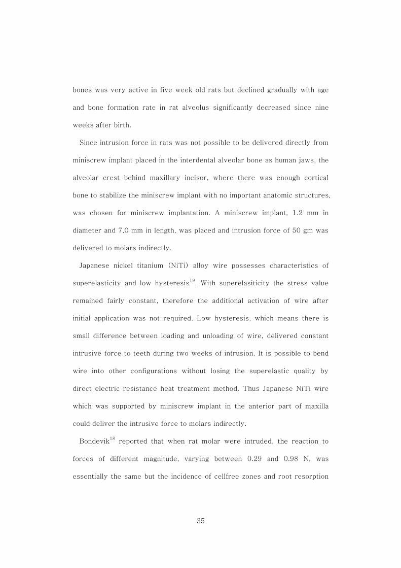
. Shimomoto et al23
reported that bone formation at the periosteal surface of the alveolar and jaw
35
bones was very active in five week old rats but declined gradually with age
and bone formation rate in rat alveolus significantly decreased since nine
weeks after birth.
Since intrusion force in rats was not possible to be delivered directly from
miniscrew implant placed in the interdental alveolar bone as human jaws, the
alveolar crest behind maxillary incisor, where there was enough cortical
bone to stabilize the miniscrew implant with no important anatomic structures,
was chosen for miniscrew implantation. A miniscrew implant, 1.2 mm in
diameter and 7.0 mm in length, was placed and intrusion force of 50 gm was
delivered to molars indirectly.
Japanese nickel titanium (NiTi) alloy wire possesses characteristics of
superelasticity and low hysteresis19
. With superelasiticity the stress value
remained fairly constant, therefore the additional activation of wire after
initial application was not required. Low hysteresis, which means there is
small difference between loading and unloading of wire, delivered constant
intrusive force to teeth during two weeks of intrusion. It is possible to bend
wire into other configurations without losing the superelastic quality by
direct electric resistance heat treatment method. Thus Japanese NiTi wire
which was supported by miniscrew implant in the anterior part of maxilla
could deliver the intrusive force to molars indirectly.
Bondevik18
reported that when rat molar were intruded, the reaction to
forces of different magnitude, varying between 0.29 and 0.98 N, was
essentially the same but the incidence of cellfree zones and root resorption

36
lacunae seemed to increase as the force increased. Based on this finding, the
intrusion force was decided to be 50 gm, which was thought to be the lowest
force level for the heat-bended NiTi wire to deliver. The intrusion appliance
was confirmed not to inhibit normal growth of rats by checking changes of
body weight.
Cementum apposition and occlusal bonding material could affect the
vertical position of molars. Cementum apposition on root apex with aging did
not make a statistically significant difference in the vertical position of
molars during experimental periods. In order to evaluate whether the bonding
materials which attached wire to occlusal tables of molars would cause the
vertical displacemenet of teeth, pre-experimental study was performed. It
was found that the occlusal bonding material did not affect the relative
vertical positions of teeth for two weeks (p > 0.05). Bresin24
reported that
lower molars to which bite block had been bonded during four weeks were
intruded in four week old rats. However, it is evident that the alveolar bone
formation rate is much higher in four week old rats in their most active
period of skeletal growth than over ten week old rats when skeletal growth is
almost finished22,23
.
Periodontal tissue changes after the application of intrusive force were
discussed in following orders; junctional and sulcular epitheliums, alveolar
bone, root resorption and periodontal ligament.
After two weeks of molar intrusion, the depth from free gingival margin to

37
the apical end of epithelium was increased and the marginal, interradicular
and apical alveolar bones were resorbed. The number of osteoclasts per unit
alveolar bone surface immediately after molar intrusion was statistically
significantly larger than other experimental and the control groups. However,
the alveolar crest height measured on periapical films immediately after
molar intrusion did not show statistically significant difference from the
control group. The findings that crown moved more apically than root apex
after molar intrusion indicated a surface root resorption on root apex
resulting in root shortening. Root resorptions partly reached into dentin were
observed on overall root surfaces, and it was confirmed from numerous root
craters seen in the 3D reconstructed CT images. Periodontal ligament was
stretched toward the direction of intrusion.
In the retention groups, intruded teeth were maintained well. The changes
due to relapse were mostly occurred during the first week of relapse.
Periodontal tissues were remodeled correspondingly to the altered teeth
position after retention or relapse. The increased depth from free gingival
margin to the apical end of epithelium was decreased again and so did the
number of osteoclasts. Surface root resorptions were repaired with
cementum and the configuration of stretched oblique and compressed apical
PDL fibers was changed similar to normal PDL.
The depth from free gingival margin to the apical end of epithelium was
increased and junctional epithelium was lengthened as teeth were intruded.

38
In every experimental specimen, the epithelium ended at the CEJ just as it
did in the control teeth, and there was no apical migration whatever. It
indicated that in normal periodontal tissue the apical end of epithelium moved
in the apical or occlusal direction with tooth as much as tooth movement.
Murakami et al25
reported the same finding from incisor intrusion in monkeys.
It was also stated that the gingiva moved in the same direction that the teeth
were intruded, but only about 60% as far. After one or two weeks of
retention, the depth from free gingival margin to the apical end of epithelium
was decreased. It could be inferred that the further recession of free gingival
margin was occurred during retention, because the apical end of epithelium
was not changed. However, the change in the relapse groups may not be the
same as that in the retention groups, because the position of the apical end of
epithelium was changed with tooth movement.
Alveolar bone resorptions in the marginal, interradicular and apical area
were induced by intrusion, and they were remodeled actively during the
initial phase of retention and looked similar to normal alveolar bone in the
later phase.
Not only alveolar bone resorptions but root resorptions were more evident
in the interradicular region as compared to the marginal and apical regions.
Several possible explanations were stated. Intrusive forces would interfere
more markedly with the blood supply and create more extensive cell free
zones in the interradicular region than in the apical and marginal regions18
. It
has been also suggested that bone morphology is a factor affecting the

39
occurrence of cellfree zones and the nature of bone resorption26
and
cementum maturity is of significance for the progression of root resorption27
.
In the two weeks of molar intrusion group, the density of osteoclasts was
higher in the interradicular alveolar crest than in other regions and a couple
of alveolar bone remnants were observed above the interradicular alveolar
crest. In the one week of retention following molar intrusion (1wk-
Retention) group, many osteoclasts were observed in the interradicular
alveolar crest as compared to the control and other retention (2wk-
Retention) and relapse (1wk-Relapse and 2wk-Relapse) groups, even
though there was no statistically significant difference.
The height of alveolar crest moved apically with molar intrusion, but there
was no statistically significant difference between immediately after molar
intrusion and the control groups. After one or two weeks of retention, the
alveolar crest between two intruded teeth was positioned apically (p < 0.05).
On the other hand, the alveolar crest between intruded maxillary 2nd
molar
and non-intruded maxillary 3rd
molar was positioned occlusally with the
relapse of tooth after one or two weeks of relapse (p < 0.05).
Numerous reports indicate that alveolar bone resorption is the consequence
of increased pressure in the periodontal ligament, while deposition of osteoid
tissue is elicited by a stretching of the fibers. Bone resorption on the alveolar
crest cannot be explained in the same way. It may be a consequence of
pressure exerted against the crest by free gingival fibers as tooth is
depressed. This was confirmed by the experiment of supracrestal

40
fiberotomy18
. Kanzaki et al4 also reported the same findings that the amount
of alveolar bone resorption was smaller in fiberotomy group compared with
nonfiberotomy group on the alveolar crest.
In the studies from molar intrusion of beagle dogs, monkeys and human,
mild root resorptions in the root apices and the furcation areas were
observed5,6,11-13
. The findings from premolar27-31
and incisor intrusions32,33
of humans indicated similar results and it was also observed in rat molars.
Most root resorptions were occurred in the apical third of root, and it
resulted in root shortening. During the first week of retention following two
weeks of molar intrusion, root resorption was aggravated a little. During the
second week of retention, additional root shortening was not observed. The
retention groups showed more apical root resorptions than the relapse
groups. Cementum immaturity may be related to apical root resorptions.
Immature cementum might be resorbed earlier than apical alveolar bone
under increased pressure around apical PDL following intrusive force. Root
resorption partly reached into dentin was repaired with cementum. Stenvik
and Mjor27
had stated that the defects created would be repaired by tissue
resembling bone and cementum if the teeth were left in situ after the force
was removed.
The oblique fibers of periodontal ligament were stretched and the apical
and interradicular fibers were compressed after intrusion. PDL was also
remodeled according to the new position of teeth and PDL after two weeks of
retention looked more similar to normal PDL than after two weeks of relapse.

41
Intrusion is not favorable for retention because tooth is moved opposite to
the physiologic movement of extrusion. Furthermore, the interradicular and
the apical periodontal tissues compressed during intrusion are reorganized
more slowly than in other sites, because repair of resorbed roots as well as
adjacent alveolar bone resorption are required simultaneously. Additionally,
intrusion is less stable than rotation and mediodistal movement because
periodontal fibers, which are generally thought to resist occlusal forces, can
also strongly resist intrusive force and an effective method for retention has
not been established for intruded molars7. In previous study, it was
demonstrated that the application of bisphosphonates in animals was an
effective pharmacologic method of retention that inhibited alveolar bone from
remodeling around moved teeth34
. It can be applied to retention of intruded
molar in animals and even humans after further experiments.
In this study, it was confirmed that rat molars were intruded using
orthodontic miniscrew implant and they were maintained to the altered
position. Periodontal tissues were remodeled to healthy periodontium after
the retention period. The changes of vertical position in the relapse groups
mostly occurred in the first week of relapse.

42
V. Conclusion
Orthodontic miniscrew implants are commonly used in current practice to
intrude molars when correcting anterior openbite. However, since there have
been few reports on the changes of the intruded teeth and periodontal tissue
during retention period following molar intrusion, relapse rates are
controversial and even the effectiveness of anterior openbite correction with
molar intrusion has been questioned. The aim of this study was to observe
periodontal tissue changes after intrusion of posterior teeth in rats using
miniscrew implant and its features of retention or relapse.
Orthodontic miniscrew implant was placed behind maxillary left incisor in a
ten week old rat and 50 gm of intrusion force was applied to the maxillary
left 1st and 2
nd molars for two weeks with Japanese NiTi wire. Periodontal
tissue changes after two weeks of molar intrusion and after a period of one
to two weeks of retention or relapse were observed. The results were as
followings;
1. When molar was intruded, mild root resorption was occurred and the
depth from free gingival margin to the apical end of epithelium was increased
due to formation of long junctional epithelium. Periodontal ligament was
stretched in the oblique portion toward the direction of intrusion and
compressed in the interradicular and the apical regions. Alveolar bone in the
marginal, interradicular and apical areas was resorbed with tooth intrusion.
43
2. After retention, craters created by surface root resorption were
repaired with cementum, consequently the size of the craters was decreased,
and shortening of the root was observed. The free gingival margin was
recessed and the periodontal ligament was remodeled correspondingly to the
altered tooth position and resulted in a close resemblance to healthy
periodontium. Alveolar crest between intruded teeth was positioned more
apically than immediately after molar intrusion. The periodontal tissues
showed more active remodeling after one week of retention compared to two
weeks of retention following molar intrusion.
3. After relapse, root resorption craters were decreased compared to
immediately after intrusion and repaired with cementum. With the relapse of
teeth, the depth from free gingival margin to the apical end of epithelium was
decreased and the stretched periodontal ligament moved occlusally. Alveolar
crest between intruded teeth showed the same vertical position as
immediately after two weeks of molar intrusion, even though the intruded
teeth moved occlusally.
It was confirmed from this study that after retention, root resorption which
occurred by molar intrusion was repaired with cementum and periodontal
tissues were remodeled correspondingly to the altered tooth position,
resulting in a final normal and healthy periodontium. Most teeth movements
of relapse occurred in the early phase, therefore initial retention is important
for the stability of treatment.
44
VI. References
1. Janssens F, Swennen G, Dujardin T, Glineur R and Malevez C. Use of an
onplant as orthodontic anchorage. Am J Orthod Dentofacial Orthop 2002;
122:566-570.
2. Kuroda S, Sakai Y, Tamamura N, Deguchi T and Takano-Yamamoto T.
Treatment of severe anterior open bite with skeletal anchorage in adults:
comparison with orthognathic surgery outcomes. Am J Orthod Dentofacial
Orthop 2007; 132:599-605.
3. Daimaruya T, Takahashi I, Nagasaka H, Umemori M, Sugawara J and
Mitani H. Effects of maxillary molar intrusion on the nasal floor and tooth
root using the skeletal anchorage system in dogs. Angle Orthod 2003;
73:158-166.
4. Kanzaki R, Daimaruya T, Takahashi I, Mitani H and Sugawara J.
Remodeling of alveolar bone crest after molar intrusion with skeletal
anchorage system in dogs. Am J Orthod Dentofacial Orthop 2007; 131:343-
351.
5. Ari-Demirkaya A, Masry MA and Erverdi N. Apical root resorption of
maxillary first molars after intrusion with zygomatic skeletal anchorage.
Angle Orthod 2005; 75:761-767.
6. Daimaruya T, Nagasaka H, Umemori M, Sugawara J and Mitani H. The
influences of molar intrusion on the inferior alveolar neurovascular bundle

45
and root using the skeletal anchorage system in dogs. Angle Orthod 2001;
71:60-70.
7. Sugawara J, Baik UB, Umemori M, Takahashi I, Nagasaka H, Kawamura H
and Mitani H. Treatment and posttreatment dentoalveolar changes following
intrusion of mandibular molars with application of a skeletal anchorage
system (SAS) for open bite correction. Int J Adult Orthodon Orthognath Surg
2002; 17:243-253.
8. Park YC, Lee HA, Choi NC and Kim DH. Open bite correction by intrusion
of posterior teeth with miniscrews. Angle Orthod 2008; 78:699-710.
9. Chang YJ, Lee HS and Chun YS. Microscrew anchorage for molar intrusion.
J Clin Orthod 2004; 38:325-330.
10. Kuroda S, Katayama A and Takano-Yamamoto T. Severe anterior open-
bite case treated using titanium screw anchorage. Angle Orthod 2004;
74:558-567.
11. Carrillo R, Rossouw PE, Franco PF, Opperman LA and Buschang PH.
Intrusion of multiradicular teeth and related root resorption with mini-screw
implant anchorage: a radiographic evaluation. Am J Orthod Dentofacial Orthop
2007; 132:647-655.
12. Carrillo R, Buschang PH, Opperman LA, Franco PF and Rossouw PE.
Segmental intrusion with mini-screw implant anchorage: a radiographic
evaluation. Am J Orthod Dentofacial Orthop 2007; 132:576 e571-576.
13. Ohmae M, Saito S, Morohashi T, Seki K, Qu H, Kanomi R, Yamasaki KI,
Okano T, Yamada S and Shibasaki Y. A clinical and histological evaluation of

46
titanium mini-implants as anchors for orthodontic intrusion in the beagle dog.
Am J Orthod Dentofacial Orthop 2001; 119:489-497.
14. Wilmes B, Rademacher C, Olthoff G and Drescher D. Parameters affecting
primary stability of orthodontic mini-implants. J Orofac Orthop 2006;
67:162-174.
15. Park HS, Jeong SH and Kwon OW. Factors affecting the clinical success
of screw implants used as orthodontic anchorage. Am J Orthod Dentofacial
Orthop 2006; 130:18-25.
16. Serra G, Morais LS, Elias CN, Meyers MA, Andrade L, Muller C and
Muller M. Sequential bone healing of immediately loaded mini-implants. Am J
Orthod Dentofacial Orthop 2008; 134:44-52.
17. Freire JN, Silva NR, Gil JN, Magini RS and Coelho PG. Histomorphologic
and histomophometric evaluation of immediately and early loaded mini-
implants for orthodontic anchorage. Am J Orthod Dentofacial Orthop 2007;
131:704 e701-709.
18. Bondevik O. Tissue changes in the rat molar periodontium following
application of intrusive forces. Eur J Orthod 1980; 2:41-49.
19. Miura F, Mogi M and Ohura Y. Japanese NiTi alloy wire: use of the direct
electric resistance heat treatment method. Eur J Orthod 1988; 10:187-191.
20. Richtsmeier JT, Baxter LL and Reeves RH. Parallels of craniofacial
maldevelopment in Down syndrome and Ts65Dn mice. Dev Dyn 2000;
217:137-145.
21. Adams N and Boice R. A longitudinal study of dominance in an outdoor

47
colony of domestic rats. J Comp Psychol 1983; 97: 24-33.
22. Hughes PCR and Tanner JM. The assessment of skeletal maturity in the
growing rat. J Anat 1970; 106:371-402.
23. Shimomoto Y, Chung CJ, Iwasaki-Hayashi Y, Muramoto T and Soma K.
Effects of occlusal stimuli on alveolar/jaw bone formation. J Dent Res 2007;
86:47-51.
24. Bresin A. Effects of masticatory muscle function and bite-raising on
mandibular morphology in the growing rat. Swed Dent J Suppl 2001; 1-49.
25. Murakami T, Yokota S and Takahama Y. Periodontal changes after
experimentally induced intrusion of the upper incisors in Macaca fuscata
monkeys. Am J Orthod Dentofacial Orthop 1989; 95:115-126.
26. Reitan K. Clinical and histologic observations on tooth movement during
and after orthodontic treatment. Am J Orthod 1967; 53:721-745.
27. Stenvik A and Mjor IA. Pulp and dentine reactions to experimental tooth
intrusion. A histologic study of the initial changes. Am J Orthod 1970;
57:370-385.
28. Dellinger EL. A histologic and cephalometric investigation of premolar
intrusion in the Macaca speciosa monkey. Am J Orthod 1967; 53:325-355.
29. Faltin RM, Arana-Chavez VE, Faltin K, Sander FG and Wichelhaus A.
Root resorptions in upper first premolars after application of continuous
intrusive forces. Intra-individual study. J Orofac Orthop 1998; 59:208-219.
30. Harris DA, Jones AS and Darendeliler MA. Physical properties of root
cementum: part 8. Volumetric analysis of root resorption craters after
48
application of controlled intrusive light and heavy orthodontic forces: a
microcomputed tomography scan study. Am J Orthod Dentofacial Orthop
2006; 130:639-647.
31. Ku MS and Park YC. A scanning electron microscopic study of the root
resorption and repair in bicuspid intrusion. Kor J Orthod 1988; 18: 387-404.
32. McFadden WM, Engstrom C, Engstrom H and Anholm JM. A study of the
relationship between incisor intrusion and root shortening. Am J Orthod
Dentofacial Orthop 1989; 96:390-396.
33. Parker RJ and Harris EF. Directions of orthodontic tooth movements
associated with external apical root resorption of the maxillary central
incisor. Am J Orthod Dentofacial Orthop 1998; 114:677-683.
34. Igarashi K, Mitani H, Adachi H and Shinoda H. Anchorage and retentive
effects of a bisphosphonate (AHBuBP) on tooth movements in rats. Am J
Orthod Dentofacial Orthop 1994; 106:279-289.

49
국문요약
백서 구치의 압하 후 보정 및 재발 양상
연세대학교 대학원 치의학과
(지도 박영철 교수)
최 윤 정
교정용 미니스크류 임플란트를 이용하여 여러 구치부 치아들을 압하시킴으로써
전치부 개방교합을 치료하는 것이 보편적인 치료 방법이 되어가고 있다. 그러나
압하된 치아 및 그 주위 조직이 보정 기간 중 일어나는 과정에 대해서는 연구된
바가 거의 없다. 본 연구는 백서 구치를 압하시킨 후 보정 혹은 재발시켰을 때 조
직 반응을 연구하고 기간에 따른 변화 양상을 관찰하는 것을 그 목적으로 하였다.
교정용 미니스크류 임플란트를 10주된 백서의 상악 왼쪽 전치 후방 치조정에 식
립하고, 압하력을 전달하도록 Japanese NiTi wire를 미니스크류 임플란트에 연결
하여 상악 왼쪽 제 1, 2 대구치에 2주간 50 gm의 압하력을 가하였다. 구치를 압
하시킨 직후의 조직 변화 및 구치 압하 후 1, 2주 동안 그 위치를 유지시키거나
재발시켰을 때의 조직 변화를 관찰하였다.
구치 압하로 경미한 치근 흡수가 나타났으며, 압하된 치아를 보정 혹은 재발시키
는 동안 치근 흡수 부위는 백악질에 의해 재생되었다. 압하된 치아 주위의 치조골
에서 활발한 골 재형성 및 개조 현상이 관찰되었으며, 특히 치근 사이의 치조골에

50
서 가장 활발하게 나타났다. 구치 압하로 치조정 높이는 감소하였으나 통계적 유
의차를 보이지는 않았다. 그러나 유지 기간 후 압하된 두 치아 사이의 치조정은
치근첨 방향으로 이동되었다 (p < 0.05). 상피의 최하방점은 법랑-백악 경계를
따라서 이동하였으며 그 결과 구치 압하로 인한 긴 접합상피가 형성되었다 (p <
0.05). 치주인대는 인장부는 신장되고, 압박부는 눌린 형태가 관찰되었다. 그러나
보정 기간을 거치면서 치은연이 퇴축되고 치주인대는 재형성되어 건강하고 정상
적인 치주조직을 나타내었다. 재발에 의한 대부분의 치아 이동은 초기에 발생하므
로 초기 보정 기간이 치료의 안정성을 위해 중요할 것이다.
핵심되는 말 : 구치부 압하, 보정, 재발, 전치부 개방교합, 절대적 고정원, 교정용
미니스크류 임플란트