periostin plays a critical role in the cell cycle in lung
TRANSCRIPT

RESEARCH Open Access
Periostin plays a critical role in the cellcycle in lung fibroblastsTomohito Yoshihara1, Yasuhiro Nanri1, Satoshi Nunomura1, Yukie Yamaguchi2, Carol Feghali-Bostwick3,Keiichi Ajito4, Shoichi Murakami4, Masaaki Mawatari5 and Kenji Izuhara1*
Abstract
Background: Idiopathic pulmonary fibrosis (IPF) is a devastating disease with a median survival of only three to 5years. Fibroblast proliferation is a hallmark of IPF as is secretion of extracellular matrix proteins from fibroblasts.However, it is still uncertain how IPF fibroblasts acquire the ability to progressively proliferate. Periostin is amatricellular protein highly expressed in the lung tissues of IPF patients, playing a critical role in the pathogenesisof pulmonary fibrosis. However, it remains undetermined whether periostin affects lung fibroblast proliferation.
Methods: In this study, we first aimed at identifying periostin-dependently expressed genes in lung fibroblastsusing DNA microarrays. We then examined whether expression of cyclins and CDKs controlling cell cycleprogression occur in a periostin-dependent manner. We next examined whether downregulation of cellproliferation-promoting genes by knockdown of periostin or integrin, a periostin receptor, using siRNA, is reflectedin the cell proliferation of lung fibroblasts. We then looked at whether lung fibroblasts derived from IPF patientsalso require periostin for maximum proliferation. We finally investigated whether CP4715, a potent inhibitor againstintegrin αVβ3 (a periostin receptor), which we have recently found blocks TGF-β signaling, followed by reducedBLM-induced pulmonary fibrosis in mice, can block proliferation of lung fibroblasts derived from IPF patients.
Results: Many cell-cycle–related genes are involved in the upregulated or downregulated genes by periostinknockdown. We confirmed that in lung fibroblasts, periostin silencing downregulates expression of several cell-cycle–related molecules, including the cyclin, CDK, and, E2F families, as well as transcription factors such as B-MYB andFOXM1. Periostin or integrin silencing slowed proliferation of lung fibroblasts and periostin silencing increased thedistribution of the G0/G1 phase, whereas the distribution of the G2/M phase was decreased. Lung fibroblasts derivedfrom IPF patients also required periostin for maximum proliferation. Moreover, CP4715 downregulated proliferationalong with expression of cell-cycle–related genes in IPF lung fibroblasts as well as in normal lung fibroblasts.
Conclusions: Periostin plays a critical role in the proliferation of lung fibroblasts and the present results provide us asolid basis for considering inhibitors of the periostin/integrin αVβ3 interaction for the treatment of IPF patients.
Keywords: Idiopathic pulmonary fibrosis, Periostin, Fibroblast, Proliferation, Cell cycle, Integrin
BackgroundIdiopathic pulmonary fibrosis (IPF) is a characteristicallyprogressive chronic lung disease, with irreversible lungscarring and the histological features of interstitial fibrosisof lung tissues, and unknown etiology [1]. IPF is devastat-ing; the median survival of IPF patients is 3 to 5 years. Ab-errant activation of the lung epithelium triggers pulmonary
fibrosis, producing mediators of fibroblast migration, pro-liferation, and differentiation into active myofibroblasts.The fibroblasts and myofibroblasts then secrete exagger-ated amounts of extracellular matrix (ECM) proteins,which then remodel the lung architecture.Fibroblast proliferation is a hallmark of IPF, as is the
secretion of ECM proteins from fibroblasts [2]. It is stilluncertain whether, in the lung tissues of IPF patients, theprogressive proliferation of fibroblasts is programmed infibroblasts themselves or is in some way influenced by theextracellular milieu. Several trials have been performed to
© The Author(s). 2020 Open Access This article is distributed under the terms of the Creative Commons Attribution 4.0International License (http://creativecommons.org/licenses/by/4.0/), which permits unrestricted use, distribution, andreproduction in any medium, provided you give appropriate credit to the original author(s) and the source, provide a link tothe Creative Commons license, and indicate if changes were made. The Creative Commons Public Domain Dedication waiver(http://creativecommons.org/publicdomain/zero/1.0/) applies to the data made available in this article, unless otherwise stated.
* Correspondence: [email protected] of Medical Biochemistry, Department of Biomolecular Sciences,Saga Medical School, 5-1-1 Nabeshima, Saga 849-8501, JapanFull list of author information is available at the end of the article
Yoshihara et al. Respiratory Research (2020) 21:38 https://doi.org/10.1186/s12931-020-1299-0

compare gene profiles in fibroblasts derived from IPFpatients and normal donors [3–5], showing that gene ex-pression patterns differ in these two types of fibroblasts.Expression of fibrosis-related genes (IGFBP3, IGFBP7,LOX, and POSTN) chemokines (CCL2, CCL8, andCCL26), and growth factors such as FGF7 are upregulatedin IPF fibroblasts. Several proliferation-related genes suchas WNT5A and RGCC are sporadically observed in thegene profiles of upregulated genes in IPF fibroblasts. How-ever, we are far from understanding how IPF fibroblastsacquire the ability to progressively proliferate.Periostin encoded by the POSTN gene is a matricellular
protein of 93.3 kDa in size belonging to the fasciclin familyand is involved in the pathogenesis of various inflamma-tory and fibrotic diseases by accelerating inflammation orfibrosis [6, 7]. We and others have demonstrated that peri-ostin is highly expressed in the lung tissue of IPF patients[8–12]. It is of note that expression of periostin is signifi-cant in fibroblastic foci in which fibrosis is active and thatupon stimulation by either IL-4 or IL-13 periostin can bedetected in the supernatant of lung fibroblasts, but not ofairway epithelial cells [8–10, 13], suggesting that fibro-blasts are main sources of periostin in lung. Moreover, weand another group have shown that a genetic deficiency ofperiostin or the administration of neutralizing antibodies(Abs) against periostin protected mice from bleomycin(BLM)-induced pulmonary fibrosis [10, 14], suggesting thesignificance of periostin in generating pulmonary fibrosis.Periostin acts by binding several integrin molecules—αVβ1, αVβ3, αVβ5, α6β4, and αMβ2—on cell surfaces [7]. Wehave previously shown that periostin derived from fibro-blasts acts on fibroblasts by co-operating with inflamma-tory cytokines such as TNF-α activating NF-κB, followedby inducing pro-inflammatory cytokines or chemokines[14]. This is one underlying mechanism by which perios-tin causes pulmonary fibrosis. Moreover, we have recentlyfound that cross-talk between TGF-β, a critical mediatorfor pulmonary fibrosis, and periostin via αVβ3 integrin isimportant for generating pulmonary fibrosis [15]. How-ever, it has remained undetermined whether or how peri-ostin affects the proliferation of fibroblasts.In this study, we first aimed to identify periostin-
dependently expressed genes in lung fibroblasts, compar-ing the gene profile in periostin-silenced fibroblasts andfound that many cell-cycle–related genes are involved inthis profile. Accordingly, periostin- or integrin αVβ3-si-lenced fibroblasts showed slower proliferation. Lung fibro-blasts derived from IPF patients also required periostin formaximum proliferation. Moreover, an inhibitor of integrinαVβ3, a periostin receptor, downregulated proliferationalong with expression of cell-cycle–related genes in IPFlung fibroblasts as well as in normal lung fibroblasts.These results offer the first formal proof that periostinplays a critical role in the proliferation of lung fibroblasts.
MethodsCell cultureMRC-5 cells (Riken BioResource Center, Tsukuba, Japan)were maintained as previously described [16]. NHLFs(normal human lung fibroblasts) were purchased fromLonza (Basel, Switzerland). RNA extracts were applied toquantitative reverse transcription PCR (qRT-PCR). Fiveclones of lung fibroblasts were cultured from the explantedlungs of IPF patients undergoing lung transplantation andnine clones of lung fibroblasts were also cultured fromnormal donor lungs that were not used for transplantation[3, 17].
Knockdown of mRNA by siRNAsiRNA oligonucleotides were purchased from Dharma-con/GE Healthcare (Lafayette, CO, USA). Cells weretransfected with ON-TARGET plus siRNA for POSTN,ITGAV, ITGB3, or control at the indicated concentrationsand for the indicated times in the presence of RNAiMAXreagent (Thermo Fisher Scientific, Rockford, IL, USA).
DNA microarray analysisMRC-5 cells were transfected with 10 nM periostin siRNAfor 48 h. Total RNA with an RNA integrity numbergreater than 9.2 was applied to Agilent Expression Array(SurePrint G3 Human GE8x60K v2 Microarray, TakaraBio, Shiga, Japan). The calculated relative signal intensityvalues were presented on a heat map and subjected toMultiExperiment Viewer (MeV) v4.9 software (Dana-Far-ber Cancer Institute, Boston, MA, USA). For gene ontol-ogy analysis, the Database for Annotation, Visualization,and Integrated Discovery (DAVID) tool (National CancerInstitute, Frederick, MD, USA) was used. This databaseincludes the Gene Ontology Database (http://geneontol-ogy.org/, GSE132917).
Cell proliferation assayAfter incubation in serum-free medium for 24 h, cellswere treated with control or with periostin siRNA. Cellproliferation was evaluated using a Cell Counting kit-8(Dojindo, Kumamoto, Japan) or BrdU Cell ProliferationELISA Kit (Abcam, Cambridge, UK).
Flow cytometryTo analyze cell death, the cells were treated with 50 μg/mL cycloheximide (Wako, Osaka, Japan) or 50 ng/mLTNF-α (PeproTech, Rocky Hill, NJ, USA) or either con-trol siRNA or periostin siRNA for the indicated times.After harvested cells were labeled with annexin V FITC(Apoptosis Detection Kit I, BD Biosciences, Tokyo,Japan) and propidium iodide (PI, Sigma-Aldrich, SanDiego, CA, USA), the cells were subjected to flow cy-tometry analysis using FACSCalibur (BD Biosciences).
Yoshihara et al. Respiratory Research (2020) 21:38 Page 2 of 12

For cell cycle analysis, MRC-5 cells and NHLFs weretreated with control siRNA or periostin siRNA and thenwere fixed in ice-cold 70% ethanol for 1 h. After washing,cells were incubated in PI staining buffer (50 μg/mL PI,0.1 mg/mL RNase A and 0.5% Triton X-100) for 30min at37 °C in the dark. After staining with PI (Sigma-Aldrich),the DNA content was analyzed by flow cytometry.
qRT-PCRqRT-PCR was performed as previously described [15].Primers for qRT-PCR are described in Additional file 2:Table S1.
ELISAELISA for human periostin was performed using two ratanti-human periostin mAbs, SS18A and SS17B (Shino-test, Tokyo, Japan), as previously reported [15.
Adhesion assayMRC-5 cells were transfected with 10 nM periostin siRNAfor 72 h. Detached cells in the culture medium were collectedto the tube and centrifuged. The cells were resuspended inthe culture medium and counted the number of cells.
Recombinant human periostin proteinRecombinant human periostin protein was purchasedfrom R&D systems (Minneapolis, MN, USA). Ten μg/mL of recombinant human periostin protein was addedfor the cell proliferation assay.
Transient transfectionOverexpression of periostin was performed as previouslydescribed [15].
αVβ3 inhibitorCP4715, an αVβ3 integrin inhibitor, was prepared as pre-viously described [18–21]. In some experiments, MRC-5cells and IPF lung fibroblasts were treated with 1 μMCP4715 dissolved in DMSO for 24 h.
Statistical analysisData are presented as mean ± SD. Statistical analyseswere performed using the Prism 5.0 software (GraphPadSoftware, La Jolla, CA, USA). Significance was assessedusing an unpaired or paired Student’s t-test. Values ofP < 0.05 were considered statistically significant.
ResultsComparison of the gene profiles affected by periostinsilencing in lung fibroblastsTo comprehensively identify genes expressed dependentlyon periostin in lung fibroblasts, we compared the gene ex-pression profiles with or without knockdown of periostin inlung fibroblasts (MRC-5 cells). A total of 58,717 probes were
tested using DNA microarrays; 1558 genes were downregu-lated to less than one third and 2097 genes were upregulatedby more than three-fold, respectively, by knockdown of peri-ostin (Additional file 3: Table S2). When we applied thesegenes to DAVID analysis to enrich biological functions inthis group, a GO TERM, “cell cycle”, was ranked as thehighest position (Fig. 1a). Downregulated genes included cellcycle-progression genes―cyclins, cyclin-dependent kinases(CDKs), cell division cycle (CDC) molecules, and severaltranscriptional factors―whereas upregulated genes includedthe cell cycle-suppressing genes p53 and TGF-β–relatinggenes (Fig. 1b). These results suggest that periostin plays arole in progressing the cell cycle in lung fibroblasts.
Effect of periostin silencing on expression of cell cycle-related genes in lung fibroblastsCell cycle progression is strictly controlled by the complexof cyclins and CDKs; cyclin D/CDK 4 or 6 promotes pro-gression into the G1 phase, cyclin E/CDK2 elicits G1/Stransition, cyclin A/CDK 2 ensures progression in S andG2, and cyclin B/CDK1 brings about progression into theM phase [22, 23]. Moreover, it is known that synthesis ofmost cell cycle proteins is regulated at the transcriptionallevel [24]. We first examined whether expression of cyclinsand CDKs controlling cell cycle progression occur in aperiostin-dependent manner. In this experiment, wethroughout analyzed mRNA expression of the indicatedmolecules. In MRC-5 cells, periostin knockdown downreg-ulated expression of cyclins E2, A2, and B1, but not of cyc-lin D1; and CDKs 1, 2, and 6, but not 4 (Fig. 2a). Severalmembers of the E2F family (E2F1–3) are known to pro-mote the expression of G1 and S phase genes, whereas B-MYB and FOXM1 promote the expression of G2 and Mphase genes [24–26]. Knockdown of periostin downregu-lated expression of E2F2 and of both B-MYB and FOXM(Fig. 2a). Expression of E2F1 and E2F3 tended to decrease,although not to a point of statistical significance. We con-firmed decrease of periostin protein by the knockdown ofperiostin (Fig. 2b). However, we could not observe upregu-lation of the indicated genes by overexpression of periostinin MRC-5 cells (Additional file 1: Figure S1). These resultssuggest that expression of periostin is critical for broad ex-pression of cell-cycle–promoting genes.
Periostin or integrin silencing slows cell proliferation inlung fibroblastsTo examine whether downregulation of cell proliferation-promoting genes by knockdown of periostin is reflected inthe cell proliferation of lung fibroblasts, we assessed the ef-fect of periostin silencing on the proliferation of MRC-5 cellsand NHLFs. The number of cells treated with siRNA forperiostin decreased significantly compared to the controlcells at 96 h after the start of incubation in MRC-5 cells(49.8 ± 9.4%, P < 0.05), and at 48 h (27.3 ± 7.2%, P < 0.05) and
Yoshihara et al. Respiratory Research (2020) 21:38 Page 3 of 12

72 h (28.5 ± 6.5%, P < 0.05) in NHLFs, respectively (Fig. 3a).We confirmed that periostin silencing did not weaken adhe-sion of MRC-5 cells (3.467 × 10³ ± 242.5 for control vs.2.494 × 103 ± 231.6 for periostin). Moreover, we confirmeddownregulation of cell proliferation by periostin silencing bythe BrdU assay (Fig. 3b). However, we could not observe en-hancement of cell proliferation of MRC-5 cells by adding re-combinant periostin protein (Additional file 1: Figure S2).To exclude the possibility that the decrease of cultured
cell numbers by periostin knockdown is due to induced celldeath, we next investigated whether periostin silencingcauses cell death, using double staining of annexin V andPI. We confirmed that addition of both cycloheximide andTNF-α increased the annexin V+ and PI− fraction, suggest-ing induction of apoptosis (Fig. 3c). In contrast, knockdownof periostin did not change the numbers of either annexinV+ or PI+ fraction (Fig. 3d), suggesting that decreased peri-ostin in lung fibroblasts does not cause cell death.Moreover, we next examined the effect of silencing of
αVβ3 integrin, the dominat periostin receptor on lung fibro-blasts [15, 27] on the proliferation of MRC-5 cells. Knock-down of integrin αV as much as periostin significantly, andknockdown of integrin β3 to a lesser extent, decreased cellproliferation at 72 h (Fig. 3e). Although αVβ3 integrin may
play an important role in cell proliferation by binding someligands other periostin, these results strongly support thatthe interaction between periostin and integrin would beimportant for cell proliferation of lung fibroblasts.
The effect of periostin on cell cycle in lung fibroblastsGiven that periostin silencing affects expression of cell-cycle–promoting genes and slows cell proliferation in lungfibroblasts, we examined whether periostin silencing af-fects distribution of cell cycle in lung fibroblasts. We con-firmed that starvation of MRC-5 cells increased thedistributions in the G0/G1 phase and decreased those ofthe G2/M phase (Additional file 1: Figure S3). Periostin si-lencing increased the distributions in the G0/G1 phasecompared to control cells (66.1% vs. 60.1% for MRC-5cells, 84.6% vs. 78.6% for NHLFs, Fig. 4), whereas the dis-tributions in the G2/M phase were decreased (29.1% vs.35.2% for MRC-5 cells, 11.4% vs. 14.6% for NHLFs) inboth MRC-5 cells and NHLF. The distribution of the Sphase either was not changed or decreased slightly (4.7%vs. 4.8% for MRC-5 cells, 6.8% vs. 4.1% for NHLFs). Theseresults suggest that periostin silencing affects the distribu-tion of cell cycle, particularly at the G1/S checkpoint, anddrives the cells into G1 arrest, slowing cell proliferation.
0 1 2 3 4 5 6 7 8-log P value
Cell cycle
Progesterone-mediated oocyte maturation
Neuroactive ligand-recepter interaction
Morphine addiction
p53 signaling pathway
Nicotine addiction
Alcoholism
Oocyte meiosis
Hippo signaling pathway
TGF-β signaling pathway
10
Periostin siRNA vs. Control siRNA
Cyclin
CCNA2CCNB2CCNB1CCND2CCNE2CCNE1CCND1CCNA1CCND3
CDK1CDK2CDK6CDK4
CDK Transcription factor
E2F2MYBL2FOXM1E2F1E2F3
Suppressive molecules
CDKN2C(p18)RB1CDKN2D(p19)TGFB1CDKN2A(p16INK4)CDKN1A(p21CIP1)CDKN1B(p27KIP1)TGFB3p53TGFB2
Others
CDC14ACDC20PLK1CDC25CCDC25A
0.0 1.0 3.0
Fig. 1 Comparison of the gene profiles affected by periostin silencing in lung fibroblasts. a MRC-5 cells with control or POSTN-specific siRNA (KD)were subjected to DNA microarray analysis. Genes downregulated by less than one third or genes upregulated by more than three-fold byknockdown of periostin were applied to DAVID analysis. Highly ranked GO terms are depicted. b Heat maps depict the genes in the GO term, cellcycle, with control siRNA versus periostin siRNA
Yoshihara et al. Respiratory Research (2020) 21:38 Page 4 of 12

Effect of periostin silencing on cell proliferation in lungfibroblasts derived from IPF patientsGiven that periostin promotes cell cycle in normal lungfibroblasts, we then looked at whether lung fibroblastsderived from IPF patients also require periostin for max-imum proliferation. We first examined periostin expres-sion in five clones of lung fibroblasts derived from IPFpatients and in nine clones of those derived from normaldonors (Fig. 5a, b). Periostin expression varied amongthe clones. Although periostin tended to be more highlyexpressed, but not with statistical significance in IPFlung fibroblasts compared to normal lung fibroblasts atthe mRNA level, the amounts of secreted periostin pro-tein were not significantly different between IPF lung fi-broblasts and normal lung fibroblasts. We theninvestigated the effect of periostin silencing on the pro-liferation of IPF lung fibroblasts. Periostin silencinginhibited proliferation at 24 h (11.8 ± 13.0%, P < 0.05), at48 h (14.8 ± 9.3%, P < 0.05), and at 72 h (21.8 ± 7.2%, P <0.05, Fig. 5c). Regardless of the levels of periostin expres-sion, most cell-cycle–related genes behaved the sameway as MRC-5 cells: expression of cyclins E2, A2, andB1 in the cyclin family; CDKs 1, 2, and 6 in the CDKfamily; E2F2 in the E2F family; and B-MYB and FOXM1was downregulated by periostin silencing in IPF lung
fibroblasts as well as in MRC-5 cells (Fig. 5d). Interest-ingly, although expression of E2F1 and E2F3 tended to bedownregulated but did not reach statistical significance byperiostin silencing in MRC-5 cells, it was significantlydownregulated in IPF lung fibroblasts. These results dem-onstrate that lung fibroblasts derived from IPF patientssustain the ability to proliferate in a periostin-dependentmanner similar to normal lung fibroblasts.
The effect of CP4715, an inhibitor of integrin αVβ3, on cellproliferation in lung fibroblasts derived from IPF patientsWe have recently found that CP4715, a potent inhibitoragainst integrin αVβ3, blocks TGF-β signaling, followedby reduced BLM-induced pulmonary fibrosis in mice,suggesting that CP4715 has the potential to be devel-oped as a therapeutic agent for IPF [15]. Therefore, weinvestigated whether CP4715 can block proliferation oflung fibroblasts derived from IPF patients. CP4715inhibited proliferation of IPF lung fibroblasts after 24 h(14.6 ± 8.9%, P < 0.05), after 48 h (19.3 ± 10.1%, P < 0.05),and after 72 h (29.1 ± 12.3%, P < 0.05) at the same levelsas periostin silencing (Fig. 6a). However, the ability todownregulate cell-cycle–related genes was weaker thanperiostin silencing (Fig. 6b); CP4715 downregulated ex-pression of cyclin A2 in the cyclin family, CDK 6 in the
CCNB1CCNE2
*
CCND1 CCNA2
* *
0
noisserpxeA
NR
mevit al e
R 0.2
0.4
0.6
0.8
1.0
1.2
NS
CDK4 CDK6 CDK1CDK2
*
0
noisserpxeA
NR
mevit al e
R 0.2
0.4
0.6
0.8
1.0
1.2* *NS
E2F1 E2F2 E2F3 FOXM1MYBL2 POSTN
*
0
noisserpxeA
NR
mevit al e
R 0.2
0.4
0.6
0.8
1.0
1.2
NS NS ** *b
Con
cent
ratio
n (n
g/m
L)
siRNA control Periostin
*
0
5
10
15
20
25
30
35
Fig. 2 Effect of periostin silencing on expression of cell cycle-related genes in lung fibroblasts. MRC-5 cells were treated with 10 nM control (openbox) or periostin (closed box) siRNA. a qRT-PCR for the indicated important cell cycle progression genes were performed in periostin-silencedMRC-5 cells after 48 h. The values were adjusted by GAPDH expression, and the fold changes are shown. b Periostin protein in the supernatantafter 72 h. Values are mean ± SD of three independent experiments. *P < 0.05, NS: not significant
Yoshihara et al. Respiratory Research (2020) 21:38 Page 5 of 12

Fig. 3 (See legend on next page.)
Yoshihara et al. Respiratory Research (2020) 21:38 Page 6 of 12

CDK family, and FOXM1 as well as periostin silencingdid. However, expression of cyclins E2 and B1; CDKs 1and 2; all of E2F1, E2F2, and E2F3; and B-MYB was notchanged or tended to be downregulated without statis-tical significance by CP4715 treatment, in contrast toperiostin silencing. These results demonstrate thatCP4715 can inhibit proliferation of IPF lung fibroblastsas well as periostin silencing but has a weaker ability todownregulate cell-cycle–related genes.
DiscussionPeriostin is a matricellular protein that exerts various ef-fects on cells by binding to several integrins on the cellsurface [6]. The ability of periostin to promote cellgrowth in cancer cells has been well studied; either ex-posure of periostin, transfection of the periostin gene, orco-existence of periostin-producing cells can enhanceproliferation of cancer cells [28–33]. This may be anunderlying mechanism explaining why, in cancer, high
(See figure on previous page.)Fig. 3 Periostin silencing slows cell proliferation in lung fibroblasts. a, b, e The growth curves of MRC-5 cells or NHLF. a The cells with (dashedline) or without (solid line) treatment of periostin knockdown were plated at a density of 1.0 × 10⁴ cells/well in 96-well plates. b The cells weretreated with control siRNA or periostin siRNA for 48 h and pulsed with BrdU. After 12 h, the incorporation of BrdU was counted. Values aremean ± SD of three independent experiments. e The cells treated with siRNA for control (black solid line), periostin (black dashed line), αV integrin(gray dashed line), or β3 integrin (gray solid line) were plated at a density of 1.0 × 10⁴ cells/well in 96-well plates. The cell numbers wereevaluated at the indicated times. Values are mean ± SD of three independent experiments. *P < 0.05, **P < 0.01. c, d Flow cytometric analysis ofannexin V (horizontal) and PI (vertical) labeling is depicted. The proportions of each fraction have been inserted. The same experiments wereperformed twice. MRC-5 cells were treated with 50 μg/mL cycloheximide and 50 ng/mL TNF-α (c) or siRNA for periostin (d) for the indicated times
Fig. 4 Effect of periostin on the distribution of the cell cycle in lung fibroblasts. After MRC-5 cells and NHLFs were serum-deprived for 24 h, thecells were treated with 10 nM control or periostin siRNA for 48 h. Distribution of the cell cycle of MRC-5 cells (a) and NHLFs (b) as estimated byflow cytometry is depicted. We performed the same experiments for three times and show the representative data
Yoshihara et al. Respiratory Research (2020) 21:38 Page 7 of 12

periostin levels reflect aggressive tumor behavior,advanced stage, and poor prognosis [34]. Activation ofthe Erk pathways and the cross-talk with EGF signalshave both been proposed as the underlying mechanismof how periostin accelerates proliferation of cancer cells[31–33]. Moreover, it has been reported that periostininduces cell cycle reentry in cardiomyocytes, followed byimproving ventricular remodeling and cardiac functionafter myocardial infarction [35], although these effectsare still controversial [36]. Our present study shows that
periostin is required for maximal proliferation of normallung fibroblasts and, moreover, that IPF lung fibroblastsretain this activity. We found that neither exposure toperiostin nor overexpression of periostin enhances pro-liferation or expression of cell cycle-related genes in lungfibroblasts (Additional file 1: Figures S1 and S2). Theseresults may suggest that the cell cycle in lung fibroblastsis strictly regulated compared with cancer cells [37], andthat excess amounts of periostin do not add proliferativeeffects on lung fibroblasts in vitro. It has been reported
Fig. 5 Effect of periostin silencing on proliferation of IPF lung fibroblasts. a, b Expression of periostin RNA estimated by qRT-PCR or periostin proteinestimated by the ELISA assay. The values were adjusted by GAPDH expression, and the folds for the values are shown for qRT-PCR in panel a. Thecomplete results of nine clones of normal lung fibroblasts and five clones of IPF lung fibroblasts are depicted. Values are mean ± SD of threeindependent experiments. *P < 0.05. c The growth curves of IPF lung fibroblasts. The cells with (dashed line) or without (solid line) knockdown ofperiostin were plated at a density of 1.0 × 10⁴ cells/well in 96-well plates. The cell numbers were evaluated at the indicated times. Values are mean ±SD of three independent experiments. *P < 0.05. d IPF lung fibroblasts were treated with 10 nM control (closed circle) or periostin (closed triangle)siRNA for 48 h. qRT-PCR for the indicated important cell cycle progression genes in periostin-silenced lung fibroblasts. The values were adjusted byGAPDH expression, and the folds for the values are shown. Values are mean ± SD of three independent experiments. *P < 0.05, NS: not significant
Yoshihara et al. Respiratory Research (2020) 21:38 Page 8 of 12
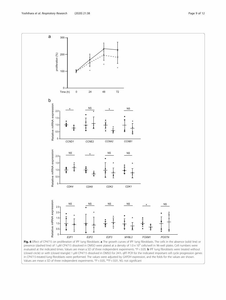
Fig. 6 Effect of CP4715 on proliferation of IPF lung fibroblasts. a The growth curves of IPF lung fibroblasts. The cells in the absence (solid line) orpresence (dashed line) of 1 μM CP4715 dissolved in DMSO were plated at a density of 1.0 × 10⁴ cells/well in 96-well plates. Cell numbers wereevaluated at the indicated times. Values are mean ± SD of three independent experiments. *P < 0.05. b IPF lung fibroblasts were treated without(closed circle) or with (closed triangle) 1 μM CP4715 dissolved in DMSO for 24 h. qRT-PCR for the indicated important cell cycle progression genesin CP4715-treated lung fibroblasts were performed. The values were adjusted by GAPDH expression, and the folds for the values are shown.Values are mean ± SD of three independent experiments. *P < 0.05, **P < 0.01, NS: not significant
Yoshihara et al. Respiratory Research (2020) 21:38 Page 9 of 12
that the negatively regulatory mechanism of cell cycle isimpaired in IPF patients [38]. In such a situation, stimu-lation by periostin may enhance cell proliferation of lungfibroblasts. The finding that lung structure is normallymaintained in periostin-deficient mice points to a dis-pensable role for periostin on proliferation of lung fibro-blasts at steady state [14]. However, given the aggressivestatus of proliferation for lung fibroblasts such as IPF,expression level of periostin may make a difference inexpansion of fibroblasts.It is widely accepted that the mitogen and integrin sig-
nals via the PI3K/Akt and Erk pathways are importantfor the transition of the cell cycle from the G1 to the Sphase, the first checkpoint of the cell cycle [39]. Integrinsignals are unique among integrin members [39, 40] andperiostin is a unique ligand for αVβ3 because periostindoes not have an RGD sequence like other ligands suchas vitronectin, osteopontin, and fibronectin. Neverthe-less, the cell cycle analysis in the present study showsthat periostin is important for the G1/S transition of thecell cycle as well as other integrin ligands (Fig. 4). In theG1 phase, the cyclin D1/CDK4 complex phosphorylatesRb protein, followed by dissociation of E2F from Rb[36]. E2F increases transcription of cyclin E followed byformation of the cyclin E/CDK2 complex, which furthersRb phosphorylation. Our present study shows that peri-ostin is important for the expression of cyclin E2, CDK2,and E2F members, but not of cyclin D1 or CDK4. Theseresults suggest that periostin promotes G1/S transitionby enhancing production of the cyclin E/CDK2 complexvia E2F members, rather than via the cyclin D/CDK4complex. Although periostin is important for expressionof the G2/M phase–related molecules―cyclin B, CDK1,B-MYB, and FOXM1―periostin silencing is unlikely tocause obvious impairment of the G2/M checkpoint.It is appreciated that fibroblasts taken from IPF patients
and cultured in vitro still retain the characteristics of the fi-broblasts in vivo in IPF patients. IPF fibroblasts display en-hanced proliferation on polymerized collagen matrices[41]. Moreover, profiles of expressed genes differ betweenIPF patients and normal donors [3–5] . These differencesinclude several signature molecules of IPF such as IGFBP-3 and lysyl oxidase [3]. Lee et al. have reported that perios-tin expression is enhanced in fibroblasts derived from IPFpatients, although expression levels of periostin varyamong the clones [5]. We observed that some clones ofIPF lung fibroblasts show high expression of periostincompared to normal lung fibroblasts, whereas there wasno statistical significance because of the heterogeneity ofIPF lung fibroblasts (Fig. 5). The concept of the heterogen-eity of IPF lung fibroblasts is consistent with our previousfinding that high expression of periostin is relatively lim-ited to the fibroblastic foci, which are not broadly observedin the lungs of IPF patients [7]. In spite of the
heterogeneity of periostin expression in IPF lung fibro-blasts, all IPF lung fibroblasts retain the effects of periostinsilencing on cell growth and expression of cell-cycle–re-lated molecules (Fig. 5). These results suggest that neitherprogramming nor the extracellular milieu in IPF affect thesignal pathway of periostin for proliferation of lungfibroblasts.Sadly, the median survival for IPF patients is only 3 to
5 years. Thus far, only two drugs, pirfenidone and ninte-danib, have been approved by by FDA to treat IPF, andthe efficacy of these drugs is limited. There is still an un-met need to develop a novel and effective therapeuticdrug to treat IPF. Given that periostin is a key moleculein the pathogenesis of pulmonary fibrosis, it is a promis-ing therapeutic target for IPF. Building on this concept,we have recently found that cross-talk between TGF-βand periostin is important for the generation of pulmon-ary fibrosis and that CP4715, a potent inhibitor of integ-rin αVβ3, improves pulmonary fibrosis in mice byinhibiting TGF-β signaling [15]. Our present studyshows that CP4715 has a potent ability to slow prolifera-tion of IPF fibroblasts, as does periostin silencing, al-though CP4715 has weaker abilities to downregulatecell-cycle–related genes than periostin silencing (Fig. 6).These results give us a basis for applying inhibitors ofthe periostin/integrin αVβ3 interaction to IPF patients.
ConclusionsIt is still uncertain how the fibroblasts in the patientswith idiopathic pulmonary fibrosis (IPF) acquire the abil-ity to progressively proliferate. Periostin is a matricellu-lar protein playing a critical role in the pathogenesis ofpulmonary fibrosis. In this study, we found that periostinplays a critical role in the proliferation of lung fibroblastsand that inhibition of the periostin/integrin αVβ3 (a peri-ostin receptor) interaction can be useful for the treat-ment of IPF patients.
Supplementary informationSupplementary information accompanies this paper at https://doi.org/10.1186/s12931-020-1299-0.
Additional file 1: Figure S1. Effect of periostin overexpression onexpression of cell cycle-related genes in lung fibroblasts. MRC-5 cells weretransiently transfected with 0.1 μg of the mock plasmid (open bar) or theexpression plasmid encoding periostin (closed bar). qRT-PCR for the indi-cated important cell cycle progression genes were performed inperiostin-overexpressed MRC-5 cells after 48 h. The values were adjustedby GAPDH expression, and the fold changes are shown. Values are mean± SD of three independent experiments. NS: not significant. Figure S2.Effect of adding recombinant periostin protein on proliferation of lung fi-broblasts. The growth curves of MRC-5 cells. (a) The normal cells in theabsence (solid line) or presence (dashed line) of recombinant periostinprotein (10 μg/mL) were plated at a density of 1.0 ×10⁴ cells/well in 96-well plates. (b) The control cells (black solid line) or periostin knockdowncells with (gray solid line) or without (black dashed line) recombinantperiostin protein (10 μg/mL) were plated at a density of 1.0 ×10⁴ cells/
Yoshihara et al. Respiratory Research (2020) 21:38 Page 10 of 12

well in 96-well plates. Cell numbers were evaluated at the indicatedtimes. Values are mean ± SD of three independent experiments. *P <0.05, NS: not significant. Figure S3. Effect of serum starvation on the dis-tribution of the cell cycle in lung fibroblasts. MRC-5 cells were cultured inthe medium with (left panel) or without (right panel) serum for 48 h. Dis-tribution of the cell cycle of MRC-5 cells as estimated by flow cytometryis depicted. We performed the same experiments for three times andshow the representative data.
Additional file 2: Table S1. Primer sequences used in qRT-PCR.
Additional file 3: Table S2. The profile of genes downregulated to lessthan one third or upregulated by more than three-fold by knockdown ofperiostin using DNA microarrays.
AbbreviationsAb: Antibody; BLM: Bleomycin; CDC: Cell division cycle; CDK: Cyclin-dependent kinase; ECM: Extracellular matrix; IPF: Idiopathic pulmonaryfibrosis; NHLF: Normal human lung fibroblast; PI: Propidium iodide; qRT-PCR: Quantitative reverse transcription PCR
AcknowledgementsWe thank Dr. Dovie R. Wylie for critical review of this manuscript. We alsothank Ms. Tomoyo Yoshida and Ms. Maki Watanabe for technical assistance.
Authors’ contributionsTY performed the experiments and evaluated the data together with SN andYN. YY and CFB obtained lung tissues from IPF patients and from normaldonor and then cultured primary lung fibroblasts. KA and SM preparedintegrin inhibitors. MM and KI wrote the manuscript. All authors commentedon the manuscript. All authors read and approved the final manuscript.
FundingThis work was supported in part by JSPS KAKENHI Grant Number#JP16H05343 (to KI).
Availability of data and materialsThe datasets used and/or analyzed during the current study available fromthe corresponding author on reasonable request.
Ethics approval and consent to participateNormal and IPF lung tissues were obtained under a protocol approved bythe University of Pittsburgh IRB.
Consent for publicationNot applicable.
Competing interestsKA and SM are employees of Meiji Seika Pharma Co. Ltd.. The other authorsdeclare no conflict of interest.
Author details1Division of Medical Biochemistry, Department of Biomolecular Sciences,Saga Medical School, 5-1-1 Nabeshima, Saga 849-8501, Japan. 2Departmentof Environmental Immuno-Dermatology, Yokohama City University GraduateSchool of Medicine, Yokohama 236-0004, Japan. 3Division of Rheumatologyand Immunology, Department of Medicine, Medical University of SouthCarolina, Charleston, SC 29425, USA. 4Pharmaceutical Research Center, MeijiSeika Pharma Co. Ltd., Yokohama 222-8567, Japan. 5Department ofOrthopaedic Surgery, Saga Medical School, Saga 849-8501, Japan.
Received: 29 July 2019 Accepted: 19 January 2020
References1. Martinez FJ, Collard HR, Pardo A, Raghu G, Richeldi L, Selman M, et al.
Idiopathic pulmonary fibrosis. Nat Rev Dis Primers. 2017;3:17074.2. Toonkel RL, Hare JM, Matthay MA, Glassberg MK. Mesenchymal stem cells
and idiopathic pulmonary fibrosis. Potential for clinical testing. Am J RespirCrit Care Med. 2013;188:133–40.
3. Hsu E, Shi H, Jordan RM, Lyons-Weiler J, Pilewski JM, Feghali-Bostwick CA.Lung tissues in patients with systemic sclerosis have gene expression
patterns unique to pulmonary fibrosis and pulmonary hypertension. ArthritisRheum. 2011;63:783–94.
4. Vuga LJ, Ben-Yehudah A, Kovkarova-Naumovski E, Oriss T, Gibson KF,Feghali-Bostwick C, et al. WNT5A is a regulator of fibroblast proliferation andresistance to apoptosis. Am J Respir Cell Mol Biol. 2009;41:583–9.
5. Lee JU, Cheong HS, Shim EY, Bae DJ, Chang HS, Uh ST, et al. Gene profile offibroblasts identify relation of CCL8 with idiopathic pulmonary fibrosis.Respir Res. 2017;18:3.
6. Izuhara K, Nunomura S, Nanri Y, Ogawa M, Ono J, Mitamura Y, et al.Periostin in inflammation and allergy. Cell Mol Life Sci. 2017;74:4293–303.
7. Izuhara K, Arima K, Ohta S, Suzuki S, Inamitsu M, Yamamoto K. Periostin inallergic inflammation. Allergol Int. 2014;63:143–51.
8. Okamoto M, Hoshino T, Kitasato Y, Sakazaki Y, Kawayama T, Fujimoto K,et al. Periostin, a matrix protein, is a novel biomarker for idiopathicinterstitial pneumonias. Eur Respir J. 2011;37:1119–27.
9. Murata K, Koga Y, Kasahara N, Hachisu Y, Nunomura S, Nakajima N, et al.Accumulation of periostin in acute exacerbation of familial idiopathicpulmonary fibrosis. J Thorac Dis. 2018;10:E587–91.
10. Naik PK, Bozyk PD, Bentley JK, Popova AP, Birch CM, Wilke CA, et al. Periostinpromotes fibrosis and predicts progression in patients with idiopathicpulmonary fibrosis. Am J Physiol Lung Cell Mol Physiol. 2018;303:L1046–56.
11. Nance T, Smith KS, Anaya V, Richardson R, Ho L, Pala M, et al. Transcriptomeanalysis reveals differential splicing events in IPF lung tissue. PLoS One.2014;9:e92111.
12. Cecchini MJ, Hosein K, Howlett CJ, Joseph M, Mura M. Comprehensive geneexpression profiling identifies distinct and overlapping transcriptionalprofiles in non-specific interstitial pneumonia and idiopathic pulmonaryfibrosis. Respir Res. 2018;19:153.
13. Takayama G, Arima K, Kanaji T, Toda S, Tanaka H, Shoji S, et al. Periostin: anovel component of subepithelial fibrosis of bronchial asthma downstreamof IL-4 and IL-13 signals. J Allergy Clin Immunol. 2006;118:98–104.
14. Uchida M, Shiraishi H, Ohta S, Arima K, Taniguchi K, Suzuki S, et al. Periostin,a matricellular protein, plays a role in the induction of chemokines inpulmonary fibrosis. Am J Respir Cell Mol Biol. 2012;46:677–86.
15. Nanri Y, Nunomura S, Terasaki Y, Yoshihara T, Hirano Y, Yokosaki Y, et al. Thecross-talk between TGF-β and periostin can be targeted for pulmonary fibrosis.Am J Respir Cell Mol Biol. 2019. https://doi.org/10.1165/rcmb.2019-0245OC.
16. Mitamura Y, Nunomura S, Nanri Y, Arima K, Yoshihara T, Komiya K, et al.Hierarchical control of interleukin 13 (IL-13) signals in lung fibroblasts bySTAT6 and SOX11. J Biol Chem. 2018;293:14646–58.
17. Pilewski JM, Liu L, Henry AC, Knauer AV, Feghali-Bostwick CA. Insulin-likegrowth factor binding proteins 3 and 5 are overexpressed in idiopathicpulmonary fibrosis and contribute to extracellular matrix deposition. Am JPathol. 2005;166:399–407.
18. Kubota D, Ishikawa M, Yamamoto M, Murakami S, Hachisu M, Katano K,et al. Tricyclic pharmacophore-based molecules as novel integrin αvβ3antagonists. Part 1: design and synthesis of a lead compound exhibitingαvβ3/αIIbβ3 dual antagonistic activity. Bioorg Med Chem. 2006;14:2089–108.
19. Ishikawa M, Kubota D, Yamamoto M, Kuroda C, Iguchi M, Koyanagi A, et al.Tricyclic pharmacophore-based molecules as novel integrin αvβ3antagonists. Part 2: synthesis of potent αvβ3/αIIbβ3 dual antagonists. BioorgMed Chem. 2006;14:2109–30.
20. Ishikawa M, Hiraiwa Y, Kubota D, Tsushima M, Watanabe T, MurakamiS, et al. Tricyclic pharmacophore-based molecules as novel integrinαvβ3 antagonists. Part III: synthesis of potent antagonists with αvβ3/αIIbβ3 dual activity and improved water solubility. Bioorg Med Chem.2006;14:2131–50.
21. Kubota D, Ishikawa M, Ishikawa M, Yahata N, Murakami S, Fujishima K, et al.Tricyclic pharmacophore-based molecules as novel integrin αvβ3antagonists. Part IV: preliminary control of αvβ3 selectivity by meta-orientedsubstitution. Bioorg Med Chem. 2006;14:4158–81.
22. Gerard C, Goldbeter A. Dynamics of the mammalian cell cycle in physiologicaland pathological conditions. Wiley Interdiscip Rev Syst Biol Med. 2016;8:140–56.
23. Lim S, Kaldis P. Cdks, cyclins and CKIs: roles beyond cell cycle regulation.Development. 2013;140:3079–93.
24. Fischer M, Muller GA. Cell cycle transcription control: DREAM/MuvB and RB-E2F complexes. Crit Rev Biochem Mol Biol. 2017;52:638–62.
25. Iaquinta PJ, Lees JA. Life and death decisions by the E2F transcriptionfactors. Curr Opin Cell Biol. 2007;19:649–57.
26. Joaquin M, Watson RJ. Cell cycle regulation by the B-Myb transcriptionfactor. Cell Mol Life Sci. 2003;60:2389–401.
Yoshihara et al. Respiratory Research (2020) 21:38 Page 11 of 12

27. Masuoka M, Shiraishi H, Ohta S, Suzuki S, Arima K, Aoki S, et al. Periostinpromotes chronic allergic inflammation in response to Th2 cytokines. J ClinInvest. 2012;122:2590–600.
28. Tai IT, Dai M, Chen LB. Periostin induction in tumor cell line explants andinhibition of in vitro cell growth by anti-periostin antibodies. Carcinogenesis.2005;26:908–15.
29. Shao R, Bao S, Bai X, Blanchette C, Anderson RM, Dang T, et al. Acquiredexpression of periostin by human breast cancers promotes tumorangiogenesis through up-regulation of vascular endothelial growth factorreceptor 2 expression. Mol Cell Biol. 2004;24:3992–4003.
30. Hong L, Sun H, Lv X, Yang D, Zhang J, Shi Y. Expression of periostin in theserum of NSCLC and its function on proliferation and migration of humanlung adenocarcinoma cell line (A549) in vitro. Mol Biol Rep. 2010;37:2285–93.
31. Kikuchi Y, Kunita A, Iwata C, Komura D, Nishiyama T, Shimazu K, et al. The nichecomponent periostin is produced by cancer-associated fibroblasts, supportinggrowth of gastric cancer through ERK activation. Am J Pathol. 2014;184:859–70.
32. Kotobuki Y, Yang L, Serada S, Tanemura A, Yang F, Nomura S, et al. Periostinaccelerates human malignant melanoma progression by modifying themelanoma microenvironment. Pigment Cell Melanoma Res. 2014;27:630–9.
33. Michaylira CZ, Wong GS, Miller CG, Gutierrez CM, Nakagawa H, Hammond R,et al. Periostin, a cell adhesion molecule, facilitates invasion in the tumormicroenvironment and annotates a novel tumor-invasive signature inesophageal cancer. Cancer Res. 2010;70:5281–92.
34. Gonzalez-Gonzalez L, Alonso J. Periostin: a matricellular protein withmultiple functions in cancer development and progression. Front Oncol.2018;8:225.
35. Kuhn B, del Monte F, Hajjar RJ, Chang YS, Lebeche D, Arab S, et al. Periostininduces proliferation of differentiated cardiomyocytes and promotes cardiacrepair. Nat Med. 2007;13:962–9.
36. Lorts A, Schwanekamp JA, Elrod JW, Sargent MA, Molkentin JD. Geneticmanipulation of periostin expression in the heart does not affect myocytecontent, cell cycle activity, or cardiac repair. Circ Res. 2009;104:e1–7.
37. Vermeulen K, Van Bockstaele DR, Berneman ZN. The cell cycle: a review ofregulation, deregulation and therapeutic targets in cancer. Cell Prolif. 2003;36:131–49.
38. Al-Tamari HM, Dabral S, Schmall A, Sarvari P, Ruppert C, Paik J, et al. FoxO3an important player in fibrogenesis and therapeutic target for idiopathicpulmonary fibrosis. EMBO Mol Med. 2018;10:276–93.
39. Moreno-Layseca P, Streuli CH. Signalling pathways linking integrins with cellcycle progression. Matrix Biol. 2014;34:144–53.
40. Schwartz MA, Assoian RK. Integrins and cell proliferation: regulation ofcyclin-dependent kinases via cytoplasmic signaling pathways. J Cell Sci.2001;114:2553–60.
41. Xia H, Diebold D, Nho R, Perlman D, Kleidon J, Kahm J, et al. Pathologicalintegrin signaling enhances proliferation of primary lung fibroblasts frompatients with idiopathic pulmonary fibrosis. J Exp Med. 2008;205:1659–72.
Publisher’s NoteSpringer Nature remains neutral with regard to jurisdictional claims inpublished maps and institutional affiliations.
Yoshihara et al. Respiratory Research (2020) 21:38 Page 12 of 12