peripheral arterial doppler
TRANSCRIPT

PERIPHERAL VASCULAR DOPPLER
DR MOHIT GOEL5 July 2013

Each normal major vessel in the human body has a characteristic flow pattern that is representable in spectral waveforms obtained with Doppler ultrasonography (US) and that reflects both the anatomic position of the vessel and the physiologic need of the organ it supplies.
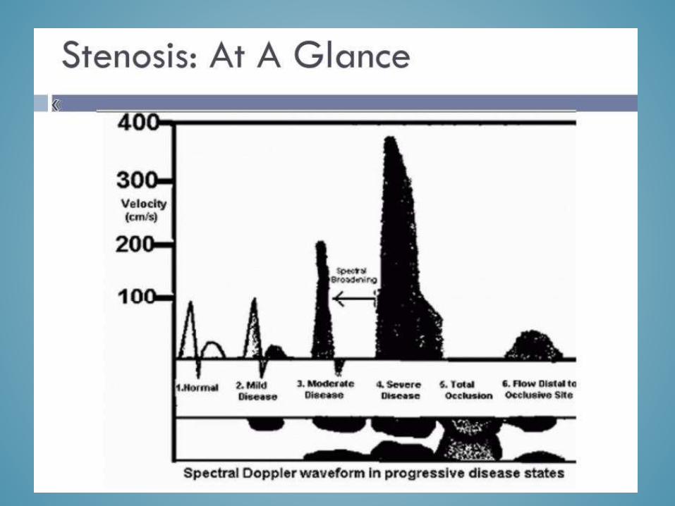
The Doppler spectrum is a time-velocity waveform that represents variation in intravascular blood flow velocities during the cardiac cycle.

Time is represented along the horizontal axis, and frequency shift (velocity) is depicted along the vertical axis.
The intensity or brightness (also referred to as the gray-scale velocity plot) of the spectral line represents the number of red blood cells that are reflecting the ultrasound beam at each velocity. The width of the spectral line represents the range of velocities within a vessel. The width may vary during the normal cardiac cycle, narrowing during systole and widening in diastole.
The spectral window is the clear black zone between the spectral line and the baseline. Widening of the spectral line and filling of the spectral window is called spectral broadening. Spectral broadening is normally seen in the presence of high flow velocity, at the branching of a vessel, or in small-diameter vessels.


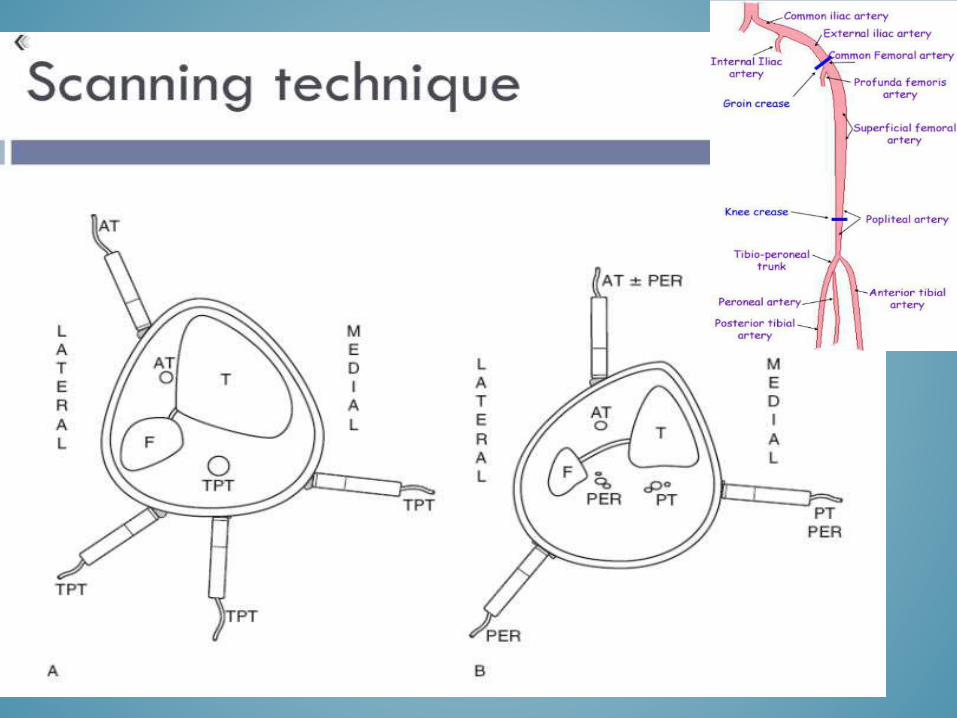
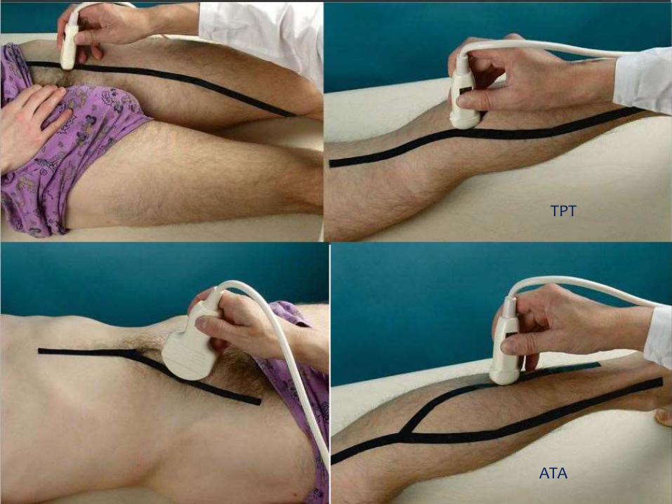
ATA
TPT


PSEUDOANEURYSMS
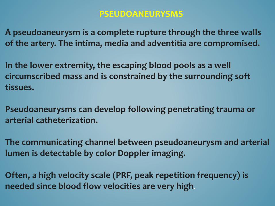
A pseudoaneurysm is a complete rupture through the three walls of the artery. The intima, media and adventitia are compromised.
In the lower extremity, the escaping blood pools as a well circumscribed mass and is constrained by the surrounding soft tissues.
Pseudoaneurysms can develop following penetrating trauma or arterial catheterization.
The communicating channel between pseudoaneurysm and arterial lumen is detectable by color Doppler imaging.
Often, a high velocity scale (PRF, peak repetition frequency) is needed since blood flow velocities are very high.

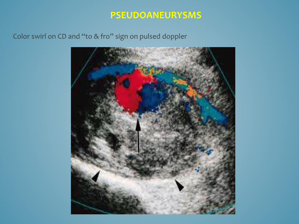
The presence of a "to-and-fro" or "forward-backward" waveform is typically seen when the Doppler gate is placed over the communicating channel of the pseudoaneurysm.
The "to" or "forward" component is due to entry of blood into the collection as the soft tissues expand to accommodate entry of blood within the pseudoaneurysm cavity. This occurs during systole.
The "fro" or "backward" component is seen during diastole as the blood stored in the cavity is ejected back into the artery.This is caused by the stored energy due to the elasticity of the surrounding soft tissues.
Pseudoaneurysms can have multiple compartments as well as be solitary . Blood flow in a pseudoaneurysm cavity has a tendency to show a swirling pattern.
PSEUDOANEURYSMS
Color swirl on CD and “to & fro” sign on pulsed doppler

classic to-and-fro waveform of a pseudoaneurysm


Arteriovenous fistulas
Arteriovenous fistulas (AVFs) can result when there is puncture of an artery and vein with a direct communication between the two injured vessels.
A significant pressure gradient will result in a focal area of significantly increased velocity at the site of the AVF with flow directed from the artery to the vein.
Color Doppler imaging of AVFs will often demonstrate a bruit artifact and/or a thrill may be palpable.

Spectral Doppler analysis of the effected artery above the AVF will demonstrate a mono-phasic continuous waveform with elevated systolic and diastolic velocities.
Flow in the injured artery distal to the AVF will generally have normal pulsatility.
Spectral Doppler analysis of the effected vein central to the AVF will demonstrate “arterialized flow” with pulsations during systole and a lack of respiratory phasicity.
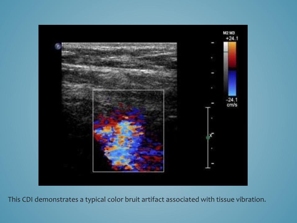
This CDI demonstrates a typical color bruit artifact associated with tissue vibration.
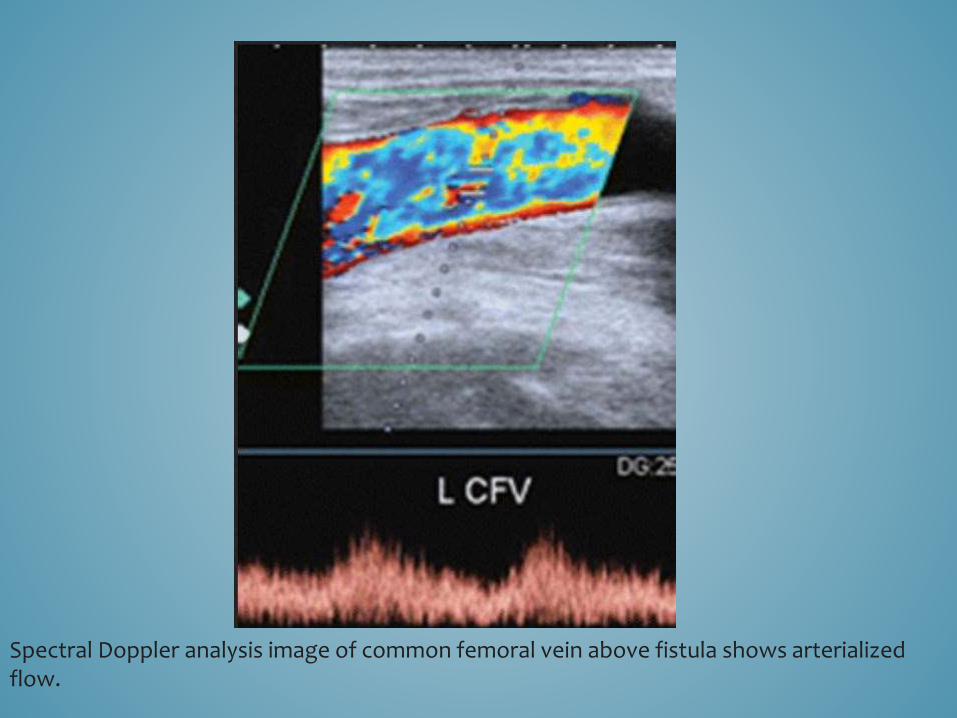
Spectral Doppler analysis image of common femoral vein above fistula shows arterialized flow.

A, Color flow Doppler image shows a high-velocity jet (arrow)from the common femoral artery (A) into the distended common femoral vein (V)

B, The arterialtype signals sampled in the common femoral vein are consistent with a large AV fistula showing an arterialized venous blood flow pattern.

THANK YOU