peripheral nervous system - linn–benton …cf.linnbenton.edu/mathsci/bio/jacobsr/upload/8 -...
TRANSCRIPT
P N S
C R A N I A L N E R V E S
S P I N A L N E R V E S
The Nervous System
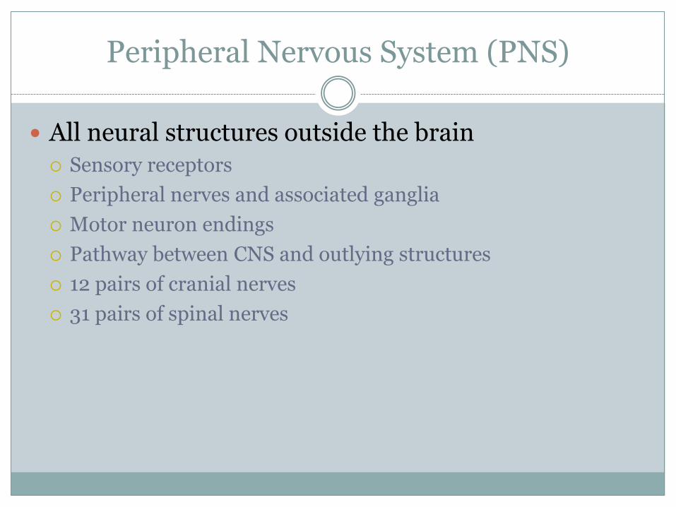
Peripheral Nervous System (PNS)
All neural structures outside the brain
Sensory receptors
Peripheral nerves and associated ganglia
Motor neuron endings
Pathway between CNS and outlying structures
12 pairs of cranial nerves
31 pairs of spinal nerves
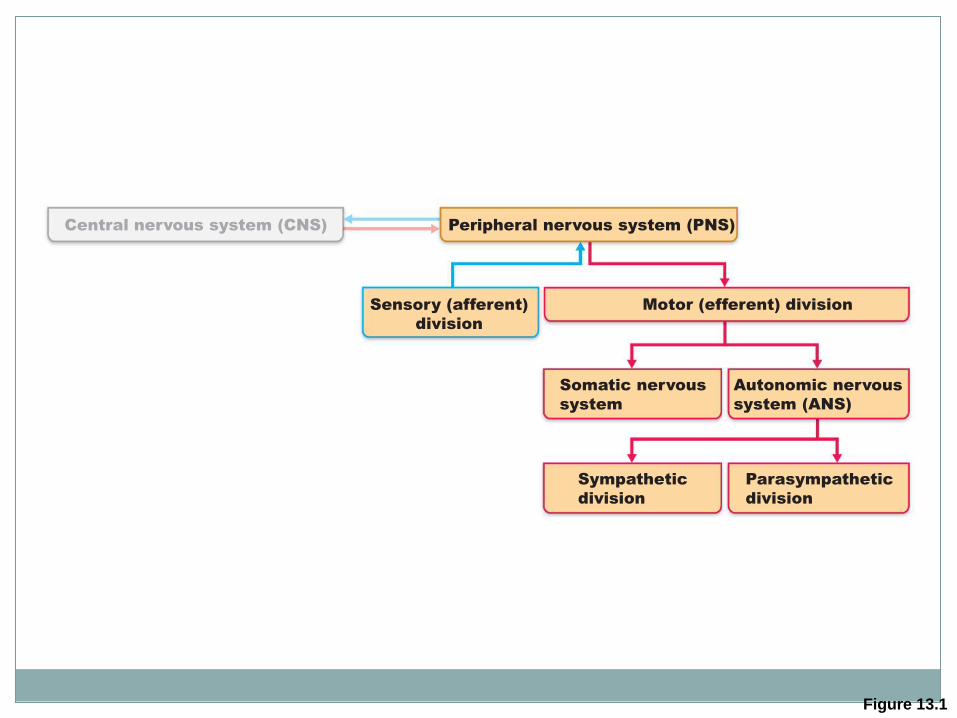
Figure 13.1
Central nervous system (CNS) Peripheral nervous system (PNS)
Motor (efferent) division Sensory (afferent)
division
Somatic nervous
system
Autonomic nervous
system (ANS)
Sympathetic
division
Parasympathetic
division
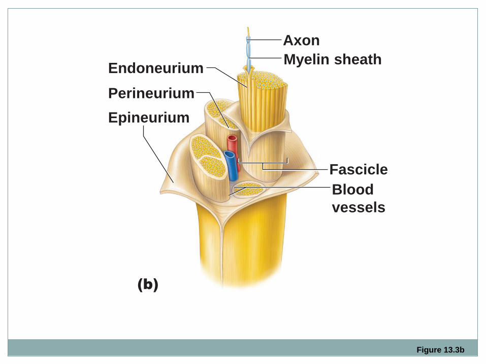
Figure 13.3b
Blood
vessels
Fascicle
Epineurium
Perineurium
Endoneurium
Axon
Myelin sheath
(b)
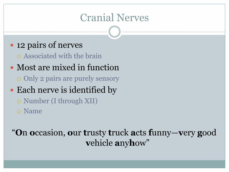
Cranial Nerves
12 pairs of nerves
Associated with the brain
Most are mixed in function
Only 2 pairs are purely sensory
Each nerve is identified by
Number (I through XII)
Name
“On occasion, our trusty truck acts funny—very good vehicle anyhow”
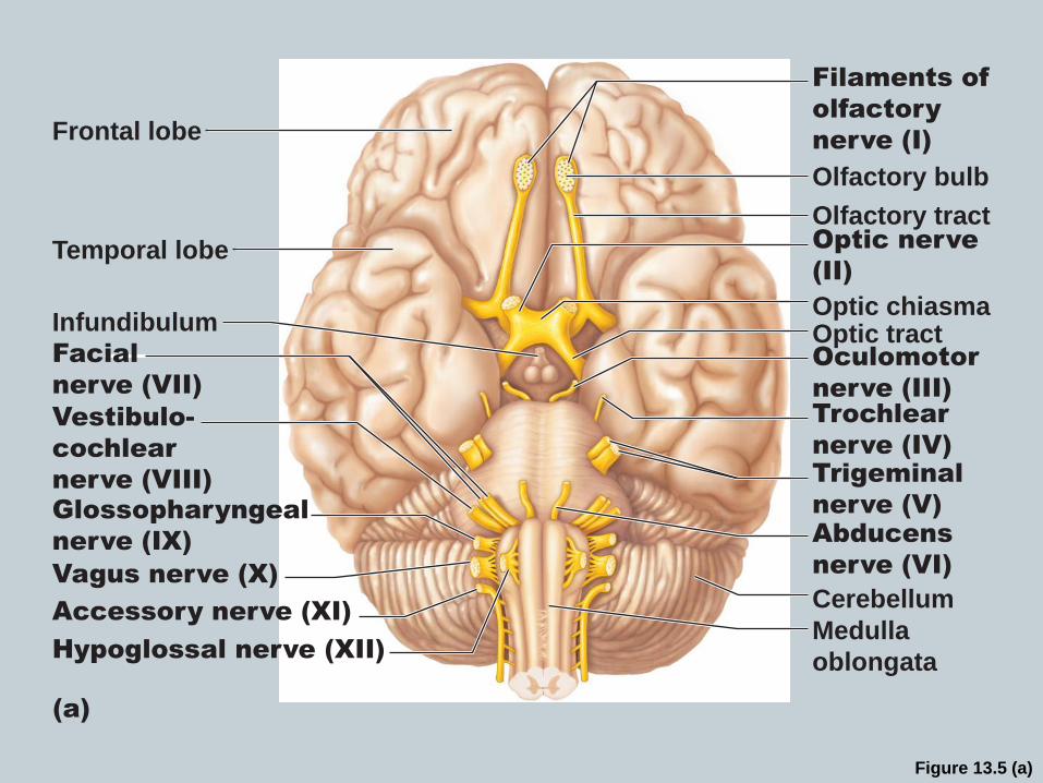
Figure 13.5 (a)
Frontal lobe
Temporal lobe
Infundibulum
Facial
nerve (VII)
Vestibulo-
cochlear
nerve (VIII)
Glossopharyngeal
nerve (IX)
Vagus nerve (X)
Accessory nerve (XI)
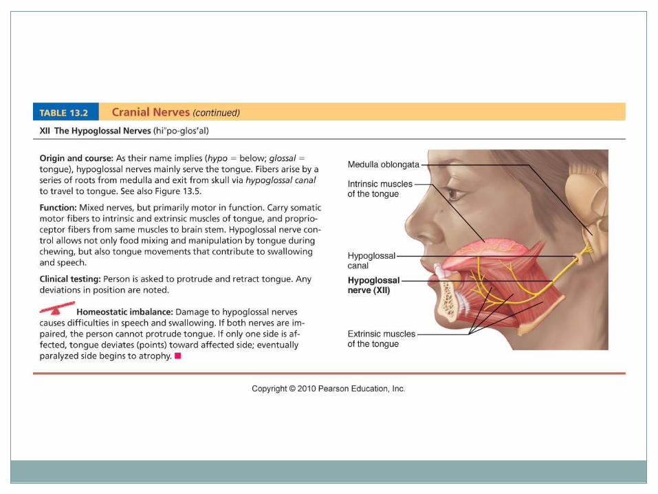
Hypoglossal nerve (XII)
(a)
Filaments of
olfactory
nerve (I)
Olfactory bulb
Olfactory tract
Optic chiasma
Optic nerve
(II)
Optic tract Oculomotor
nerve (III)
Trochlear
nerve (IV)
Trigeminal
nerve (V)
Abducens
nerve (VI)
Cerebellum
Medulla
oblongata
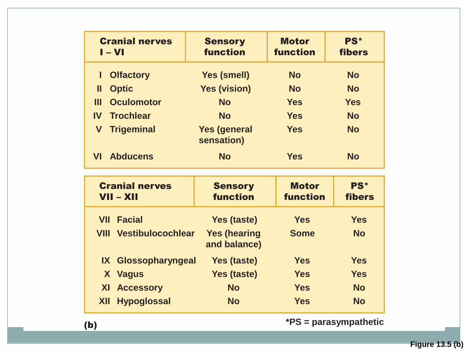
Figure 13.5 (b)
*PS = parasympathetic (b)
Cranial nerves
I – VI
I
II
III
IV
V
VI
Olfactory
Optic
Oculomotor
Trochlear
Trigeminal
Abducens
Yes (smell)
Yes (vision)
No
No
Yes (general
sensation)
No
No
No
Yes
Yes
Yes
Yes
No
No
Yes
No
No
No
Cranial nerves
VII – XII
Sensory
function
Motor
function
PS*
fibers
Sensory
function
Motor
function
PS*
fibers
VII
VIII
IX
X
XI
XII
Facial
Vestibulocochlear
Glossopharyngeal
Vagus
Accessory
Hypoglossal
Yes (taste)
Yes (hearing
and balance)
Yes (taste)
Yes (taste)
No
No
Yes
Some
Yes
Yes
Yes
Yes
Yes
No
Yes
Yes
No
No
Table 13.2
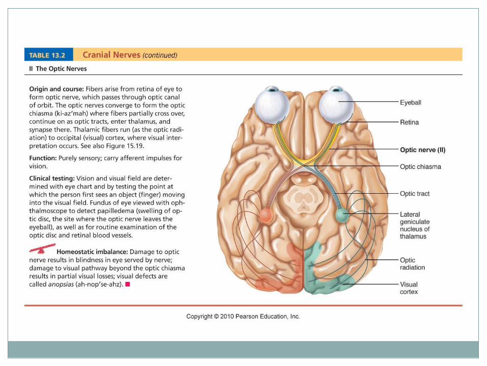
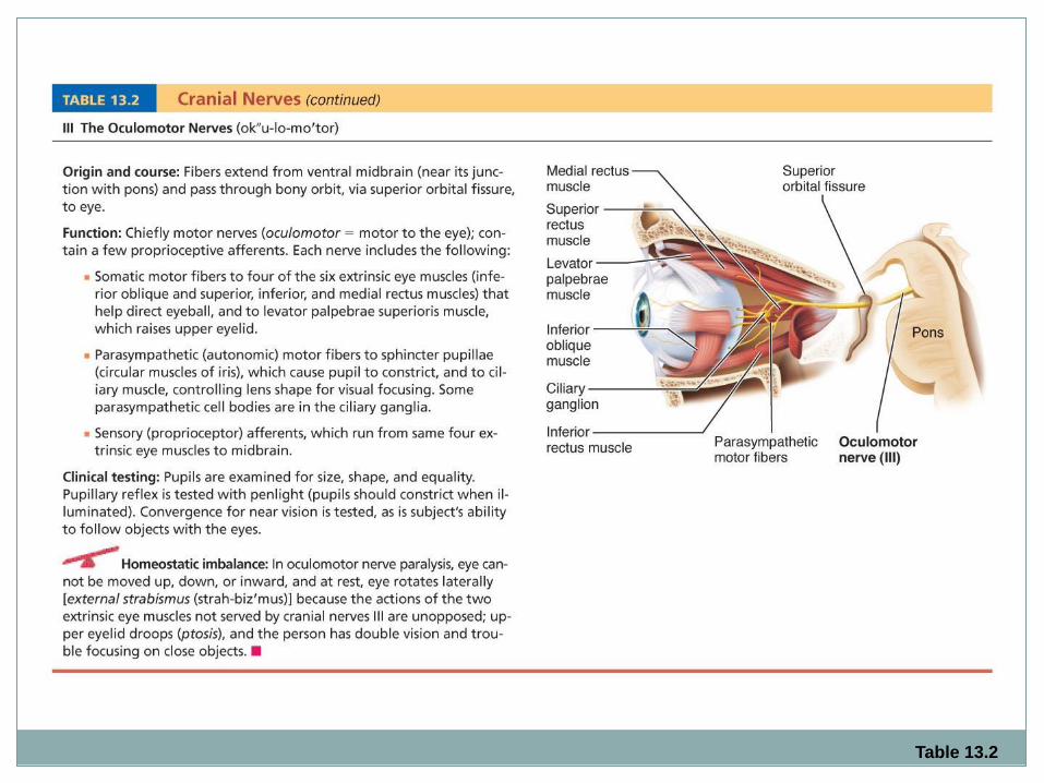
Table 13.2
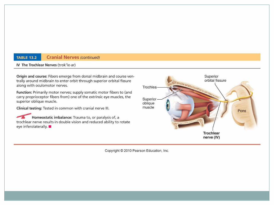
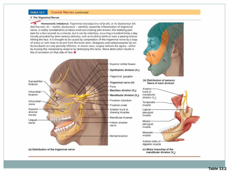
Table 13.2
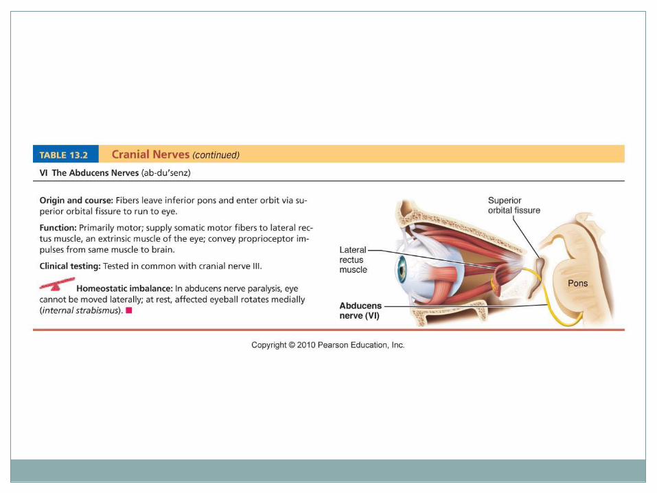
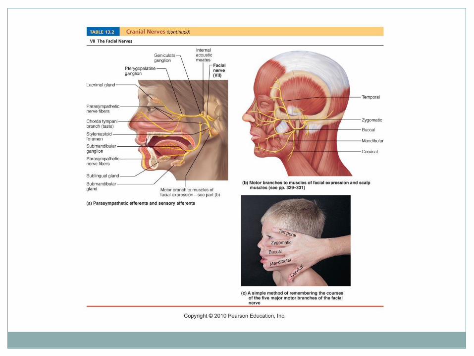
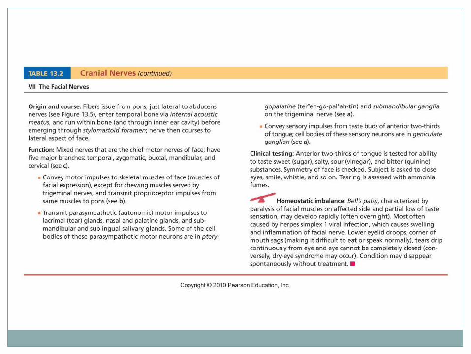
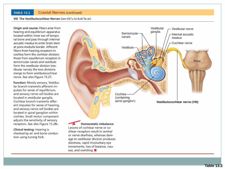
Table 13.2
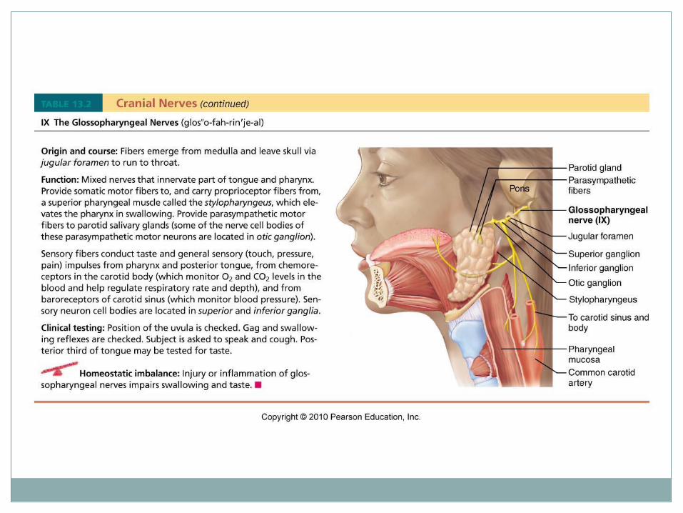
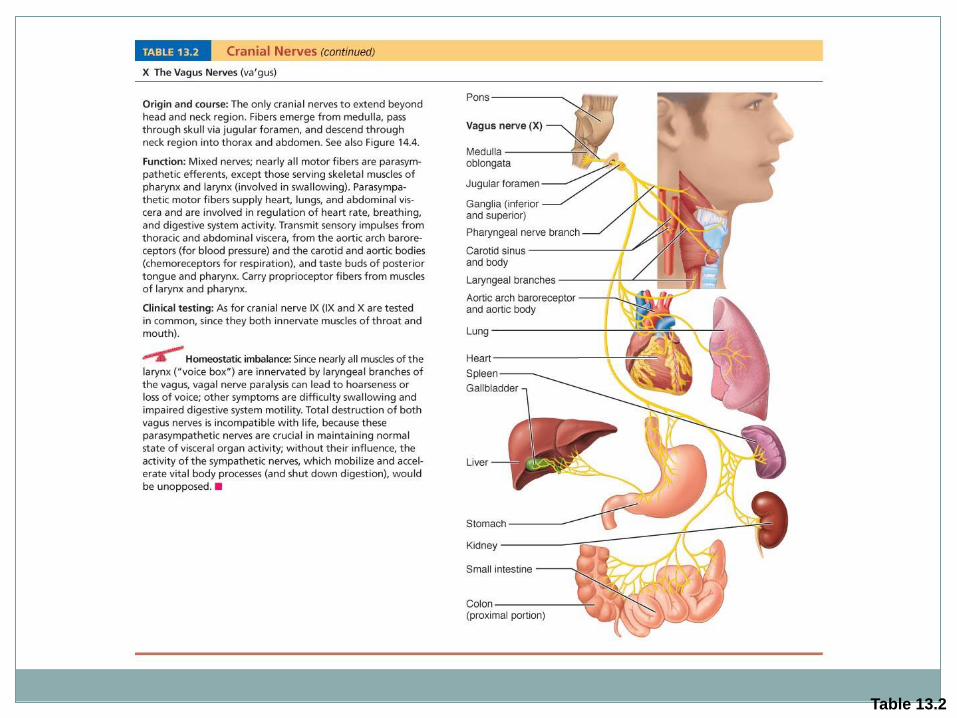
Table 13.2
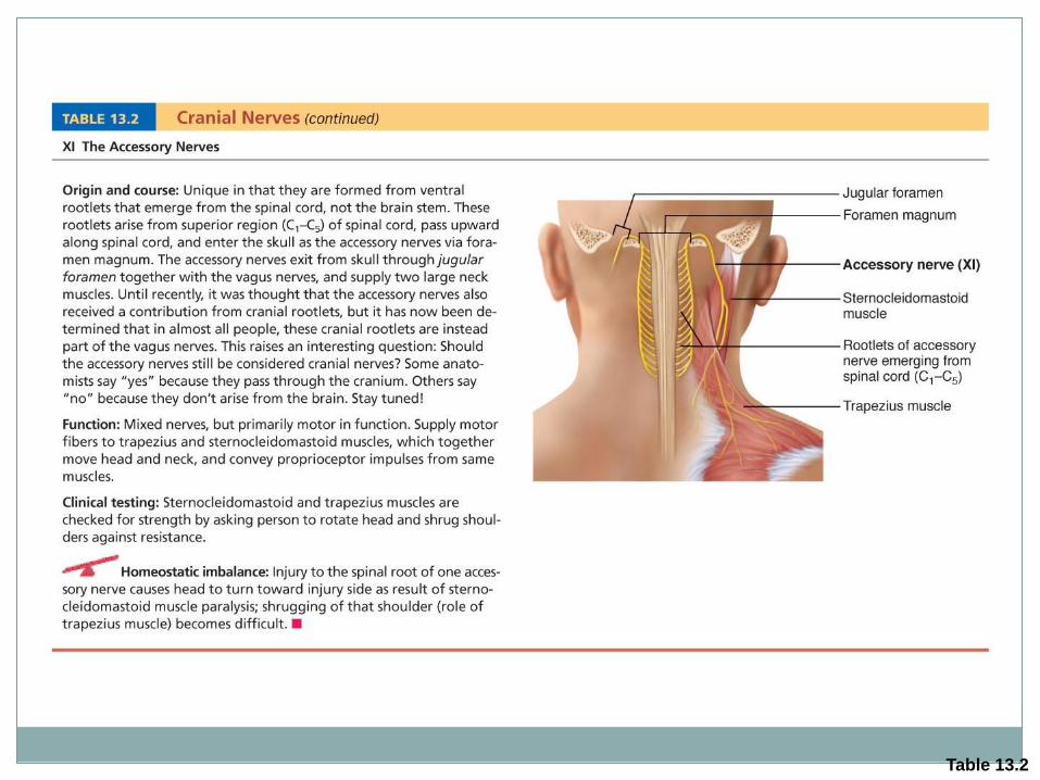
Table 13.2
Activity
Complete Cranial Nerve Chart
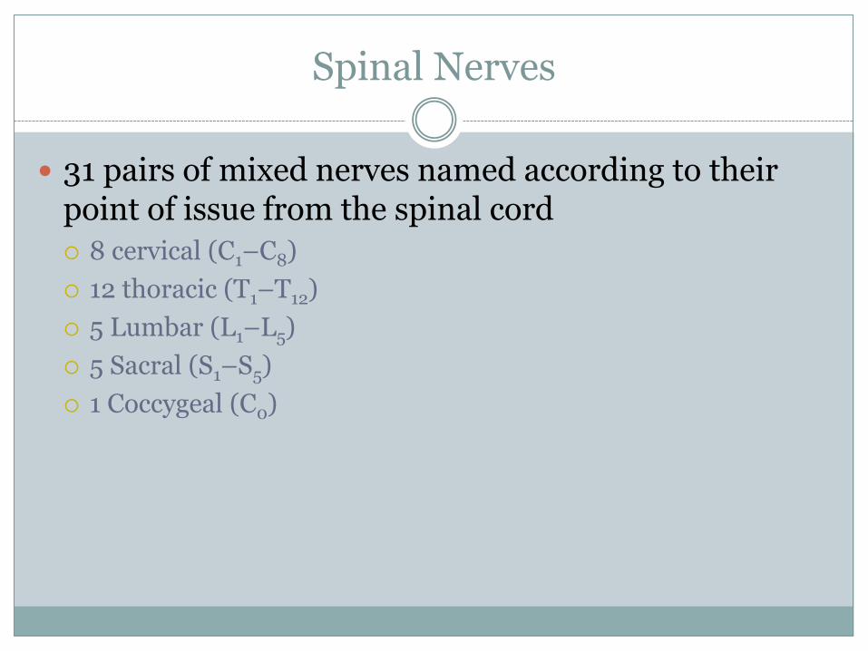
Spinal Nerves
31 pairs of mixed nerves named according to their point of issue from the spinal cord
8 cervical (C1–C8)
12 thoracic (T1–T12)
5 Lumbar (L1–L5)
5 Sacral (S1–S5)
1 Coccygeal (C0)

Figure 13.6
Cervical
nerves
C1 – C8
Thoracic
nerves
T1 – T12
Lumbar
nerves
L1 – L5
Sacral nerves
S1 – S5
Coccygeal nerve Co1
Cervical plexus
Intercostal
nerves
Cervical
enlargement
Lumbar
enlargement
Cauda equina
Brachial plexus
Lumbar plexus
Sacral plexus
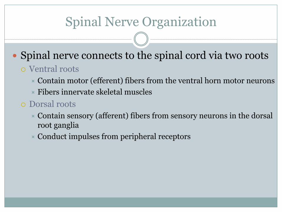
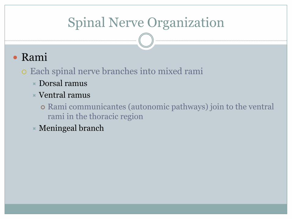
Spinal Nerve Organization
Spinal nerve connects to the spinal cord via two roots
Ventral roots
Contain motor (efferent) fibers from the ventral horn motor neurons
Fibers innervate skeletal muscles
Dorsal roots
Contain sensory (afferent) fibers from sensory neurons in the dorsal root ganglia
Conduct impulses from peripheral receptors
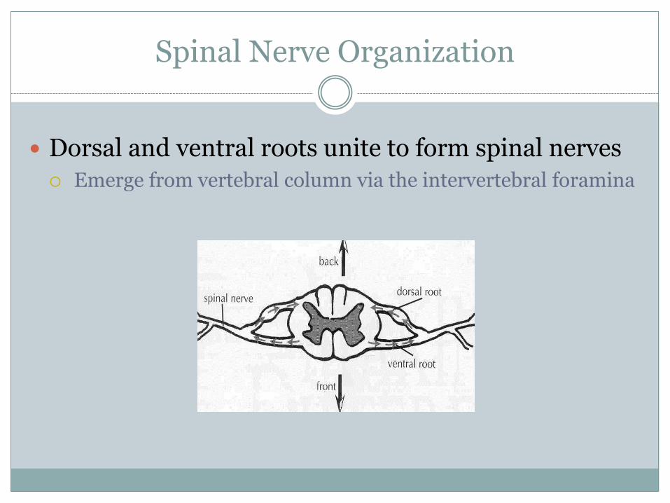
Spinal Nerve Organization
Dorsal and ventral roots unite to form spinal nerves
Emerge from vertebral column via the intervertebral foramina
Spinal Nerve Organization
Rami
Each spinal nerve branches into mixed rami
Dorsal ramus
Ventral ramus
Rami communicantes (autonomic pathways) join to the ventral rami in the thoracic region
Meningeal branch
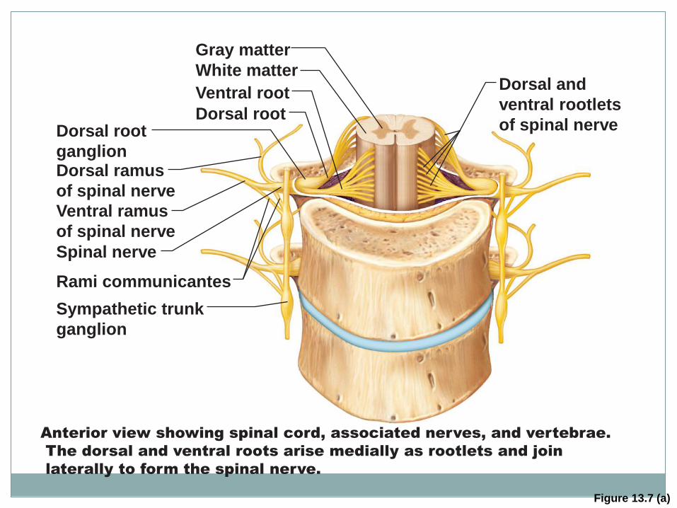
Figure 13.7 (a)
Dorsal root
ganglion
Gray matter
White matter
Ventral root
Dorsal root
Dorsal and
ventral rootlets
of spinal nerve
Dorsal ramus
of spinal nerve
Ventral ramus
of spinal nerve
Sympathetic trunk
ganglion
Spinal nerve
Rami communicantes
Anterior view showing spinal cord, associated nerves, and vertebrae.
The dorsal and ventral roots arise medially as rootlets and join
laterally to form the spinal nerve.
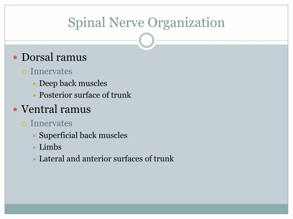
Spinal Nerve Organization
Dorsal ramus
Innervates
Deep back muscles
Posterior surface of trunk
Ventral ramus
Innervates
Superficial back muscles
Limbs
Lateral and anterior surfaces of trunk
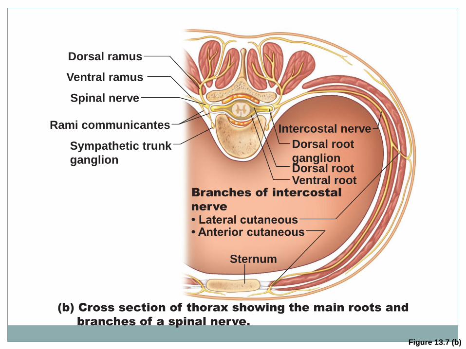
Figure 13.7 (b)
Dorsal ramus
Ventral ramus
Intercostal nerve
Spinal nerve
Rami communicantes
Dorsal root
ganglion Dorsal root Ventral root
Sympathetic trunk
ganglion
Sternum
(b) Cross section of thorax showing the main roots and
branches of a spinal nerve.
Branches of intercostal
nerve
• Lateral cutaneous • Anterior cutaneous
Distribution of Spinal Nerves
Dermatome
Area of skin innervated by the cutaneous branches of a single spinal nerve
All spinal nerves except C1 participate in dermatomes
Most dermatomes overlap
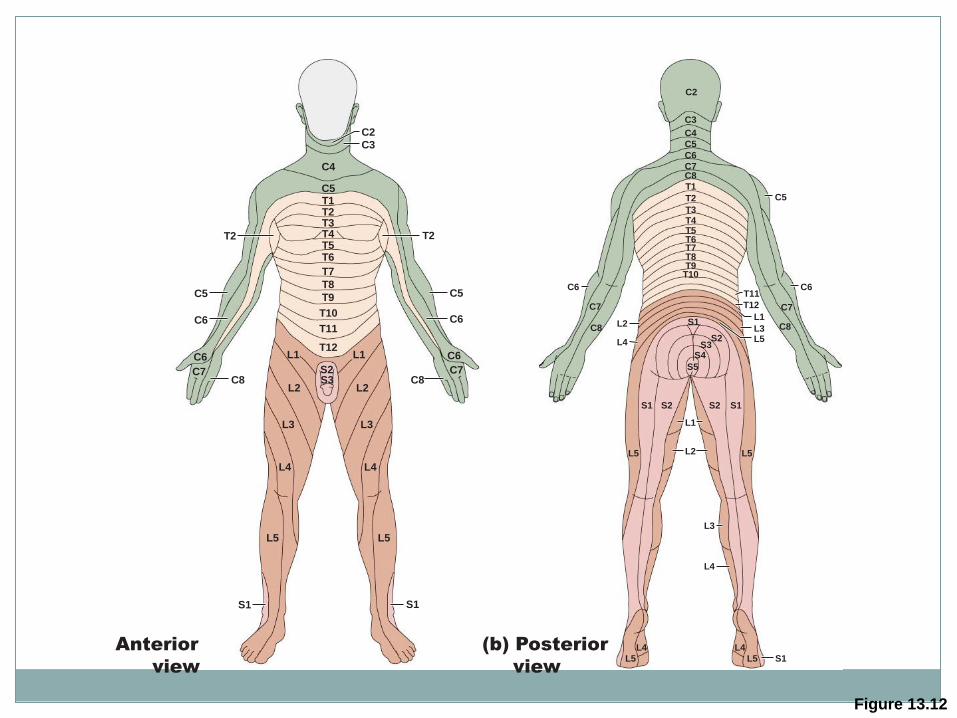
Figure 13.12
C2
C3
C4
C5
T1
T2
T2
T3
T4
T5
C6
C8
C7 C7
C6
T6
T7
T8
T9
T10
T11
T12
L1
S2
S3
L1
L2
L3
L4
L5
L2
L3
L4
L5
S1
C5
C6
C8
T2
C5
C6
S1
Anterior
view
C2
C3
C4
C5
C6
C7
C8
C8 C8
C7 C7
T1
T2
T3
T4
T5
T6
T7
T8
T9
T10
T11
T12
L1
L2 L3
S1
(b) Posterior
view
L5 S2
S1
S1
S3
S2 S1 S2
S4
S5
L5 L5
L4
L5 L5
L4
C6 C6
C5
L4
L3
L2
L1
L4
Plexuses
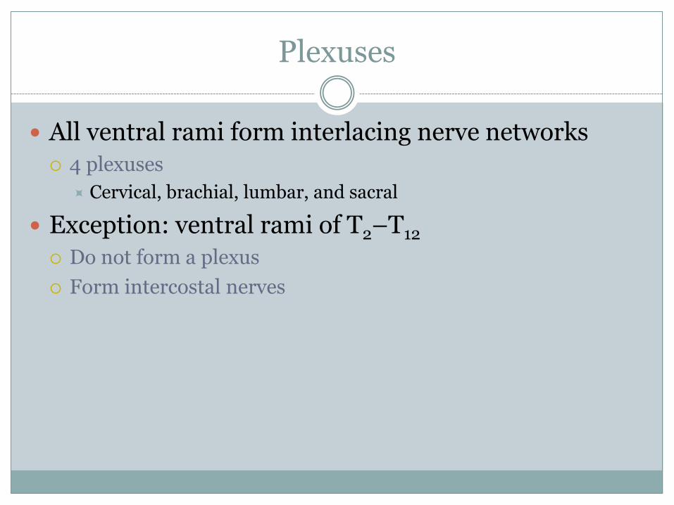
All ventral rami form interlacing nerve networks
4 plexuses
Cervical, brachial, lumbar, and sacral
Exception: ventral rami of T2–T12
Do not form a plexus
Form intercostal nerves
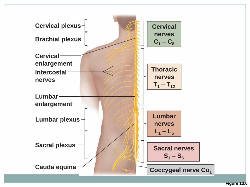
Figure 13.6
Cervical
nerves
C1 – C8
Thoracic
nerves
T1 – T12
Lumbar
nerves
L1 – L5
Sacral nerves
S1 – S5
Coccygeal nerve Co1
Cervical plexus
Intercostal
nerves
Cervical
enlargement
Lumbar
enlargement
Cauda equina
Brachial plexus
Lumbar plexus
Sacral plexus
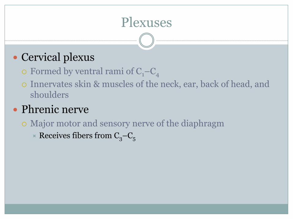
Plexuses
Cervical plexus
Formed by ventral rami of C1–C4
Innervates skin & muscles of the neck, ear, back of head, and shoulders
Phrenic nerve
Major motor and sensory nerve of the diaphragm
Receives fibers from C3–C5
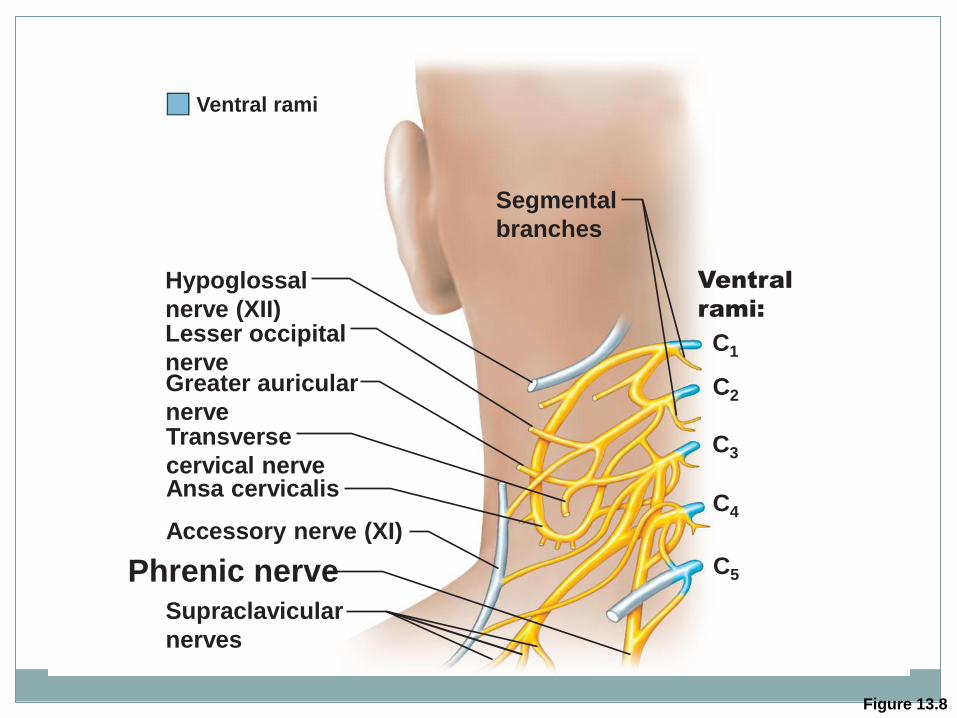
Figure 13.8
Hypoglossal
nerve (XII)
C1
C2
C3
C4
C5
Segmental
branches
Lesser occipital
nerve Greater auricular
nerve
Ansa cervicalis
Phrenic nerve Supraclavicular
nerves
Accessory nerve (XI)
Transverse
cervical nerve
Ventral
rami:
Ventral rami
Plexuses
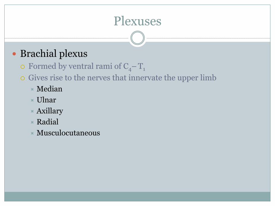
Brachial plexus Formed by ventral rami of C4– T1
Gives rise to the nerves that innervate the upper limb
Median
Ulnar
Axillary
Radial
Musculocutaneous
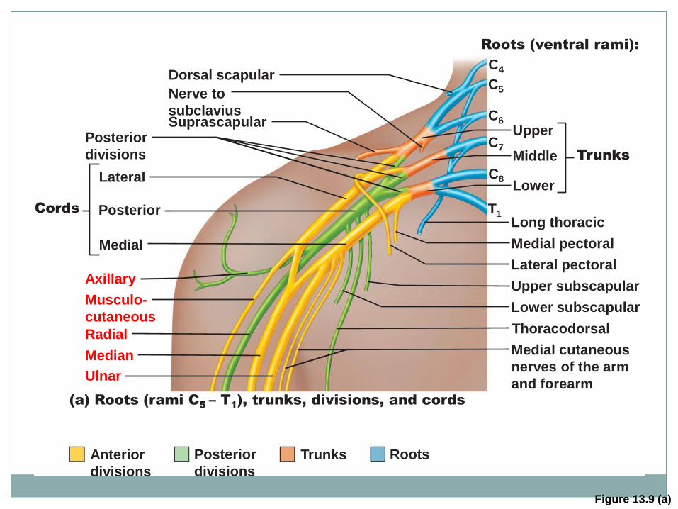
Figure 13.9 (a)
Upper
Middle Trunks
Lower
Roots (ventral rami):
Upper subscapular
Lower subscapular
Thoracodorsal
Medial cutaneous
nerves of the arm
and forearm
Long thoracic
Medial pectoral
Lateral pectoral
Nerve to
subclavius Suprascapular
Dorsal scapular
Posterior
divisions
Anterior
divisions
Lateral
Posterior Cords
Medial
Axillary
Musculo-
cutaneous
Radial
Median
Ulnar
Posterior
divisions
Trunks Roots
C4
C5
C6
C7
C8
T1
(a) Roots (rami C5 – T
1), trunks, divisions, and cords
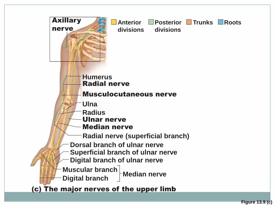
Figure 13.9 (c)
Median nerve
Musculocutaneous nerve
Radial nerve
Humerus
Ulna
Ulnar nerve
Median nerve
Radius
Radial nerve (superficial branch)
Superficial branch of ulnar nerve Dorsal branch of ulnar nerve
Digital branch of ulnar nerve
Muscular branch
Digital branch
(c) The major nerves of the upper limb
Axillary
nerve
Anterior
divisions
Posterior
divisions
Trunks Roots
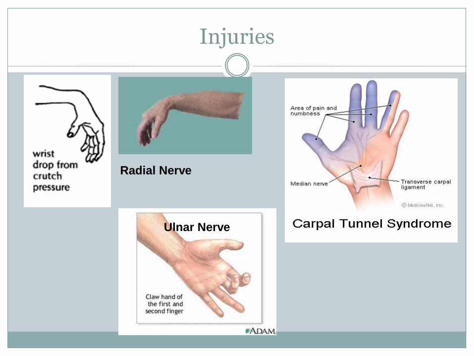
Injuries
Radial Nerve
Ulnar Nerve
Plexuses
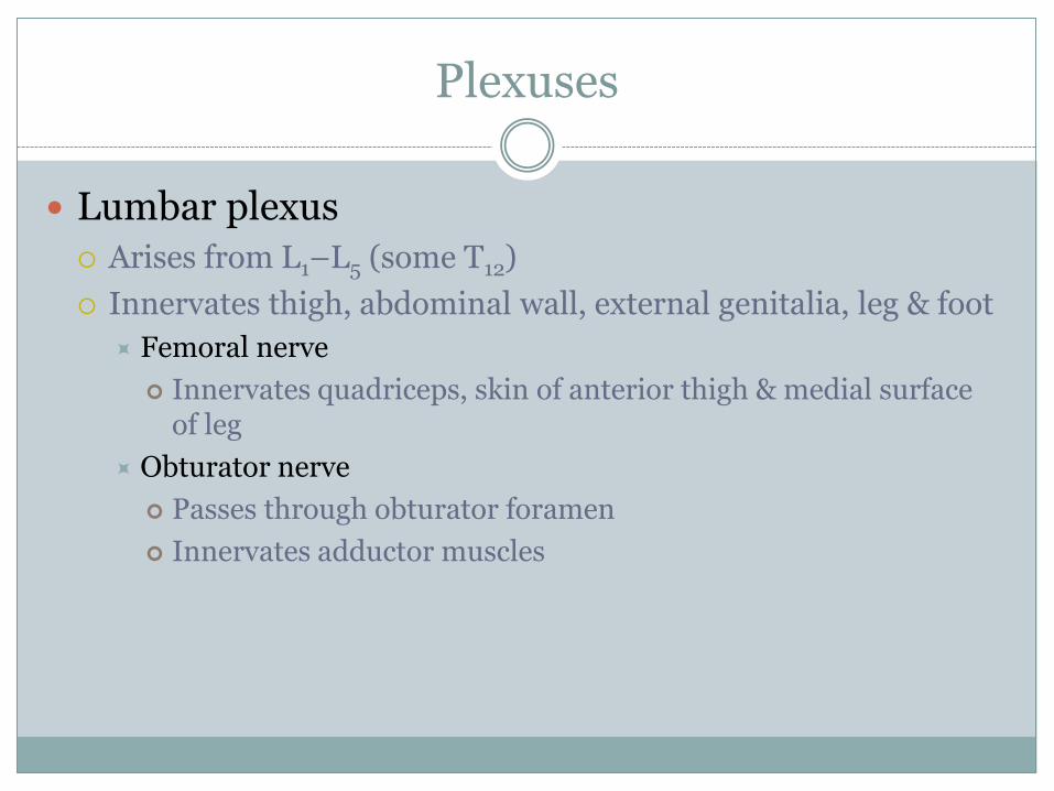
Lumbar plexus
Arises from L1–L5 (some T12)
Innervates thigh, abdominal wall, external genitalia, leg & foot
Femoral nerve
Innervates quadriceps, skin of anterior thigh & medial surface of leg
Obturator nerve
Passes through obturator foramen
Innervates adductor muscles
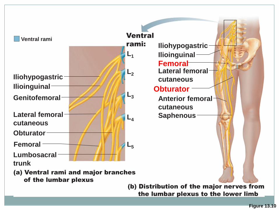
Figure 13.10
(a) Ventral rami and major branches
of the lumbar plexus
Iliohypogastric
L1
L2
L3
L4
L5
Ilioinguinal
Genitofemoral
Lateral femoral
cutaneous
Obturator
Femoral
Lumbosacral
trunk
Lateral femoral
cutaneous
Anterior femoral
cutaneous
Saphenous
Obturator
Iliohypogastric
Ilioinguinal
Femoral
Ventral rami Ventral
rami:
(b) Distribution of the major nerves from
the lumbar plexus to the lower limb
Plexuses
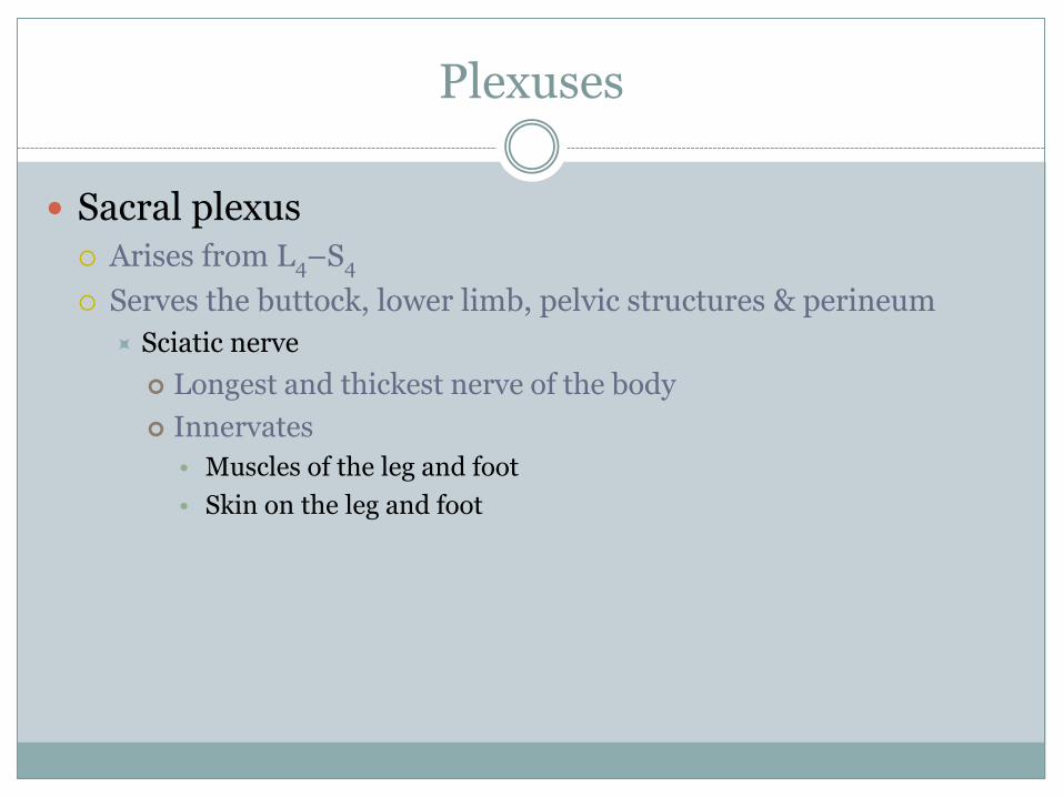
Sacral plexus Arises from L4–S4
Serves the buttock, lower limb, pelvic structures & perineum
Sciatic nerve
Longest and thickest nerve of the body
Innervates
• Muscles of the leg and foot
• Skin on the leg and foot
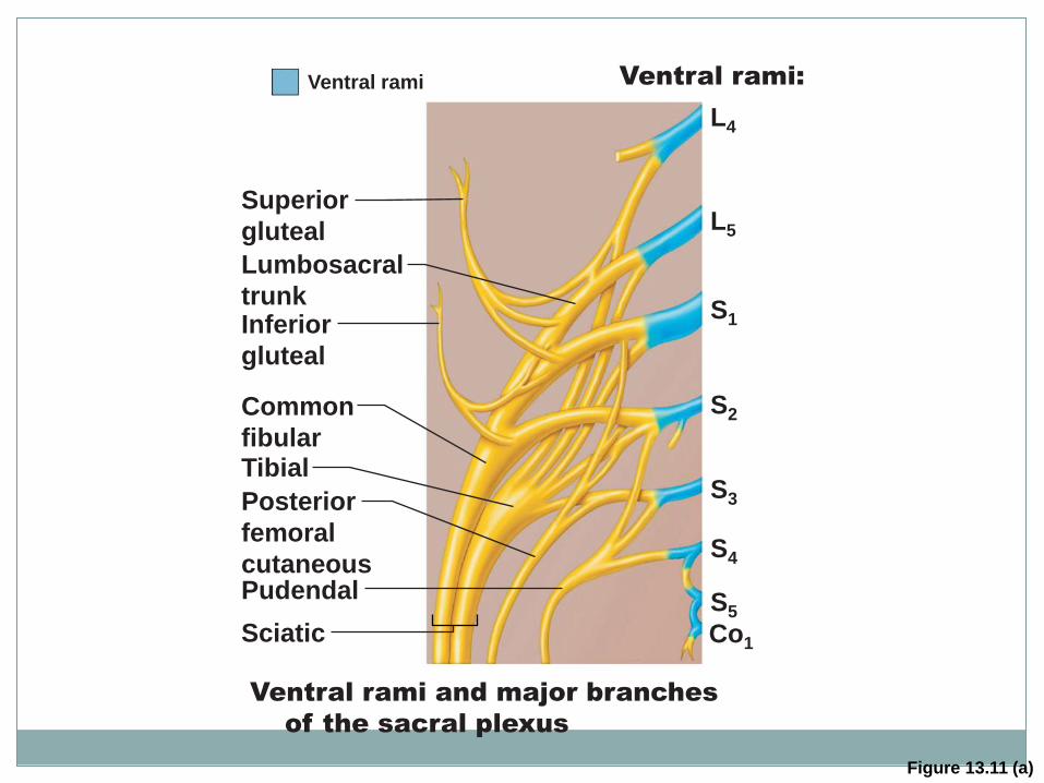
Figure 13.11 (a)
Superior
gluteal
Lumbosacral
trunk
Inferior
gluteal
Common
fibular Tibial
Posterior
femoral
cutaneous
Pudendal
Sciatic
Ventral rami and major branches
of the sacral plexus
L4
L5
S1
S2
S3
S4
S5
Co1
Ventral rami Ventral rami:
Figure 13.11 (b)
Superior gluteal
Inferior gluteal
Common fibular
Deep fibular
Superficial fibular
Plantar branches
Tibial
Sural (cut)
Posterior femoral
cutaneous
Pudendal
Sciatic
(b) Distribution of the major nerves from
the sacral plexus to the lower limb