peritoneal dialysate ef ß uent during peritonitis induces human...
TRANSCRIPT

7
Peritoneal Dialysate Effluent During Peritonitis Induces Human Cardiomyocyte Apoptosis and
Express Matrix Metalloproteinases-9
Ching-Yuang Lin and Chia-Ying Lee College of Medicine, China Medical University,
China Medical University Hospital, Taichung Taiwan
1. Introduction
Cardiovascular event and infection are the first and second leading causes of death in the peritoneal dialysis (PD) populations (Parfrey and Foley, 1999; Go et al., 2004; Schiffrin et al., 2007; USRDS, 2008); both events are closely related. PD-related peritonitis is the crucial infection in PD patients (Aslam et al., 2006; Bender et al., 2006). Peritoneal toxin should be absorbed to the systemic circulation and might induce cardiotoxicity. After an episode of severe infection in dialysis patients, risk of death from cardiovascular events is increased sevenfold for 6 months and continues to rise for up to 48 months (Ishani et al., 2005; Bender et al., 2006). It has been considered to play a significant role in up to one sixth of patient deaths occurring during the course of PD therapy (Fried et al., 1996). In 41.5% of patients with peritonitis-related mortality, immediate cause of death was a cardiovascular event (Pe´ rez Fontan et al., 2005). Clinical findings indicate that a peritonitis episode may culminate in cardiovascular event (Fried et al., 1996; Bender et al., 2006): high incidence of peritonitis is accompanied by greater risk of death (Maiorca et al., 1993; Fried et al., 1996; Piraino, 1998), and cardiovascular events contribute to risk of peritonitis-related death in patients undergoing PD (Digenis et al., 1990; Firanek et al., 1991; Lupo et al., 1994). However, the possible mechanisms connecting PD-related peritonitis and cardiac mortality have not been addressed. Growing evidence implicates cardiomyocyte apoptosis as a mechanism contributing to various types of heart disease (Olivetti et al., 1997; Haunstetter and Izumo, 1998; Narula et al., 1999). Cardiomyocyte apoptosis could result in a loss of contractile tissue, compensatory hypertrophy of myocardial cells, reparative fibrosis, and heart failure. In animal models, endotoxin (Natanson et al., 1989; Ramana et al., 2006), exotoxin (Natanson et al., 1989; Sibelius et al., 2000), and inflammatory mediator (Mann, 1999) play important roles in cardiomyocyte apoptosis. In PD patients with infectious peritonitis, expression of inflammatory mediators and cytokines increase in PD effluent (PDE) and correlate with treatment outcome (Lai et al., 2000; Wang and Lin, 2005). Yet there are no data on effects of peritonitis PD effluent (PPDE) on cardiomyocytes viability and apoptosis. Bcl-2 protein family members are the best characterized proteins that are directly involved in the regulation of apoptosis (Cory and Adams, 2002). Bcl-2 and its closest homologues,
www.intechopen.com

Progress in Peritoneal Dialysis
100
Bcl-xL and Bcl-w, potently inhibit apoptosis in response to many cytotoxic insults. Bax and Bak are well known proapoptotic members of the Bcl-2 protein family. Regulation of apoptosis is highly dependent on the ratio of anti-apoptotic to pro- apoptotic proteins. Conditions that induce myocardial stress cause complex alterations in levels of Bcl-2 family proteins (Bishopric et al., 2001). Cardiac Bcl-2 gene expression has been shown to be regulated by GATA-4 both in vitro and in vivo (Kobayashi et al., 2006). GATA-4 is a transcription factor enriched in cardiac tissue that is essential for various cardiomyocyte physiological and adaptive responses. An early event in the cardiotoxicity induced by the antitumor drug doxorubicin is GATA-4 depletion, which in turn causes cardiomyocyte apoptosis (Aries et al., 2004; Suzuki and Evans, 2004). GATA-4 has also been shown to upregulate transcription of the anti-apoptotic genes Bcl-2 (Kobayashi et al., 2006) and Bcl-xL (Aries et al., 2004; Suzuki and Evans, 2004) in cardiomyocytes, and to play a central role in regulating the survival or apoptosis of cardiomyocytes. Although previous studies have suggested the importance of apoptosis regulation and GATA-4 expression in various heart diseases, their role in PD peritonitis-related cardiotoxicity has not been elucidated. Cardiac extracellular matrix (ECM) lends structural support and integrity to the myocardium and facilitates conversion of cardiomyocyte contraction into pump function (Caulfield JB et al., 1979; Sato S et al., 1983; Thompson MM et al., 2002). Integrity of original ECM is thought to play a key role in determining extent of remolding after myocardial infarction and matrix metalloproteinases (MMPs) play crucial roles in regulation of ECM. Inflammatory response and cardiac pro-matrix metalloproteinase (MMP)-9 are critical to heart failure (Jugdutt BI, 2003; Shah AP et al., 2009). Increasing MMP-9 level and activity may develop in a special group of patients with exposure to peritonitis toxin in their cardiomyocytes. To clarify the relationship between PD-related peritonitis and high cardiac mortality, we
postulated that during PD-related peritonitis, proapoptotic pathways are activated and
MMP-9 protein is also expressed in cardiomyocytes.
To test this hypothesis, human cardiomyocytes were cultured and treated with PPDE. The
possible underlying signaling pathways of cardiotoxicity and enhancing MMP-9 expression
in cultured human cardiomyocytes after stimulated by PPDE were examined.
2. Human cardiomyocytes culture
This research was approved by the China Medical Hospital Institutional Review Board.
Written informed consent was obtained from each individual. Human cardiomyocytes
obtained from the myocardial ventricular resection specimens of patients undergoing
cardiac surgery were isolated as previously described (Hsin-Hui Wang et al., 2010).
3. Characterization of human cardiomyocytes in primary culture
To characterize cardiomyocytes, muscle markers desmin and myocyte-specific protein a-sarcomeric actinin were detected (Fig. 1). CAPON, recently documented as endogenous protein expressed in guinea pig cardiomyocytes, interacts with nitric oxide synthase to accelerate cardiac repolarization by inhibition of L-type calcium channels. Expression of endogenous CAPON protein in cultured cardiomyocytes was detected by immunofluorescent staining and confocal microscopy (Fig. 1). Both action potential duration
www.intechopen.com

Peritoneal Dialysate Effluent During Peritonitis Induces Human Cardiomyocyte Apoptosis and Express Matrix Metalloproteinases-9
101
(APD) and peak L-Type calcium current (IcaL) were APD10, APD50, APD75 and APD90: 95.4 ± 10.6, 289.2 ± 15.6, 308.2 ± 15.4, and 318.4 ± 16.4 msec, respectively, with peak IcaL density of —10.2 ± 0.9 pA/pE at ± 10 mV (n=6).
Fig. 1. Characterization of human cardiomyocytes by immunostaining of cardiomyocyte
markers. Double labeling of cardiomyocytes with WGA (red) and -sarcomeric actinin (green) (first line)or desmin (green) (second line) or CAPON (green) (third line). Negative control using cultured human skeletal muscle cells was stained with CAPON (last line).
www.intechopen.com

Progress in Peritoneal Dialysis
102
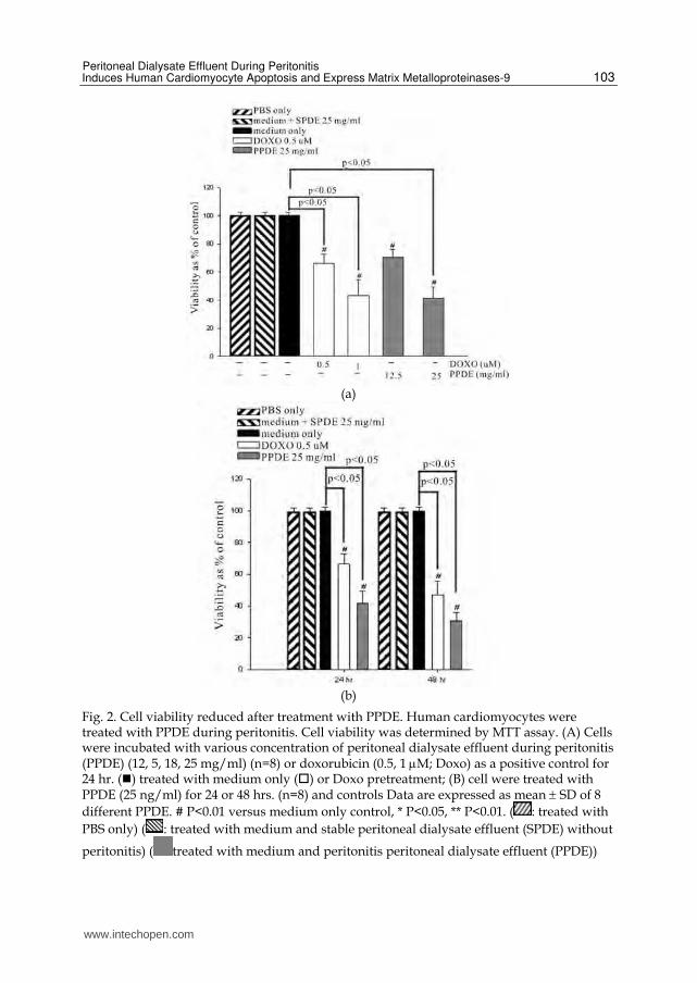
4. PPDE induces cell death in human cardiomyocytes
Cardiac cell death is believed to play a major contributory role in development and progression of myocardial dysfunction (Haunstetter and Izumo, 1998). To assess whether PPDE treatment induced cardiac cell death, cell viability were evaluated by MTT assay. Doxorubicin-induced cardiotoxicity, which has been well described (Shan et al., 1996), was used as a positive control. MTT assay showed PDE during peritonitis- and doxorubicin-induced human cardiomyocyte cell death as both dose- (Fig. 2A and Table 1) and time-dependent (Fig. 2B and Table 2). When cardiomyocytes were pre-exposed to 12.5, 18, or 25 mg/ml PDE during peritonitis for 24h, cell viabilities were 70.6 ± 5.7%, 58.7 ± 9.7%, and 41.6 ± 7.8%, respectively, all significantly lower than in cardiomyocytes without pre-treatment ( P < 0.05) (Fig. 2A). This change was even more profound in the 48 h treatment group (Fig. 2B). When cardiomyocytes were pre-exposed to 25 mg/ml PDE from stable PD patients for 24 and 48 h, cell viabilities were similar with cardiomyocytes without pre-treatment (data not shown).
% of apoptosis
Treatment 0 12.5 mg/ml 25 mg/ml
Medium only 1.0 ± 2.7 10.4 ± 2.8 11.8 ± 3.2 PBS only 2.2 ± 3.1 10.2 ± 3.2 10.8 ± 3.6 Medium + DOXO 9.7 ± 2.4 28.5 ± 4.1a 41.2 ± 4.5 a Medium + SPDE 0.7 ± 2.5 10.2 ± 2.7 12.4 ± 3.5 Medium + PPDE 9.7 ± 2.6 32.4 ± 3.8 a 48.6 ± 4.8 a
Table 1. Dose dependent manner of PPDE induced cell apoptosis in cultured human cardiomyocytes
% of apoptosis
Treatment 0 24 h 48 h
Medium only 1.0 ± 2.7 11.5 + 2.7 12.1 + 3.2 PBS only 2.2 ± 3.1 10.4 + 2.6 11.5 + 3.4 Medium + DOXO 9.7 ± 2.4 34.8 + 3.6a 42.2 + 4.2 a Medium + SPDE 0.7 ± 2.5 12.2 + 3.2 13.6 + 3.2 Medium + PPDE 9.7 ± 2.6 48.2 + 4.7 a 57.6 + 4.4 a
Table 2. Time dependent manner of PPDE induced cell apoptosis in cultured human cardiomyocytes
5. PPDE induces apoptosis in human cardiomyocytes
The above lend substantial evidence of apoptosis playing a critical role in cardiomyocyte cell death associated with several cardiac diseases (Olivetti et al., 1997; Haunstetter and Izumo, 1998). To explore whether PPDE during peritonitis challenge induces human cardiomyocyte apoptosis, we assessed apoptotic cell death by flow cytometry. Comet assays were also performed for determination of DNA damage. Doxorubicin, which can induce cardiomyocytes apopotosis (Kim et al., 2003), was used as a positive control. After cell incubation with 25 mg/ml PPDE peritonitis for 24 h, apoptosis was detected by flow cytometry (Tables 1 and 2). Analyses indicated little cardiomyocyte apoptosis with non-exposed condition (control group) and exposure with 25 mg/ml SPDE from stable PD
www.intechopen.com
Peritoneal Dialysate Effluent During Peritonitis Induces Human Cardiomyocyte Apoptosis and Express Matrix Metalloproteinases-9
103
(a)
(b)
Fig. 2. Cell viability reduced after treatment with PPDE. Human cardiomyocytes were treated with PPDE during peritonitis. Cell viability was determined by MTT assay. (A) Cells were incubated with various concentration of peritoneal dialysate effluent during peritonitis (PPDE) (12, 5, 18, 25 mg/ml) (n=8) or doxorubicin (0.5, 1 M; Doxo) as a positive control for 24 hr. () treated with medium only () or Doxo pretreatment; (B) cell were treated with PPDE (25 ng/ml) for 24 or 48 hrs. (n=8) and controls Data are expressed as mean SD of 8
different PPDE. # P<0.01 versus medium only control, * P<0.05, ** P<0.01. ( : treated with
PBS only) ( : treated with medium and stable peritoneal dialysate effluent (SPDE) without
peritonitis) ( treated with medium and peritonitis peritoneal dialysate effluent (PPDE))
www.intechopen.com

Progress in Peritoneal Dialysis
104
patients (Tables 1 and 2). By contrast, doxorubicin and PPDE induced apoptosis in 34.8–48.6% of human cardiomyocytes after treated for 24 h. Finally, PPDE induced DNA damage was determined by Comet assay (Fig. 3): higher concentrations of PPDE resulted in greater numbers of damaged cells.
Fig. 3. PPDE treatment induces apoptosis in cultured human cardiomyocytes. Cardiomyocyte DNA damage was determined by the Comet assay. Cardiomyocytes were treated with PPDE (12.5 or 25 mg/ml) or Doxo (0.5 M) as a positive control for 24 h, and then the Comet assay was performed.
6. PPDE induced Bax increase and suppression of GATA-4 expression in human cardiomyocytes
The Bcl-2 family of proteins are key regulators of the stress- induced apoptotic pathway (Bishopric et al., 2001); to determine their role in regulation of PPDE induced cardiomyocyte apoptosis, mRNA concentrations of prosurvival proteins Bcl-2 and Bcl-xL and proapoptotic protein Bax were measured in human cardiomyocytes by quantitative real-time RT-PCR (Fig. 4A). Compared to the no-exposure control group, Bcl-2/Bax and Bcl-xL/Bax ratios dropped significantly following 4 h of PPDE treatment (Fig. 4B; P < 0.05 vs. control). Western blotting analysis for Bcl-2, Bcl-xl, and Bax protein expression in the same experimental conditions obtained similar results (Fig. 6A). These data indicated that PPDE treatment decreased Bcl-2/Bax and Bcl-xL/Bax ratios, resulting in increase Bax expression in human cardiomyocytes. Transcription factor GATA-4 has been identified as a specific myocardial survival factor which induces transcription and expression of Bcl-2 and which is associated with cell survival (Ancey et al., 2002; Kitta et al., 2003; Aries et al., 2004; Suzuki and Evans, 2004). To characterize mechanisms underlying PPDE activity in human cardiomyocytes, mRNA and protein expression of GATA-4 were measured. For cardiomyocytes exposed to PPDE, GATA-4 mRNA expression decreased fivefold relative to no-exposure control cells by quantitative real-time RT-PCR (P < 0.05) (Fig. 5A). Western blots of nuclear GATA-4 protein expression in PPDE exposed human cardiomyocytes also showed lower levels than the control group (Fig. 5B,C), suggesting that PDE during peritonitis treatment decreases levels of GATA-4 gene expression in human cardiomyocytes.
7. PPDE does not contain inflammatory mediators
To evaluate whether PPDE was enriched in pro-apoptotic mediators, TRAEL, FasL, TNFa, IL-6, and IL-1 were measured by enzyme-linked immunoassay by commercial ELISA kit.
www.intechopen.com

Peritoneal Dialysate Effluent During Peritonitis Induces Human Cardiomyocyte Apoptosis and Express Matrix Metalloproteinases-9
105
(a)
(b)
Fig. 4. (A) PPDE treatment upregulates Bax gene expression in cultured human cardiomyocytes. Human cardiomyocytes were treated with or without PPDE (25 mg/ml), and then total RNA was prepared following 4 h of treatment. Bcl-2, Bcl-xL, and Bax mRNA expression levels in cardiomyocytes were determined by quantitative real-time RT-PCR. (n=8) Data are expressed as the mean SD of 8 different PPDE.* P<0.01 versus control. (B)Bcl-2/Bax and Bcl-xL/Bax ratio of experiment (A). Data are expressed as mean SD of 8
different PPDE. * P<0.01 versus control. : no treatment, medium only ; : no treatment
PBS only ; : medium + stable PDE without peritonitis(SPDE) ( treated with medium and peritonitis peritoneal dialysate effluent (PPDE).
www.intechopen.com

Progress in Peritoneal Dialysis
106
Cultured supernatant from peripheral blood mononuclear cells stimulated with lippolysaccharide was used as positive control; TRAEL, FasL, TNFa, and IL-1 were undetectable in 25 mg/ml PPDE (data not shown). The lower limit of sensitivity was 0.70 pg/ml.
Fig. 5. Effects of PPDE on expression of cardiac GATA-4 mRNA and protein. Cultured human cardiomyocytes were treated with or without PPDE (25 mg/ml), and then protein extracts and total RNA were prepared following 4 h of treatment. A: GATA-4 mRNA expression levels in cultured human cardiomyocytes as determined by quantitative real-time RT-PCR. B: Western blot showing GATA-4 protein levels. C: GATA-4 protein levels in cultured human cardiomyocytes, ascertained by densitometry. Data are expressed as mean
SD of 8 different PPDE. * P<0.01 versus control.
8. Role of ERK pathway in PPDE induced cardiotoxicity
We next examined possible signaling mechanisms regulating PPDE-induced cardiomyocyte apoptosis. The GATA-4 molecule contains putative ERK phosphorylation sites, and recent studies have shown that some survival factors (Morimoto et al., 2000; Kitta et al., 2001, 2003) induce activity of GATA-4 via MEK/ERK-dependent phosphorylation. Therefore, we explored activity of MEK/ERK signaling pathways in PPDE treated cardiomyocytes. Figure 6 shows ERK phosphorylation significantly reduced in cells exposed to PPDE peritonitis, suggesting that PPDE inhibits the ERK signaling pathway, consistent with the idea that the ERK pathway is crucial for GATA-4 activity and cardiomyocyte survival.
www.intechopen.com

Peritoneal Dialysate Effluent During Peritonitis Induces Human Cardiomyocyte Apoptosis and Express Matrix Metalloproteinases-9
107
Fig. 6. PPDE treatment reduces ERK phosyhorylation, GATA4, Bcl-2, Bcl-xL expression and
enhances Bax expression in cultured human cardiomyocytes. Cardiomyocytes were treated
with medium only, PBS only, SPDE (25 mg/ml) for 2 and 4 hours and PPDE (25 mg/ml) for
2 and 4 hours. Cell lysates were separated by SDS-PAGE and specific monoclonal antibodies
were used to detect phosphorylated and total ERK, and Bax, Bcl-2, Bcl-xL, GATA4
expression. Representative blots from 8 separate experiments were shown. Quantitative
densitometry expressed as phosphorylated protein relative to total protein. Data are
expressed as the mean SD of 8 different PPDE .* P<0.01 versus control. Medium only, PBS
only and SPDE (25 mg/ml) in medium were used as negative controls.
9. PPDE induces MMP-9 expression in cultured human cardiomyocytes
Expression of MMP-9 protein was noted, accompanied by enhanced MyD88, IRAK1 and
NF-kB phosphorylation in cultured human cardiomyocytes treated by PPDE.(Fig. 7). We
also found MMP-9 co-localized with macrophages and myofibroblasts in autopy specimens
of cardiac tissue from patients with end-stage renal disease by immunostaining using
confocal microscopic imaging. (Fig. 8)
www.intechopen.com

Progress in Peritoneal Dialysis
108
Fig. 7. Effects of PPDE (25 mg/ml) on the expression of MyD88, IRAK-1, MMP-9 protein and NFκB phosphorylation. Cultured human cardiomyocytes were treated with medium only, medium + SPDE (25 mg/ml) and medium + PPDE (25 mg/ml), and then protein extracted for western blot analysis. Data are expressed as the mean±SD of 8 different PPDE. *P<0.01 versus control.
10. PPDE induces MMP-9 expression via MyD88/IRAK1/p65 signal pathway
To determine whether MyD88-dependent signaling pathway in cultured human cardiomyocytes will be modulate by PPDE, we studied MyD88 protein expression in cultured human cardiomyocytes after treated with PPDE. The result showed cultured human cardiomyocytes express MyD88 after PPDE treatment (Fig. 9). To further study MyD88-dependent signaling pathway in cultured human cardiomyocytes after treated with PPDE, we compared the difference of cytoplasm IRAK1-1 expression with and without PPDE treatment in cultured human cardiomyocytes. PPDE enhanced cytoplasmic IRAK1 expression when compared to the SPDE and medium only (Fig. 10). NF-κB is an important transcription factor to induce chronic inflammation in myocarditis; and contains a transactivation domain which is involved in the MyD88-dependent pathway that produces many pro-inflammatory cytokines. Increased nuclear NF-κB/p65 expression with PPDE treatment in cultured human cardiomyocytes was observed (Fig. 11).
www.intechopen.com

Peritoneal Dialysate Effluent During Peritonitis Induces Human Cardiomyocyte Apoptosis and Express Matrix Metalloproteinases-9
109
CAPON
MMP-9
nuclei
Merge
Fig. 8. MMP-9 co-localized with cardiomyocytes in cardiac tissue of patient with uremic cardiomyopathy. Double labeling of cardiomyocytes with CAPON(green), MMP-9(red) and nuclei(blue).
www.intechopen.com

Progress in Peritoneal Dialysis
110
Fig. 9. Cultured human cardiomyocytes express MyD88 after PPDE (25 mg/ml) stimulation. Representative western blot results of 8 different PPDE were shown.
Fig. 10. Increased cytoplasm interleukin receptor-associated kinase-1 (IRAK-1) expression in cultured human cardiomyocytes after PPDE (25 mg/ml) stimulation. Representative western blot results of 8 different PPDE were shown.
www.intechopen.com

Peritoneal Dialysate Effluent During Peritonitis Induces Human Cardiomyocyte Apoptosis and Express Matrix Metalloproteinases-9
111
Fig. 11. Cultured human cardiomyocytes were stimulated with PPDE (25 mg/ml) for shown and expression of nuclear NF-κB/p65 was detected by western blot. Increased expression of nuclear NF-κB/p65 was noted. Representative western blot results of 8 different PPDE were shown.
11. Conclusion
Our study demonstrated that PPDE contains potent pro-apoptotic factors and causes an
imbalance between proapoptotic and prosurvival pathways, inducing apoptosis in human
cardiomyocytes. This study revealed a possible mechanism of PD-related, peritonitis-
induced cardiotoxicity. These novel findings constitute the first direct evidence linking PD
peritonitis and cardiomyocyte apoptosis. Cardiovascular events are the major cause of death
in PD patients with peritonitis. Our findings demonstrated the central role of apoptosis in
PD peritonitis-associated cardiovascular events, and provided an explanation for the high
incidents of cardiovascular events in PD-related peritonitis.
Cardiomyocyte death is important in the pathogenesis of cardiac disease in end-stage renal
disease (Parfrey and Foley, 1999). Cardiomyocyte death induces LV compensatory LV
hypertrophy, and eventually leading to dilatation with systolic dysfunction. LV
hypertrophy appears to be an important, independent, determinant of survival in patients
with end stage renal diseases (Silberberg et al., 1989). Our study yields direct cellular
evidence of PPDE from PD patients as cardiotoxic. In end-stage renal disease,
cardiomyocyte death may be caused by continual LV overload, decreased large and small
coronary vessel perfusion, hyperparathyroidism, and malnutrition (Parfrey and Foley,
1999). Our data provide another possible cause of cardiac cell death in patients undergoing
PD.
www.intechopen.com

Progress in Peritoneal Dialysis
112
Previous studies suggested that the elevated plasma concentration of MMP-9 were likely due to an enhanced release from the infarcted myocardium (Inokubo, Y., et al., 2001; Rosemberg, G.A. et al., 1996). Furthermore, it is well documented that both neutrophil and MMP-9 have a synergistic effect in inflammatory injury (Steinberg, J. et al., 2001). In our previous studies, we also found MMP-9 co-localized with macrophages and myofibroblasts in human myocardial infarcted tissue. In the present study we demonstrated MMP-9 co-localized in human uremic myocardium. These results suggest both macrophage from post myocardial infarction to heart failure and activated cardiomyocytes can express MMP-9 protein. The increase of MMP-9 activity and levels not only can be used as the marker of post myocardial infarction, but also involved the pathogenesis of cardiac remodeling. Our results provided some new information in terms of diagnostic and therapeutic implications in a special group of patients with uremic cardiomyopathy in PD patients. In conclusion, we demonstrated for the first time that PPDE contains potent pro-apoptotic factors that regulate expression of GATA-4 and Bcl-2 families, inducing cultured cardiomyocyte apoptosis. MMP-9 expression provided some new information of cardiac remodeling. Findings illustrate a pivotal role of apoptosis and cardiac remodeling in PD peritonitis-associated cardiovascular events, explain high cardiac mortality in PD-related peritonitis, and pinpoint apoptotic events as a marker and potential therapeutic target for PD peritonitis-induced cardiotoxicity.
12. References
Ancey C, Corbi P, Froger J, Delwail A, Wijdenes J, Gascan H, Potreau D, Lecron JC. (2002). “Secretion of IL-6, IL-II and LIF by human cardiomyocytes in primary culture.” Cytokine 18:199–205.
Aries A, Paradis P, Lefebvre C, Schwartz RJ, Nemer M. (2004). “Essential role of GATA-4 in cell survival and drug-induced cardiotoxicity.” Proc Natl Acad Sci USA 101:6975–6980.
Aslam N, Bernardini J, Fried L, Burr R, Piraino B. (2006). “Comparison of infectious complications between incident hemodialysis and peritoneal dialysis patients.” Clin J Am Soc Nephrol 1:1226–1233.
Bender FH, Bernardini J, Piraino B. (2006). “Prevention of infectious complications in peritoneal dialysis: Best demonstrated practices.” Kidney Int 103(Suppl): S44–S54.
Bishopric NH, Andreka P, Slepak T, Webster KA. (2001). “Molecular mechanisms of apoptosis in the cardiac myocyte.” Curr Opin Pharmacol 1:141–150.
Caulfield JB, Borg TK. (1979). “The collagen network of the heart.” Lab Invest 40: 364-72. Cory S, Adams JM. (2002). “The Bcl2 family: Regulators of the cellular life-or-death switch.”
Nat Rev Cancer 2:647–656. Firanek CA, Vonesh EF, Korbet SM. (1991). “Patient and technique survival among an urban
population of peritoneal dialysis patients: An 8-year experience.” Am J Kidney Dis 18:91–96.
Fried LF, Bernardini J, Johnston JR, Piraino B. (1996). “Peritonitis influences mortality in peritoneal dialysis patients.” J Am Soc Nephrol 7:2176–2I82.
Go AS, Chertow GM, Fan D, McCulloch CE, Hsu CY. (2004). “Chronic kidney disease and the risks of death, cardiovascular events, and hospitalization.” N Engl J Med 351:1296–1305.
www.intechopen.com

Peritoneal Dialysate Effluent During Peritonitis Induces Human Cardiomyocyte Apoptosis and Express Matrix Metalloproteinases-9
113
Haunstetter A, Izumo S. (1998). “Apoptosis: Basic mechanisms and implications for cardiovascular disease. Circ Res 82:1111–1129.
Inokubo, Y., Hanada, H. and Ishizaka, H. (2001). “Plasma levels of Matrix metalloproteinase-9 and tissue inhibitor of metalloproteinase-1 are increased in the coronary circulation in patients with acute coronary syndrome.” Am. Heart J. 141:211-217.
Ishani A, Collins AJ, Herzog CA, Foley RN. (2005). “Septicemia, access and cardiovascular disease in dialysis patients: The USRDS Wave 2 study.” Kidney Int 68:311–318.
Jugdutt BI. (2003). “Ventricular remodeling after infarction and the extracellular collagen Matrix: when is enough enough?” Circulation 108: 1395-403.
Kim Y, Ma AG, Kitta K, Fitch SN, Ikeda T, Ihara Y, Simon AR, Evans T, Suzuki YJ. (2003). “Anthracycline-induced suppression of GATA-4 transcription factor: Implication in the regulation of cardiac myocyte apoptosis.” Mol Pharmacol 63:368–377.
Kitta K, Clement SA, Remeika J, Blumberg JB, Suzuki YJ. (2001). “Endothelin-I induces phosphorylation of GATA-4 transcription factor in the HL-I atrial-muscle cell line.” Biochem J 359:375–380.
Kitta K, Day RM, Kim Y, Torregroza I, Evans T, Suzuki YJ. (2003). “Hepatocyte growth factor induces GATA-4 phosphorylation and cell survival in cardiac muscle cells.” J Biol Chem 278:4705–4712.
Kobayashi S, Lackey T, Huang Y, Bisping E, Pu WT, Boxer LM, Liang Q. (2006). “Transcription factor gata4 regulates cardiac BCL2 gene expression in vitro and in vivo.” FASEB J 20:800– 802.
Lai KN, Lai KB, Lam CW, Chan TM, Li FK, Leung JC. (2000). “Changes of cytokine profiles during peritonitis in patients on continuous ambulatory peritoneal dialysis.” Am J Kidney Dis 35:644–652.
Lupo A, Tarchini R, Carcarini G, Catizone L, Cocchi R, De Vecchi A, Viglino G, Salomone M, Segoloni G, Giangrande A. (1994). “Long-term outcome in continuous ambulatory peritoneal dialysis: A I0-year-survey by the Italian Cooperative Peritoneal Dialysis Study Group.” Am J Kidney Dis 24:826–837.
Mann DL. (1999). “Inflammatory mediators in heart failure: Homogeneity through heterogeneity.” Lancet 353:1812–1813.
Morimoto T, Hasegawa K, Kaburagi S, Kakita T, Wada H, Yanazume T, Sasayama S. (2000). “Phosphorylation of GATA-4 is involved in alpha I-adrenergic agonist-responsive transcription of the endothelin-I gene in cardiac myocytes.” J Biol Chem 275:13721– 13726.
Narula J, Pandey P, Arbustini E, Haider N, Narula N, Kolodgie FD, Dal Bello B, Semigran MJ, Bielsa-Masdeu A, Dec GW, Israels S, Ballester M, Virmani R, Saxena S, Kharbanda S. (1999). “Apoptosis in heart failure: Release of cytochrome c from mitochondria and activation of caspase-3 in human cardiomyopathy.” Proc Natl Acad Sci USA 96:8144–8149.
Natanson C, Danner RL, Elin RJ, Hosseini JM, Peart KW, Banks SM, MacVittie TJ, Walker RI, Parrillo JE. (1989). “Role of endotoxemia in cardiovascular dysfunction and mortality. Escherichia coli and Staphylococcus aureus challenges in a canine model of human septic shock.” J Clin Invest 83:243–251.
Olivetti G, Abbi R, Quaini F, Kajstura J, Cheng W, Nitahara JA, Quaini E, Di Loreto C, Beltrami CA, Krajewski S, Reed JC, Anversa P. (1997). “Apoptosis in the failing human heart.” N Engl J Med 336:1131–1141.
www.intechopen.com

Progress in Peritoneal Dialysis
114
Parfrey PS, Foley RN. (1999). “The clinical epidemiology of cardiac disease in chronic renal failure.” J Am Soc Nephrol 10:1606–1615.
Pe´ rez Fontan M, Rodr´ıguez-Carmona A, Garc´ıa-Naveiro R, Rosales M, Villaverde P, Valde´ s F. (2005). “Peritonitis-related mortality in patients undergoing chronic peritoneal dialysis.” Perit Dial Int 25: 274–284.
Ramana KV, Willis MS, White MD, Horton JW, DiMaio JM, Srivastava D, Bhatnagar A, Srivastava SK. (2006). “Endotoxin-induced cardiomyopathy and systemic inflammation in mice is prevented by aldose reductase inhibition.” Circulation 114:1838–1846.
Rosemberg, G.A., Navratil, M., Barone, F. and Feuerstein, G. (1996). “Proteolytic cascade enzymes increase in focal cerebral ischemia in rat.” J. Cereb. Blood Flow Metab. 16: 360-366.
Sato S, Ashraf M, Millard RW. (1983). “Connective tissue changes in early ischemia of porcine myocardium: an ultrastructural study.” J Mol Cell Cardiol 15: 261-75.
Schiffrin EL, Lipman ML, Mann JF. (2007). “Chronic kidney disease: Effects on the cardiovascular system.” Circulation 116:85–97.
Shah AP, Niemann JT, Youngquist S, Heyming T, Rosborough JP. (2009). “Plasma endothelin-1 level at the onset of ischemic ventricular fibrillation predicts resuscitation outcome.” Resuscitation 80: 580-3.
Shan K, Lincoff AM, Young JB. (1996). “Anthracycline-induced cardiotoxicity.” Ann Intern Med 125:47–58.
Sibelius U, Grandel U, Buerke M, Mueller D, Kiss L, Kraemer HJ, Braun-Dullaeus R, Haberbosch W, Seeger W, Grimminger F. (2000). “Staphylococcal alpha-toxin provokes coronary vasoconstriction and loss in myocardial contractility in perfused rat hearts: Role of thromboxane generation.” Circulation 101:78–85.
Silberberg JS, Barre PE, Prichard SS, Sniderman AD. (1989). “Impact of left ventricular hypertrophy on survival in end-stage renal disease.” Kidney Int 36:286–290.
Steinberg, J., Fink, G., Picone, A ., Searles, B., Schiller, H,. Lee, H.M. and Nieman, G. (2001). “Evidence of increased matrix metalloproteinase-9 concentration in patients following cardiopulmonary bypass.” J. Extra.Corpor. Technol. 33: 218-222.
Suzuki YJ, Evans T. (2004). “Regulation of cardiac myocyte apoptosis by the GATA-4 transcription factor.” Life Sci 74:1829–1838.
Thompson MM, Squire IB. (2002). “Matrix metalloproteinase-9 expression after myocardial infarction: physiological or pathological?” Cardiovasc Res 54: 495-8.
U.S. Renal Data System. (2008). USRDS 2008 Annual Data Report: Atlas of Chronic Kidney Disease and End-Stage Renal Disease in the United States, National Institutes of Health, National Institute of Diabetes and Digestive and Kidney Diseases, Bethesda, MD.
Wang HH, Lin CY. (2005). “Interleukin-I2 and -I8 levels in peritoneal dialysate effluent correlate with the outcome of peritonitis in patients undergoing peritoneal dialysis: Implications for the Type I/Type II T-cell immune response.” Am J Kidney Dis 46:328– 338.
Wang HH, Li PC, Huang HJ, Lee TY, Lin CY. (2011). “Peritoneal Dialysate Effluent During Peritonitis Induces Human Cardiomyocytes Apoptosis by Regulating the Expression of GATA-4 and Bcl-2 Families” J.cell. Physiol. 226(1):94-102.
www.intechopen.com

Progress in Peritoneal DialysisEdited by Dr. Ray Krediet
ISBN 978-953-307-390-3Hard cover, 184 pagesPublisher InTechPublished online 17, October, 2011Published in print edition October, 2011
InTech EuropeUniversity Campus STeP Ri Slavka Krautzeka 83/A 51000 Rijeka, Croatia Phone: +385 (51) 770 447 Fax: +385 (51) 686 166www.intechopen.com
InTech ChinaUnit 405, Office Block, Hotel Equatorial Shanghai No.65, Yan An Road (West), Shanghai, 200040, China
Phone: +86-21-62489820 Fax: +86-21-62489821
Progress in Peritoneal Dialysis is based on judgement of a number of abstracts, submitted by interestedpeople involved in various aspects of peritoneal dialysis. The book has a wide scope, ranging from in-vitroexperiments, mathematical modelling, and clinical studies. The interested reader will find state of the artessays on various aspects of peritoneal dialysis relevant to expand their knowledge on this underusedmodality of renal replacement therapy.
How to referenceIn order to correctly reference this scholarly work, feel free to copy and paste the following:
Ching-Yuang Lin and Chia-Ying Lee (2011). Peritoneal Dialysate Effluent During Peritonitis Induces HumanCardiomyocyte Apoptosis and Express Matrix Metalloproteinases-9, Progress in Peritoneal Dialysis, Dr. RayKrediet (Ed.), ISBN: 978-953-307-390-3, InTech, Available from: http://www.intechopen.com/books/progress-in-peritoneal-dialysis/peritoneal-dialysate-effluent-during-peritonitis-induces-human-cardiomyocyte-apoptosis-and-express-m

© 2011 The Author(s). Licensee IntechOpen. This is an open access articledistributed under the terms of the Creative Commons Attribution 3.0License, which permits unrestricted use, distribution, and reproduction inany medium, provided the original work is properly cited.