peritoneopericardial communication after aortic valve replacement in a peritoneal dialysis patient
TRANSCRIPT
CASE REPORT
Peritoneopericardial communication after aortic valvereplacement in a peritoneal dialysis patient
Satoshi Morimoto • Chikara Nakano • Kazunori Someya •
Mitsutaka Nakahigashi • Hiroko Ueda • Makiko Kusabe •
Tatsuyori Morita • Atsuhiro Ichihara • Toshiji Iwasaka
Received: 20 August 2013 / Accepted: 30 April 2014
� Japanese Society of Nephrology 2014
Abstract A 73-year-old male undergoing peritoneal
dialysis (PD) for end-stage renal disease due to diabetic
nephropathy was diagnosed with aortic stenosis and was
admitted to our hospital in September, 2009. The patient
underwent replacement of the ascending aorta with an
artificial blood vessel plus aortic valve replacement without
any notable complications. PD was restarted 3 days after
the surgery and large amounts of light red fluid from the
drain placed in the pericardium were observed just after
resumption of PD solution. The patient was diagnosed with
peritoneopericardial communication. PD was discontinued
and hemodialysis was performed only with intermittent
lavage of the peritoneal cavity. The amount of drainage
was spontaneously decreased, and on the 17th day after
surgery, PD was resumed. The patient is undergoing PD
without recurrence of peritoneopericardial communication,
59 months after the onset of symptoms. Peritoneopericar-
dial communication in a patient with PD developing after
open-heart surgery is rare because such a case has been
documented in only one case report. However, since
massive pericardial effusion may cause severe cardiac
problems, we consider that the communication between the
peritoneal cavity and the pericardium needs to be checked
for in patients with PD after cardiac surgery.
Keywords Cardiotomy � Aortic stenosis � Hemodialysis �Pericardium � Diaphragm
Introduction
Peritoneopericardial communication is a rare complication
of open-heart surgery in patients with peritoneal dialysis
(PD). However, this condition is important because it may
cause severe cardiac problems due to massive pericardial
effusion. We report here a case of peritoneopericardial
communication following cardiotomy, in which a PD
patient temporarily required hemodialysis (HD), but could
finally undergo PD with conservative treatment.
Case report
The patient was a 73-year-old male. His past history
included, lung cancer at 65 years of age (treated with left
upper lobe pneumonectomy), gastric cancer at 70 years of
age (treated with gastrotomy), and early colon cancer at
72 years of age (treated with colonic polypectomy). He had
no history of thoracic injury or pleuroperitoneal or perito-
neopericardial communication. PD for end-stage renal dis-
ease as diabetic nephropathy was started in February, 2007.
He was diagnosed with aortic stenosis by echocardiography
and admitted to our hospital for aortic valve replacement in
September, 2009. Findings on admission were as follows:
height, 170.9 cm; weight, 67.0 kg; BMI, 22.9 kg/m2; body
temperature, 36.2�C; blood pressure, 152/81 mmHg; pulse
rate, 78/min; no rales in lung fields; grade III/VI systolic
ejection murmur (Levine’s classification) at the apex; no
abdominal abnormality; no abnormalities at the PD catheter
insertion site; and no edema. The laboratory test results on
admission are shown in Table 1. Aortic stenosis (left
S. Morimoto � C. Nakano � K. Someya � M. Nakahigashi �H. Ueda � M. Kusabe � T. Morita � T. Iwasaka
Second Department of Internal Medicine, Kansai Medical
University, Moriguchi, Japan
S. Morimoto (&) � A. Ichihara
Department of Medicine II, Tokyo Women’s Medical
University, 8-1 Kawada-cho, Shinjuku-ku, Tokyo 162-8666,
Japan
e-mail: [email protected]
123
CEN Case Rep
DOI 10.1007/s13730-014-0125-2
ventricular outflow tract pressure gradient 57 mmHg; aortic
valve area 0.96 cm2) and grade II aortic insufficiency were
observed on echocardiography.
The operative procedure involved replacement of the
ascending aorta with an artificial blood vessel plus aortic
valve replacement. First, median sternotomy was per-
formed, but it was difficult to pump blood into the aorta
and to block the blood flow due to strong calcification of
the ascending aorta. Next, blood was pumped into the right
femoral artery and removal from the right atrium was
performed to establish cardiopulmonary bypass, and then
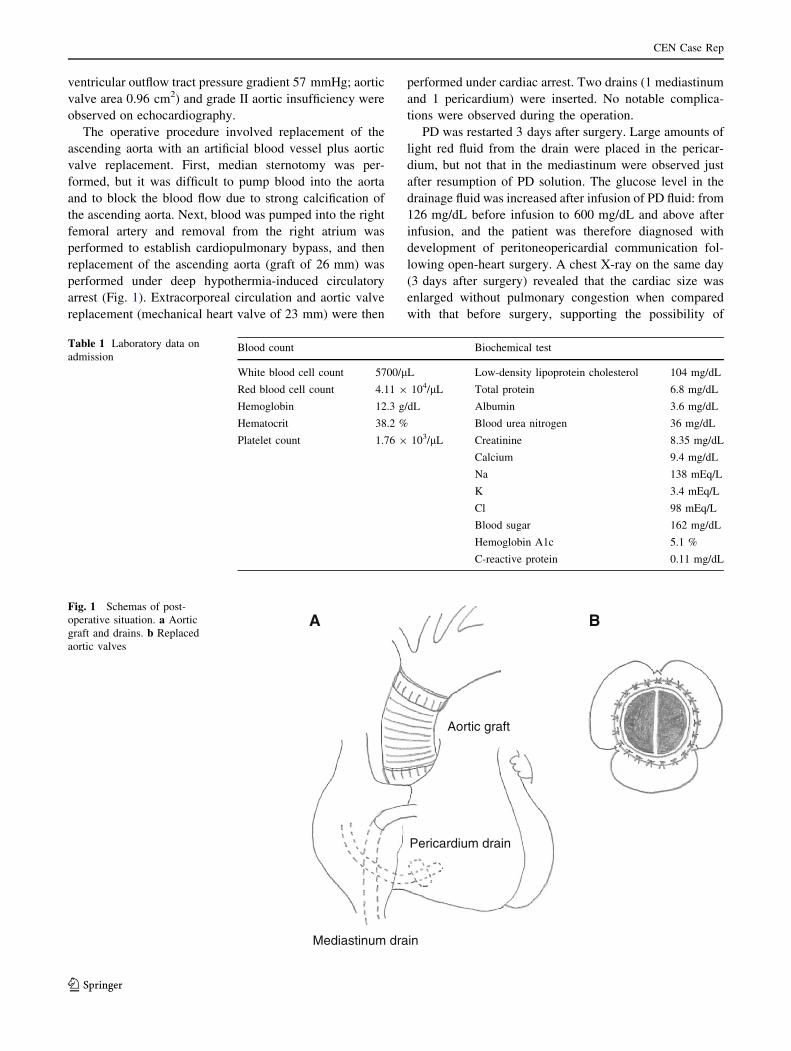
replacement of the ascending aorta (graft of 26 mm) was
performed under deep hypothermia-induced circulatory
arrest (Fig. 1). Extracorporeal circulation and aortic valve
replacement (mechanical heart valve of 23 mm) were then
performed under cardiac arrest. Two drains (1 mediastinum
and 1 pericardium) were inserted. No notable complica-
tions were observed during the operation.
PD was restarted 3 days after surgery. Large amounts of
light red fluid from the drain were placed in the pericar-
dium, but not that in the mediastinum were observed just
after resumption of PD solution. The glucose level in the
drainage fluid was increased after infusion of PD fluid: from
126 mg/dL before infusion to 600 mg/dL and above after
infusion, and the patient was therefore diagnosed with
development of peritoneopericardial communication fol-
lowing open-heart surgery. A chest X-ray on the same day
(3 days after surgery) revealed that the cardiac size was
enlarged without pulmonary congestion when compared
with that before surgery, supporting the possibility of
Table 1 Laboratory data on
admissionBlood count Biochemical test
White blood cell count 5700/lL Low-density lipoprotein cholesterol 104 mg/dL
Red blood cell count 4.11 9 104/lL Total protein 6.8 mg/dL
Hemoglobin 12.3 g/dL Albumin 3.6 mg/dL
Hematocrit 38.2 % Blood urea nitrogen 36 mg/dL
Platelet count 1.76 9 103/lL Creatinine 8.35 mg/dL
Calcium 9.4 mg/dL
Na 138 mEq/L
K 3.4 mEq/L
Cl 98 mEq/L
Blood sugar 162 mg/dL
Hemoglobin A1c 5.1 %
C-reactive protein 0.11 mg/dL
A B
Mediastinum drain
Pericardium drain
Aortic graft
Fig. 1 Schemas of post-
operative situation. a Aortic
graft and drains. b Replaced
aortic valves
CEN Case Rep
123
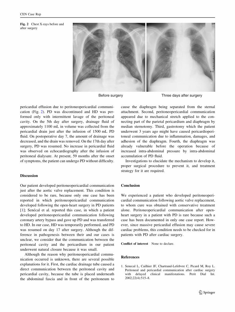
pericardial effusion due to peritoneopericardial communi-
cation (Fig. 2). PD was discontinued and HD was per-
formed only with intermittent lavage of the peritoneal
cavity. On the 5th day after surgery, drainage fluid of
approximately 1100 mL in volume was collected from the
pericardial drain just after the infusion of 1500 mL PD
fluid. On postoperative day 7, the amount of drainage was
decreased, and the drain was removed. On the 17th day after
surgery, PD was resumed. No increase in pericardial fluid
was observed on echocardiography after the infusion of
peritoneal dialysate. At present, 59 months after the onset
of symptoms, the patient can undergo PD without difficulty.
Discussion
Our patient developed peritoneopericardial communication
just after the aortic valve replacement. This condition is
considered to be rare, because only one case has been
reported in which peritoneopericardial communication
developed following the open-heart surgery in PD patients
[1]; Senecal et al. reported this case, in which a patient
developed peritoneopericardial communication following
coronary artery bypass and gave up PD and was transferred
to HD. In our case, HD was temporarily performed, and PD
was resumed on day 17 after surgery. Although the dif-
ference in pathogenesis between their and our cases is
unclear, we consider that the communication between the
peritoneal cavity and the pericardium in our patient
underwent natural closure because it was small.
Although the reason why peritoneopericardial commu-
nication occurred is unknown, there are several possible
explanations for it. First, the cardiac drainage tube caused a
direct communication between the peritoneal cavity and
pericardial cavity, because the tube is placed underneath
the abdominal fascia and in front of the peritoneum to
cause the diaphragm being separated from the sternal
attachment. Second, peritoneopericardial communication
appeared due to mechanical stretch applied to the con-
necting part of the parietal pericardium and diaphragm by
median sternotomy. Third, gastrotomy which the patient
underwent 3 years ago might have caused pericardioperi-
toneal communication due to inflammation, damages, and
adhesion of the diaphragm. Fourth, the diaphragm was
already vulnerable before the operation because of
increased intra-abdominal pressure by intra-abdominal
accumulation of PD fluid.
Investigations to elucidate the mechanism to develop it,
proper surgical procedure to prevent it, and treatment
strategy for it are required.
Conclusion
We experienced a patient who developed peritoneoperi-
cardial communication following aortic valve replacement,
to whom cure was obtained with conservative treatment
alone. Peritoneopericardial communication after open-
heart surgery in a patient with PD is rare because such a
case has been documented in only one case report. How-
ever, since massive pericardial effusion may cause severe
cardiac problems, this condition needs to be checked for in
patients with PD after cardiac surgery.
Conflict of interest None to declare.
References
1. Senecal L, Cailhier JF, Chartrand-Lefebvre C, Picard M, Roy L.
Peritoneal and pericardial communication after cardiac surgery
with delayed clinical manifestations. Perit Dial Int.
2002;22(4):515–8.
Before surgery Three days after surgery
Fig. 2 Chest X-rays before and
after surgery
CEN Case Rep
123