persistent neonatal hypoglycemia - smgebooks.com€¦ · hypoglycemia should be considered as a...
TRANSCRIPT
1Hypoglycemia- Causes and Occurrences | www.smgebooks.comCopyright Yadav D.This book chapter is open access distributed under the Creative Commons Attribution 4.0 International License, which allows users to download, copy and build upon published articles even for commercial purposes, as long as the author and publisher are properly credited.
Persistent Neonatal Hypoglycemia
ABSTRACTHypoglycemia is the most common metabolic alteration in neonatal period. Most of these
neonates have transient hypoglycemia. However, persistent and recurrent hypoglycemia in neonates is usually caused by endocrine or metabolic disorders. These babies require frequent glucose monitoring to prevent hypoglycemic brain injury and advanced investigations for diagnosis of underlying etiology. An approach to diagnosis and management of persistent neonatal hypoglycemia has been discussed in present review.
Dinesh Yadav1*1Neonatology Division, Kailash Hospital and research centre, Behror, Rajasthan, India
*Corresponding author: Dinesh Yadav, Yadav Hospital, Behror, Rajasthan, India, Tel: +91 9462725425; Email: [email protected]
Published Date: July 25, 2016
Gr upSM

2Hypoglycemia- Causes and Occurrences | www.smgebooks.comCopyright Yadav D.This book chapter is open access distributed under the Creative Commons Attribution 4.0 International License, which allows users to download, copy and build upon published articles even for commercial purposes, as long as the author and publisher are properly credited.
INTRODUCTIONHypoglycemia is the most common biochemical finding in the neonatal period. Both healthy
and sick neonates can be affected by hypoglycemia, most commonly during the first few days of life. Incidence of neonatal hypoglycemia is variable in different parts of the world, depending on definition of the condition and the methods of glucose estimation. The overall incidence has been estimated at 1 to 5 per 1,000 live births, but it is higher (up to 30%) in high risk populations. For example, 8% of large-for-gestational-age infants (primarily infants of diabetic mothers) and 15% of preterm infants and infants who have Intrauterine Growth Retardation (IUGR) have been reported having hypoglycemia [1].
Distinguishing between transitional neonatal glucose regulation in normal newborns and hypoglycemia that persists or occurs for the first time beyond the first 3 days of life is important for prompt diagnosis and effective treatment to prevent long term adverse events. However, the approach to diagnosis and management of persistent hypoglycemia is different and has been discussed in present review.
DEFINITION OF HYPOGLYCEMIAClinical hypoglycemia is defined as a plasma glucose concentration low enough to cause
symptoms and/or signs of impaired brain function. Guidelines in adults emphasize the value of Whipple’s triad for confirming hypoglycemia: symptoms and/or signs consistent with hypoglycemia, documented low Plasma Glucose (PG) concentration, and relief of signs/ symptoms when plasma glucose concentration is restored to normal. However, it may be difficult to recognize in neonates because the signs and symptoms are nonspecific and overlapping with many co-existing neonatal conditions. Besides this, PG concentrations are usually lower in first 24-48 hours of life during normal neonatal transition from intrauterine to extrauterine life [2].
Hypoglycemia cannot be defined as a specific PG concentration, because: (i) thresholds for specific brain responses to hypoglycemia occur across a range of PG concentrations, and these thresholds can be altered by the presence of alternative fuels, such as ketones, and by recent antecedent hypoglycemia; (ii) it is not possible to identify a single PG value that causes brain injury, and the extent of injury is influenced by other factors, such as duration and degree of hypoglycemia; and (iii) potential artefacts and technical factors that lead to inaccuracies in glucose determination may complicate the interpretation of any single PG value [2]. Hence, a numerical definition for neonatal hypoglycemia remains controversial even today. The critical level below which the incidence of brain injury increases depends on a number of factors including gestational age, co-existing conditions (e.g. birth asphyxia, neonatal sepsis), availability of alternate energy fuels and the baby’s energy demands [3].
Hypoglycemia should be considered as a failure of normal metabolic adaptation and a more pragmatic approach has been adopted at defining operational thresholds, which might help to guide the clinician in terms of practical management [4]. An “operational threshold” is that
3Hypoglycemia- Causes and Occurrences | www.smgebooks.comCopyright Yadav D.This book chapter is open access distributed under the Creative Commons Attribution 4.0 International License, which allows users to download, copy and build upon published articles even for commercial purposes, as long as the author and publisher are properly credited.
concentration of plasma or whole blood glucose at which clinicians should consider intervention, based on the evidence currently available in the literature. In an expert recommendation by Cornblath et al, it had been suggested that in any infant who shows neurologic symptoms and/or signs suggestive of hypoglycemia, intervention should be taken if the blood sugar values are less than 2.5 mmol/L. In asymptomatic “at-risk” infants, urgent interventions should be considered to raise blood sugar levels if they are less than 2.0 mmol/L on two consecutive occasions or if a single blood sugar level is less than 1.0 mmol/L [5].
Srinivasan et al, published data on blood glucose levels after evaluating term infants who were appropriate for gestational age weighing between 2,500 and 4,000 g. In this data, nadir of blood glucose was between 1 and 2 hours, and there was a significant rise during the 3rd hour. They suggested “cut off values” for blood glucose concentration of less than 1.9 mmol/L in the first 3 hours, 2.2 mmol/L between 3 and 24 hours, and 2.5 mmol/L after 24 hours of postnatal life to be considered in the hypoglycemic range [6]. Heck and Erenberg also evaluated the blood glucose concentration in term infants during the first 48 hours of life and concluded that values of 2.2 mmol/L from 3 to 24 hours and 2.5 mmol/L after 24 hours should be considered as hypoglycemia. However, in infants with suspected Hyperinsulinemic Hypoglycemia (HH) blood glucose levels should be maintained >3.5 mmol/L due to lack of alternative energy fuels [7].
Hence, current evidence does not support a single, specific concentration of glucose that can discriminate euglycemia from hypoglycemia or can predict that acute or chronic irreversible neurologic damage will result. Therefore, a significantly low concentration of glucose in plasma should be reliably established and treated to restore glucose values to a normal physiologic range [8]. PG values below 2.2 mmol/L in first 24 hours and below 2.5 mmol/L after 24 hours of life should be considered as threshold for clinical intervention till further evidence is generated, which is further guided by clinical symptoms.
NORMAL NEONATAL GLUCOSE TRANSITIONDuring fetal life, glucose passively diffuses across the placenta, using a concentration gradient,
while insulin cannot cross the placenta; therefore, the fetus must secrete insulin independently. With the clamping of the umbilical cord, the placental supply of glucose ceases abruptly, while fetal insulin secretion continues. After birth, PG concentration falls rapidly to as low as 30 mg/dL, reaching a nadir at 1 hour of age and then stabilizing by ~3 hours of age spontaneously or in response to milk feeds in healthy full-term infants. These low glucose levels are usually transient, asymptomatic and part of normal adaptation to postnatal life. This fall in glucose levels that is noticed after birth appears essential to facilitate the physiological transition for neonatal survival, which includes acceleration of glucose production by gluconeogenesis, stimulation of appetite, adaptation to fast/feed cycles, and promotion of oxidative fat metabolism using lipid from fat stores and ingested milk feeds [9]. Release of counter regulatory hormones (glucagon and cortisol) along with endogenous glucose production through gluconeogenesis and glycogenolysis, helps to restore the normal plasma glucose levels. In healthy neonates, early initiation of feeding

4Hypoglycemia- Causes and Occurrences | www.smgebooks.comCopyright Yadav D.This book chapter is open access distributed under the Creative Commons Attribution 4.0 International License, which allows users to download, copy and build upon published articles even for commercial purposes, as long as the author and publisher are properly credited.
also helps in maintaining normal serum glucose concentrations [10]. Hypoglycemia occurring during these initial few hours of life because of a slow or immature fasting adaptation process is known as transitional or transient hypoglycemia.
Normal adaptive process is different in term and preterm babies. A normal term infant accomplishes this transition via a series of metabolic and hormonal adaptations which allow it an independent existence. The metabolism in the newborn depends greatly on the function of vital enzymes: hepatic glycogen phosphorylase for glycogenolysis, phosphoenolpyruvate carboxykinase for gluconeogenesis, and carnitine acyltransferase for ketogenesis. Endocrine changes occur soon after birth with a decrease in plasma insulin levels and increases of catecholamine and glucagon. Glucagon, as the major glucoregulatory hormone which induces glycogenolysis, gluconeogenesis, and ketogenesis in liver. Catecholamines, cortisol, and growth hormone do play a key role in glucose homeostasis as counter-regulatory hormones. While on breast feeds, when blood glucose concentrations are low, these infants will have high ketone body and lactate levels, which act as alternate energy substrates for the brain. However, formula-fed infants generate lower ketone body levels compared to breast-fed infants [4].
This fetal-neonatal adaptation fails to a variable extent in preterm, Small-for-Gestational-Age (SGA) infants and babies with Intrauterine Growth Retardation (IUGR), that’s why these babies are at high risk for neonatal hypoglycemia. Preterm and IUGR babies have limited energy stores along with immature metabolic pathways, leading to a larger fall in PG levels in first few hours after birth. These babies are capable of gluconeogenesis and lipolysis, with some differences and restrictions. Because they have limited glycogen liver storage depots (since this takes place mainly during the third trimester), glycogenolysis is limited, therefore gluconeogenesis (from glycerol, alanine and lactate) is the main pathway for glucose production. Because gluconeogenesis requires some time to begin, in the absence of adequate glycogenolysis, these babies are at higher risk of hypoglycemia if exogenous glucose is not administered. The fat depot in an infant born at 28 weeks of gestation is only 2% of total body weight (7 times less than in term infants); therefore the degree of lipolysis is also decreased. Preterm infants are therefore at higher risk for hypoglycaemia because of their limitations for adequate glucose metabolism, but also because other clinical conditions which are associated with hypoglycemia are common in this population, such as perinatal asphyxia, hypoxia, sepsis, and hypothermia. Similarly, infants with IUGR also have lesser energy depots. Gluconeogenesis has been shown to occur effectively from glycerol in these infants (50% of glycerol is converted to glucose and this rate increases in babies who do not receive extra parenteral glucose infusion) and from pyruvate. Lipolysis also occurs but is limited because it correlates with birth weight, and it is therefore reduced in IUGR babies [11].
TRANSIENT HYPOGLYCEMIATransient hypoglycemia usually occurs during first few hours of life because of slow or
immature fasting adaptation process and usually resolves with feeding or intravenous dextrose.
5Hypoglycemia- Causes and Occurrences | www.smgebooks.comCopyright Yadav D.This book chapter is open access distributed under the Creative Commons Attribution 4.0 International License, which allows users to download, copy and build upon published articles even for commercial purposes, as long as the author and publisher are properly credited.
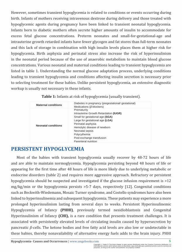
However, sometimes transient hypoglycemia is related to conditions or events occurring during birth. Infants of mothers receiving intravenous dextrose during delivery and those treated with hypoglycemic agents during pregnancy have been linked to transient neonatal hypoglycemia. Infants born to diabetic mothers often secrete higher amounts of insulin to accommodate for excess fetal glucose concentrations. Preterm neonates and small-for-gestational-age and intrauterine growth retarded infants have fewer glycogen and fat stores than full-term neonates and this lack of storage in combination with high insulin levels places them at higher risk for hypoglycemia. Birth asphyxia and perinatal stress also increase the risk of hyperinsulinism in the neonatal period because of the use of anaerobic metabolism to maintain blood glucose concentrations. Various neonatal and maternal conditions leading to transient hypoglycemia are listed in table 1. Understanding the normal glucose adaptation process, underlying conditions leading to transient hypoglycemia and conditions affecting insulin secretion is necessary prior to selecting treatment for these babies. Unlike persistent hypoglycemia, an extensive diagnostic workup is usually not necessary in these infants.
Table 1: Infants at risk of hypoglycemia (usually transient).
Maternal conditions Diabetes in pregnancy (pregestational/ gestational)Medications (β-blockers)
Neonatal conditions
PrematurityIntrauterine Growth Retardation (IUGR)Small for gestational age (SGA)Large for gestational age (LGA)Perinatal asphysiaHemolytic disease of newbornNeonatal sepsisPolycythemiaPost-exchange transfusionParenteral nutrition
PERSISTENT HYPOGLYCEMIAMost of the babies with transient hypoglycemia usually recover by 48-72 hours of life
and are able to maintain normoglycemia. Hypoglycemia persisting beyond 48 hours of life or appearing for the first time after 48 hours of life is more likely due to underlying metabolic or endocrine disorders (table 2) and requires more aggressive approach. Refractory or persistent hypoglycemia should be suspected and investigated if the glucose infusion requirement is >12 mg/kg/min or the hypoglycemia persists >5-7 days, respectively [12]. Congenital conditions such as Beckwith-Wiedemann, Mosaic Turner syndrome, and Costello syndromes have also been linked to hyperinsulinemia and subsequent hypoglycemia. These patients may experience a more prolonged hyperinsulinism lasting from several days to weeks. Persistent Hyperinsulinemic Hypoglycemia of Infancy (PHHI), previously termed nesidioblastosis and Congenital Hyperinsulinism of Infancy (CHI), is a rare condition that presents treatment challenges. It is associated with persistently elevated levels of circulating insulin caused by hypersecretion by pancreatic β-cells. The ketone bodies and free fatty acid levels are also low or undetectable in these babies, thereby nonavailability of alternative energy fuels adds to the brain injury. PHHI

6Hypoglycemia- Causes and Occurrences | www.smgebooks.comCopyright Yadav D.This book chapter is open access distributed under the Creative Commons Attribution 4.0 International License, which allows users to download, copy and build upon published articles even for commercial purposes, as long as the author and publisher are properly credited.
may present in the first few days of life or may present later in infancy. Many different metabolic disorders can also lead to persistent hypoglycemia, including glycogen storage disorders, disorders of gluconeogenesis, and fatty acid oxidation defects. In hypoketotic conditions, such as hyperinsulinism or fatty acid oxidation disorders, ketones and lactate are not available as alternate cerebral energy fuels, resulting in higher risk of brain energy failure [10]. Congenital Adrenal Hyperplasia (CAH) and hypopituitarism can also present with intractable hypoglycemia. Similarly, disorders of amino acid and carnitine metabolism and glucose transporter defects are rare causes of persistent hypoglycemia.
Table 2: Endocrine and Metabolic causes of neonatal hypoglycemia (usually persistent).
Endocrine disorders
HyperinsulinismPrimary- mutations in ABCC8, KCNJ11,
GCK, HADH, SCL16A1, HNF4A, UCP2 gene
Secondary- Infants of Diabetic Mother (IDM) Erythroblastosis fetalis Syndromes- Beckwith-wiedemann, Soto’s, Timothy, Laron
HypopituitarismIsolated growth hormone deficiencyAdrenal disorders-
Congenital adrenal hyperplasia ACTH deficiency Familial glucocorticoid deficiency Cortisol deficiency
Disorders of gluconeogenesisFructose-1,6 bisphosphatase deficiencyPhosphoenolpyruvate carboxykinase deficiencyPyruvate carboxulase deficiency
Disorders of galactose metabolism GalactosemiaDisorders of hepatic glycogen synthesis Glycogen storage diseaseDisorders of fructose metabolism Hereditary fructose intolerance
Disorders of aminoacid metabolism
Maple syrup urine diseasePropionic academiaMethylmalonic acidemiaTyrosinemia
Disorders of fatty acid metabolism
Defects in β-oxidationMCAD deficiencyLCHAD deficiencySCHAD deficiency
Disorders of carnitine metabolism
Primary carnitine deficiencyCPT-I deficiencyCACT deficiencyCPT-II deficiency
Ketogenesis and ketone body utilization defects
HMG CoA synthase deficiencyHMG CoA lyase deficiencyβ-ketothiolase deficiencySCOT deficiency
Glucose transporter defects GLUT 1/2/3 transporter defects
Abbreviations: Adrenocorticotropic Hormone (ACTH); Carnitine-Acylcarnitine Translocase (CACT); Carnitine Palmitoyltransferase (CPT); Long-Chain 3-Hydroxyacyl-Coenzyme A Dehydrogenase (LCHAD); Medium-Chain Acyl-Coa Dehydrogenase (MCAD); 3-Hydroxyacyl-Coenzyme A Dehydrogenase (SCHAD); Succinyl-CoA: 3-Ketoacid-Coenzyme A Transferase 1 (SCOT).
7Hypoglycemia- Causes and Occurrences | www.smgebooks.comCopyright Yadav D.This book chapter is open access distributed under the Creative Commons Attribution 4.0 International License, which allows users to download, copy and build upon published articles even for commercial purposes, as long as the author and publisher are properly credited.
GENETICS OF PERSISTENT HYPERINSULINEMIC HYPOGLYCEMIA OF INFANCY
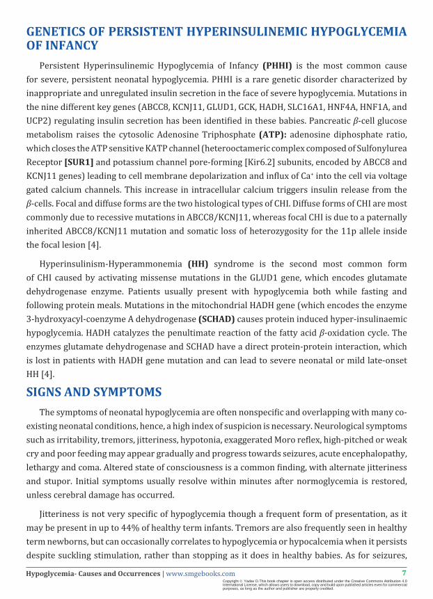
Persistent Hyperinsulinemic Hypoglycemia of Infancy (PHHI) is the most common cause for severe, persistent neonatal hypoglycemia. PHHI is a rare genetic disorder characterized by inappropriate and unregulated insulin secretion in the face of severe hypoglycemia. Mutations in the nine different key genes (ABCC8, KCNJ11, GLUD1, GCK, HADH, SLC16A1, HNF4A, HNF1A, and UCP2) regulating insulin secretion has been identified in these babies. Pancreatic β-cell glucose metabolism raises the cytosolic Adenosine Triphosphate (ATP): adenosine diphosphate ratio, which closes the ATP sensitive KATP channel (heterooctameric complex composed of Sulfonylurea Receptor [SUR1] and potassium channel pore-forming [Kir6.2] subunits, encoded by ABCC8 and KCNJ11 genes) leading to cell membrane depolarization and influx of Ca+ into the cell via voltage gated calcium channels. This increase in intracellular calcium triggers insulin release from the β-cells. Focal and diffuse forms are the two histological types of CHI. Diffuse forms of CHI are most commonly due to recessive mutations in ABCC8/KCNJ11, whereas focal CHI is due to a paternally inherited ABCC8/KCNJ11 mutation and somatic loss of heterozygosity for the 11p allele inside the focal lesion [4].
Hyperinsulinism-Hyperammonemia (HH) syndrome is the second most common form of CHI caused by activating missense mutations in the GLUD1 gene, which encodes glutamate dehydrogenase enzyme. Patients usually present with hypoglycemia both while fasting and following protein meals. Mutations in the mitochondrial HADH gene (which encodes the enzyme 3-hydroxyacyl-coenzyme A dehydrogenase (SCHAD) causes protein induced hyper-insulinaemic hypoglycemia. HADH catalyzes the penultimate reaction of the fatty acid β-oxidation cycle. The enzymes glutamate dehydrogenase and SCHAD have a direct protein-protein interaction, which is lost in patients with HADH gene mutation and can lead to severe neonatal or mild late-onset HH [4].
SIGNS AND SYMPTOMSThe symptoms of neonatal hypoglycemia are often nonspecific and overlapping with many co-
existing neonatal conditions, hence, a high index of suspicion is necessary. Neurological symptoms such as irritability, tremors, jitteriness, hypotonia, exaggerated Moro reflex, high-pitched or weak cry and poor feeding may appear gradually and progress towards seizures, acute encephalopathy, lethargy and coma. Altered state of consciousness is a common finding, with alternate jitteriness and stupor. Initial symptoms usually resolve within minutes after normoglycemia is restored, unless cerebral damage has occurred.
Jitteriness is not very specific of hypoglycemia though a frequent form of presentation, as it may be present in up to 44% of healthy term infants. Tremors are also frequently seen in healthy term newborns, but can occasionally correlates to hypoglycemia or hypocalcemia when it persists despite suckling stimulation, rather than stopping as it does in healthy babies. As for seizures,
8Hypoglycemia- Causes and Occurrences | www.smgebooks.comCopyright Yadav D.This book chapter is open access distributed under the Creative Commons Attribution 4.0 International License, which allows users to download, copy and build upon published articles even for commercial purposes, as long as the author and publisher are properly credited.
these may present very early after hypoglycemia appears, but normally occur after at least 12 hours of recurrent or persistent hypoglycemia. Other clinical manifestations include tachypnea, cyanosis (due to apnea, autonomic response or decreased pulmonary flow) or apnea spells. Difficulty to suck and feeding problems may also occur. Autonomic alterations are frequent, such as hypothermia or unstable temperature, pallor, profuse sweating or bradycardia [11].
DIAGNOSISA detailed history related to pregnancy (diabetes/diet/insulin), delivery (asphyxia), gestational
age (Small or large for gestational age), and birth weight (low birth weight/macrosomia) is essential. Family history of diabetes (gestational/pregestational) may point toward HNF4α mutations and parental consanguinity might point toward a recessively inherited cause of the hypoglycemia. Similarly, history of infantile seizures in siblings may also indicate inherited causes of the hypoglycemia. History should also include the episode’s timing and its relationship to food. Macrosomia, macroglossia, ear pits, hemihypertrophy, and omphalocele may suggest Beckwith-Wiedemann syndrome, a condition with uniparental disomy of chromosome 11, with a high predisposition for hyperinsulinism. Midline defects, micropenis, and hypoglycemia point to congenital hypopituitarism. Hypoglycemia, dehydration, and shock in the presence of ambiguous genitalia point to a diagnosis of congenital adrenal hyperplasia. Presence of hepatomegaly may indicate glycogenoses. Sudden cardiorespiratory collapse, acidosis, and hypoglycemia following feeds in an otherwise healthy neonate may indicate underlying inborn errors of metabolism.
LABORATORY DIAGNOSISFor neonates suspected to be at high risk of having a persistent hypoglycemic disorder, diagnostic
evaluation is suggested beyond 48 hours of age so that the period of transient hypoglycemia has passed. Point-of-care meters provide a convenient screening method for detecting hypoglycemia at bedside, but their accuracy is limited to approximately +10-15 mg/dL (0.6-0.8 mmol/L) in the range of hypoglycemia. Therefore, before establishing a diagnosis of hypoglycemia, it is essential to confirm low PG concentration using a clinical laboratory method. Important considerations are that whole blood glucose values are ~15% lower than PG concentrations, and that because of red cell glycolysis, delays in processing can reduce the glucose concentration by up to 6 mg/dL/hour (0.3 mmol/L/hour) [2].
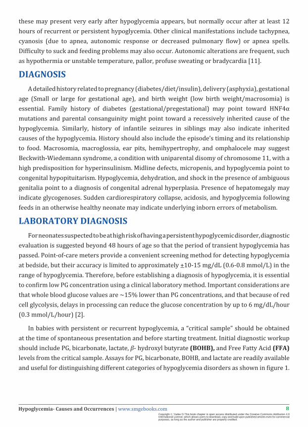
In babies with persistent or recurrent hypoglycemia, a “critical sample” should be obtained at the time of spontaneous presentation and before starting treatment. Initial diagnostic workup should include PG, bicarbonate, lactate, β- hydroxyl butyrate (BOHB), and Free Fatty Acid (FFA) levels from the critical sample. Assays for PG, bicarbonate, BOHB, and lactate are readily available and useful for distinguishing different categories of hypoglycemia disorders as shown in figure 1.
9Hypoglycemia- Causes and Occurrences | www.smgebooks.comCopyright Yadav D.This book chapter is open access distributed under the Creative Commons Attribution 4.0 International License, which allows users to download, copy and build upon published articles even for commercial purposes, as long as the author and publisher are properly credited.
Figure 1: Metabolic clues to diagnosis of persistent hypoglycemia from critical sample.
Abbreviations: Bicarbonate (HCO3), β-hydroxy bytyrate (BOHB); Free Fatty Acids (FFA); Growth Hormone (GH)
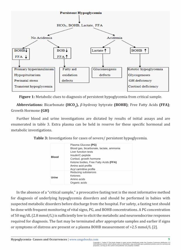
Further blood and urine investigations are dictated by results of initial assays and are enumerated in table 3. Extra plasma can be held in reserve for these specific hormonal and metabolic investigations.
Table 3: Investigations for cases of severe/ persistent hypoglycemia.
Blood
Plasma Glucose (PG)Blood gas, bicarbonate, lactate, ammoniaLiver function testsInsulin/C-peptideCortisol, growth hormoneKetone bodies, Free Fatty Acids (FFA)Amino acid profileAcyl carinitine profile
Urine
Reducing substancesKetonesAmino acidsOrganic acids
In the absence of a “critical sample,” a provocative fasting test is the most informative method for diagnosis of underlying hypoglycemia disorders and should be performed in babies with suspected metabolic disorders before discharge from the hospital. For safety, a fasting test should be done with frequent monitoring of vital signs, PG, and BOHB concentrations. A PG concentration of 50 mg/dL (2.8 mmol/L) is sufficiently low to elicit the metabolic and neuroendocrine responses required for diagnosis. The fast may be terminated after appropriate samples and earlier if signs or symptoms of distress are present or a plasma BOHB measurement of >2.5 mmol/L [2].
10Hypoglycemia- Causes and Occurrences | www.smgebooks.comCopyright Yadav D.This book chapter is open access distributed under the Creative Commons Attribution 4.0 International License, which allows users to download, copy and build upon published articles even for commercial purposes, as long as the author and publisher are properly credited.
For suspected hyperinsulinism, the fasting test can be terminated when the PG concentration is <50 mg/dL (<2.8 mmol/L) with administration of glucagon (1 mg IV, intramuscularly, or subcutaneously) to evaluate the glycemic response. An exaggerated glycemic response (>30 mg/dL [>1.7 mmol/L]) is nearly pathognomonic of hyperinsulinism. Whenever assessing the possibility of hypoglycemia due to hyperinsulinism, it is important to include plasma BOHB and FFA (both inappropriately low; BOHB <1.5 mmol/L [<15 mg/dL] and FFA <1.0-1.5 mmol/L [<28-42 mg/dL]), because plasma insulin concentration is sometimes not above the lower limit of detection. Based on the suspected diagnosis, additional tests performed on specimens obtained at the time that fasting should be considered, such as growth hormone, cortisol, total and free carnitine, acyl-carnitine profile, C-peptide, proinsulin, and drug screening.
MANAGEMENTThe underlying cause of hypoglycemia and severity will also dictate the approach to
management. For neonates with a suspected or confirmed hyperinsulinemic hypoglycemia, the goal of treatment should be to maintain a PG concentration >70 mg/dL (3.9 mmol/L). For high-risk neonates without a suspected congenital hypoglycemia disorder, the goal of treatment should be to maintain a PG concentration >50 mg/dL (>2.8 mmol/L) for those aged <48 hours and >60 mg/dL (>3.3 mmol/L) for those aged >48 hours [2].
For disorders such as hyperinsulinism, the aim is to prevent recurrent hypoglycemia that increases the risk of subsequent, possibly unrecognized, hypoglycemic episodes and Neonatal Hypoglycemic Brain Injury (NHBI). For disorders such as defects in glycogen metabolism and gluconeogenesis, maintenance of PG concentration in the normal range prevents metabolic acidosis and growth failure, and possibly the development of long-term complications. Any episode of severe symptomatic hypoglycemia should be rapidly corrected with IV dextrose infusion. The initial dose is 200 mg/kg, followed by infusion of 10% dextrose at a maintenance rate for age. In cases of hyperinsulinism, glucagon can be expected to raise PG concentration to normal or above within 10-15 minutes and to maintain that concentration for at least 1 hour. Doses of 0.5-1.0 mg (independent of weight), given IV, intramuscularly, or subcutaneously, are usually effective.
Long-term therapy for hypoglycemia disorders should be based on the specific aetiology of the disorder, in consultation with experts and with careful consideration of patient and family preferences. Many drug therapies are available for babies with persistent hypoglycemia including diazoxide, glucocorticoids, somatostatin analogues and Nifedipine [10]. Their doses, route of administration and adverse effects are described in table 4. Nutritional therapy is the cornerstone of treatment for disorders of glycogen metabolism or hereditary fructose intolerance. Some milder disorders may be treated adequately by avoidance of prolonged fasting. Patients diagnosed with ketotic hypoglycemia who have recurrent episodes should be re-evaluated for glycogen storage disorders, and MCT1 gene deficiency.

11Hypoglycemia- Causes and Occurrences | www.smgebooks.comCopyright Yadav D.This book chapter is open access distributed under the Creative Commons Attribution 4.0 International License, which allows users to download, copy and build upon published articles even for commercial purposes, as long as the author and publisher are properly credited.
Agent Doses Administration Side-effects
Diazoxide 10-15 mg/kg/day(max 30 mg/kg/day) Oral (q 8 hourly) Hirsuitism, fluid retention, cardiac failure,
GI disturbances
GlucagonBolus: 0.5-1 mg
Infusion: 1-20 mcg/kg/hour
Intermittent bolus Continuous infusion Hyponatremia, thrombocytopenia
DexamethasoneHydrocortisone
0.25 mg/kg/dose1-2.5 mg/kg/dose
Intravenous q 12 hourlyIntravenous q 6hourly
Growth suppressionhypertension
Octreotide7-12 mcg/kg/day(max 40 mcg/kg/
day)Subcutaneous q 4-6 hourly cholelithiasis
Calcium channel blockers (Nifedipine) 0.25-0.8 mg/ kg/ day Orally q 8 hourly, experimental
drug Hypotension, long term safety not known
Table 4: Pharmacologic therapies for persistent neonatal hypoglycemia.
MANAGEMENT OF PHHIThe risk for brain injury and subsequent neurodevelopment handicap is significantly greater
with hyperinsulinemic hypoglycemia. It may persist for many weeks to months and then remit spontaneously, particularly in growth retarded and stressed neonates. In such infants, the hypoglycemia nearly always responds to medications like diazoxide [12]. Once the diagnosis of PHHI is confirmed (detectable insulin/C-peptide in the face of hypoglycemia, hypoketonemia, hypofattyacidemia, or glycemic response to glucagon in an infant receiving a glucose infusion rate of more than 8 mg/kg/min), diazoxide is the drug of choice (5-20 mg/kg/day given orally). A thiazide diuretic (7-10 mg/kg/day) is combined to combat the fluid retention side effect of diazoxide. Diazoxide, being a K+-ATP channel agonist, keeps the channel open and prevents depolarization of the β-cell and thereby reducing insulin secretion. In diazoxide unresponsive cases, intravenous glucagon (5-10 μg/kg/hr) and/or octreotide (5-30 μg/kg/day) can be used to stabilize the blood glucose levels in addition to high concentrations of dextrose infusion. Long acting (once-a-month) somatostatin analogue, lanreotide acetate, is a safe alternative to octreotide pump therapy. Nifedipine, being a calcium channel antagonist, has been tried with variable response in the literature [4].
Several forms of congenital hyperinsulinism also present with hypoglycemia in neonates that does not remit. Depending on the type of genetic mutation, hypoglycemia in these infants with congenital hyperinsulinism may be controlled medically or may require surgery. The extent of surgery required in infants unresponsive to diazoxide is dependent upon whether the histological subtype is focal or diffuse Genetic studies should be done to look for the common genetic mutations in ABCC8 and KCNJ11 genes [12]. Once the genetic study gives an indication of the type of disease, an [18F] fluoro-L-DOPA positron emission tomography/computed tomography scan can be done to localize the focal lesion, which is curable by lesionectomy. In diffuse disease, the treatment option is still a near total pancreatectomy (95%-98%) (as shown in Figure 2) [13]. Patients who undergo pancreatectomy are at a high risk for developing diabetes mellitus and exocrine pancreatic insufficiency later in life and need to be kept in long-term follow up [14].

12Hypoglycemia- Causes and Occurrences | www.smgebooks.comCopyright Yadav D.This book chapter is open access distributed under the Creative Commons Attribution 4.0 International License, which allows users to download, copy and build upon published articles even for commercial purposes, as long as the author and publisher are properly credited.
Figure 2: Management of Persistent Hyperinsulinemic Hypoglycemia of Infancy.
Abbreviations: PET- positron emission tomography.
NEONATAL HYPOGLYCEMIC BRAIN INJURY (NHBI)Experts agree that the neurological disabilities associated to neonatal hypoglycaemia depend
on gestational and chronological age and associated risk factors such as Hypoxic- Ischemic Encephalopathy (HIE), sepsis and impaired perfusion. NHBI is more commonly seen after persistent and severe hypoglycemia.
During the periods of hypoglycemia, several neuroprotective mechanisms are believed to play a role. Substitution of alternative cerebral substrates – lactate, ketone bodies, pyruvate, amino acids, free fatty acids, and glycerol – in the face of low blood glucose levels is the first mechanism to prevent neuronal injury. These alternative energy fuels are transported across the blood-brain barrier by monocarboxylate glycoprotein transporters (MCT 1 and 2). Healthy term

13Hypoglycemia- Causes and Occurrences | www.smgebooks.comCopyright Yadav D.This book chapter is open access distributed under the Creative Commons Attribution 4.0 International License, which allows users to download, copy and build upon published articles even for commercial purposes, as long as the author and publisher are properly credited.
infants can mount the counter-regulatory ketogenic response, whereas preterm and SGA infants fail to trigger an adequate response. Secondly, an increase in serum epinephrine concentration and the cerebral blood flow has been reported during hypoglycemic episodes. Thirdly, it has been postulated that an immature brain normally needs decreased cerebral energy fuels in the newborn period compared to a child or adult. As the brain matures the need for energy fuels increases. Lastly, limited glycogen stores in astrocytes provide an immediate supply of glucose to the neurons during episodes of neuroglycopenia. These mechanisms can protect the brain for limited periods, but these protective mechanisms fail when faced with recurrent and persistent hypoglycemia, resulting in permanent neuronal injury. Moreover, recurrent episodes of insulin-induced hypoglycemia have been shown to blunt or completely prevent the secretion of counter-regulatory hormones, termed as “Hypoglycaemia-Associated Autonomic Failure (HAAF) [4].
It is unquestionably clear that hypoglycemia causes neonatal encephalopathy and may result in permanent brain injury. However, the specific mechanisms responsible and exact reason for the vulnerability of the occipital and parietal lobes to hypoglycemic injury is not clear. Animal work has shown significant reduction of regional cerebral glucose use in the occipital lobes during periods of hypoglycemia, which may render these regions more vulnerable. Other causative factors predisposing to occipital injury could be intensive neuronal migration and synaptogenesis occurring in the neonatal period. As compared against other areas of the cerebral cortex, the fourth lamina of the visual cortex is thicker with more neurons and synapses, thus requiring more energy and also being more susceptible to damage. Occipital lobes have well-developed excitatory amino acid receptors, which are excited by high levels of aspartate during hypoglycemia. Increased activation of the N-methyl-D-aspartate receptors due to excitatory amino acids induces excessive influx of sodium and calcium ions into the cells and alters the transmembrane ion gradient. Oxygen free radical injury could be another mechanism in NHBI. The increased mitochondrial reactive oxygen species production could result in alterations in brain structure and function due to oxidant injury to mitochondrial proteins and DNA or changes in oxidant-sensitive signal transduction pathways in the brain [15].
Diffuse white matter involvement particularly in the watershed areas is more common with hypoxic ischemia, whereas cortical injury is more commonly seen in NHBI. In NHBI, the cerebellum and brainstem are often spared and hemorrhagic lesions are not present. Rarely, abnormal changes have been observed in the deeper gray matter nuclei (basal ganglia, thalami), internal capsule, splenium of the corpus callosum, and the corona radiate).
No uniform diagnostic criteria for NHBI are available. Wang et al, has proposed criteria for NHBI, which includes documented biochemical neonatal hypoglycemia: clinical symptoms such as paroxysmal cyanosis, tremors, convulsions, apnea, and decreased responsiveness; exclusion of other disorders (eg: hypoxic-ischemic encephalopathy and infectious diseases) that may cause brain injury); and confirmatory findings on brain imaging [16].
14Hypoglycemia- Causes and Occurrences | www.smgebooks.comCopyright Yadav D.This book chapter is open access distributed under the Creative Commons Attribution 4.0 International License, which allows users to download, copy and build upon published articles even for commercial purposes, as long as the author and publisher are properly credited.
Vast majority of healthy term newborns with isolated glucose levels under the target of 45 mg/dL will have a normal neurological prognosis. However, even asymptomatic hypoglycemia has been reported to result in abnormal evoked potentials, increased incidence of minimal brain dysfunction, and deficits in attention, motor control, and perception at later age. Therefore, treatment needs to be considered even in babies with asymptomatic hypoglycemia [4].
Cranial ultrasound and computed tomography scans of the brain lack sensitivity and specificity, and have been replaced by MRI for diagnosing NHBI. In order to detect acute manifestations, MRI should be performed between 3 and 7 days after the hypoglycemic insult, and the study should include Diffusion Weighted Imaging (DWI). DWI plays an essential role in the early diagnosis and prognosis of NHBI. Such images done within 7 days revealed marked parieto-occipital hyperintensity signals which were not detected in the T1 and T2 weighted images of conventional MRI scans [15]. Magnetic resonance spectroscopy studies following the acute phase of hypoglycemia have revealed increased lactate and free fatty acid peaks, reduced acetyl aspartic acid peaks, and altered ATP/lactic acid ratio indicating altered brain function.
CONCLUSIONThough hypoglycemia is the most common neonatal metabolic alteration, yet accurate
thresholds are still not available. Transient hypoglycemia is a common phenomenon in high risk babies and it doesn’t require extensive investigations. However, babies with persistent or recurrent hypoglycemia are at risk for hypoglycemic neonatal brain injury. Hence, this subset of neonates requires close monitoring, diagnostic evaluation from critical sample and advanced investigations to find out underlying metabolic or endocrine disorder. Specific management depends on underlying cause.
References1. McGowan JE. Neonatal Hypoglycemia. Pediatrics in Review. 1999; 20: e6.
2. Thornton PS, Stanley CA, De Leon DD, Harris D, Haymond MW, et al. Recommendations from the Pediatric Endocrine Society for Evaluation and Management of Persistent Hypoglycemia in Neonates, Infants, and Children. J Pediatr. 2015; 167: 238-245.
3. Marles SL, Casiro OG. Persistent neonatal hypoglycemia: Diagnosis and management. Paediatr Child Health. 1998; 3: 16-19.
4. Chandran S, Rajadurai VS, Haium AAA, Hussain K. Current perspectives on neonatal hypoglycemia, its management, and cerebral injury risk. Research and Reports in Neonatology. 2015; 5: 17-30.
5. Cornblath M, Hawdon JM, Williams AF, Aynsley-Green A, Ward-Platt MP, et al. Controversies Regarding Definition of Neonatal Hypoglycemia: Suggested Operational Thresholds. Pediatr. 2000; 105: 1141-1145.
6. Srinivasan G, Pildes RS, Cattamanchi G, Voora S, Lilien LD. Plasma glucose values in normal neonates: a new look. J Pediatr. 1986; 109: 114-117.
7. Heck LJ, Erenberg A. Serum glucose levels in term neonates during the first 48 hours of life. J Pediatr. 1987; 110: 119-122.
8. Adamkin DH, Polin R. Neonatal hypoglycemia: is 60 the new 40? The questions remain the same. J Perinatol. 2016; 36: 10-12.
9. Committee on Fetus and Newborn, Adamkin DH. Postnatal glucose homeostasis in late-preterm and term infants. Pediatrics. 2011; 127: 575-579.
10. Sweet CB, Grayson S, Polak M. Management strategies for neonatal hypoglycemia. J Pediatr Pharmacol Ther. 2013; 18: 199-208.
15Hypoglycemia- Causes and Occurrences | www.smgebooks.comCopyright Yadav D.This book chapter is open access distributed under the Creative Commons Attribution 4.0 International License, which allows users to download, copy and build upon published articles even for commercial purposes, as long as the author and publisher are properly credited.
11. Blanca A´lvarez Ferna´ndez and Irene Cuadrado Pe´rez. Neonatal Hypoglycemia - Current Concepts, Hypoglycemia - Causes and Occurrences, Prof. Everlon Rigobelo (editor), ISBN: 978-953-307-657-7, In Tech. 2011.
12. Praveen Kumar and Shiv Sajan Saini. An Update on Neonatal Hypoglycemia, Hypoglycemia – Causes and Occurrences, Prof. Everlon Rigobelo (editor), ISBN: 978-953-307-657-7, InTech. 2011.
13. Kapoor RR, Flanagan SE, James C, Shield J, Ellard S, et al. Hyperinsulinaemic hypoglycaemia. Arch Dis Child. 2009; 94: 450-457.
14. Yadav D, Dhingra B, Kumar S, Kumar V, Dutta AK. Persistent hyperinsulinemic hypoglycemia of infancy. J Pediatr Endocrinol Metab. 2012; 25: 591-593.
15. Su J, Wang L. Research advances in neonatal hypoglycemic brain injury. Transl Pediatr. 2012; 1: 108-115.
16. Wang L, Fan G, Ji X, Sun B, Guo Q. qMRI findings of brain damage due to neonatal hypoglycemia. Zhonghua Fang She Xue Za Zhi. 2009; 43: 42-45.