pet imaging of transgene expression - semantic scholar · pet imaging of transgene expression ......
TRANSCRIPT
REVIEWS
PET Imaging of Transgene Expression
Duncan C. MacLaren, Tatsushi Toyokuni, Simon R. Cherry, Jorge R. Barrio,Michael E. Phelps, Harvey R. Herschman, and Sanjiv S. Gambhir
A vital step in transgenic animal study and gene therapy isthe ability to assay the extent of transgene expression.Unfortunately, classic methods of assaying transgeneexpression require biopsies or death of the subject. We aredeveloping techniques to noninvasively and repetitivelydetermine the location, duration, and magnitude of trans-gene expression in living animals. This will allow inves-tigators and clinicians to assay the effectiveness of theirparticular experimental and therapeutic paradigms. Ofradionuclide (single photon emission computed tomogra-phy, positron emission tomography [PET]), optical (greenfluorescent protein, luciferase), and magnetic (magneticresonance imaging) approaches, only the radionuclideapproach has sufficient sensitivity and quantitation tomeasure the expression of genes in vivo. We describe theinstrumentation involved in high resolution PET scanning.We also describe the principles of PET reporter gene/reporter probe in vivo imaging, the development of two invivo reporter gene imaging systems, and the validation ofour ability to noninvasively, quantitatively, and repeti-tively image gene expression in murine viral gene transferand transgenic models. We compare the two reporter genesystems and discuss their utility for the study of transgenicanimals and gene therapies. Finally, we mention alterna-tive approaches to image gene expression by using radio-labeled antibody fragments to image specific proteins andradiolabeled oligonucleotides to image RNA messagesdirectly. Biol Psychiatry 2000;48:337–348 ©2000 So-ciety of Biological Psychiatry
Key Words: Transgene imaging, PET, gene expression,herpes simplex virus thymidine kinase, dopamine 2 recep-tor, reporter gene
Introduction
In this review we describe the technology involved inmonitoring reporter gene expression in living animals,
using positron emission tomography (PET). First weaddress the biological and clinical significance of studyinggene expression in mouse model systems and how itapplies to human diseases. Then we cover the technologyinvolved in imaging gene expression using classic (e.g.,b-galactosidase [bgal], alkaline phosphatase [AP], lucif-erase, fluorescent proteins) and radionuclide (e.g., PET)methods. We also describe and compare current PETreporter gene imaging methods presently in use to quan-titatively assay gene expression in living animals. We thencomment on potential new technologies to image geneexpression and, finally, discuss the general state, utility,and future of using PET to image gene expression.
Human Diseases and Mouse Model Systems
Advances in molecular biology have allowed us to inves-tigate the function of genes and the role of variousregulatory/promoter regions in human diseases by study-ing their role in mouse model systems. For example,mutations in the p53 gene can lead to many cancers in bothhuman and murine subjects (Culver and Blaese 1994;Dasika et al 1999; Lewin 1994). Studies of the promoterand regulatory regions of genes in mouse models have alsoled to an understanding of human diseases where theexpression of the gene under consideration is eitherattenuated or accentuated. Following gene therapy inanimal models of Parkinson’s and Alzheimer’s diseasescould lead to treatments for human patients.
Reporter Genes
The role and function of the promoter and regulatory regionsof genes are often measured by their regulated expression ofa reporter gene (e.g.,bgal and AP; Forss-Petter et al 1990;Lewin 1994; Naciff et al 1999). The reporter gene can eitherbe fused to the gene of interest to make a chimeric protein ofquestionable function or be expressed as a separate protein. Inthe case where the reporter gene is expressed as a separateprotein, its expression can be controlled by the same butseparate promoter, or the reporter gene can be expressed aspart of a bicistronic message through the use of an internal
From The Crump Institute for Biological Imaging (DCM, TT, SRC, MEP, HRH,SSG), the UCLA/DOE Laboratory of Structural Biology & Molecular Medi-cine (DCM, TT, SRC, JRB, MEP, HRH, SSG), the Department of Molecular& Medical Pharmacology (DCM, TT, SRC, JRB, MEP, HRH, SSG), theMolecular Biology Institute (DCM, HRH), the UCLA–Jonsson ComprehensiveCancer Center (HRH, SSG), and the Department of Biomathematics (MEP,SSG), UCLA School of Medicine, Los Angeles, California.
Address reprint requests to Duncan C. MacLaren, University of California at LosAngeles, Dept. of Pharmacology, 611 Charles E. Young Dr. East, 341 BoyerHall, Los Angeles CA 90095-1570.
Received February 3, 2000; revised May 31, 2000; accepted June 14, 2000.
© 2000 Society of Biological Psychiatry 0006-3223/00/$20.00PII S0006-3223(00)00970-7
ribosome entry site (IRES; Levenson et al 1998). Unfortu-nately, assaying these classic reporter gene methods requiresbiopsies or even death of the subject, and thus leaves out thepossibility of true noninvasive longitudinal studies. Methodsto image gene expression in animals that are small or largelytransparent to visible light include the use of fluorescentproteins (e.g., green fluorescent protein; Misteli and Spector1997) and luciferases (firefly and renilla luciferases; Nish-iyama et al 1985). Technologies have also been developed toqualitatively image luciferase gene expression in small ani-mals such as mice by using systemic delivery of an enzyme
substrate (e.g., luciferin) and extremely sensitive cameras(Contag et al 1998; Sweeney et al 1999). A method appliedmore recently to noninvasively, repetitively, and quantita-tively image gene expression uses PET reporter genes
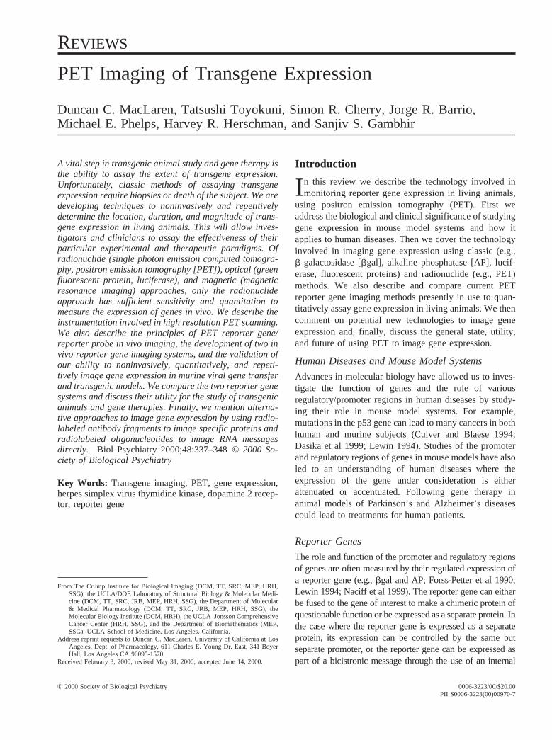
Figure 1. (A) Schematic for imaging probe-modifying positron emission tomography (PET) reporter gene (PRG) expression. Theenzymatic reporter gene complex is transfected into target cells. Inside the transfected cell, the transgene is transcribed to messengerRNA (mRNA) and then translated to the enzyme. The radiolabeled probe is modified by the enzyme and “trapped” within the cell.Thus, after a period of time where the unbound label is “washed” from the system, the magnitude of reporter probe accumulation inthe cell reflects the level of enzyme activity and level of PRG expression.(B) Schematic for imaging receptor/ligand-based PRGexpression. The reporter gene once delivered to a cell by a vector of choice is transcribed to mRNA and then translated to aligand-binding protein or receptor. Accumulation of radiolabeled probe by the protein in or on the cell reflects the reporter geneexpression level.
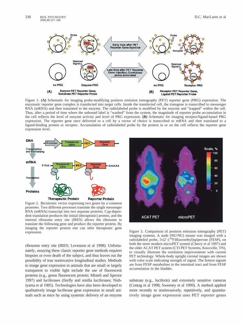
Figure 2. Bicistronic vector expressing two genes by a commonpromoter. Two different processes translate the single messengerRNA (mRNA) transcript into two separate proteins. Cap-depen-dent translation produces the initial (therapeutic) protein, and theinternal ribosome entry site (IRES) allows the ribosome totranslate the following gene and produce the reporter protein. Byimaging the reporter protein one can infer therapeutic geneexpression. Figure 3. Comparison of positron emission tomography (PET)
imaging systems. A nude (NU/NU) mouse was imaged with aradiolabeled probe, 3-(29-[18F]fluoroethyl)spiperone (FESP), onboth the more modern microPET system (Cherry et al 1997) andthe older ACAT PET system (CTI PET Systems, Knoxville, TN),to visually illustrate the resolution improvements with currentPET technology. Whole-body upright coronal images are shownwith color scale indicating strength of signal. The hottest signalsare from FESP metabolites in the intesitnal tract and from FESPaccumulation in the bladder.
338 D.C. MacLaren et alBIOL PSYCHIATRY2000;48:337–348
and radio-labeled PET reporter probes (for reviews, seeGambhir et al 1999a; Herschman et al 2000).
PET Reporter Genes
Positron emission tomography reporter genes encodereceptors that bind positron-emitting ligand probes orenzymes that modify the positron-emitting substrateprobes to produce sequestered positron-emitting prod-ucts. Cells expressing the PET reporter gene willsequester the radiolabel of the PET reporter probe 1) asa ligand bound to the PET reporter receptor or 2) as a“trapped” product of the enzymatic reaction of the PETreporter enzyme (Figure 1; also reviewed in Gambhir etal 1999a; Herschman et al 2000). Ideally, those cells notexpressing the PET reporter genes will not retain thePET reporter probe. Since positron-emitting radionu-clides result in the creation of high-energy gamma rays(511 keV) by positron– electron annihilation, the animalis largely transparent to the wavelength of the radiationproduced, and visualization of radiolabeled probe/li-gand accumulation is readily obtained in even deep andvisually opaque tissues. Emission computed tomogra-phy then allows quantitative imaging of the accumula-tion of the PET reporter probe and, in turn, theexpression levels of the PET reporter gene. Since PETimaging does not require obtaining tissue samples fromthe subject, this system is noninvasive and can be usedto repetitively measure reporter gene expression invivo.
It is also important to note that radionuclide-basedmethods offer significant advantages over optical- (Contaget al 1998) and magnetic resonance imaging–based (Bog-danov and Weissleder 1998) approaches for imagingreporter gene expression. Radionuclide-based methods
offer the highest level of sensitivity for imaging relativelylow levels of reporter gene expression—as low as 10212
mol/L of radiolabeled substrate (Phelps 1991; Phelps et al1986). This high degree of sensitivity may allow the use ofrelatively weak promoters and the imaging of relativelylow levels of gene expression. Furthermore, radionuclide-based methods are highly quantitative. Since PET allowsfor the quantitation of absolute levels of radionuclideprobes, dynamic imaging and kinetic modeling allow us toobtain the rate constants of the underlying biochemicalprocesses (Green et al 1998; Huang and Phelps 1986).
One of the most basic considerations is whether thereporter gene is endogenous or exogenous for the organismunder scrutiny. Expression of endogenous genes has theadvantage of not inducing an immune response and thusallowing for repeated studies. A possible complication inusing an endogenous gene as a PET reporter transgene is ahigh background or erroneous signal due to its inherentexpression. Exogenous genes, especially in gene therapy use,have the disadvantage of inciting an immune response thatmight limit their repeated application, unless, of course, onewishes to image the immune response itself. Exogenous PETreporter genes, with the appropriate probe, have the advan-tage of only producing signal in the tissues in which they areexpressed. An ideal reporter gene for longitudinal studiesshould therefore produce no immune response and not benormally expressed in the organism—or at least in theorgan(s) under consideration.
Direct and Indirect Reporter Transgene Strategies
Direct imaging strategies are based on imaging the trans-gene product directly by binding of a radiolabeled ligandor probe directly to the gene product (e.g., messengerRNA [mRNA], dopamine 2 receptor [D2R]; MacLaren et
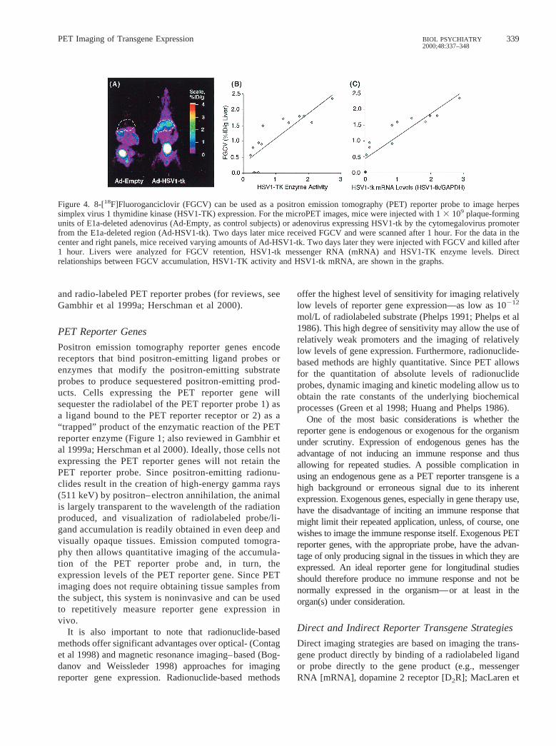
Figure 4. 8-[18F]Fluoroganciclovir (FGCV) can be used as a positron emission tomography (PET) reporter probe to image herpessimplex virus 1 thymidine kinase (HSV1-TK) expression. For the microPET images, mice were injected with 13 109 plaque-formingunits of E1a-deleted adenovirus (Ad-Empty, as control subjects) or adenovirus expressing HSV1-tk by the cytomegalovirus promoterfrom the E1a-deleted region (Ad-HSV1-tk). Two days later mice received FGCV and were scanned after 1 hour. For the data in thecenter and right panels, mice received varying amounts of Ad-HSV1-tk. Two days later they were injected with FGCV and killed after1 hour. Livers were analyzed for FGCV retention, HSV1-tk messenger RNA (mRNA) and HSV1-TK enzyme levels. Directrelationships between FGCV accumulation, HSV1-TK activity and HSV1-tk mRNA, are shown in the graphs.
PET Imaging of Transgene Expression 339BIOL PSYCHIATRY2000;48:337–348
al 1999). Another “direct” approach involves an enzymaticstrategy where a radiolabeled probe is modified andtrapped in cells expressing the PET reporter gene (e.g.,HSV1-tk; Gambhir et al 1998a, 1999d, 2000a; Tjuvajev etal 1995, 1998).
Most therapeutic transgenes do not lend themselves todirect imaging of the transgene product. This is becausemost therapeutic transgene products lack appropriate ra-diolabeled probes. In addition, it would be very timeconsuming and inefficient to develop and validate newprobes for each therapeutic transgene. Alternatively, it isboth feasible and reasonable to develop and validate“indirect” imaging strategies using a reporter gene incombination with a therapeutic gene. The advantage ofthis paradigm is that a given reporter gene can be coupledwith any therapeutic gene. Two strategies have beendiscussed: one uses a fusion gene containing complemen-tary DNA from both reporter and therapeutic genes, andthe second strategy has a separate reporter gene andtherapeutic gene on the same mRNA transcript (cis-linked). Both strategies are based on demonstrating aproportional and constant relationship in the coexpressionof two transgenes over a wide range of expression levels.
Researchers have described and validated the propor-tional expression of twocis-linked genes, using an IRESelement within a single bicistronic transcription unit (Tju-vajev et al 1999; Yu et al 2000). The IRES elementenables translation initiation at a downstream start codonwithin the bicistronic mRNA, thus permitting gene coex-pression by cap-dependent translation of the first cistronand cap-independent, IRES-mediated translation of thesecond cistron (Figure 2; Ghattas et al 1991; Jackson andKaminski 1995; Pelletier 1988; Sachs et al 1997). Thesestudies show that coexpression of the genes is proportionaland quantitative and also demonstrate the potential formonitoring therapeutic gene transfer and expression bynoninvasive imaging ofcis-linked PET reporter genes(Tjuvajev et al 1999; Yu et al 2000).
PET Instrumentation
Because of their short breeding span, extensively charac-terized genetics, and readily manipulatable genome, micehave become the primary platform for most whole-animalresearch in gene expression, gene transfer, and models ofhuman disease. The desire to image mouse models ofhuman disease has led to rapidly increasing interest andefforts in developing imaging technologies that can mea-sure the distribution of radiolabeled tracers in vivo in themouse (Weber and Ivanovic 1999). Autoradiography is awell-established technique that requires killing the animalof interest (Lear 1986, 197–235) and placing tissue slicesin direct contact with analog or digital film. Although
autoradiography has and continues to play a key role,noninvasive approaches are highly desirable for applica-tions in which the same animal needs to be repeatedlyevaluated, or for applications in which it is too expensiveto study large sets of animals at various time points.Furthermore, for human applications, although tissue bi-opsies can be performed, whole-body noninvasive imag-ing is much more compatible with the patient’s comfortand safety. In essence, the goal for radionuclide-basedimaging instrumentation has been to develop a noninva-sive in vivo analogue of autoradiography, with sufficientspatial resolution to resolve the structures of interest in amouse and with sufficient sensitivity that high signal-to-noise images can be obtained.
Radiotracer imaging technologies that can measure thedistribution of radiolabeled tracers in the human body arewidely available and have a wide range of clinical andresearch applications. Two classes of clinical nuclearimaging systems exist—those designed to image singlegamma–emitting radionuclides (e.g., technetium-99m, io-dine-131) and those designed to image positron-emittingradionuclides (e.g., fluorine-18, carbon-11, nitrogen 13,oxygen-15, copper-64, iodine-124). The former is knownas single photon imaging or, when performed tomographi-cally, single photon emission computed tomography(SPECT). The latter is known as PET. In general, PET hasgreater spatial resolution and higher sensitivity and iseasier to quantify than SPECT. For the sake of brevity, thisreview focuses on PET, though most of the discussion isalso applicable to other radionuclide systems.
The realization of the potential power of using PETreporter genes in animal research, together with severaltechnological innovations, has led to the development ofdedicated animal PET scanners by a number of researchcenters in the past 5 years. The first system designedspecifically for rodent imaging was the RAT-PET systemdeveloped at Hammersmith Hospital (Bloomfield et al1995). Although this system was limited to a resolution inthe 3–4-mm range, it established the principle of using adedicated PET scanner for imaging small animals. RAT-PET, despite its relatively coarse spatial resolution, clearlydemonstrated the ability of PET to obtain relevant biolog-ical information from the dopaminergic system of the ratusing highly specific radiotracers (Fricker et al 1997;Hume et al 1996). These results encouraged a number ofresearch groups worldwide to develop very high resolutionPET systems (Cherry et al 1997), with a focus on theopportunities afforded by the sophistication of geneticmanipulation techniques in the mouse (Fries et al 1997;Jeavons et al 1999; Marriott et al 1994; Weber et al 1997).The latest example of a dedicated small animal PETsystem is the microPET system (Cherry et al 1997). The
340 D.C. MacLaren et alBIOL PSYCHIATRY2000;48:337–348
microPET scanner has a reconstructed image resolution of1.8 mm in all three axes and has been shown to be fullyquantitative (Chatziioannou et al 1999). The volumetricresolution is more than an order of magnitude better thanstate-of-the-art clinical PET systems, as illustrated by theimages in Figure 3.
Gene Therapy and Reporter Genes
Our understanding of molecular biology in the last de-cades allowed us to create transgenic animals as well as tointroduce genes into living animals (i.e., gene therapy). Avital step in either of these processes is the ability to assayfor the expression of the transgene. Thus, the use of PETreporter genes can play critical roles in developing genetherapies by allowing researchers to determine the loca-tion, duration, and expression level of the transferred DNAand, specifically, 1) develop vector modifications to im-prove delivery, 2) control expression levels, and 3) im-prove treatments to control duration of expression. Appli-cations of somatic gene transfer technology to treatdiseases are at the forefront of gene therapy applications;as a result, these issues are of great interest to life scientistsand clinicians. The repeatability, quantifiability, and highsensitivity of PET reporter gene systems should lead torapid advancements in science and medicine.
Herpes Simplex Virus 1 ThymidineKinase (HSV1-tk), an Enzymatic PETReporter Gene
Herpes simplex virus 1 thymidine kinase, like mammalianTKs, phosphorylates thymidine, but unlike mammalianTKs, HSV1-TK has relaxed substrate specificity and sophosphorylates thymidine analogues (e.g., 5-iodo-29-fluoro-29deoxy-1-b-D-arabino-furanosyl-uracil [FIAU]) aswell as acycloguanosine analogues (e.g., acyclovir, ganci-clovir [GCV], penciclovir [PCV]; Namavari et al 2000).Cellular enzymes then convert acycloguanosine mono-phosphates and the monophosphate of FIAU to di- andtriphosphates, which have been shown to kill cells byincorporation as chain-terminating derivatives and/or byinhibition of DNA polymerase; however, at the concen-trations of tracer used for imaging by PET, the derivativeshave no discernable effect upon the cells or the health ofpatients (for review, see Gambhir et al 2000b). Herpessimplex virus 1 thymidine kinase has been extensivelystudied; it is nontoxic in humans and is currently beingused as a “susceptibility” gene (in combination with GCV)in clinical gene therapy protocols. Herpes simplex virus 1thymidine kinase can be used as a reporter gene as well asa therapeutic gene (Borrelli et al 1988; Culver et al 1992;Moolten 1997; Moolten and Wells 1990). In gene therapy
protocols using HSV1-tk as a susceptibility gene, identi-fying the location and magnitude of HSV1-TK expressionby noninvasive imaging would provide a highly desirablemeasure of expression (following successful gene trans-fection) and a basis from which the timing of GCVtreatment can be optimized. This represents an idealsituation where the therapeutic and reporter genes are thesame, and is an example of a direct imaging approach.
Two main categories of substrates have been investi-gated as reporter probes for imaging HSV1-tk reportergene expression: derivatives of uracil nucleoside (e.g.,FIAU radiolabeled with iodine; Morin et al 1997; Tjuvajevet al 1995) and derivatives of guanosine radiolabeled withfluorine-18 or carbon-11 (Alauddin et al 1996; Barrio et al1996a, 1996b, 1996c, 1997; Monclus et al 1995). Thesetwo major classes of reporter probes share the ability to bephosphorylated by HSV1-TK, leading to their accumula-tion in cells by DNA polymerase.
Thymidine Derivative Reporter Probes
Using a cell culture model, researchers investigated threecompounds (FIAU, iododeoxyuridine, and GCV) as po-tential HSV1-tk reporter probes and found FIAU to havethe best imaging potential, based upon its in vitro charac-teristics for HSV1-TK and its ability to be labeled withseveral different nuclides (Tjuvajev et al 1995). Radiola-beled FIAU has been used in cell culture and in vivo as anagent for imaging gene expression by both SPECT andPET systems (Tachizawa et al 1981; Tjuvajev et al 1995,1996). Imaging HSV1-TK expression in cancer patientsundergoing combined HSV1-tk–GCV gene therapy withPET or SPECT has also been evaluated and shown to befeasible (Blasberg and Tjuvajev 1997).
Guanosine Derivative Reporter Probes
Another approach developed to image reporter gene ex-pression also relies on the HSV1-tk reporter gene bututilizes acycloguanosine (e.g., radiolabeled GCV/PCV)derivatives as reporter probes (Gambhir et al 1998a,1999d). The choice of acycloguanosines as potentialprobes was based on their ability to be radiolabeled withthe short half-life (110 min) isotope fluorine-18, thusallowing rapid repeated PET imaging of HSV1-tk geneexpression. Initial cell culture uptake experiments with8-[18F]fluoroacyclovir showed poor performance and werenot pursued further (Srinivasan et al 1996). Subsequentinvestigations using 8-[14C]-GCV and 8-[18F]fluoroganci-clovir (FGCV) as reporter probes showed better perfor-mance (Gambhir et al 1998a, 1999d). In further studies totest reporter probes in vivo for HSV1-tk, a replication-deficient adenovirus expressing the HSV1-tk PET reportergene was injected into mice. Because adenovirus accumu-
PET Imaging of Transgene Expression 341BIOL PSYCHIATRY2000;48:337–348
lates on hepatocytes in large part due to the presence ofcoxsackie and adenoviral receptors (Haisma et al 1999),the majority of the injected adenovirus (.95%) infects theliver. Administration of radiolabeled reporter probesshowed accumulation of PET signal only in livers express-ing HSV1-TK (Figure 4), thus validating the applicabilityof using radiolabeled acycloguanosine derivatives to im-age HSV1-tk as a PET reporter gene (Gambhir et al 1998a,
1998b, 1999c, 1999e). Panels B and C of Figure 4 alsoshow the linear relationship between the amount of FGCVsignal to the HSV1-tk activity and mRNA levels.
Reporter probes using fluorine-18 in the side chain ofGCV (FHPG; Alauddin et al 1996; Bading et al 1997;Monclus et al 1997) and in the side chain of PCV (FHBG;Alauddin and Conti 1998) are also being studied and haveshown that high specific activity (250–500 Ci/mmol)
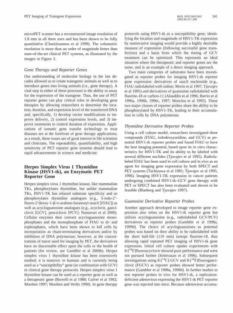
Figure 5. Fluoropenciclovir (FPCV) and the side chain of PCV(FHBG) positron emission tomography (PET) images of a trans-genic mouse expressing herpes simplex virus 1 thymidine kinase(HSV1-tk) in the liver. We studied a transgenic mouse in which thealbumin promoter drives the HSV1-tk reporter gene. The mouse wasimaged on day 0 with a microPET 1 hour after administration ofFPCV and on day 1 with FHBG. Both images are displayed usingthe same common global maximum and illustrate the higher percentinjected dose retained per gram of liver tissue (%ID/g) whenutilizing FHBG. There is significantly greater hepatic accumulationwhen using FHBG (8–11% ID/g), as compared with FPCV (3–6%ID/g).
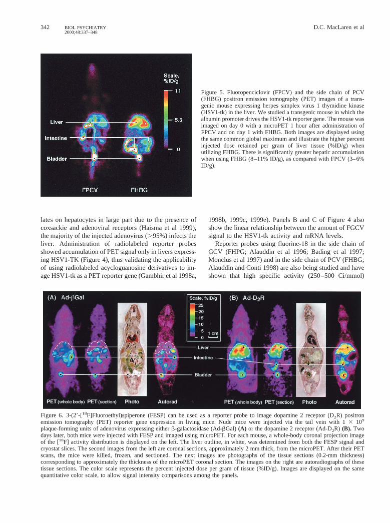
Figure 6. 3-(29-[18F]Fluoroethyl)spiperone (FESP) can be used as a reporter probe to image dopamine 2 receptor (D2R) positronemission tomography (PET) reporter gene expression in living mice. Nude mice were injected via the tail vein with 13 109
plaque-forming units of adenovirus expressing eitherb-galactosidase (Ad-bGal) (A) or the dopamine 2 receptor (Ad-D2R) (B). Twodays later, both mice were injected with FESP and imaged using microPET. For each mouse, a whole-body coronal projection imageof the [18F] activity distribution is displayed on the left. The liver outline, in white, was determined from both the FESP signal andcryostat slices. The second images from the left are coronal sections, approximately 2 mm thick, from the microPET. After their PETscans, the mice were killed, frozen, and sectioned. The next images are photographs of the tissue sections (0.2-mm thickness)corresponding to approximately the thickness of the microPET coronal section. The images on the right are autoradiographs of thesetissue sections. The color scale represents the percent injected dose per gram of tissue (%ID/g). Images are displayed on the samequantitative color scale, to allow signal intensity comparisons among the panels.
342 D.C. MacLaren et alBIOL PSYCHIATRY2000;48:337–348

[18F]fluorinated acycloguanosines can be synthesized inrelatively high yields (5–15 mCi). The side chain of GCVand FHBG have been studied in cell culture models and invivo, and are also well suited for imaging HSV1-tk geneexpression (Gambhir et al 2000b).
Comparison of HSV1-tk Reporter Probes
Direct comparison of all the radiolabeled probes for invivo imaging of HSV1-tk reporter gene expression is anecessary step for optimizing this PET reporter system.The UCLA group has compared uptake of FGCV,FPCV, FHBG, FHPG, and [14C]-FIAU in C6 cellsexpressing HSV1-tk. These preliminary data show thatFIAU and FHBG are the better candidates for imagingHSV1-tk reporter gene expression because of their 1)long half-life in vivo, 2) low nonspecific retention, and3) high specific retention. The true utility of alternativeHSV-TK probes must ultimately be evaluated in wholeanimals, as issues such as stability, substrate competi-tion, routes of clearance, and rates of cellular transportall come into play. A transgenic mouse model in whichthe albumin promoter drives the HSV1-tk reporter geneis being studied at UCLA (Gambhir et al 2000b;Herschman et al 2000). These transgenic mice havebeen imaged by PET and clearly demonstrate accumu-lation of the FPCV (3– 6% injected dose retained pergram of tissue [ID/g]) and FHBG (8 –11% ID/g) re-porter probes in the mouse liver (Figure 5). Futurestudies that directly compare all prospective acy-cloguanosines and thymidine analogues in whole ani-mals will help to better define the advantages anddisadvantages for each probe.
Mutant HSV1-tk Reporter Gene Approaches
Researchers at UCLA are also investigating mutantHSV1-tk reporter genes to further enhance the sensitivityof the HSV1-tk reporter assays (Gambhir et al 2000a). Weare using a mutated HSV1-TK enzyme (HSV1-sr39TK)that utilizes GCV and PCV substrates more effectively andthymidine less effectively than the wild-type HSV1-TKenzyme (Black et al 1996). Mouse models with anadenovirus expressing this mutant HSV1-TK enzymedemonstrated improved imaging sensitivity (equivalent totwo- to threefold) with both FGCV and FPCV as PETreporter probes. Additional studies with FHBG show afurther enhancement by a factor of;2 as well. Takentogether, the mutant HSV1-tk with FHBG should offer agreater than fourfold improvement in sensitivity and placeit on par with the sensitivity of the D2R/FESP PETreporter system.
D2R, a Receptor PET Reporter Gene
Another PET reporter gene system investigated uses D2Rand the radiolabeled ligand 3-(29-[18F]fluoroethyl)spiper-one (FESP), a positron-emitting analogue of the dopamineantagonist spiperone. The latter was originally developedas a probe to image, by PET, the D2Rs of the striatum(Barrio et al 1989), where concentrations of D2R as low as2–20 nmol/L can be detected. This PET reporter genesystem was also investigated because of the high bindingaffinities of D2R to FESP and, unlike substrates forHSV1-tk, the ability of FESP to diffuse into every tissue inthe body (Barrio et al 1989).
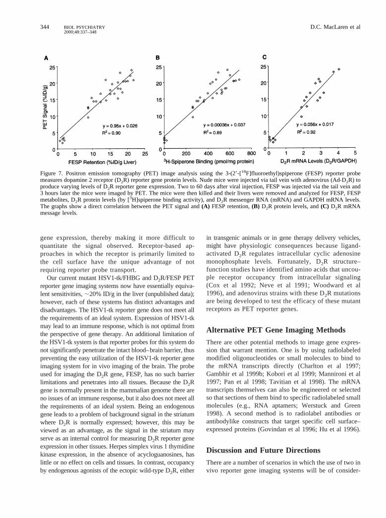
As for the HSV1-tk system, we used adenovirusdelivery of the D2R reporter gene to demonstrate thenoninvasive, repetitive, and quantitative ability of theD2R/FESP PET reporter gene/PET reporter probe invivo imaging system. To determine whether PET canquantitatively monitor hepatic D2R expression in ade-novirus-infected mice, animals were injected with vary-ing amounts of D2R-expressing orbgal-expressing(control) virus, then injected with FESP and imaged byPET (Figure 6; MacLaren et al 1999). After scanning,the mice were killed and liver samples were analyzedfor 1) FESP retention by ether extraction and [fluorine-18] well counting, 2) functional D2R protein levels by[3H]spiperone binding using a conventional receptorbinding assay, and 3) D2R mRNA levels by quantitativeNorthern blots. The fluorine-18 signal retained in liver,as determined by region of interest measurements ofPET images of living mice, is proportional to both theamount of hepatic FESP present and functional D2Rprotein levels (Figure 7). In vivo PET analysis ofhepatic D2R reporter gene expression accurately reflectsin vitro determinations of D2R levels and validates theuse of this PET reporter gene/probe system.
Comparison of HSV1-tk and D2R ReporterGene Systems
The reporter gene assays developed to date fall into thetwo main categories enzyme based (e.g., HSV1-tk) andreceptor based (e.g., D2R). Each of these assays hassome distinct features that deserve special comment. Anenzyme-based approach has the theoretical advantage ofsignal amplification, since one molecule of reporterenzyme is capable of acting on many molecules ofreporter probes. Most receptor-based assays, such as theD2R system, are capable of only a one-to-one stochio-metric interaction of reporter ligand with receptor.Enzyme-based approaches will always require intracel-lular transport of the reporter substrate, and the rate oftransport may change independent of levels of reporter
PET Imaging of Transgene Expression 343BIOL PSYCHIATRY2000;48:337–348
gene expression, thereby making it more difficult toquantitate the signal observed. Receptor-based ap-proaches in which the receptor is primarily limited tothe cell surface have the unique advantage of notrequiring reporter probe transport.
Our current mutant HSV1-tk/FHBG and D2R/FESP PETreporter gene imaging systems now have essentially equiva-lent sensitivities,;20% ID/g in the liver (unpublished data);however, each of these systems has distinct advantages anddisadvantages. The HSV1-tk reporter gene does not meet allthe requirements of an ideal system. Expression of HSV1-tkmay lead to an immune response, which is not optimal fromthe perspective of gene therapy. An additional limitation ofthe HSV1-tk system is that reporter probes for this system donot significantly penetrate the intact blood–brain barrier, thuspreventing the easy utilization of the HSV1-tk reporter geneimaging system for in vivo imaging of the brain. The probeused for imaging the D2R gene, FESP, has no such barrierlimitations and penetrates into all tissues. Because the D2Rgene is normally present in the mammalian genome there areno issues of an immune response, but it also does not meet allthe requirements of an ideal system. Being an endogenousgene leads to a problem of background signal in the striatumwhere D2R is normally expressed; however, this may beviewed as an advantage, as the signal in the striatum mayserve as an internal control for measuring D2R reporter geneexpression in other tissues. Herpes simplex virus 1 thymidinekinase expression, in the absence of acycloguanosines, haslittle or no effect on cells and tissues. In contrast, occupancyby endogenous agonists of the ectopic wild-type D2R, either
in transgenic animals or in gene therapy delivery vehicles,might havephysiologic consequences because ligand-activated D2R regulates intracellular cyclic adenosinemonophosphate levels. Fortunately, D2R structure–function studies have identified amino acids that uncou-ple receptor occupancy from intracellular signaling(Cox et al 1992; Neve et al 1991; Woodward et al1996), and adenovirus strains with these D2R mutationsare being developed to test the efficacy of these mutantreceptors as PET reporter genes.
Alternative PET Gene Imaging Methods
There are other potential methods to image gene expres-sion that warrant mention. One is by using radiolabeledmodified oligonucleotides or small molecules to bind tothe mRNA transcripts directly (Charlton et al 1997;Gambhir et al 1999b; Kobori et al 1999; Mannironi et al1997; Pan et al 1998; Tavitian et al 1998). The mRNAtranscripts themselves can also be engineered or selectedso that sections of them bind to specific radiolabeled smallmolecules (e.g., RNA aptamers; Werstuck and Green1998). A second method is to radiolabel antibodies orantibodylike constructs that target specific cell surface–expressed proteins (Govindan et al 1996; Hu et al 1996).
Discussion and Future Directions
There are a number of scenarios in which the use of two invivo reporter gene imaging systems will be of consider-
Figure 7. Positron emission tomography (PET) image analysis using the 3-(29-[18F]fluoroethyl)spiperone (FESP) reporter probemeasures dopamine 2 receptor (D2R) reporter gene protein levels. Nude mice were injected via tail vein with adenovirus (Ad-D2R) toproduce varying levels of D2R reporter gene expression. Two to 60 days after viral injection, FESP was injected via the tail vein and3 hours later the mice were imaged by PET. The mice were then killed and their livers were removed and analyzed for FESP, FESPmetabolites, D2R protein levels (by [3H]spiperone binding activity), and D2R messenger RNA (mRNA) and GAPDH mRNA levels.The graphs show a direct correlation between the PET signal and(A) FESP retention,(B) D2R protein levels, and(C) D2R mRNAmessage levels.
344 D.C. MacLaren et alBIOL PSYCHIATRY2000;48:337–348
able utility in research protocols. For example, the abilityto image two distinct reporter genes in vivo will allowdirect comparisons, in the same individual, of alternativegene delivery vehicles (e.g., viruses, liposomes) in somaticgene transfer protocols. Many combinatorial applicationsof these in vivo PET reporter gene imaging systems arelikely to emerge as their availability and utility becomeapparent. The advantages of high sensitivity, quantitativecapability, and direct ability to translate the developedassays from animal to human studies will keep radionu-clide-based approaches at the forefront of imaging geneexpression for transgenic and gene therapy studies.
Continued development of reporter genes and corre-sponding reporter probes will be needed to produce moreoptimal assays; however, for many applications the sys-tems developed to date appear sufficiently robust. Themajor applications of PET reporter gene imaging in vivoare likely to be 1) repetitive, quantitative monitoring of thelocation, duration, and extent of gene expression fromgene therapy vehicles, using bicistronic vectors that ex-press therapeutic and reporter gene products from acommon transcript, and 2) the evaluation of gene functionand the role of promoter/regulatory elements by reportergene expression in transgenic animals during longitudinalexperiments.
There are also improvements in PET instrumenta-tion—a “second-generation” microPET instrument, cur-rently being developed, will provide a much higher reso-lution of 1 mm for each axis and be able to scan an entiremouse in 10 min. Positron emission tomography technol-ogy is still a long way from reaching the resolution andsensitivity limits imposed by geometric considerations andthe physics of the positron annihilation process (Chatziio-annou et al 1999; Cherry et al 1997). The performance ofdedicated animal PET scanners is likely to improvesubstantially in the next few years and will be accompa-nied by a reduction in both their size and their cost.
The combination of improved PET reporter gene imag-ing systems and PET instrumentation will allow investi-gators to readily image and quantitatively evaluate geneexpression in transgenic animals and gene therapy sub-jects. This will ultimately allow doctors to follow theeffectiveness of gene therapy for many disorders, includ-ing neurodegenerative diseases such as Parkinson’s andAlzheimer’s.
These studies were supported by Department of Energy contract DE-FC03-87ER60615, National Institutes of Health Grant No. RO1CA82214-01 (SSG), National Institutes of Health Grant No. RO1CA84572-01 (HRH), the University of California Biotechnology Pro-gram, the UCLA Gene Medicine Program, the Dana Foundation, and theUCLA–Jonsson Comprehensive Cancer Center.
ReferencesAlauddin MM, Conti PS (1998): Synthesis and preliminary
evaluation of 9-(4-[18F]-fluoro-3-hydroxymethylbutyl)gua-nine ([18F]FHBG): A new potential imaging agent for viralinfection and gene therapy using PET.Nucl Med Biol25:175–180.
Alauddin MM, Conti PS, Mazza SM, Hamzeh FM, Lever JR(1996): Synthesis of 9-[(3-[18F]fluoro-1-hydroxy-2-propoxy)methyl]guanine ([18F]FHPG): A potential imaging agent ofviral infection and gene therapy using PET.Nucl Med Biol23:787–792.
Bading JR, Alauddin MM, Fissekis JD, Kirkman E, Raman RK,Conti PS (1997): Pharmacokinetics of F-18 fluorohydroxy-propoxymethylguanine (FHPG).J Nucl Med38:43P.
Barrio JR, Namavari M, Phelps ME, Satyamurthy N (1996a):Elemental fluorine to 8-fluorpurines in one step.J Am ChemSoc118:10408–10411.
Barrio JR, Namavari M, Phelps ME, Satyamurthy N (1996b):Regioselective fluorination of substituted guanines with di-lute F2: A facile entry of 8-fluoroguanine derivatives.JOrganic Chem61:6084–6085.
Barrio JR, Namavari M, Satyamurthy N, Srinivasan A, Her-schman H, Gambhir S (1996c): 8-[F-18]fluoroacyclovir: Anin vivo probe for gene expression with PET.J Nucl Med37:193P.
Barrio JR, Namavari M, Srinivasan A, Gambhir S, Cherry S,Herschman H, et al (1997): Carbon-8 radiofluorination ofpurines: A general approach to probe design for gene therapyin humans.J Labelled Compounds Radiopharmaceuticals40:348.
Barrio JR, Satyamurthy N, Huang SC, Keen RE, Nissenson CH,Hoffman JM, et al (1989): 3-(29-[18F]fluoroethyl)spiperone:In vivo biochemical and kinetic characterization in rodents,nonhuman primates, and humans.J Cereb Blood Flow Metab9:830–839.
Black ME, Newcomb TG, Wilson H-MP, Loeb LA (1996):Creation of drug-specific hepes simplex virus type 1 thymi-dine kinase mutant for gene therapy.Proc Natl Acad SciU S A93:3525–3529.
Blasberg R, Tjuvajev J (1997): In vivo monitoring of genetherapy by radiotracer imaging. In: Semmler W, SchwaigerW, editors.Ernst Shering Research Foundation Workshop22: Impact of Molecular Biology and New Technical Devel-opments on Diagnostic Imaging.Berlin: Springer Verlag,161–189.
Bloomfield PM, Rajeswaran S, Spinks TJ, Hume SP, Myers R,Ashworth S, et al (1995): The design and physical character-istics of a small animal positronemission tomograph.PhysMed Biol 40:1105–1126.
Bogdanov A, Weissleder R (1998): The development of in vivoimaging systems to study gene expression.Trends Biotechnol16:5–10.
Borrelli E, Heyman R, Hsi M, Evans RM (1988): Targeting of aninducible toxic phenotype in animal cells.Proc Natl Acad SciU S A85:7572–7576.
Charlton J, Sennello J, Smith D (1997): In vivo imaging ofinflammation using an aptamer inhibitor of human neutrophilelastase.Chem Biol4:809–816.
Chatziioannou AF, Cherry SR, Shao Y, Silverman RW, Meadors
PET Imaging of Transgene Expression 345BIOL PSYCHIATRY2000;48:337–348
K, Farquhar TH, et al (1999): Performance evaluation ofmicroPET: A high resolution LSO PET scanner for animalimaging.J Nucl Med40:1164–1175.
Cherry SR, Shao Y, Silverman RW, Meadors K, Siegel S,Chatziioannou AF, et al (1997): MicroPET: A high resolutionPET scanner for imaging small animals.IEEE Trans Nucl Sci44:1161–1166.
Contag PR, Olomu IN, Stevenson DK, Contag CH (1998):Bioluminescent indicators in living mammals.Nat Med4:245–247.
Cox BA, Henningsen RA, Spanoyannis A, Neve RL, Neve KA(1992): Contributions of conserved serine residues to theinteractions of ligands with dopamine D2 receptors.J Neu-rochem59:627–635.
Culver KW, Blaese RM (1994): Gene therapy for cancer.TrendsGenet10:174–178.
Culver KW, Ram Z, Walbridge S, Ishii H, Oldfield EH, BlaiseRM (1992): In vivo gene transfer with retroviral vector-producer cells for treatment of experimental brain tumors.Science256:1550–1552.
Dasika GK, Lin SC, Zhao S, Sung P, Tomkinson A, Lee EY(1999): DNA damage-induced cell cycle checkpoints andDNA strand break repair in development and tumorigenesis.Oncogene20:7883–7899.
Forss-Petter S, Danielson PE, Catsicas S, Battenberg E, PriceJ, Nerenberg M, Sutcliffe JG (1990): Transgenic miceexpressing beta-galactosidase in mature neurons underneuron-specific enolase promoter control.Neuron5:187–197.
Fricker RA, Torres EM, Hume SP, Myers R, Opacka-JuffreyJ, Ashworth S, et al (1997): The effects of donor stage onthe survival and function of embryonic striatal grafts in theadult rat brain. II. Correlation between positron emissiontomography and reaching behaviour.Neuroscience79:711–721.
Fries O, Bradbury SM, Gebauer J, Holl I, Lorenz E, Renker D,et al (1997): A small animal PET prototype based on LSOcrystals read out by avalanche photodiodes.Nucl Inst Meth-odsA387:220–224.
Gambhir SS, Barrio J, Wu L, Iyer M, Namavari M, Satyamur-thy N, et al (1998a): Imaging of adenoviral directed herpessimplex virus type 1 thymidine kinase gene expression inmice with ganciclovir.J Nucl Med39:2003–2011.
Gambhir SS, Barrio JR, Bauer E, Iyer M, Namavari M,Satyamurthy N, et al (1998b): Radiolabeled penciclovir: Anew reporter probe with improved imaging properties overganciclovir for imaging herpes-simplex virus type 1 thy-midine kinase reporter gene expression.J Nucl Med39:53P.
Gambhir SS, Barrio JR, Herschman HR, Phelps ME (1999a):Assays for noninvasive imaging of reporter gene expression.Nucl Med Biol26:481–490.
Gambhir SS, Barrio JR, Herschman HR, Phelps ME (1999b):Imaging gene expression: Principles and assays.J NuclCardiol 6:219–233.
Gambhir SS, Barrio JR, Iyer M, Namavari M, Satyamurthy N,Toyokuni T, et al (1999c): In vivo validation of PET reportergene/reporter probe assay for herpes simplex virus type 1thymidine kinase with 8-[F-18]-fluoropenciclovir.J NuclMed 40:25–26.
Gambhir SS, Barrio JR, Phelps ME, Iyer M, Namavari M,Satyamurthy N, et al (1999d): Imaging adenoviral-directedreporter gene expression in living animals with positronemission tomography.Proc Natl Acad Sci U S A 96:2333–2338.
Gambhir SS, Bauer E, Black M, Liang Q, Kokoris MS, BarrioJR, et al (2000a): A mutant herpes simplex virus type 1thymidine kinase reporter gene shows improved sensitivityfor imaging reporter gene expression with positron emis-sion tomography.Proc Natl Acad Sci U S A97:2785–2790.
Gambhir SS, Herschman HR, Cherry SR, Barrio JR, Satyamur-thy N, Toyokuni T, et al (2000b): Imaging transgene expres-sion with radionuclide imaging technologies.Neoplasia2:118–138.
Gambhir SS, MacLaren DC, Barrio JR, Toyokuni T, Satyamur-thy N, Nguyen K, et al (1999e, May): Noninvasive andrepeated imaging of reporter gene expression in living miceutilizing positron emission tomography. Poster presented atthe annual meeting of the American Society of Gene Therapy,Washington, DC.
Ghattas IR, Sanes JR, Majors JE (1991): The encephalomyo-carditis virus internal ribosomal entry site allows efficientcoexpression of two genes from a recombinant provirus incultured cells and in embryos.Mol Cell Biol 11:5848 –5859.
Govindan SV, Goldenberg DM, Grebenau RC, Hansen HJ,Griffiths GL (1996): Thiolations, 99mTc labelings, and ani-mal in vivo biodistributions of divalent monoclonal antibodyfragments.Bioconjug Chem7:290–297.
Green LA, Gambhir SS, Barrio JR, Bauer E, Nguyen K,Namavari M, et al (1998): Tracer kinetic modeling of8-(F18)-fluoroganciclovir PET data: A new tracer formeasuring reporter gene expression.J Nucl Med39(suppl):10P.
Haisma H, Pinedo H, Rijswijk A, der Meulen-Muileman I,Sosnowski BA, Ying W, et al (1999): Tumor-specific genetransfer via an adenoviral vector targeted to the pan-carci-noma antigen EpCAM.Gene Ther6:1469–1474.
Herschman HR, MacLaren DC, Iyer M, Namavari M, BobinskiK, Green LA, et al (2000): Seeing is believing: Non-invasive,quantitative and repetitive imaging of reporter gene expres-sion in living animals, using positron emission tomography.J Neurosci Res59:699–705.
Hu S, Shively L, Raubitschek A, Sherman M, Williams LE,Wong JY, et al (1996): Minibody: A novel engineeredanti-carcinoembryonic antigen antibody fragment (single-chain Fv-CH3) which exhibits rapid, high-level targeting ofxenografts.Cancer Res56:3055–3061.
Huang SC, Phelps ME (1986): Chapter 7. In: Phelps ME,Mazziota J, Schelbert HR, editors.Positron EmissionTomography and Autoradiography, Principles and Appli-cations for the Brain and Heart.New York: Raven.
Hume SP, Lammertsma AA, Myers R, Rajeswaran S, Bloom-field PM, Ashworth S, et al (1996): The potential of high-resolution positron emission tomography to monitor striataldopaminergic function in rat models of disease.J NeurosciMethods67:103–112.
Jackson RJ, Kaminski A (1995): Internal initiation of translationin eukaryotes: The picornovirus paradigm and beyond.RNA1:985–1000.
346 D.C. MacLaren et alBIOL PSYCHIATRY2000;48:337–348
Jeavons AP, Chandler RA, Dettmmar CAR (1999): A 3DHIDAC-PET camera with sub-millimetre resolution forimaging small animals.IEEE Trans Nucl Sci46:468 – 473.
Kobori N, Imahori Y, Mineura K, Ueda S, Fujii R (1999):Visualization of mRNA expression in CNS using 11C-labeledphosphorothioate oligodeoxynucleotide.Neuroreport10:2971–2974.
Lear JL (1986):Principles of Single and Multiple RadionuclideAutoradiography.New York: Raven.
Levenson VV, Transue ED, Roninson IB (1998): Internal ribo-somal entry site-containing retroviral vectors with greenfluorescent protein and drug resistance markers.Hum GeneTher 9:1233–1236.
Lewin B (1994):Genes V.New York: Oxford University Press.
MacLaren DC, Gambhir SS, Satyamurthy N, Barrio JR, Sharf-stein S, Toyokuni T, et al (1999): Repetitive, non-invasiveimaging of the dopamine D2 receptor as a reporter gene inliving animals.Gene Ther6:785–791.
Mannironi C, Di NA, Fruscoloni P, Tocchini VG (1997): In vitroselection of dopamine RNA ligands.Biochemistry36:9726–9734.
Marriott CJ, Cadorette JE, Lecomete R, Scasnar V, Rousseau J,van Lier JE (1994): High-resolution PET imaging and quan-titation of pharmaceutical biodistributions in a small animalusing avalance photodiode detectors.J Nucl Med35:1390–1396.
Misteli T, Spector D (1997): Applications of the green fluores-cent protein in cell biology and biotechnology.Nat Biotech-nol 15:961–964.
Monclus M, Luxen A, Cool V, Damhaut P, Velu T, Goldman S(1997): Development of a positron emission tomographyradiopharmaceutical for imaging thymidine kinase gene ex-pression: Synthesis and in vitro evaluation of 9-{3-[18F]fluoro-1-hydroxy-2-propoxy)methyl}guanine. BioorgMed Chem Lett7:1879–1882.
Monclus M, Luxen A, Van Naemen J, et al (1995): Develop-ment of PET radiopharmaceuticals for gene therapy: Syn-thesis of 9-((1-(18F)fluoro-3-hydroxy-2-propoxy)methyl)guanine. J Labelled Compounds Radiopharmaceuticals37:193–195.
Moolten FL (1997): Suicide genes for cancer therapy.Sci Med4:16–25.
Moolten FL, Wells JM (1990): Curability of tumors bearingherpes thymidine kinase genes transfected by retroviral vec-tors.J Natl Cancer Inst82:297–300.
Morin KW, Atrazheva ED, Knaus EE, Wiebe LI (1997): Syn-thesis and celluar uptake of 29-substituted analogues of(E)-5-(2-[125I]iodovinyl-29-deoxyuridine in tumor cellstransduced with the herpes simplex type-1 thymidine kinasegene. Evaluation as probes for monitoring gene therapy.J Med Chem40:2184–2190.
Naciff JM, Behbehani MM, Misawa H, Dedman JR (1999):Identification and transgenic analysis of a murine promoterthat targets cholinergic neuron expression.J Neurochem72:17–28.
Namavari M, Barrio JR, Toyokuni T, Gambhir SS, Cherry SR,Herschman HR, et al (2000): Synthesis of 8-[18F] fluorogua-nine derivatives: In vivo probes imaging gene expression withPET.Nucl Med Biol27:157–162.
Neve KA, Cox BA, Henningsen RA, Spanoyannis A, Neve RL(1991): Pivotal role for aspartate-80 in the regulation ofdopamine D2 receptor affinity for drugs and inhibition ofadenylyl cyclase.Mol Pharmacol39:733–739.
Nishiyama T, Kawamura Y, Kawamoto K, Matsumura H,Yamamoto N, Ito T, et al (1985): Antineoplastic effects of5-fluorocytosine in combination with cytosine deaminasecapsules.Cancer Res45:1753–1761.
Pan D, Gambhir SS, Toyokuni T, Iyer MR, Acharya N, PhelpsME, Barrio JR (1998): Rapid synthesis of a 59-fluorinatedoligodeoxy-nucleotide: A model antisense probe for use inimaging with positron emission tomography (PET).BioorgMed Chem Lett8:1317–1320.
Pelletier S (1988): Internal initiation of translation of eukaryoticmRNA directed by a sequence derived from poliovirus RNA.Nature334:320.
Phelps ME (1991): PET: A biological imaging technique.Neu-rochem Res16:929–994.
Phelps ME, Schelbert HR, Mazziotta J, editors (1986):Positron Emission Tomography and Autoradiography,Principles and Applications for the Brain and Heart.NewYork: Raven.
Sachs AB, Sarnow P, Hentze MW (1997): Starting at thebeginning, middle, and end: Translation initiation in eu-karyotes.Cell 89:831–838.
Srinivasan A, Gambhir SS, Green LA, Cherry SR, Sharfstein S,Barrio JR, et al (1996): A PET reporter gene (PRG)/PETreporter probe (PRP) technology for repeatedly imaging geneexpression in living animals.J Nucl Med37:107P.
Sweeney TJ, Maila¨nder V, Tucker AA, Olomu AB, Zhang W,Cao Y, et al (1999): Visualizing the kinetics of tumor-cellclearance in living animals.Proc Natl Acad Sci U S A96:12044–12049.
Tachizawa H, Sudo K, Sasano H, Sano M (1981): Dispositionand metabolism of timiperone in the rat, dog and monkey.Drug Metab Dispos9:442–448.
Tavitian B, Terrazzino S, Kuhnast B, Marzabal S, Stettler O,Dolle F, et al (1998): In vivo imaging of oligonucleotideswith positron emission tomography.Nat Med4:467– 471.
Tjuvajev JG, Avril N, Oku T, Sasajima T, Miyagawa T, Joshi R,et al (1998): Imaging herpes virus thymidine kinase genetransfer and expression by positron emission tomography.Cancer Res58:4333–4341.
Tjuvajev JG, Callegari A, Lindsley L, Joshi R, Balatoni J, FinnR, et al (1999): A general approach to the non-invasiveimaging of transgenes using cis-linked herpes simplex virusthymidine kinase.Neoplasia1:315–320.
Tjuvajev JG, Finn R, Watanabe K, Joshi R, Oku T, KennedyJ, et al (1996): Noninvasive imaging of herpes virusthymidine kinase gene transfer and expression: A potentialmethod for monitoring clinical gene therapy.Cancer Res56:4087– 4095.
Tjuvajev JG, Stockhammer G, Desai R, Uehara H, WatanabeK, Gansbacher B, Blasberg RG (1995): Imaging theexpression of transfected genesin vivo. Cancer Res55:6126 – 6132.
Weber DA, Ivanovic M (1999): Ultra-high-resolution imaging ofsmall animals: Implications for preclinical and research stud-ies.J Nucl Cardiol6:332–344.
PET Imaging of Transgene Expression 347BIOL PSYCHIATRY2000;48:337–348

Weber S, Terstegge A, Herzon H, Reinartz R, Reinhart P,Rongen F, et al (1997): The design of an animal PET:Flexible geometry for achieving optimal spatial resolutionor high sensitivity.IEEE Trans Med Imaging16:684 – 689.
Werstuck G, Green MR (1998): Controlling gene expression inliving cells through small molecule-RNA interactions.Sci-ence282:296–298.
Woodward R, Roley C, Daniell S, Naylor LH, Strange PG
(1996): Investigation of the role of conserved serineresidues in the long form of the rat D2 dopamine receptorusing site-directed mutagenesis.J Neurochem66:394 –402.
Yu Y, Annala AJ, Barrio JR, Toyokuni T, Satyamurthy N,Namavari M, et al (2000): Quantitation of target geneexpression by imaging reporter gene expression in livinganimals.Nat Med6:933–937.
348 D.C. MacLaren et alBIOL PSYCHIATRY2000;48:337–348