pharmacokinetics and mechanisms of action of carbon … · 5-1 chapter 5 pharmacokinetics and...
TRANSCRIPT

5-1
CHAPTER 5
Pharmacokinetics and Mechanisms of Action ofCarbon Monoxide
5.1 IntroductionBasic research on the physiology, pharmacokinetics, and toxicology of carbon monoxide (CO) that
ended in the late seventies was followed by studies focused primarily on the cardiopulmonary effects of COas an ambient air pollutant. Although research in this area continues, more recent studies have refocusedon the mechanisms of action and pathophysiological effects of CO at a cellular level and on its role as acytotoxic agent and neural messenger. In this chapter, the sections discussing basic pharmacokinetics drawheavily from Chapter 9 of the previous CO criteria document (U.S. Environmental Protection Agency,1991). However, all sections were revised and consolidated, many were expanded, and several new sectionswere added. In particular, sections on tissue production and metabolism of CO and intracellular effects ofCO have been revised extensively and expanded. The new section on conditions affecting uptake andelimination of CO discusses the influence of physical activity, altitude, physical characteristics, and healthstatus on carboxyhemoglobin (COHb) formation. Also, new sections on the mechanisms of CO and areview of the developing concepts have been added.
Although the focus of this document is on the effects of ambient and near ambient levels of COleading to low COHb levels (#5%), this chapter discusses, where appropriate, findings of a selected numberof human studies carried out at moderate COHb levels (#20%). Also discussed are observations from alimited number of relevant animal studies at even higher COHb levels. The purpose for the inclusion ofsuch observations from human studies at higher CO concentrations, and animal studies in general, is tofacilitate the understanding of CO kinetics, related pathophysiologic processes, and mechanisms ofcytotoxicity. Despite much higher CO uptake and elimination rates in animal species than in humans,primarily because of substantially higher ventilation rates, the laboratory animal data still fill, although onlypartially, the knowledge gaps for which no human data are available in these areas of research. Over therange of CO concentrations that are most relevant experimentally to typical environmental CO exposures(e.g., 50 to 500 ppm), the rate of both CO uptake and elimination in mammals is inversely proportional tobody mass (i.e., the smaller the animal, the faster the rate [Klimisch et al., 1975; Tyuma et al., 1981]). Overthis same range of CO concentrations, the most widely used predictive model of COHb formation, theCoburn-Forster-Kane (CFK) equation, accurately predicts the resulting COHb levels not only in humansubjects, but also in laboratory rats and mice (Tyuma et al., 1981; Benignus and Annau, 1994; Kimmelet al., 1999). Thus, despite many well identified interspecies differences in the toxicokinetics of CO, thebasic mechanisms of CO toxicity between laboratory animals and humans are similar and, in many respects,close to identical. Although a more detailed discussion of interspecies differences as they relate to humansmay aid in interpretation of data and elucidation of mechanisms, it is not essential for understanding thematerial presented in this chapter and is well beyond the scope of this document (see Chapter 1). Despiteinterspecies differences, especially in the uptake and elimination kinetics of CO, extrapolation ofobservations from animals to man as applied in this chapter, even with its many assumptions, may be useful

5-2
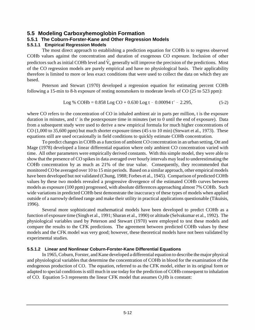
Figure 5-1. Diagrammatic presentation of CO uptake andelimination pathways and CO body stores.Source: Adapted from Coburn (1967).
in identifying potential pathophysiologic and histotoxic processes associated with ambient or near ambientCO exposure.
5.2 Absorption, Distribution, and Pulmonary Elimination5.2.1 Pulmonary Uptake
Although CO is not one of the respiratory gases, the similarity of physico-chemical properties ofCO and oxygen (O2) permits an extension of the findings of studies on the kinetics of transport of O2 tothose of CO. The rate of formation and elimination of COHb, its concentration in blood, and its catabolismis controlled by numerous physical factors and physiological mechanisms. The relative contribution ofthese mechanisms to the overall COHb kinetics will depend on the environmental conditions, the physicalactivity of an individual, and many other physiological processes, some of which are complex and stillpoorly understood (see Section 5.4 for details). All of the pulmonary uptake occurs at the respiratorybronchioles and alveolar ducts and sacs. The rate of CO uptake depends on the rate of COHb formation.At the low concentration of CO in inhaled air, the rate of uptake and the rate of COHb formation could, forall practical purposes, be considered to be qualitatively the same.
5.2.1.1 Mass Transfer of Carbon MonoxideThe mass transport of CO between the
airway opening (mouth and nose) and the redblood cell (RBC) hemoglobin (Hb) ispredominantly controlled by physical processes.The CO transfer to the Hb-binding sites isaccomplished in two sequential steps:(1) transfer of CO in a gas phase, between theairway opening and the alveoli, and (2) transferin a “liquid” phase, across the air-bloodinterface, including the RBC. In the gas phase,the key mechanisms of transport are convectiveflow, by the mechanical action of the respiratorysystem, and diffusion in the acinar zone of thelung (Engel et al., 1973). Subsequent moleculardiffusion of CO across the alveolo-capillarymembrane along the CO pressure gradient,plasma, and RBC is the virtual mechanism of theliquid phase. The principal transport pathwaysand body stores of CO are shown in Figure 5-1(Coburn, 1967).
5.2.1.2 Effects of Dead Space and Ventilation/Perfusion RatioThe effectiveness of alveolar gas exchange depends on effective gas mixing and matching of
ventilation and perfusion. During normal tidal breathing, the inhaled gas is not distributed uniformly acrossthe tracheobronchial tree. With increased inspiratory flow, as during exercise, intrapulmonary gasdistribution becomes more uniform, but gas concentration inhomogeneity still will persist. Considering thatalmost 90% of gas is contained within the acinar zone of the lung, any increase in gas inhomogeneity in thisterminal region will have about the same negative effect as an additional increase in the alveolar dead spaceor a decrease in the alveolo-capillary diffusion capacity (Engel et al., 1973).

5-3
The inefficiency of gas mixing and a consequent decrease in the effectiveness of alveolar gasexchange is aggravated by ventilation/perfusion ( 0VA / 0Q) mismatch. Because of the gravity dependence ofventilation and even more of perfusion in an upright posture, regional 0VA / 0Q ratios will range from 0.6 (atthe base of the lung) to 3.0 (at the apex), the overall value being 0.85. As a result, the 0VA/ 0Q ratio is theprincipal variable controlling gas exchange, and any inequalities not only will impair transfer of gases tothe blood but also will interfere with unloading of gases from the blood into the alveolar air. In humans,a change in posture to recumbent or exercise will increase the uniformity of 0VA / 0Q ratios and promote moreefficient gas exchange, whereas increased resting lung volume, increased airway resistance, decreased lungcompliance, and, generally, any lung abnormality will aggravate 0VA / 0Q ratio inequality.
The simplest indicator of the 0VA / 0Q ratio inequalities is the volume of physiological dead space (VD),which comprises both the anatomical and alveolar dead space. The alveolar dead space results fromreduced perfusion of alveoli, relative to their ventilation (Singleton et al., 1972). Both right-to-left andphysiological shunts under normal conditions contribute little to 0VA / 0Q inequality (West, 1990a).An increase in tidal volume or respiratory frequency, or both, will increase moderately to substantially theVD in healthy subjects and in individuals with lung function impairment, respectively (Lifshay et al., 1971).
5.2.1.3 Lung Diffusion of Carbon MonoxideThe next step in the transfer of gases across the alveolar air-Hb barrier is accomplished by gas
diffusion, which is an entirely passive process. To reach the Hb-binding sites, CO and other gas moleculeshave to diffuse across the alveolo-capillary membrane, through the plasma, across the RBC membrane, and,finally, into the RBC stroma before reaction between CO and Hb can take place. The molecular transferacross the membrane and the blood phase is governed by general physico-chemical laws, particularly byFick’s first law of diffusion (West, 1990b). The exchange and equilibration of gases between the twocompartments (air and blood) is very rapid. The dominant driving force is a partial pressure differential ofCO across this membrane; for example, inhalation of a bolus of air containing levels of CO above bloodbaseline rapidly increases blood COHb. The rapidity of CO binding to Hb keeps a low partial pressure ofCO within the RBC, thus maintaining a high pressure differential between air and blood and consequentdiffusion of CO into blood. Subsequent inhalation of CO-free air reverses the gradient (higher CO pressureon the blood side than alveolar air), and CO is released into alveolar air. The air-blood gradient for COpressure is usually much higher than the blood-air gradient; therefore, CO uptake will be a proportionatelyfaster process than CO elimination. The rate of CO release also will be affected by back pressure fromendogenous production of CO.
Diurnal variations in CO diffusion capacity of the lung (DLCO) related to variations in Hbconcentration have been reported in normal, healthy subjects (Frey et al., 1987). Others found the changesto be related also to physiological factors such as oxyhemoglobin (O2Hb), COHb, partial pressure ofalveolar carbon dioxide (CO2), ventilatory pattern, O2 consumption, blood flow, functional residualcapacity, etc. (Forster, 1987). Diffusion capacity seems to be relatively independent of lung volume withinthe mid-range of vital capacity. However, at extreme volumes, the differences in diffusion rates could besignificant; at total lung capacity, diffusion is higher, whereas, at residual volume, it is lower than theaverage (McClean et al., 1981). In a supine position at rest, DLCO has been shown to be significantlyhigher than that at rest in a sitting position (McClean et al., 1981). Carbon monoxide diffusion capacityincreases with exercise, and, at maximum work rates, diffusion will be maximal regardless of body position.This increase is attained not only by increases in both the diffusing capacity of the alveolar-capillarymembrane and the pulmonary capillary blood flow (Stokes et al., 1981) but also by increased.VA /
.Q uniformity (Harf et al., 1978). Under pathologic conditions, where several components of the air-
blood interface may be affected severely, and the .
VA /.Q ratio inequality also may increase (as in emphysema,
and fibrosis, or edema), both the uptake and elimination of CO will be affected (Barie et al., 1994).

5-4
Figure 5-2. Oxyhemoglobin dissociation curve of normalhuman blood, of blood containing 50% COHb, and of bloodwith only 50% Hb because of anemia. See the text foradditional details.Source: U.S. Environmental Protection Agency (1991).
5.2.2 Tissue Uptake5.2.2.1 The Lung
Although the lung in its function as a transport system for gases is exposed continuously to CO, verylittle CO actually diffuses into the lung tissue itself (as dissolved CO), except for the alveolar region whereit diffuses across the lung tissue and into blood. The epithelium of the conductive zone (nasopharynx andlarge airways) presents a significant barrier to diffusion of CO. Therefore, diffusion and gas uptake by thetissue, even at high CO concentration, will be slow; most of this small amount of CO will be dissolved inthe mucosa of the airways. Diffusion into the submucosal layers and interstitium will depend on theconcentration and duration of CO exposure and on the relative surface area. Experimental exposures of theoronasal cavity in monkeys to very high concentrations of CO (>400 ppm) for a very short period of time(5 s) increased the blood COHb level to <3.5%. Comparative exposures of the whole lung, however,elevated COHb to almost 60% (Schoenfisch et al., 1980). Thus, diffusion of CO across the airway mucosawill contribute little, if at all, to overall COHb concentration.
5.2.2.2 The BloodThe rate of CO binding with Hb is about 20% slower, and the rate of dissociation from Hb is an
order of magnitude slower than are these rates for O2. However, the CO chemical affinity (represented bythe Haldane coefficient, M) for Hb is about 218 (210 to 250) times greater than that of O2 (Roughton, 1970;Rodkey et al., 1969). Under steady-state conditions (gas exchange between blood and atmosphere remainconstant), one part of CO and 218 parts of O2 would form equal parts of O2Hb and COHb, which would beachieved by breathing air containing 21% oxygen and 650 ppm CO. Moreover, the ratio of COHb to O2Hbis proportional to the ratio of their respective partial pressures, PCO and PO2. The relationship between theaffinity constant M and PO2 and PCO, first expressed by Haldane (1897-1898), has the following form:
COHb / O Hb = M (PCO / ).2 2× PO (5-1)
At equilibrium, when Hb is maximally saturated by O2 and CO at their respective gas tensions, the M valuefor all practical purposes is independent of pH, CO2, temperature, and 2,3-diphosphoglycerate (Wymanet al., 1982; Grønlund and Garby, 1984).
Under dynamic conditions, competitivebinding of O2 and CO to Hb is complex; simplysaid, the greater the number of heme moleculesbound to CO, the greater is the affinity of freehemes for O2. However, CO not only occupiesO2-binding sites, molecule for molecule, thusreducing the amount of available O2, but alsoalters the characteristic relationship betweenO2Hb and PO2, which, in normal blood, isS-shaped. Figure 5-2 illustrates the basicmechanisms of CO toxicity operating at any COconcentration. The a and a! points represent thearterial values of PO2. The v represents thevenous PO2 of healthy individuals afterextraction of 5 vol % of O2. With increasingconcentration of COHb in blood, thedissociation curve is shifted gradually to the left,and its shape is transformed into a nearrectangular hyperbola. Because the shift occurs

5-5
over a critical saturation range for release of O2 to tissues, a reduction in O2Hb by CO binding will havemore severe effects on the release of O2 than the equivalent reduction in Hb caused by anemia. Thus, in
an acute anemia patient (50% of Hb) at a venous PO2 of 26 torr (v!!!!1 ), 5 vol % of O2 (50% desaturation) wasextracted from blood, an amount sufficient to sustain tissue metabolism. In contrast, in a person poisoned
with CO (50% COHb), the venous PO2 will have to drop to 16 torr (v!!!!2 ; severe hypoxia) to release the same,5 vol % O2. Any higher demand on O2 under these conditions (e.g., by exercise) might result in brainoxygen depletion and loss of consciousness of the CO-poisoned individual.
Because so many cardiopulmonary factors determine COHb formation, the association betweenCOHb concentration in blood and duration of exposure is not linear but S-shaped. With progression ofexposure, the initial slower COHb formation gradually accelerates, but, as COHb approaches equilibrium,the build-up slows down again. The S-shape form becomes more pronounced with higher CO levels or withexercise (Benignus et al., 1994; Tikuisis et al., 1992).
As Figure 5-1 shows, CO not only is exchanged between alveolar air and blood but also isdistributed by blood to other tissues. Studies on dogs (Coburn, 1967; Luomanmäki and Coburn, 1969)found that, over the range of 2 to 35% COHb, an average of 77% of total body CO remains in the vascularcompartment. The rest of CO diffused to extravascular tissues, primarily skeletal muscle where it is boundto myoglobin (Mb). Compared to dogs, the extravascular CO stores in men are smaller and account for 10to 15% of total body CO, and less than 1% of the body CO stores appears to be physically dissolved in bodyfluids (Coburn, 1970a). Similar to animals, no shift between blood and extravascular compartments in menwas found at low (<4%) COHb.
5.2.2.3 Heart and Skeletal MuscleMyoglobin, as a respiratory hemoprotein of muscular tissue, will undergo a reversible reaction with
CO in a manner similar to O2. Greater affinity of O2 for Mb than Hb (hyperbolic versus S-shapeddissociation curve) is, in this instance, physiologically beneficial because a small drop in tissue PO2 willrelease a large amount of O2 from oxymyoglobin. The main function of Mb is thought to be a temporarystore of O2 and to act as a diffusion facilitator between Hb and the tissues (Peters et al., 1994).
Myoglobin has a CO affinity constant approximately eight-times lower than Hb (M = 20 to 40versus 218, respectively) (Haab and Durand-Arczynska, 1991; Coburn and Mayers, 1971). As with Hb, thecombination velocity constant between CO and Mb is only slightly lower than that for O2, but thedissociation velocity constant is much lower than that for O2. The combination of greater affinity (Mb is90% saturated at PO2 of 20 mmHg) and lower dissociation velocity constant for CO favors retention of COin the muscular tissue. Thus, a considerable amount of CO potentially can be stored in the skeletal muscle(Luomanmäki and Coburn, 1969). The binding of CO to Mb (carboxymyoglobin [COMb]) in heart andskeletal muscle in vivo has been demonstrated at levels of COHb below 2% in heart and 1% in skeletalmuscle (Coburn, 1973; Coburn and Mayers, 1971). At rest, the COMb/COHb ratio (0.4 to 1.2) does notincrease with an increase in COHb up to 50% saturation and appears to be independent of the duration ofexposure (Sokal et al., 1984). During exercise, the relative rate of CO binding increases more for Mb thanfor Hb, and CO will diffuse from blood to skeletal muscle (Werner and Lindahl, 1980); consequently, theCOMb/COHb will increase for both skeletal and cardiac muscles (Sokal et al., 1986). A similar shift in COhas been observed under hypoxic conditions because a fall in myocyte intracellular PO2 below a criticallevel will increase the relative affinity of Mb to CO (Coburn and Mayers, 1971). Consequent reduction inO2 storage capacity of Mb may have a profound effect on the supply of O2 to the tissue. Apart from Hb andMb, other hemoproteins will react with CO; however, the exact role of such compounds on O2-CO kineticsstill needs to be ascertained. For more discussion on this topic, see Section 5.6.1.

5-6
5.2.2.4 The Brain and Other TissuesThe concentration of CO in brain tissue has been found to be about 30- to 50-times lower than that
in blood. During the elimination of CO from the brain, the above ratio of concentrations was stillmaintained (Sokal et al., 1984). However, the energy requirement of brain tissue is very high and variesgreatly with local metabolism. Because oxygen demand also is coupled to local functional activity, whichat times may be very high, and because the brain’s oxygen storage is minimal, any degree of hypoxia ifuncompensated will have a detrimental effect on brain function. The primary effects of low ambientconcentrations of CO on other organs (e.g., liver, kidney) is via hypoxic mechanisms (see Section 6.6).
5.2.3 Pulmonary and Tissue EliminationAn extensive amount of data available on the rate of CO uptake and the formation of COHb contrast
sharply with the limited information available on the dynamics of CO washout from body stores and blood.Although almost all of the studies investigating CO elimination pattern and processes were done atmoderate COHb levels (#20%), the physiologic mechanisms involved in CO elimination kinetics also areeffected at lower blood COHb, including levels resulting from ambient exposures (#5%). The eliminationrate of CO from an equilibrium state will follow a monotonically decreasing, second-order (logarithmic orexponential) function (Pace et al., 1950). The rate, however, may not be constant when the steady-stateconditions have not yet been reached. Particularly after very short and high CO exposures, it is possiblethat COHb decline could be biphasic, and it can be approximated best by a double-exponential function;the initial rate of decline or “distribution” might be considerably faster than the later “elimination” phase(Wagner et al., 1975). The reported divergence of the COHb decline rate in blood and in exhaled airsuggests that the CO elimination rates from extravascular pools are slower than those reported for blood(Landaw, 1973). Although the absolute elimination rates are associated positively with the initialconcentration of COHb, the relative elimination rates appear to be independent of the initial concentrationof COHb (Wagner et al., 1975).
The same factors that govern CO uptake will affect CO elimination. This suggests that the CFKmodel (see Section 5.5.1) may be suitable to predict CO elimination as well. Surprisingly, few studiestested this application. When breathing air, the CFK model predicted very well the COHb decline.However, at a higher partial pressure of O2 in humidified inspired air (PIO2) or under hyperbaricO2 conditions, the key CFK equation parameters, particularly the DLCO value, must be adjusted forhyperoxic conditions so that CFK will predict more accurately the elimination of CO (Tikuisis, 1996;Tikuisis et al., 1992; Tyuma et al., 1981). The half-time of CO disappearance from blood under normalrecovery (air) showed a considerable between-individual variance. For COHb concentrations of 2 to 10%,the half-time ranged from 3 to 5 h (Landaw, 1973); others reported the range to be 2 to 6.5 h for slightlyhigher initial concentrations of COHb (Peterson and Stewart, 1970). The CO elimination half-time innonsmokers is considerably longer in men (4.5 h) than in women (3.2 h). During sleep, the elimination rateslowed in both sexes, but, in men, it became almost twice as slow (8.0 h) as during waking hours. Althoughno ventilation variables were obtained during the study, the day-to-night differences have been attributedto lower ventilation rates at sleep. The authors speculate that the sex differences in elimination half-timeare related to the skeletal muscle mass and intrinsically to the amount of Mb (Deller et al., 1992). The half-time elimination rate appears to be independent of the CO exposure source (e.g., fire, CO intoxication).Normobaric O2 administered to fire victims and CO-poisoned individuals resulted in about the same COelimination half-time, 91 and 87 min, respectively (Levasseur et al., 1996).
Increased inhaled concentrations of O2 accelerated elimination of CO; by breathing 100% O2, thehalf-time was shortened by almost 75% (Peterson and Stewart, 1970). The average half-life of COHb inindividuals with very low COHb level (1.16%) breathing hyperbaric O2 was 26 min, compared with 71 minwhen breathing normobaric O2 (Jay and McKindly, 1997). The elevation of PO2 to 3 atm reduced thehalf-time to about 20 min, which is approximately a 14-fold decrease over that seen when breathing room

5-7
air (Britten and Myers, 1985; Landaw, 1973). Although the washout of CO can be somewhat acceleratedby an admixture of 5% CO2 in O2, hyperbaric O2 treatment is more effective in facilitating displacementof CO. Therefore, hyperbaric oxygen is used as a treatment of choice in CO poisoning. A mathematicalmodel of COHb elimination that takes into account PIO2 has been developed but not yet validated (Singhet al., 1991; Selvakumar et al., 1993).
5.3 Tissue Production and Metabolism of Carbon MonoxideIn the process of natural degradation of RBC Hb to bile pigments, a carbon atom is separated from
the porphyrin nucleus and, subsequently, is catabolized by heme oxygenase (HO) into CO. The major siteof heme breakdown and, therefore, the major production organ of endogenous CO is the liver (Berk et al.,1976). The spleen and the erythropoietic system are other important catabolic generators of CO. Becausethe amount of porphyrin breakdown is stoichiometrically related to the amount of endogenously formedCO, the blood level of COHb or the concentration of CO in the alveolar air have been used with mixedsuccess as quantitative indices of the rate of heme catabolism (Landaw et al., 1970; Solanki et al., 1988).Diurnal variations in endogenous CO production are significant, reaching a maximum around noon and aminimum around midnight (Levitt et al., 1994; Mercke et al., 1975a). Week-to-week variations of COproduction are greater than day-to-day or within-day variations for both males and females (Lynch andMoede, 1972; Mercke et al., 1975b).
Any disturbance leading to accelerated destruction of RBCs and accelerated breakdown of otherhemoproteins would lead to increased production of CO. Hematomas, intravascular hemolysis of RBCs,blood transfusion, and ineffective erythropoiesis all will elevate COHb concentration in blood. In females,COHb levels fluctuate with the menstrual cycle; the mean rate of CO production in the premenstrual,progesterone phase is almost doubled (Delivoria-Papadopoulos et al., 1974; Mercke and Lundh, 1976).Neonates and pregnant women also showed a significant increase in endogenous CO production related toincreased breakdown of RBCs. Degradation of RBCs under pathologic conditions such as anemia(hemolytic, sideroblastic, and sickle cell), thalassemia, Gilbert’s syndrome with hemolysis, and otherhematological diseases also will accelerate CO production (Berk et al., 1974; Solanki et al., 1988).In patients with hemolytic anemia, the CO production rate was 2- to 8-times higher, and blood COHbconcentration was 2- to 3-times higher than in healthy individuals (Coburn et al., 1966). Anemias also maydevelop under many pathophysiologic conditions characterized by chronic inflammation, such as malignanttumors or chronic infections (Cavallin-Ståhl et al., 1976) (see also Section 5.4.3).
Not all endogenous CO comes from RBC degradation. Other hemoproteins, such as Mb,cytochromes, peroxidases, and catalase, contribute approximately 20 to 25% to the total amount of CO(Berk et al., 1976). Approximately 0.4 mL/h of CO is formed by Hb catabolism, and about 0.1 mL/horiginates from non-Hb sources (Coburn et al., 1963, 1964). This will result in a blood COHb concentrationbetween 0.4 and 0.7% (Coburn et al., 1965).
A large variety of drugs will affect endogenous CO production. Generally, any drug that willincrease bilirubin production, primarily from the catabolism of Hb, will promote endogenous productionof CO. Nicotinic acid (Lundh et al., 1975), allyl-containing compounds (acetamids and barbiturates)(Mercke et al., 1975c), diphenylhydantoin (Coburn, 1970b), progesterone (Delivoria-Papadopoulos et al.,1974), and contraceptives (Mercke et al., 1975b) all will elevate tissue bilirubin and, subsequently, COproduction.
Another mechanism that will increase CO production is a stimulation of HO and subsequentdegradation of cytochrome P-450-dependent, mixed-function oxidases. Several types of compounds, suchas a carbon disulfide and sulfur-containing chemicals (parathion and phenylthiourea), will act on differentmoieties of the P-450 system leading to an increase in endogenous CO (Landaw et al., 1970). Other sourcesof CO involving HO activity include auto-oxidation of phenols, photooxidation of organic compounds, and

5-8
lipid peroxidation of cell membrane lipids (Rodgers et al., 1994). The P-450 system also is involved inoxidative dehalogenation of dihalomethanes, widely used solvents in homes and industry (Kim and Kim,1996). Metabolic degradation of these solvents and other xenobiotics results in the formation of CO thatcan lead to very high (>10%) COHb levels (Manno et al., 1992; Pankow, 1996).
Ascent to high elevations will increase the endogenous level of COHb in both humans and animals(McGrath, 1992; McGrath et al., 1993). The baseline COHb level has been shown to be positivelydependent on altitude (McGrath, 1992). Assuming the same endogenous production of CO at altitude asat sea level, the increase in COHb most likely is consequent to a decrease in PO2 (McGrath et al., 1993).Whether other variables, such as an accelerated metabolism or a greater pool of Hb, transient shifts in bodystores, or a change in the elimination rate of CO are contributing factors, remains to be explored. Animalstudies suggest that the elevated basal COHb production is not a transient phenomenon but persists througha long-term adaptation period (McGrath, 1992).
In recent years, new discoveries in molecular biology identified the CO molecule as being involvedin many physiological responses, such as smooth muscle relaxation, inhibition of platelet aggregation, andas a neural messenger in the brain (for details, see Sections 5.6 and 5.7). Most recently, several studiesreported yet another function of CO, that of a possible marker of inflammation in individuals with upperrespiratory tract infection (Yamaya et al., 1998) and bronchiectasis (Horvath et al., 1998a) and in asthmatics(Zayasu et al., 1997; Horvath et al., 1998b). In the Zayasu et al. (1997) study, the investigators found thatexhaled concentrations of CO in asthmatics taking corticosteroids were about the same as in healthyindividuals (1.7 and 1.5 ppm, respectively), whereas, in asthmatics who did not use corticosteroids, theaverage CO concentration was 5.7 ppm. The authors speculate that one of the anti-inflammatory effectsof corticosteroids is the down-regulation of HO. Whether asthmatics have an increased COHb level wasnot measured in this study or reported in other studies. Patients with chronic inflammatory lung disease,such as bronchiectasis may produce a substantial amount of CO (e.g., 11.8 ppm). As with asthma, inductionof heme oxygenase appears to be the primary mechanism involved in the production of CO (Horvath et al.,1998a,b). Critical illness also seems to be associated with elevated production of CO (Meyer et al., 1998).When compared with controls, ill patients (not characterized) had higher COHb in both arterial and centralvenous blood not attributable to an elevated inspired concentration of O2 used to treat patients. Moreover,the higher COHb in arterial blood than in central venous blood measured in both ill and control individualshas lead the authors to speculate that a positive arterio-venous COHb difference results from theup-regulation of the inducible isoform of heme oxygenase (HO-1) in the lung and subsequent productionof CO (see Section 5.6.4).
5.4 Conditions Affecting Carbon Monoxide Uptake and Elimination5.4.1 Environment and Activity
During exercise, increased demand for O2 requires adjustment of the cardiopulmonary system, sothat an increased demand for O2 is met with an adequate supply of O2. Depending on the intensity ofexercise, the physiologic changes may range from minimal, involving primarily the respiratory system, tosubstantial, involving extensively the respiratory, cardiovascular, and other organ systems, inducing localas well as systemic changes. Exercise will improve the 0VA / 0Q ratio in the lung, increase the respiratoryexchange ratio (RER), increase cardiac output, increase DLCO, mobilize RBC reserves from the spleen, andinduce other compensatory changes. Heavy exercise will cause a decrease in plasma volume, leading tohemoconcentration and a subsequent decrease in blood volume. Of the many mechanisms operating duringexercise, the two most important physiologic variables are (1) the alveolar ventilation (0VA) and (2) cardiacoutput. Although some physiologic changes during exercise may impair CO loading into blood (e.g.,relative decrease in DLCO during severe exercise), the majority of the changes will facilitate CO transport.Thus, by increasing gas exchange efficiency, exercise also will promote CO uptake. Consequently, the rates

5-9
of CO uptake and COHb formation will be proportional to the intensity of exercise. During a transitionperiod from rest to exercise while exposed to CO (500 ppm/10 min), the diffusing capacity and CO uptakewere reported to rise faster than O2 consumption for each exercise intensity (Kinker et al., 1992).
Apart from physiological factors, the concentration of CO, as well as the rate of change of COconcentration in an individual’s immediate environment, can have a significant impact on COHb. Forexample, at intersections with idling and accelerating cars, pedestrians will be exposed for a short periodof time to higher CO concentrations than those present at other places on the same street. Around home,an individual working with a chain saw, lawnmower, or other gasoline-powered tools will be exposedtransiently to higher, and occasionally to much higher (e.g., breathing near the exhaust of a chain saw),concentrations of CO (up to 400 ppm) (Bünger et al., 1997). In indoor environments, exposure to elevatedCO from unventilated gas appliances or from environmental tobacco smoke may increase transiently theCOHb level of a previously unexposed individual. Occupationally, there are many instances and conditionsunder which workers may be exposed briefly to moderate-to-high levels of CO from operating equipmentor other sources. Despite the shortness of each exposure episode, such transients may result in a relativelyhigh COHb concentration. As an example, exposure for 5 min or less of a resting individual to 7,600 ppmCO in inhaled air will result in almost 20% COHb (Benignus et al., 1994). On repeated brief exposures tohigh CO, the COHb will increase further until the concentrations in inhaled CO and in blood reachequilibrium. Once the distribution of CO within body stores is complete, the COHb will remain constant,unless the ambient CO concentration changes (either up or down) again. As is the CO uptake, so is theelimination of CO from blood governed by the gas concentration gradient between blood and alveolar air.However, the elimination of CO from blood is a much slower process (see Section 5.2.3) and, therefore,will take many hours of breathing clean air before the baseline COHb value is reached.
Recently, a unique source of CO exposure was identified. It has been found repeatedly that the useof volatile anesthetics (fluranes) in closed-circuit anesthetic machines, when CO2 absorbent (soda lime) isdry, can result in a significant production of CO caused by a degradation of the anesthetic and subsequentexposure of a patient to CO (up to 7% COHb) (Woehlck et al., 1997a,b).
5.4.2 AltitudeAltitude may have a significant influence on the COHb kinetics (U.S. Environmental Protection
Agency, 1978). These changes are consequent to compensatory and adaptive physiologic mechanisms.At sea level, at a body temperature of 37 EC, barometric pressure (PB) of 760 torr, and air (gas) saturatedwith water vapor (BTPS conditions) the PIO2 is 149 torr. At an altitude of 3,000 m (9,840 ft; PB = 526 torr),the PIO2 is only 100 torr, resulting in an acute hypoxic hypoxia. Direct measurements of blood gases onover 1,000 nonacclimatized individuals at this altitude found the partial pressure of O2 in alveolar air to beonly 61 torr (Boothby et al., 1954). The hypoxic drive will trigger a complement of physiologicalcompensatory mechanisms (to maintain O2 transport and supply), the extent of which will depend onelevation, exercise intensity, and the length of a stay at the altitude. During the first several days, thepulmonary ventilation at a given O2 uptake (work level) will increase progressively until a newquasi-steady-state level is achieved (Bender et al., 1987; Burki, 1984). The DLCO will not changesubstantially at elevations below 2,200 m but was reported to increase above that altitude, and thespirometric lung function will be reduced as well (Ge et al., 1997). The maximal aerobic capacity and totalwork performance will decrease, and the RER will increase (Horvath et al., 1988). Redistribution of bloodfrom skin to organs and within organs from blood into extravascular compartments, as well as an increasein cardiac output, will promote CO loading (Luomanmäki and Coburn, 1969). Because of a decrease inplasma volume (hemoconcentration), the Hb concentration will be higher than at sea level (Messmer, 1982).The blood electrolytes and acid-base equilibrium will be readjusted, facilitating transport of O2. Thus, forthe same CO concentration as at sea level, these compensatory changes will favor CO uptake and COHbformation (Burki, 1984). By the same token, the adaptive changes will affect not only CO uptake but CO

5-10
elimination as well. Carboxyhemoglobin levels at altitude have been shown to be increased in bothlaboratory animals and humans (McGrath, 1992; McGrath et al., 1993). Breathing CO (9 ppm) at rest ataltitude has produced higher COHb levels than those at sea level (McGrath et al., 1993). Surprisingly,exercise in a CO atmosphere (50 to 150 ppm) at altitude appeared either to suppress COHb formation orto shift the CO storage, or both. The measured COHb levels were lower than those found under similarconditions of exercise and exposure at sea level (Horvath et al., 1988).
The short-term acclimatization (within a week or two) will stabilize the compensatory changes.During a prolonged stay at high altitude (over a few months), most of the early adaptive changes graduallywill revert to the sea level values, and long-term adaptive changes, such as an increase in tissue capillarityand Mb content in the skeletal muscle, begin to take place. Smokers appear to tolerate short-term hypoxichypoxia caused by high altitude (7,620 m [25,000 ft]) much better than nonsmokers, who experience moresevere subjective symptoms and a greater decline in task performance (Yoneda and Watanabe, 1997).Perhaps smokers, because of chronic hypoxemia (because of chronically elevated COHb), develop partialtolerance to hypoxic hypoxia. Although the mechanisms of COHb formation in hypoxic hypoxia and COhypoxia are different, the resultant decrease in O2 saturation and activation of compensatory mechanisms(e.g., an increased cerebral blood flow) appear to be at least additive (McGrath, 1988).Psychophysiological studies, in particular, seem to support the possibility of physiological equivalency ofhypoxic effects, whether induced by altitude at equlibrium or ambient CO concentration. However, it mustbe remembered that, although some of the mechanisms of action of hypoxic hypoxia and CO hypoxia arethe same, CO elicits other toxic effects not necessarily related to O2 transport mechanisms (Ludbrook et al.,1992; Zhu and Weiss, 1994). Recently, Kleinman et al. (1998) demonstrated that the effects of CO andsimulated altitude were not synergistic but additive.
5.4.3 Physical CharacteristicsPhysical characteristics (e.g., sex, age, race, pregnancy) are not known to directly modify the basic
mechanisms of CO uptake and COHb formation and elimination. However, the baseline values of manycardiopulmonary variables that may influence COHb kinetics are known to change with physicalcharacteristics.
The CO uptake and elimination rates either at rest or with exercise decrease with age. During thegrowing years (2 to 16 years of age), the COHb elimination half-time increases rapidly with age in bothsexes and is relatively shorter for boys than for girls. Beyond teenage years, the half-time for COelimination continues to grow longer but at a lower rate. In contrast to the adolescent period, the COHbhalf-life during the adult years was found to be persistently shorter (.6%) in females than that in males(Joumard et al., 1981). Furthermore, it has been well established that the DLCO decreases with age(Guénard and Marthan, 1996). The rate of DLCO decline is lower in middle-aged women than it is in men;however, at older ages, the rates evened and are about the same for both sexes (Neas and Schwartz, 1996).The decrease in DLCO, combined with an increase in 0VA / 0Q mismatch, which increases with age, means thatit will take longer to both load and eliminate CO from blood.
In pregnancy, increased requirement for iron and hemodilution may lead to iron deficiency andanemia (for further details see Sections 6.4 and 7.7.1). Pregnant women who smoked showed a morepronounced shift of the O2 dissociation curve to the left (.5% COHb) than one would expect from thesame COHb concentration in nonpregnant women. Thus, increased O2 affinity, combined with decreasedO2-carrying capacity of blood of CO-exposed women, may promote fetal hypoxia (Grote et al., 1994).Animal studies found that protein deficiency in pregnant mice had no modulating effect on maternal COHbbut resulted in a greater concentration of placental COHb (Singh et al., 1992, 1993; Singh and Moore-Cheatum, 1993).
Young women were found to be more resistant to altitude hypoxia than were men, but thephysiological factors for this difference remain unexplored (Horvath et al., 1988). Carboxyhemoglobin

5-11
levels, although elevated at altitude, were found to be about the same for both males and females (McGrathet al., 1993).
Whether the dynamics of COHb formation and elimination or the absolute COHb levels for thesame exposure conditions are different in any way between races have not been studied. Blacks have lowerdiffusion capacity than whites (Neas and Schwartz, 1996), which transiently will slow CO loading andunloading. It also is well documented that the black population has a higher incidence of sickle cell anemia,which may be a risk factor for CO hypoxia (see Section 5.4.4 below).
5.4.4 Health StatusAn individual with any pathophysiologic condition that reduces the blood O2 content will be at a
greater risk from CO exposure because additional reduction in the O2-carrying capacity of blood resultingfrom COHb formation will increase hypoxemia. Depending on the severity of initial hypoxia, exposure toCO may lower the O2 content to the point where O2 delivery to the tissues becomes insufficient.
One group of disorders that encompasses a range of etiologically varied diseases characterized bya reduction in total blood Hb and subsequent insufficiency to meet O2 demands are the anemias. Anemiais a result of either impaired formation of RBCs or increased loss or destruction of RBCs. The formercategory includes disorders of altered O2 affinity, methemoglobinemias, and diseases with functionallyabnormal and unstable Hb. By far, the most prevalent disorder in this group is a single-point mutation ofHb, causing sickle cell diseases, the most typical of which is a sickle cell anemia. The O2-carrying capacityof individuals afflicted with sickle cell anemia is reduced not only because of a smaller amount of Hb, butalso the O2 dissociation curve is shifted to the right, reducing the O2 affinity as well. Initial compensationinvolves primarily the cardiovascular system. The cardiac output will increase as both heart rate and strokevolume increase.
The opposite condition of anemia is polycythemia, an increased number of RBCs in blood.Although in polycythemia the total amount of Hb generally is elevated, under certain conditions the arterialO2 saturation may be decreased, leading to a higher risk of additional hypoxia when exposed to CO (Fosteret al., 1978; Stork et al., 1988).
A distinctive characteristic of chronic obstructive pulmonary disease (COPD) is increased VD and0VA / 0Q inequality (Marthan et al., 1985). Subsequently, impaired gas mixing because of poorly ventilatedlung zones will result in decreased arterial O2 saturation and hypoxemia. These pathophysiologic conditionswill slow both CO uptake and elimination. Any COHb formation will further lower the O2 content of bloodand increase hypoxemia. Because COPD patients very often operate at the limit of their O2 transportcapability, exposure to CO may severely compromise tissue oxygenation.
Because O2 extraction by the myocardium is high, a greater O2 demand by the myocardium ofhealthy individuals is met by an increased coronary blood flow. Patients with coronary artery disease(CAD) have a limited ability to increase coronary blood flow in response to increased O2 demand duringphysical activity. If this compensatory mechanism is further compromised by decreased O2 saturation fromCO inhalation, the physical activity of patients with CAD may be restricted severely consequent to morerapid development of myocardial ischemia.
Individuals with congestive heart failure, right-to-left shunt in congenital heart disease, orcerebrovascular disease also may be at a greater risk from CO exposure because of already compromisedO2 delivery.
5-12
5.5 Modeling Carboxyhemoglobin Formation5.5.1 The Coburn-Forster-Kane and Other Regression Models5.5.1.1 Empirical Regression Models
The most direct approach to establishing a prediction equation for COHb is to regress observedCOHb values against the concentration and duration of exogenous CO exposure. Inclusion of other
predictors such as initial COHb level and 0VA generally will improve the precision of the predictions. Mostof the CO regression models are purely empirical and have no physiological basis. Their applicabilitytherefore is limited to more or less exact conditions that were used to collect the data on which they arebased.
Peterson and Stewart (1970) developed a regression equation for estimating percent COHbfollowing a 15-min to 8-h exposure of resting nonsmokers to moderate levels of CO (25 to 523 ppm):
Log % COHb = 0.858 Log CO + 0.630 Log t ! 0.00094 tN ! 2.295, (5-2)
where CO refers to the concentration of CO in inhaled ambient air in parts per million, t is the exposureduration in minutes, and tN is the postexposure time in minutes (set to 0 until the end of exposure). Datafrom a subsequent study were used to derive a new empirical formula for much higher concentrations ofCO (1,000 to 35,600 ppm) but much shorter exposure times (45 s to 10 min) (Stewart et al., 1973). Theseequations still are used occasionally in field conditions to quickly estimate COHb concentration.
To predict changes in COHb as a function of ambient CO concentration in an urban setting, Ott andMage (1978) developed a linear differential equation where only ambient CO concentration varied withtime. All other parameters were empirically derived constants. With this simple model, they were able toshow that the presence of CO spikes in data averaged over hourly intervals may lead to underestimating theCOHb concentration by as much as 21% of the true value. Consequently, they recommended thatmonitored CO be averaged over 10 to 15 min periods. Based on a similar approach, other empirical modelshave been developed but not validated (Chung, 1988; Forbes et al., 1945). Comparison of predicted COHbvalues by these two models revealed a progressive divergence of the estimated COHb curves betweenmodels as exposure (100 ppm) progressed, with absolute differences approaching almost 7% COHb. Suchwide variations in predicted COHb best demonstrate the inaccuracy of these types of models when appliedoutside of a narrowly defined range and make their utility in practical applications questionable (Tikuisis,1996).
Several more sophisticated mathematical models have been developed to predict COHb as afunction of exposure time (Singh et al., 1991; Sharan et al., 1990) or altitude (Selvakumar et al., 1992). Thephysiological variables used by Peterson and Stewart (1970) were employed to test these models andcompare the results to the CFK predictions. The agreement between predicted COHb values by thesemodels and the CFK model was very good; however, these theoretical models have not been validated byexperimental studies.
5.5.1.2 Linear and Nonlinear Coburn-Forster-Kane Differential EquationsIn 1965, Coburn, Forster, and Kane developed a differential equation to describe the major physical
and physiological variables that determine the concentration of COHb in blood for the examination of theendogenous production of CO. The equation, referred to as the CFK model, either in its original form oradapted to special conditions is still much in use today for the prediction of COHb consequent to inhalationof CO. Equation 5-3 represents the linear CFK model that assumes O2Hb is constant:

5-13
Vd[COHb]
dt V
[COHb] P c O
[O Hb] M
11
D CO
1
V
P CO1
D CO
1
V
,bt
CO0 2
2
L A
I
L A
= −+
++
•
• •
(5-3)
where Vb is blood volume in milliliters; [COHb]t is the COHb concentration at time t in milliliters CO per
milliliter blood, standard temperature and pressure, dry (STPD); 0VCO is the endogenous CO production ratein milliliters per minute, STPD; [COHb]0 is the COHb concentration at time zero in milliliters CO permilliliter blood, STPD; [O2Hb] is the oxyhemoglobin concentration in milliliters O2 per milliliter blood,
STPD; PcGO2 is the average partial pressure of O2 in lung capillaries in millimeters of mercury; 0VA is thealveolar ventilation in milliliters per minute, STPD; DLCO is the lung diffusing capacity of CO in millilitersper minute per millimeter of mercury, STPD; and PICO is the CO partial pressure in inhaled air inmillimeters of mercury. The model also assumes an instant equilibration of gases in the lung, COHbconcentration between venous and arterial blood, and COHb concentrations between blood andextravascular tissues. Because O2 and CO combine with Hb from the same pool, higher COHb values doaffect the amount of Hb available for bonding with O2. Such interdependence can be modeled bysubstituting (1.38 Hb ! [COHb]) for [O2Hb], where Hb refers to the number of grams of Hb per milliliterof blood (Tikuisis et al., 1987a). The CFK differential equation (Equation 5-3) then becomes nonlinear:
d[COHb]
dt
V
V
1
VP CO
[COHb] P cO
[O Hb] Mt CO
b bI
0 2
2
= + −
•
β, (5-4)
where $ is (1/DLCO) + (PB ! 47)/ 0VA, and PB is the barometric pressure in millimeters of mercury. Thenonlinear CFK model is more accurate physiologically but has no explicit solution. Therefore, interactiveor numerical integration methods must be used to solve the equation (Muller and Barton, 1987; Johnsonet al., 1992). One of the requirements of the method is that the volumes of all gases be adjusted to the sameconditions (e.g., STPD) (Muller and Barton, 1987; Tikuisis et al., 1987a,b).
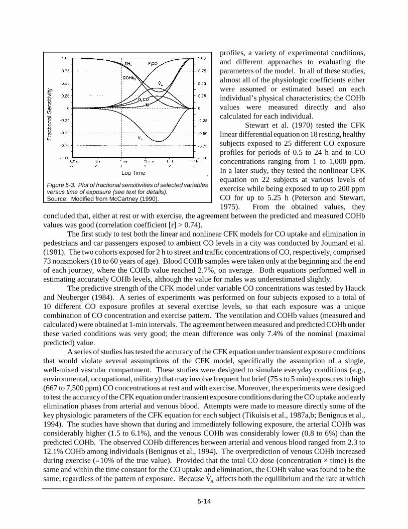
In general, the linear CFK equation is a better approximation to the nonlinear equation during theuptake of CO than during the elimination of CO. As long as the linear CFK equation is used to predictCOHb levels at or below 6% COHb, the solution to the nonlinear CFK model will deviate no more than±0.5% COHb (Smith, 1990). Over the years, it has been empirically determined that minute ventilationand the DLCO have the greatest influence on the CO uptake and elimination. The relative importance ofother physiologic variables will vary with exposure conditions and health status. A comprehensiveevaluation of fractional sensitivities of physiologic variables for both the linear and nonlinear CFKequations shows that each variable will exert its maximal influence at different times of exposure(McCartney, 1990). The analysis found that only the fractional concentration of CO in inhaled air, in partsper million (FICO), and VCO will not affect the rate at which equilibrium is reached. Figure 5-3 illustratesthe temporal changes in fractional sensitivities of the principal determinants of CO uptake for the linearform of the CFK equation; THb is the total blood concentration of Hb. The fractional sensitivity of unitymeans that, for example, a 5% error in the selected variable induces a 5% error in predicted COHb valueby the nonlinear model.
5.5.1.3 Confirmation Studies of Coburn-Forster-Kane ModelsSince the publication of the original paper (Coburn et al., 1965), several investigators have
tested the fit of both the linear and nonlinear CFK model to experimental data using different CO exposure
5-14
Figure 5-3. Plot of fractional sensitivities of selected variablesversus time of exposure (see text for details).Source: Modified from McCartney (1990).
profiles, a variety of experimental conditions,and different approaches to evaluating theparameters of the model. In all of these studies,almost all of the physiologic coefficients eitherwere assumed or estimated based on eachindividual’s physical characteristics; the COHbvalues were measured directly and alsocalculated for each individual.
Stewart et al. (1970) tested the CFKlinear differential equation on 18 resting, healthysubjects exposed to 25 different CO exposureprofiles for periods of 0.5 to 24 h and to COconcentrations ranging from 1 to 1,000 ppm.In a later study, they tested the nonlinear CFKequation on 22 subjects at various levels ofexercise while being exposed to up to 200 ppmCO for up to 5.25 h (Peterson and Stewart,1975). From the obtained values, they
concluded that, either at rest or with exercise, the agreement between the predicted and measured COHbvalues was good (correlation coefficient [r] > 0.74).
The first study to test both the linear and nonlinear CFK models for CO uptake and elimination inpedestrians and car passengers exposed to ambient CO levels in a city was conducted by Joumard et al.(1981). The two cohorts exposed for 2 h to street and traffic concentrations of CO, respectively, comprised73 nonsmokers (18 to 60 years of age). Blood COHb samples were taken only at the beginning and the endof each journey, where the COHb value reached 2.7%, on average. Both equations performed well inestimating accurately COHb levels, although the value for males was underestimated slightly.
The predictive strength of the CFK model under variable CO concentrations was tested by Hauckand Neuberger (1984). A series of experiments was performed on four subjects exposed to a total of10 different CO exposure profiles at several exercise levels, so that each exposure was a uniquecombination of CO concentration and exercise pattern. The ventilation and COHb values (measured andcalculated) were obtained at 1-min intervals. The agreement between measured and predicted COHb underthese varied conditions was very good; the mean difference was only 7.4% of the nominal (maximalpredicted) value.
A series of studies has tested the accuracy of the CFK equation under transient exposure conditionsthat would violate several assumptions of the CFK model, specifically the assumption of a single,well-mixed vascular compartment. These studies were designed to simulate everyday conditions (e.g.,environmental, occupational, military) that may involve frequent but brief (75 s to 5 min) exposures to high(667 to 7,500 ppm) CO concentrations at rest and with exercise. Moreover, the experiments were designedto test the accuracy of the CFK equation under transient exposure conditions during the CO uptake and earlyelimination phases from arterial and venous blood. Attempts were made to measure directly some of thekey physiologic parameters of the CFK equation for each subject (Tikuisis et al., 1987a,b; Benignus et al.,1994). The studies have shown that during and immediately following exposure, the arterial COHb wasconsiderably higher (1.5 to 6.1%), and the venous COHb was considerably lower (0.8 to 6%) than thepredicted COHb. The observed COHb differences between arterial and venous blood ranged from 2.3 to12.1% COHb among individuals (Benignus et al., 1994). The overprediction of venous COHb increasedduring exercise (.10% of the true value). Provided that the total CO dose (concentration × time) is thesame and within the time constant for the CO uptake and elimination, the COHb value was found to be thesame, regardless of the pattern of exposure. Because 0VA affects both the equilibrium and the rate at which

5-15
it is achieved, inconsistencies in the estimates or conversion of gas volumes (ATPS and BTPS to STPD)will affect the predicted values. The interindividual and intraindividual disparities between measured andpredicted COHb values were attributed primarily to delays in mixing of arterial and venous blood anddifferences in cardiac output; but, other factors, such as lung wash-in, also contribute to this phenomenon.Modification of the CFK equation by adjusting for regional differences in blood flow produced a model thatpredicted with much greater accuracy both the arterial (<0.7% COHb difference) and venous (<1% COHbdifference) COHb during transient uptake and elimination of CO from blood (Smith et al., 1994).
Although the CO concentrations used in these studies are several orders of magnitude higher thanthe usual CO concentrations found in ambient air, under certain conditions (see Section 5.4.1), people canbe exposed briefly (<10 min) to such (or even higher) levels of CO in their immediate environment.Because the physiologic mechanisms (but not the kinetics) of COHb formation are independent of COconcentration, high COHb transients, particularly in at-risk individuals, could be of clinical importance.Even briefly, higher arterial COHb may lead to functional impairment of the hypoxia-sensitive heart andbrain (see Sections 5.2.2.3 and 5.2.2.4). In these situations, the predicted instantaneous arterial COHb levelwill be substantially underestimated.
5.5.1.4 Application of Coburn-Forster-Kane ModelsTo obviate measurements of CFK equation parameters, many of which are complex techniques,
attempts were made to simplify the CFK equation, because it may be difficult or even impossible tomeasure directly some of these parameters, particularly during physical activity. In one study, by relatingphysiological parameters to the O2 uptake by the body, which was in turn related to an activity level, asimplified linear form of the CFK model was developed (Bernard and Duker, 1981). The model was usedsubsequently to draw simple nomograms of predictive relationships between pairs of variables, but theaccuracy of the nomograms was not tested experimentally.
The need for more accurate COHb prediction under more complex physiologic or exposureconditions requires either modification or expansion of the CFK model. Benignus (1995) combined aphysiological model of respiratory gas exchange, MACPUF (Ingram et al., 1987), with the CFK model.The new model allows for continuous output and input of 60 cardiopulmonary variables, including FICO.The usefulness of the model is particularly in its ability to continuously update COHb concentration inresponse to dynamically changing physiologic parameters. The model also allows COHb prediction underconditions that otherwise would be very difficult to duplicate in the laboratory.
A fundamental modification of the CFK model was made by Hill et al. (1977) to study the effectsof CO inspired by the mother on the level of fetal COHb. The Hill equation combines the CFK equation(for maternal COHb) with a term denoting COHb transfer from a placenta into the fetus. Comparativeevaluation of predicted and measured fetal COHb concentrations under time-varying and steady-stateconditions in both women and animals showed acceptable agreement only under steady-state conditions(Hill et al., 1977; Longo and Hill, 1977).
As mentioned in Section 5.5.1.3, Smith et al. (1994) expanded the CFK model to allow forprediction of arterial and venous COHb during transient CO uptake and early elimination phases. Themodel incorporated regional differences in blood flow, particularly in the forearm, because the forearm isused most frequently for blood sampling. This more elaborate model performed extremely well inpredicting blood COHb. Although the model was validated on a small number of subjects using the sameexperimental setting, the validation was not performed under more demanding conditions of physicalactivity and varying CO concentrations.
To accurately predict COHb in individuals exposed to dihalomethanes, which are a source ofendogenous CO (see Section 5.3), the CFK model was extended to account for the CO production causedby oxidation of a parent chemical (Andersen et al., 1991). The model developed and validated on ratsemployed a variety of exposure scenarios to dichloromethane. It subsequently was tested on six volunteers

5-16
exposed to dichloromethane, and, after adjustment of a few parameters, the COHb level was predictedremarkably well. After further validation, this model has potential use in predicting accurately COHbcaused by exogenous and endogenous CO originating from different sources (e.g., Hb degradation,metabolism of dihalomethanes, inhaled CO).
Reexpression of the solution of the CFK model from percent COHb to parts per million of COallows the examination of a variety of CO concentration profiles, while keeping a simple preselected targetCOHb as a constant. Application of the transformed model to urban hourly averaged CO concentrationsthat just attained alternative 1-h and 8-h CO National Ambient Air Quality Standards (NAAQS) showedthat, depending on the air quality pattern used, between 0.01 to 10% of the population may exceed a target2.1% COHb level in blood without ambient CO concentrations ever exceeding the standard. By includingtransients, the models predicted COHb more accurately, particularly when built into the 8-h runningaverages (Venkatram and Louch, 1979; Biller and Richmond, 1982, 1992). Actually, the ambient COconcentrations could be averaged over any time period less than or equal to the half-life of COHb (Saltzmanand Fox, 1986).
5.6 Intracellular Effects of Carbon Monoxide5.6.1 Introduction
Traditional concepts for CO pathophysiology have been based on the high affinity of CO fordeoxyhemoglobin and consequent reduction of O2 delivery. This mechanism is relevant for high COconcentrations, but it is less likely to be relevant to the concentrations of CO currently found in the ambientenvironment. This section will summarize recently published information on biochemical mechanisms thatis not related to an impairment of oxygen delivery from elevations in COHb. Some of the studies outlinedin this section were done with cells in culture and others with laboratory rats. To be relevant to humanexposures from environmental contamination, it is important to note what concentrations of CO are likelyto occur in vivo. Lung parenchyma represents a special situation where cells may be exposed to ambientCO without the reduction in concentration associated with Hb-bound CO. Elsewhere in the body, only afraction of COHb will dissociate to elevate extravascular CO concentrations. This elevation is in the rangeof approximately 2 to 10 nmol when the COHb concentration is from 0.8 to 3.8% (Coburn, 1970a; Göthertet al., 1970). The COHb values near steady-state conditions in laboratory rats are close to values forhumans (Kimmell et al., 1999). This strengthens the potential for human relevance in recent animal studiesthat show that newly identified biochemical mechanisms do have adverse physiological effects. However,caution still is warranted because direct evidence for the occurrence of these mechanisms in humans hasnot been shown.
5.6.2 Inhibition of Hemoprotein FunctionCarbon monoxide can inhibit a number of hemoproteins found in cells, such as Mb, cytochrome
c oxidase, cytochrome P-450, dopamine $ hydroxylase, and tryptophan oxygenase (Coburn and Forman,1987). Inhibition of these enzymes could have adverse effects on cell function.
Carbon monoxide acts as a competitive inhibitor, hence biological effects depend on the partialpressures of both CO and O2. The cellular hemoprotein with the highest relative affinity for CO over thatfor O2 is Mb. Carbon monoxide will inhibit Mb-facilitated oxygen diffusion, but physiological compromiseis seen only with high concentrations of COMb. Wittenberg and Wittenberg (1993) found that high-energyphosphate production was inhibited in isolated cardiac myocytes, maintained at a physiologically relevantoxygen concentration, when COMb exceeded 40%. The authors estimated that formation of sufficientCOMb to impair oxidative phosphorylation in vivo would require a COHb level of 20 to 40%.
Coefficients for binding CO versus O2 among cytochrome P-450-like proteins vary between 0.1 andapproximately 12, and there have been recent discussions suggesting that CO-mediated inhibition of these

5-17
proteins could cause smooth muscle relaxation in vivo (Coburn and Forman, 1987; Wang et al., 1997a;Wang, 1998). The issue relates to inhibition of cytochrome P-450-dependent synthesis of several potentvasoconstricting agents (Wang, 1998). Vasodilation has been shown via this mechanism with highconcentrations of CO (ca. 90,000 ppm) (Coceani et al., 1988). It is unclear, however, whether this couldarise under physiological conditions and CO concentrations produced endogenously. The competitionbetween CO and O2 for cytochrome c oxidase was well outlined in the previous review (U.S. EnvironmentalProtection Agency, 1991), but some additional information has been published since then. Based on itsWarburg partition coefficient of between 5 and 15, CO binding is favored only in situations where oxygentension is extremely low (Coburn and Forman, 1987). Carbon monoxide binding to cytochrome c oxidasein vivo will occur when COHb is high (ca. 50%), a level that causes both systemic hypotension as well asimpaired oxygen delivery (Brown and Piantadosi, 1992). Mitochondrial dysfunction, possibly linked tocytochrome inhibition, has been shown to inhibit energy production, and it also may be related to enhancedfree radical production (Piantadosi et al., 1995, 1997a). There has been no new information published sincethe last air quality criteria document that pertains to the effects of CO on dopamine $ hydroxylase ortryptophan oxygenase.
5.6.3 Free Radical ProductionLaboratory animal studies indicate that nitrogen- and oxygen-based free radicals are generated
in vivo during CO exposures. Exposure to CO at concentrations of 20 ppm or more for 1 h will causeplatelets to become a source of the nitric oxide free radical (CNO) in the systemic circulation of rats (Thomet al., 1994; Thom and Ischiropoulos, 1997). Studies with cultured bovine pulmonary endothelial cells havedemonstrated that exposures to CO at concentrations as low as 20 ppm cause cells to release CNO, and theexposure will cause death by a CNO process that is manifested 18 to 24 h after the CO exposure (Thom etal., 1997; Thom and Ischiropoulos, 1997). The mechanism is based on elevations in steady-state CNOconcentration and production of peroxynitrite (Thom et al., 1994, 1997). Peroxynitrite is a relativelylong-lived, strong oxidant that is produced by the near diffusion-limited reaction between CNO andsuperoxide radical (Huie and Padmaja, 1993).
The mechanism by which CO concentrations of 11 nmol or more cause an elevation of steady-stateCNO concentrations appears to be based on altered intracellular “routing” of CNO in endothelial cells andplatelets. It is well established that the association and dissociation rate constants of CNO with hemoproteinsexceed the rate constants for O2 or CO (Gibson et al., 1986). However, Moore and Gibson (1976) foundthat when CO was incubated with nitrosyl (CNO)-Mb or CNO-Hb, CO slowly displaced the CNO. Carbonmonoxide replacement occurred even when there was excess CNO-heme protein, and replacement rates wereenhanced by increasing the CO concentration or by carrying out the reaction in the presence of agents suchas thiols, which will react with the liberated CNO. These conditions, including the presence of thiols, existin cells exposed to environmentally relevant concentrations of CO. Exposures to up to 1,070 nmol CO donot alter the rate of production of CNO by platelets and endothelial cells, yet liberation of CNO was enhancedby CO (Thom and Ischiropoulos, 1997; Thom et al., 1994; Thom et al., 1997).
Carbon monoxide will increase the concentration of CNO available to react with in vivo targets inboth lung and brain, based on electron paramagnetic resonance studies with rats exposed to 50 ppm CO ormore (Ischiropoulos et al., 1996; Thom et al., 1999a). The concentrations of nitric oxide synthase isoformsin lung were not altered because of CO, and the mechanism for elevation in CNO was thought to be the sameas that found in isolated cells (Thom et al., 1994, 1997). Exposure to 50 to 100 ppm CO also will increasehydrogen peroxide (H2O2) production in lungs of rats (Thom et al., 1999a). The phenomenon depended onCNO production, as it was inhibited in rats pretreated with NTnitro-L-arginine methyl ester, a nitric oxidesynthase inhibitor. Production of CNO-derived oxidants also is increased in CO-exposed rats, based onmeasurements of nitrotyrosine, a major product of the reaction of peroxynitrite with proteins (Ischiropouloset al., 1996; Thom et al., 1998, 1999a,b).

5-18
The mechanism for enhanced H2O2 production in lungs of CO-exposed rats is not clear. It ispossible that CNO or peroxynitrite may perturb mitochondrial function. Peroxynitrite inhibits electrontransport at complexes I through III, and CNO targets cytochrome oxidase (Cassina and Radi, 1996;Lizasoain et al., 1996; Poderoso et al., 1996). It is important to note, however, that alterations inmitochondrial function and an increase of cellular H2O2 were not found in studies where cultured bovineendothelial cells were exposed to similar CO concentrations (Thom et al., 1997). An alternative possiblemechanism to mitochondrial dysfunction is that exposure to CO may inhibit antioxidant defenses.Mechanisms linked to elevations in CNO could be responsible for inhibiting one or more enzymes. Nitricoxide-derived oxidants can inhibit manganese superoxide dismutase and glyceraldehyde-3-phosphatedehydrogenase and deplete cellular stores of reduced glutathione (Ischiropoulos et al., 1992; Luperchioet al., 1996).
Exposure to high CO concentrations (2,500 to 10,000 ppm) cause mitochondria in brain cells togenerate hydroxyl-like radicals (Piantadosi et al., 1995, 1997a). An additional source of partially reducedO2 species found in animals exposed to CO is xanthine oxidase. Conversion of xanthine dehydrogenase,the enzyme normally involved with uric acid metabolism, to xanthine oxidase, the radical-producing formof the enzyme, occurred in the brains of rats exposed to approximately 3,000 ppm CO (Thom, 1992).Lower CO concentrations did not trigger this change. Therefore, xanthine oxidase is unlikely to be a freeradical source following exposures to CO at concentrations found in ambient air. Moreover, enzymeconversion was not a primary effect of CO; rather, it occurred only following sequestration and activationof circulating leukocytes (Thom, 1993).
5.6.4 Stimulation of Guanylate CyclaseIn recent years, CO has been demonstrated to play a physiological role in vasomotor control and
neuronal signal transduction (Morita et al., 1995; Ingi et al., 1996). Carbon monoxide is producedendogenously by oxidation of organic molecules, but the predominant source is from the degradation ofheme (Rodgers et al., 1994). The rate-limiting enzyme for heme metabolism is heme oxygenase (HO),which converts heme to biliverdin, free iron, and CO. Three isoforms of HO have been characterized. TheHO-1 is an inducible enzyme found in vascular endothelial cells, smooth muscle cells, bronchoalveolarepithelium, and pulmonary macrophages. The HO-1 is induced by its substrate, heme, as well as CNO,H2O2, several cytokines, and lipopolysaccharide (Arias-Díaz et al., 1995; Durante et al., 1997; Morita et al.,1995; Motterlini et al., 1996). The HO-2 is a constitutive enzyme found in certain neurons within thecentral nervous system, testicular cells, and vascular smooth muscle cells (Marks, 1994). Little is knownabout HO-3, which recently was identified in homogenates from a number of organs (McCoubrey et al.,1997).
A main physiological role for CO is thought to be regulation of soluble guanylate cyclase activity.Both CO and CNO can activate guanylate cyclase, although activation by CO is approximately 30-fold lower(Stone and Marletta, 1994). In neuronal cells possessing both heme oxygenase and nitric oxide synthase,regulation of cyclic guanosine monophosphate (cGMP) synthesis is mediated in a reciprocal fashion byproducing either CO or CNO (Ingi et al., 1996; Maines et al., 1993). A compensatory interrelationshipbetween nitric oxide synthase and heme oxygenase also has been found in endothelial cells and activatedmacrophages, although its functional significance is unknown (Kurata et al., 1996; Seki et al., 1997).In macrophages, cGMP synthesis promotes chemotaxis, and cGMP-mediated synthesis and secretion oftumor necrosis factor " has been linked to both CO and CNO (Arias-Díaz et al., 1995; Belenky et al., 1993).Carbon monoxide causes smooth muscle relaxation by stimulating soluble guanylate cyclase (Utz andUllrich, 1991; Wang et al., 1997b). Smooth muscle relaxation also may occur because of activation ofcalcium dependent potassium channels, although this effect may be linked to guanylate cyclase activity(Trischmann et al., 1991; Wang et al., 1997a). Carbon monoxide-mediated smooth muscle relaxation isinvolved with control of microvascular hepatic portal blood flow (Goda et al., 1998; Pannen and Bauer,

5-19
1998) and suppressing contractions in the gravid uterus (Acevedo and Ahmed, 1998). It also may play arole in gastrointestinal motility (Farrugia et al., 1998).
5.7 Mechanisms of Carbon Monoxide Toxicity5.7.1 Alterations in Blood Flow
Carbon monoxide from environmental pollution may exert similar effects in vivo to those ofendogenously produced CO, because the nanomolar tissue concentrations resulting from inhalation of COare comparable or greater than concentrations produced by cells possessing heme oxygenase. Liverparenchyma has been estimated to generate approximately 0.45 nmol CO/gram liver/min (Goda et al.,1998). Carbon monoxide synthesis by smooth muscle cells is approximately 8 pmol/mg protein/min forhuman aorta and 23 to 37 pmol/mg protein/min for rat aorta (Cook et al., 1995; Grundemar et al., 1995).Carbon monoxide production by unstimulated pulmonary macrophages is 3.6 pmol/mg protein/min, and,after stimulation with lipopolysaccharide, it increases to about 5.1 pmol/mg protein/min (Arias-Díaz et al.,1995). The rate of synthesis of CO varies widely for nerve cells. Cerebellar granule cells generateapproximately 3 fmol/mg protein/min, olfactory nerve cells produce 4.7 pmol/mg protein/min, and ratcerebellar homogenates can generate as much as 56.6 pmol/mg protein/min (Ingi and Ronnett, 1995; Ingiet al., 1996; Maines, 1988; Nathanson et al., 1995).
Vasodilation is a well-established effect caused by exposure to environmental CO. At high COconcentrations, on the order of 500 to 2,000 ppm, the mechanism is related to impairment of O2 delivery(Kanten et al., 1983; MacMillan, 1975). However, a portion of the observed increases in cerebral bloodflow are independent of perturbations in O2 supply (Koehler et al., 1982). In a setting where cellularoxidative metabolism was not impaired by CO, elevations in cerebral blood flow appeared to be mediatedby CNO (Meilin et al., 1996). Whether the mechanism was the same as that outlined in the section above,which causes oxidative stress, remains to be determined.
It is unclear whether disturbances in vascular tone by environmental CO is a generalized, systemicresponse, and the impact of variables such as the duration of exposure have not been adequatelyinvestigated. Although cerebral vasodilation mediated by CNO was reported with exposures to 1,000 ppmCO, that level of exposure did not alter pulmonary vasoconstriction in an isolated-perfused rat lung model(Cantrell and Tucker, 1996). Exposure to 150,000 ppm CO caused no changes in pulmonary artery pressurein isolated blood-perfused lungs, although CO did inhibit hypoxic pulmonary vasoconstriction (Tamayo etal., 1997). Humans exposed to CO for sufficient time to achieve COHb levels of approximately 8% werenot found to have alterations in forearm blood flow, blood pressure, or heart rate (Hausberg and Somers,1997).
Animals exposed to high CO concentrations (e.g., 3,000 to 10,000 ppm) have diminished organblood flow, which contributes to CO-mediated tissue injury (Brown and Piantadosi, 1992; Ginsberg andMeyers, 1974; Okeda et al., 1981; Song et al., 1983; Thom, 1990). The mechanism is based on CO-mediated hypoxic stress and cardiac dysfunction; therefore, these effects do not arise at CO concentrationsrelevant to ambient air quality.
5.7.2 Mitochondrial Dysfunction and Altered Production of High-Energy IntermediatesWhen exposed to 10,000 ppm CO, rats exhibit impaired high-energy phosphate synthesis and
production of hydroxyl free radicals because of mitochondrial dysfunction (Brown and Piantadosi, 1992;Piantadosi et al., 1995). Exposure to 2,500 ppm CO also will cause hydroxyl radicals to be produced,apparently by mitochondria, because of a process that could not be related to hypoxic stress (Piantadosiet al., 1997a). Evidence for mitochondrial dysfunction has not been observed in vivo at lower COconcentrations. However, under conditions of high metabolic demand, exposure to even 1,000 ppm CO

5-20
in the absence of an overt hypoxic stress will result in impaired energy production in brain (Meilin et al.,1996).
Carbon monoxide binding to mitochondrial cytochromes of respiring cells in vitro has beendocumented only when either the CO concentration was extraordinarily high, or O2 tension was extremelylow, such that the CO/O2 ratio exceeded 12:1 (Coburn and Forman, 1987). Following CO exposure andremoval to fresh air, CO diffuses out from cells, and mitochondrial function is restored. This process isenhanced by inspiration of hyperbaric oxygen (Brown and Piantadosi, 1992). Studies in mice indicate thathigh CO concentrations inhibit synthesis of high-energy phosphates during exposure to 5,000 ppm CO for15 min and these changes do not persist following removal to fresh air (Matsuoka et al., 1993).In summary, mitochondrial dysfunction and impaired high-energy phosphate synthesis have been shownby several independent laboratories to occur during exposures to high CO concentrations. Currentinformation suggests that this alteration does not occur at CO concentrations relevant to ambient air quality,and that changes in energy production are not persistent for long periods of time following CO exposure.
5.7.3 Vascular Insults Associated with Exposure to Carbon MonoxideThere are two primary variables that impact on CO toxicity. One is the concentration of CO, the
other is the duration of exposure. Traditionally, these two variables have been viewed as merely alternativeways of elevating COHb concentration in the body. The concentration of CO breathed dictates the durationof exposure required to achieve a particular blood level of COHb or tissue level of CO. This view ispredicated on the notion that CO pathophysiology is determined by its binding to one or anotherhemoprotein and to inhibition of oxygen delivery or oxidative metabolism.
There is a substantial body of literature to suggest that, at least with regard to vascular effects, theduration of exposure has a greater impact on the magnitude of CO pathophysiology than what is predictedbased on the concentration of CO that is inspired. For example, the lungs are the first site for potentialaction of environmental CO. Results from investigations have been conflicting regarding the risk forpulmonary injury from CO. Because of the lack of consensus and also the absence of a recognizedbiochemical mechanism, low concentrations of CO have been viewed as posing little risk to lung physiology(U.S. Environmental Protection Agency, 1991, 1992). When animals have been exposed to high COconcentrations sufficient to raise COHb levels to 56 to 90%, exposures have lasted for only minutes becauseof the hypoxic stress. In these studies, evidence of increased capillary permeability was inconsistent (Feinet al., 1980; Niden and Schulz, 1965; Penney et al., 1988), and no other alterations in lung physiology weredetected (Fisher et al., 1969; Robinson et al., 1985; Shimazu et al., 1990; Sugi et al., 1990). In contrast,when human beings or experimental animals were exposed to CO for many hours at a time, capillaryleakage of macromolecules from the lungs and systemic vasculature has been documented, but the presenceof hypoxic stress was questioned (Kjeldsen et al., 1972; Maurer, 1941; Parving et al., 1972; Siggaard-Andersen et al., 1968).
In light of the physiological role for CO in vasomotor control, protracted exposures may be proneto disturb vascular homeostasis, giving rise to pathophysiological responses. Monkeys exposed to 250 ppmCO for 2 weeks exhibited coronary artery damage consisting of subendothelial edema, fatty streaking, andlipid-loaded cells (Thomsen, 1974). This study and others (Armitage et al., 1976; Davies et al., 1976;Turner et al., 1979; Webster et al., 1968) have suggested a link between atherosclerosis and chronic COexposure. However, other studies have failed to find evidence for an association (Hugod et al., 1978; Pennet al., 1992).
Carbon monoxide may cause vascular insults. Leakage of albumin and leukocyte sequestration havebeen shown following exposures of rats to 50 ppm or more for 1 h, and the process is mediated byCNO-derived oxidants (Ischiropoulos et al., 1996; Thom, 1993; Thom et al., 1998, 1999a,b). Brain oxidativestress associated with this mechanism has been shown with rats exposed to 1,000 to 3,000 ppm CO for 1 h(Ischiropoulos et al., 1996; Thom, 1993). However, it is unclear whether the flux of CNO, resulting from

5-21
exposures to lower CO concentrations contribute to oxidative or nitrosative stress in vivo. Importantdifferences in the patterns of leakage from pulmonary and systemic vascular beds suggest that they may becaused by different mechanisms. For example, systemic vascular leakage was present for several hours afterCO exposure, and the leakage resolved within 18 h, whereas pulmonary vascular leakage was notmeasurable until 18 h after CO exposure, and it resolved by 48 h. Both pulmonary and systemic vascularleakage occurred after hour-long exposures to CO, but not when exposures lasted for only 30 min, andvascular changes were not different whether rats were exposed to just 50 ppm or as much as 1,000 ppm CO.These are recent observations, and further investigations are required before their relevance toenvironmental CO contamination can be assessed adequately. Moreover, it should be emphasized that thevascular leakage observed in lungs and systemic microvasculature following exposures to CO atconcentrations as low as 50 ppm may have no pathophysiological impact if regional lymphatics can sustaina higher flow so that edema does not occur (Thom et al., 1998, 1999a,b).
5.8 Other Effects of Carbon MonoxideAmong the most concerning pathophysiological effects of CO is its propensity for causing brain
damage. There has been considerable effort focused on potential mechanisms for this process. With regardto ambient air standards, however, it is important to note that recent studies were done with high COconcentrations. Carbon monoxide poisoning is not a “pure” pathological process, as injuries may beprecipitated by a combination of cardiovascular effects linked to hypoperfusion or frank ischemia, COHb-mediated hypoxic stress, and intracellular effects, including free radical production and oxidative stress.For example, CO poisoning causes elevations of glutamate and dopamine in experimental models andhuman fatalities (Arranz et al., 1997; Ishimaru et al., 1991, 1992; Nabeshima et al., 1990, 1991; Newby etal., 1978; Piantadosi et al., 1997b). These elevations occur because of the CO-associated cardiovascularcompromise and, possibly, other direct CO-mediated effects. Based on the effects of agents that block theN-methyl-D-aspartate (NMDA) receptor, elevations of glutamate in experimental CO poisoning have beenlinked to a delayed type (but not an acute type) of amnesia, to loss of CA1 neurons in the hippocampus ofmice, and to loss of glutamate-dependent cells in the inner ear of rats (Ishimaru et al., 1991, 1992; Liu andFechter, 1995; Nabeshima et al., 1990, 1991). Antioxidants can protect against CO-mediated cytotoxicityof glutamate-dependent nerve cells (Fechter et al., 1997). Mechanisms of glutamate neurotoxicity includeexcessive calcium influx, free-radical-mediated injury that may include calcium-calmodulin-dependentactivation of cytosolic NO synthase, and lipid peroxidation. Moderate stimulation by excitatory aminoacids may cause mitochondrial dysfunction with impaired synthesis of adenosine triphosphate andproduction of reactive O2 species (Beal, 1992). Cell death can be through necrosis or programmed celldeath, depending on the intensity of the stimulus (Gwag et al., 1995). There also may be a synergistic injurywith other forms of oxidative stress because reactive O2 species can intensify excitotoxicity (Bridges et al.,1991; Pellegrini-Giampietro et al., 1990). Glutamate also can injure cells in the central nervous system thatdo not have NMDA receptors by competing for cysteine uptake, which inhibits synthesis of glutathione(Lipton et al., 1997; Murphy et al., 1989; Oka et al., 1993).
5.9 SummaryThe most prominent pathophysiological effect of CO is hypoxemia caused by avid binding of CO
to Hb. Formation of COHb reduces O2-carrying capacity of blood and impairs release of O2 from O2Hb totissues. Failure of vasodilation to compensate causes tissue hypoxia. In addition to tissue hypoxia, ultimatediffusion of CO to cells may affect adversely their function. The brain and heart tissues are particularlysensitive to CO-induced hypoxia and cytotoxicity. The rate of COHb formation and elimination dependson many physical and physiological factors. The same factors that govern CO uptake determine CO

5-22
elimination as well. The flow of CO between blood and either alveolar air or the tissues, and vice versa,is governed by the CO concentration gradient between these compartments and becomes the rate-determining step in the mass transfer of CO for COHb formation and elimination. Because of a smallblood-to-air CO gradient and tight binding of CO to Hb, the elimination half-time is quite long, varyingfrom 2.0 to 6.5 h. Apart from the CO concentration in ambient air, the principal determinants of CO uptakeare minute ventilation and lung diffusion capacity. Thus, any physiological conditions that affect thesevariables (e.g., exercise, age) also will affect the kinetics of COHb. Both the physical and physiologicalvariables have been integrated into many empirical and mathematical models of COHb formation andelimination under static and dynamic conditions of ambient CO concentration and physiologic function.The nonlinear CFK equation is the most widely used predictive model of COHb formation, and it still isconsidered the best all-around model for COHb prediction. Altitude may have a significant influence onCOHb kinetics. The effects of hypoxic hypoxia (altitude) and CO-induced hypoxia appear to be additive.Adaptation to altitude will moderate COHb formation. In addition to exogenous sources of CO, the gas alsois produced endogenously through catabolism of Hb, metabolic processes of drugs, and degradation ofinhaled solvents and other xenobiotics. This last source may lead to very high (up to 50%) COHbconcentrations. Many disorders, particularly anemias of any etiology, will predispose affected individualsto CO hypoxia. Furthermore, patients with a variety of cardiopulmonary diseases (e.g., COPD, CAD) andchronic inflammatory diseases may be at increased risk because of elevated COHb.
Apart from impaired O2 delivery to the tissues because of COHb formation, recent studies of COpathophysiology suggest cytotoxic effects independent of O2. New investigations have expanded theunderstanding of CO in two areas. First, there is a growing recognition of the role that CO may play innormal neurophysiology and in microvascular vasomotor control. The impact of CO from ambient air onthese processes has not been investigated adequately; hence, there is insufficient information available toinfluence decisions on ambient air quality standards. The second area of investigation of CO is related toits propensity for causing free-radical-mediated changes in tissues. Mechanisms for these changes havebeen linked both to mitochondria and to a CO-mediated disturbance of intracellular “buffering” ofendogenously generated free radicals (e.g., CNO). The role these mechanisms play in pathophysiologycurrently is being investigated. Where dose-response studies are available, the concentrations of CO thatcause adverse effects in animals exceed current NAAQS.
ReferencesAcevedo, C. H.; Ahmed, A. (1998) Hemeoxygenase-1 inhibits human myometrial contractility via carbon monoxide and is
upregulated by progesterone during pregnancy. J. Clin. Invest. 101: 949-955.Andersen, M. E.; Clewell, H. M., III; Gargas, M. L.; MacNaughton, M. G.; Reitz, R. H.; Nolan, R. J.; McKenna, M. J. (1991)
Physiologically based pharmacokinetic modeling with dichloromethane, its metabolite, carbon monoxide, and bloodcarboxyhemoglobin in rats and humans. Toxicol. Appl. Pharmacol. 108: 14-27.
Arias-Díaz, J.; Vara, E.; García, C.; Villa, N.; Balibrea, J. L. (1995) Evidence for a cyclic guanosine monophosphate-dependent,carbon monoxide-mediated, signaling system in the regulation of TNF-" production by human pulmonary macrophages.Arch. Surg. (Chicago) 130: 1287-1293.
Armitage, A. K.; Davies, R. F.; Turner, D. M. (1976) The effects of carbon monoxide on the development of atherosclerosis inthe White Carneau pigeon. Atherosclerosis 23: 333-344.
Arranz, B.; Blennow, K.; Eriksson, A.; Månsson, J.-E.; Marcusson, J. (1997) Serotonergic, noradrenergic, and dopaminergicmeasures in suicide brains. Biol. Psychiatry 41: 1000-1009.
Barie, P. S.; Wu, W.; Hariri, R. J.; Halebian, P. H.; Shires, G. T. (1994) Alterations of pulmonary gas exchange after superimposedcarbon monoxide poisoning in acute lung injury. Surgery (St. Louis) 115: 678-686.
Beal, M. F. (1992) Does impairment of energy metabolism result in excitotoxic neuronal death in neurodegenerative illnesses?Ann. Neurol. 31: 119-130.
Belenky, S. N.; Robbins, R. A.; Rubinstein, I. (1993) Nitric oxide synthase inhibitors attenuate human monocyte chemotaxis invitro. J. Leukocyte Biol. 53: 498-503.

5-23
Bender, P. R.; Weil, J. V.; Reeves, J. T.; Moore, L. G. (1987) Breathing pattern in hypoxic exposures of varying duration. J. Appl.Physiol. 62: 640-645.
Benignus, V. A. (1995) A model to predict carboxyhemoglobin and pulmonary parameters after exposure to O2, CO2, and CO.Aviat. Space Environ. Med. 66: 369-374.
Benignus, V. A.; Annau, Z. (1994) Carboxyhemoglobin formation due to carbon monoxide exposure in rats. Toxicol. Appl.Pharmacol. 128: 151-157.
Benignus, V. A.; Hazucha, M. J.; Smith, M. V.; Bromberg, P. A. (1994) Prediction of carboxyhemoglobin formation due totransient exposure to carbon monoxide. J. Appl. Physiol. 76: 1739-1745.
Berk, P. D.; Rodkey, F. L.; Blaschke, T. F.; Collison, H. A.; Waggoner, J. G. (1974) Comparison of plasma bilirubin turnover andcarbon monoxide production in man. J. Lab. Clin. Med. 83: 29-37.
Berk, P. D.; Blaschke, T. F.; Scharschmidt, B. F.; Waggoner, J. G.; Berlin, N. I. (1976) A new approach to quantitation of thevarious sources of bilirubin in man. J. Lab. Clin. Med. 87: 767-780.
Bernard, T. E.; Duker, J. (1981) Modeling carbon monoxide uptake during work. Am. Ind. Hyg. Assoc. J. 42: 361-364.Biller, W. F.; Richmond, H. M. (1982) Sensitivity analysis on Coburn model predictions of COHb levels associated with
alternative CO standards. Research Triangle Park, NC: U.S. Environmental Protection Agency, Office of Air QualityPlanning and Standards; EPA contract no. 68-02-3600.
Biller, W. F.; Richmond, H. M. (1992) COHb module for a probabilistic CO NEM. In: Johnson, T.; Capel, J.; Paul, R.; Wijnberg,L. Estimation of carbon monoxide exposures and associated carboxyhemoglobin levels in Denver residents using aprobabilistic version of NEM. Research Triangle Park, NC: U.S. Environmental Protection Agency, Office of Air QualityPlanning and Standards; contract no. 68-D0-0062; appendix C.
Boothby, W. M.; Lovelace, W. R., II; Benson, O. O., Jr.; Strehler, A. F. (1954) Volume and partial pressures of respiratory gasesat altitude. In: Boothby, W. M., ed. Respiratory physiology in aviation. Randolph Field, TX: U.S. Air Force School ofAviation Medicine; pp. 39-49.
Bridges, R. J.; Koh, J.-Y.; Hatalski, C. G.; Cotman, C. W. (1991) Increased excitotoxic vulnerability of cortical cultures withreduced levels of glutathione. Eur. J. Pharmacol. 192: 199-200.
Britten, J. S.; Myers, R. A. M. (1985) Effects of hyperbaric treatment on carbon monoxide elimination in humans. UnderseaBiomed. Res. 12: 431-438.
Brown, S. D.; Piantadosi, C. A. (1992) Recovery of energy metabolism in rat brain after carbon monoxide hypoxia. J. Clin. Invest.89: 666-672.
Bünger, J.; Bombosch, F.; Mesecke, U.; Hallier, E. (1997) Monitoring and analysis of occupational exposure to chain sawexhausts. Am. Ind. Hyg. Assoc. J. 58: 747-751.
Burki, N. K. (1984) Effects of acute exposure to high altitude on ventilatory drive and respiratory pattern. J. Appl. Physiol. Respir.Environ. Exercise Physiol. 56: 1027-1031.
Cantrell, J. M.; Tucker, A. (1996) Low-dose carbon monoxide does not reduce vasoconstriction in isolated rat lungs. Exp. LungRes. 22: 21-32.
Cassina, A.; Radi, R. (1996) Differential inhibitory action of nitric oxide and peroxynitrate on mitochondrial electron transport.Arch. Biochem Biophys. 328: 309-316.
Cavallin-Ståhl, E.; Mercke, C.; Lundh, B. (1976) Carbon monoxide production in patients with breast carcinoma. Br. J. Haematol.32: 177-182.
Chung, S. J. (1988) Formulas predicting carboxyhemoglobin resulting from carbon monoxide exposure. Vet. Hum. Toxicol.30: 528-532.
Coburn, R. F. (1967) Endogenous carbon monoxide production and body CO stores. Acta Med. Scand. Suppl. 472: 269-282.Coburn, R. F. (1970a) The carbon monoxide body stores. In: Coburn, R. F., ed. Biological effects of carbon monoxide. Ann. N. Y.
Acad. Sci. 174: 11-22.Coburn, R. F. (1970b) Enhancement by phenobarbital and diphenylhydantoin of carbon monoxide production in normal man.
N. Engl. J. Med. 283: 512-515.Coburn, R. F. (1973) Mean myoglobin oxygen tension in skeletal and cardiac muscle. In: Bicher, H. I.; Bruley, D. F., eds. Oxygen
transport to tissue: instrumentation, methods, and physiology. New York, NY: Plenum Press; pp. 571-577. (Advances inexperimental medicine and biology: v. 37A-B).
Coburn, R. F.; Forman, H. J. (1987) Carbon monoxide toxicity. In: Fishman, A. P.; Farhi, L. E.; Tenney, S. M.; Geiger, S. R., eds.Handbook of physiology: a critical, comprehensive presentation of physiological knowledge and concepts. Section 3: therespiratory system. Volume IV. Gas exchange. Bethesda, MD: American Physiological Society; pp. 439-456.
Coburn, R. F.; Mayers, L. B. (1971) Myoglobin O2 tension determined from measurements of carboxymyoglobin in skeletalmuscle. Am. J. Physiol. 220: 66-74.
Coburn, R. F.; Blakemore, W. S.; Forster, R. E. (1963) Endogenous carbon monoxide production in man. J. Clin. Invest.42: 1172-1178.
Coburn, R. F.; Williams, W. J.; Forster, R. E. (1964) Effect of erythrocyte destruction on carbon monoxide production in man.J. Clin. Invest. 43: 1098-1103.

5-24
Coburn, R. F.; Forster, R. E.; Kane, P. B. (1965) Considerations of the physiological variables that determine the bloodcarboxyhemoglobin concentration in man. J. Clin. Invest. 44: 1899-1910.
Coburn, R. F.; Williams, W. J.; Kahn, S. B. (1966) Endogenous carbon monoxide production in patients with hemolytic anemia.J. Clin. Invest. 45: 460-468.
Coceani, F.; Breen, C. A.; Lees, J. G.; Falck, J. R.; Olley, P. M. (1988) Further evidence implicating a cytochrome P-450-mediatedreaction in the contractile tension of the lamb ductus arteriosus. Circ. Res. 62: 471-477.
Cook, M. N.; Nakatsu, K.; Marks, G. S.; McLaughlin, B. E.; Vreman, H. J.; Stevenson, D. K.; Brien, J. F. (1995) Heme oxygenaseactivity in the adult rat aorta and liver as measured by carbon monoxide formation. Can. J. Physiol. Pharmacol.73: 515-518.
Davies, R. F.; Topping, D. L.; Turner, D. M. (1976) The effect of intermittent carbon monoxide exposure on experimentalatherosclerosis in the rabbit. Atherosclerosis 24: 527-536.
Delivoria-Papadopoulos, M.; Coburn, R. F.; Forster, R. E. (1974) Cyclic variation of rate of carbon monoxide production innormal women. J. Appl. Physiol. 36: 49-51.
Deller, A.; Stenz, R.; Forstner, K.; Konrad, F. (1992) Die elimination von kohlenmonoxydhamoglobin - geschlechtsspezifischeund zirkadiane einflusse [The elimination of carboxyhemoglobin COHB sex-specific and circadian influences in healthyvolunteers]. Infusionstherapie 19: 121-126.
Durante, W.; Kroll, M. H.; Christodoulides, N.; Peyton, K. J.; Schafer, A. I. (1997) Nitric oxide induces heme oxygenase-1 geneexpression and carbon monoxide production in vascular smooth muscle cells. Circ. Res. 80: 557-564.
Engel, L. A.; Wood, L. D. H.; Utz, G.; Macklem, P. T. (1973) Gas mixing during inspiration. J. Appl. Physiol. 35: 18-24.Farrugia, G.; Miller, S. M.; Rich, A.; Liu, X.; Maines, M. D.; Rae, J. L.; Szurszewski, J. H. (1998) Distribution of heme oxygenase
and effects of exogenous carbon monoxide in canine jejunum. Am. J. Physiol. 274: G350-G358.Fechter, L. D.; Liu, Y.; Pearce, T. A. (1997) Cochlear protection from carbon monoxide exposure by free radical blockers in the
guinea pig. Toxicol. Appl. Pharmacol. 142: 47-55.Fein, A.; Grossman, R. F.; Jones, J. G.; Hoeffel, J.; McKay, D. (1980) Carbon monoxide effect on alveolar epithelial permeability.
Chest 78: 726-731.Fisher, A. B.; Hyde, R. W.; Baue, A. E.; Reif, J. S.; Kelly, D. F. (1969) Effect of carbon monoxide on function and structure of
the lung. J. Appl. Physiol. 26: 4-12.Forbes, W. H.; Sargent, F.; Roughton, F. J. W. (1945) The rate of carbon monoxide uptake by normal men. Am. J. Physiol.
143: 594-608.Forster, R. E. (1987) Diffusion of gases across the alveolar membrane. In: Fishman, A. P.; Farhi, L. E.; Tenney, S. M.; Geiger,
S. R., eds. Handbook of physiology: a critical, comprehensive presentation of physiological knowledge and concepts.Section 3: the respiratory system. Volume IV. Gas exchange. Bethesda, MD: American Physiological Society; pp. 71-88.
Foster, L. J.; Corrigan, K.; Goldman, A. L. (1978) Effectiveness of oxygen therapy in hypoxic polycythemic smokers. Chest73: 572-576.
Frey, T. M.; Crapo, R. O.; Jensen, R. L.; Elliott, C. G. (1987) Diurnal variation of the diffusing capacity of the lung: is it real?Am. Rev. Respir. Dis. 136: 1381-1384.
Ge, R.-L.; Matsuzawa, Y.; Takeoka, M.; Kubo, K.; Sekiguchi, M.; Kobayashi, T. (1997) Low pulmonary diffusing capacity insubjects with acute mountain sickness. Chest 111: 58-64.
Gibson, Q. H.; Olson, J. S.; McKinnie, R. E.; Rohlfs, R. J. (1986) A kinetic description of ligand binding to sperm whalemyoglobin. J. Biol. Chem. 261: 10,228-10,236.
Ginsberg, M. D.; Myers, R. E. (1974) Experimental carbon monoxide encephalopathy in the primate. I. Physiologic and metabolicaspects. Arch. Neurol. 30: 202-208.
Goda, N.; Suzuki, K.; Naito, M.; Takeoka, S.; Tsuchida, E.; Ishimura, Y.; Tamatani, T.; Suematsu, M. (1998) Distribution of hemeoxygenase isoforms in rat liver: topographic basis for carbon monoxide-mediated microvascular relaxation. J. Clin. Invest.101: 604-612.
Göthert, M.; Lutz, F.; Malorny, G. (1970) Carbon monoxide partial pressure in tissue of different animals. Environ. Res.3: 303-309.
Grønlund, J.; Garby, L. (1984) Numerical values of the classical Haldane coefficient. J. Appl. Physiol. 57: 850-859.Grote, J.; Dall, P.; Oltmanns, K.; Stolp, W. (1994) The effect of increased blood carbon monoxide levels on the hemoglobin
oxygen affinity during pregnancy. In: Vaupel, P.; Zander R.; Bruley, D. F., eds. Oxygen transport to tissue XV.Proceedings of the twentieth annual meeting of the International Society on Oxygen Transport to Tissue; August 1992;Mainz, Germany. New York, NY: Plenum Press; pp. 145-150. (Advances in experimental medicine and biology: v. 345).
Grundemar, L.; Johansson, M.-B.; Ekelund, M.; Högestätt, E. D. (1995) Haem oxygenase activity in blood vessel homogenatesas measured by carbon monoxide production. Acta Physiol. Scand. 153: 203-204.
Guénard, H.; Marthan, R. (1996) Pulmonary gas exchange in elderly subjects. Eur. Respir. J. 9: 2573-2577.Gwag, B. J.; Lobner, D.; Koh, J. Y.; Wie, M. B.; Choi, D. W. (1995) Blockade of glutamate receptors unmasks neuronal apoptosis
after oxygen-glucose deprivation in vitro. Neuroscience 68: 615-619.

5-25
Haab, P. E.; Durand-Arczynska, W. Y. (1991) Carbon monoxide effects on oxygen transport. In: Crystal, R. G.; West, J. B.;Barnes, P. J.; Cherniack, N. S.; Weibel, E. R., eds. The lung: scientific applications, v. 2. New York, NY: Raven Press;pp. 1267-1275.
Haldane, J. (1897-1898) Some improved methods of gas analysis. J. Physiol. (London) 22: 465-480.Harf, A.; Pratt, T.; Hughes, J. M. B. (1978) Regional distribution of 0VA/ 0Q in man at rest and with exercise measured with
krypton-81m. J. Appl. Physiol.: Respir. Environ. Exercise Physiol. 44: 115-123.Hauck, H.; Neuberger, M. (1984) Carbon monoxide uptake and the resulting carboxyhemoglobin in man. Eur. J. Appl. Physiol.
53: 186-190.Hausberg, M.; Somers, V. K. (1997) Neural circulatory responses to carbon monoxide in healthy humans. Hypertension
29: 1114-1118.Hill, E. P.; Hill, J. R.; Power, G. G.; Longo, L. D. (1977) Carbon monoxide exchanges between the human fetus and mother:
a mathematical model. Am. J. Physiol. 232: H311-H323.Horvath, S. M.; Agnew, J. W.; Wagner, J. A.; Bedi, J. F. (1988) Maximal aerobic capacity at several ambient concentrations of
carbon monoxide at several altitudes. Cambridge, MA: Health Effects Institute; research report no. 21.Horvath, I.; Loukides, S.; Wodehouse, T.; Kharitonov, S. A.; Cole, P. J.; Barnes, P. J. (1998a) Increased levels of exhaled carbon
monoxide in bronchiectasis: a new marker of oxidative stress. Thorax 53: 867-870.Horvath, I.; Donnelly, L. E.; Kiss, A.; Paredi, P.; Kharitonov, S. A.; Barnes, P. J. (1998b) Raised levels of exhaled carbon
monoxide are associated with an increased expression of heme oxygenase-1 in airway macrophages in asthma: a newmarker of oxidative stress. Thorax 53: 668-672.
Hugod, C.; Hawkins, L. H.; Kjeldsen, K.; Thomsen, H. K.; Astrup, P. (1978) Effect of carbon monoxide exposure on aortic andcoronary intimal morphology in the rabbit. Atherosclerosis 30: 333-342.
Huie, R. E.; Padmaja, S. (1993) The reaction of NO with superoxide. Free Radical Res. Commun. 18: 195-199.Ingi, T.; Ronnett, G. V. (1995) Direct demonstration of a physiological role for carbon monoxide in olfactory receptor neurons.
J. Neurosci. 15: 8214-8222.Ingi, T.; Cheng, J.; Ronnett, G. V. (1996) Carbon monoxide: an endogenous modulator of the nitric oxide-cyclic GMP signaling
system. Neuron 16: 835-842.Ingram, D.; Dickinson, C. J.; Ahmed, K. (1987) MACPUF software package. London, United Kingdom: Centre for Health
Informatics and Multiprofessional Education (CHIME), University College London, Medical School and Health SciencesCentre, McMaster University. Available: www.chime.ucl.ac.uk/Models/macpuf.htm.
Ischiropoulos, H.; Zhu, L.; Chen, J.; Tsai, M.; Martin, J. C.; Smith, C. D.; Beckman, J. S. (1992) Peroxynitrate-mediated tyrosinenitration catalyzed by superoxide dismutase. Arch. Biochem. Biophys. 298: 431-437.
Ischiropoulos, H.; Beers, M. F.; Ohnishi, S. T.; Fisher, D.; Garner, S. E.; Thom, S. R. (1996) Nitric oxide production andperivascular tyrosine nitration in brain after carbon monoxide poisoning in the rat. J. Clin. Invest. 97: 2260-2267.
Ishimaru, H.; Nabeshima, T.; Katoh, A.; Suzuki, H.; Fukuta, T.; Kameyama, T. (1991) Effects of successive carbon monoxideexposures on delayed neuronal death in mice under the maintenance of normal body temperature. Biochem. Biophys. Res.Commun. 179: 836-840.
Ishimaru, H.; Katoh, A.; Suzuki, H.; Fukuta, T.; Kameyama, T.; Nabeshima, T. (1992) Effects of N-methyl-D-aspartate receptorantagonists on carbon monoxide-induced brain damage in mice. J. Pharmacol. Exp. Ther. 261: 349-352.
Jay, G. D.; McKindley, D. S. (1997) Alterations in pharmacokinetics of carboxyhemoglobin produced by oxygen under pressure.Undersea Hyperbaric Med. 24: 165-173.
Johnson, T.; Capel, J.; Paul, R.; Wijnberg, L. (1992) Estimation of carbon monoxide exposures and associated carboxyhemoglobinlevels in Denver residents using a probabilistic version of NEM. Research Triangle Park, NC: U.S. EnvironmentalProtection Agency, Office of Air Quality Planning and Standards; contract no. 68-D0-0062.
Joumard, R.; Chiron, M.; Vidon, R.; Maurin, M.; Rouzioux, J.-M. (1981) Mathematical models of the uptake of carbon monoxideon hemoglobin at low carbon monoxide levels. Environ. Health Perspect. 41: 277-289.
Kanten, W. E.; Penney, D. G.; Francisco, K.; Thill, J. E. (1983) Hemodynamic responses to acute carboxyhemoglobinemia in therat. Am. J. Physiol. 244: H320-H327.
Kim, S. K.; Kim, Y. C. (1996) Effect of a single administration of benzene, toluene or m-xylene on carboxyhaemoglobin elevationand metabolism of dichloromethane in rats. J. Appl. Toxicol. 16: 437-444.
Kimmel, E. C.; Carpenter, R. L.; Reboulet, J. E.; Still, K. R. (1999) A physiological model for predicting carboxyhemoglobinformation from exposure to carbon monoxide in rats. J. Appl. Physiol. 86: 1977-1983.
Kinker, J. R.; Haffor, A.-S.; Stephan, M.; Clanton, T. L. (1992) Kinetics of CO uptake and diffusing capacity in transition fromrest to steady-state exercise. J. Appl. Physiol. 72: 1764-1772.
Kjeldsen, K.; Astrup, P.; Wanstrup, J. (1972) Ultrastructural intimal changes in the rabbit aorta after a moderate carbon monoxideexposure. Atherosclerosis 16: 67-82.
Kleinman, M. T.; Leaf, D. A.; Kelly, E.; Caiozzo, V.; Osann, K.; O'Niell, T. (1998) Urban angina in the mountains: effects ofcarbon monoxide and mild hypoxemia on subjects with chronic stable angina. Arch. Environ. Health 53: 388-397.

5-26
Klimisch, H.-J.; Chevalier, H.-J.; Harke, H.-P.; Dontenwill, W. (1975) Uptake of carbon monoxide in blood of miniature pigs andother mammals. Toxicology 3: 301-310.
Koehler, R. C.; Jones, M. D., Jr.; Traystman, R. J. (1982) Cerebral circulatory response to carbon monoxide and hypoxic hypoxiain the lamb. Am. J. Physiol. 243: H27-H32.
Kurata, S.; Matsumoto, M.; Yamashita, U. (1996) Concomitant transcriptional activation of nitric oxide synthase and hemeoxygenase genes during nitric oxide-mediated macrophage cytostasis. J. Biochem. 120: 49-52.
Landaw, S. A. (1973) The effects of cigarette smoking on total body burden and excretion rates of carbon monoxide. J. Occup.Med. 15: 231-235.
Landaw, S. A.; Callahan, E. W., Jr.; Schmid, R. (1970) Catabolism of heme in vivo: comparison of the simultaneous productionof bilirubin and carbon monoxide. J. Clin. Invest. 49: 914-925.
Levasseur, L.; Galliot-Guilley, M.; Richter, F.; Scherrmann, J. M.; Baud, F. J. (1996) Effects of mode of inhalation of carbonmonoxide and of normobaric oxygen administration on carbon monoxide elimination from the blood. Hum. Exp. Toxicol.15: 898-903.
Levitt, M. D.; Ellis, C.; Levitt, D. G. (1994) Diurnal rhythm of heme turnover assessed by breath carbon monoxide concentrationmeasurements. J. Lab. Clin. Med. 124: 427-431.
Lifshay, A.; Fast, C. W.; Glazier, J. B. (1971) Effects of changes in respiratory pattern on physiological dead space. J. Appl.Physiol. 31: 478-483.
Lipton, S. A.; Kim, W.-K.; Choi, Y.-B.; Kumar, S.; D'Emilia, D. M.; Rayudu, P. V.; Arnelle, D. R.; Stamler, J. S. (1997)Neurotoxicity associated with dual actions of homocysteine at the N-methyl-D-aspartate receptor. Proc. Natl. Acad. Sci.U. S. A. 94: 5923-5928.
Liu, Y.; Fechter, L. D. (1995) MK-801 protects against carbon monoxide-induced hearing loss. Toxicol. Appl. Pharmacol.132: 196-202.
Lizasoain, I.; Moro, M. A.; Knowles, R. G.; Darley-Usmar, V.; Moncada, S. (1996) Nitric oxide and peroxynitrate exert distincteffects on mitochondrial respiration which are differentially blocked by glutathione or glucose. Biochem. J. 314: 877-880.
Longo, L. D.; Hill, E. P. (1977) Carbon monoxide uptake and elimination in fetal and maternal sheep. Am. J. Physiol.232: H324-H330.
Ludbrook, G. L.; Helps, S. C.; Gorman, D. F.; Reilly, P. L.; North, J. B.; Grant, C. (1992) The relative effects of hypoxic hypoxiaand carbon monoxide on brain function in rabbits. Toxicology 75: 71-80.
Lundh, B.; Cavallin-Ståhl, E.; Mercke, C. (1975) Nicotinic acid and the endogenous production of carbon monoxide. Acta Med.Scand. 197: 173-176.
Luomanmäki, K.; Coburn, R. F. (1969) Effects of metabolism and distribution of carbon monoxide on blood and body stores.Am. J. Physiol. 217: 354-363.
Luperchio, S.; Tamir, S.; Tannenbaum, S. R. (1996) NO-induced oxidative stress and glutathione metabolism in rodent and humancells. Free Radical Biol. Med. 21: 513-519.
Lynch, S. R.; Moede, A. L. (1972) Variation in the rate of endogenous carbon monoxide production in normal human beings.J. Lab. Clin. Med. 79: 85-95.
MacMillan, V. (1975) Regional cerebral blood flow of the rat in acute carbon monoxide intoxication. Can. J. Physiol. Pharmacol.53: 644-650.
Maines, M. D. (1988) Heme oxygenase: function, multiplicity, regulatory mechanisms, and clinical applications. FASEB J.2: 2557-2568.
Maines, M. D.; Mark, J. A.; Ewing, J. F. (1993) Heme oxygenase, a likely regulator of cGMP production in the brain: inductionin vivo of HO-1 compensates for depression in NO synthase activity. Mol. Cell. Neurosci. 4: 398-405.
Manno, M.; Rugge, M.; Cocheo, V. (1992) Double fatal inhalation of dichloromethane. Hum. Exp. Toxicol. 11: 540-545.Marks, G. S. (1994) Heme oxygenase: the physiological role of one of its metabolites, carbon monoxide and interactions with zinc
protoporphyrin, cobalt protoporphyrin and other metalloporphyrins. Cell. Mol. Biol. (Paris) 40: 863-870.Marthan, R.; Castaing, Y.; Manier, G.; Guenard, H. (1985) Gas exchange alterations in patients with chronic obstructive lung
disease. Chest 87: 470-475.Matsuoka, M.; Igisu, H.; Tanaka, I.; Hori, H.; Koga, M. (1993) Brain energy metabolites in mice after an acute exposure to carbon
monoxide. Res. Commun. Chem. Pathol. Pharmacol. 81: 15-20.Maurer, F. W. (1941) The effects of carbon monoxide anoxemia on the flow and composition of cervical lymph. Am. J. Physiol.
133: 170-179.McCartney, M. L. (1990) Sensitivity analysis applied to Coburn-Forster-Kane models of carboxyhemoglobin formation. Am. Ind.
Hyg. Assoc. J. 51: 169-177.McClean, P. A.; Duguid, N. J.; Griffin, P. M.; Newth, C. J. L.; Zamel, N. (1981) Changes in exhaled pulmonary diffusing capacity
at rest and exercise in individuals with impaired positional diffusion. Clin. Respir. Physiol. 17: 179-186.McCoubrey, W. K., Jr.; Huang, T. J.; Maines, M. D. (1997) Isolation and characterization of a cDNA from the rat brain that
encodes hemoprotein heme oxygenase-3. Eur. J. Biochem. 247: 725-732.McGrath, J. J. (1988) Carbon monoxide studies at high altitude. Neurosci. Biobehav. Rev. 12: 311-314.

5-27
McGrath, J. J. (1992) Effects of altitude on endogenous carboxyhemoglobin levels. J. Toxicol. Environ. Health 35: 127-133.McGrath, J. J.; Schreck, R. M.; Lee, P. S. (1993) Carboxyhemoglobin levels in humans: effects of altitude. Inhalation Toxicol.
5: 241-249.Meilin, S.; Rogatsky, G. G.; Thom, S. R.; Zarchin, N.; Guggenheimer-Furman, E.; Mayevsky, A. (1996) Effects of carbon
monoxide on the brain may be mediated by nitric acid. J. Appl. Physiol. 81: 1078-1083.Mercke, C.; Lundh, B. (1976) Erythrocyte filterability and heme catabolism during the menstrual cycle. Ann. Intern. Med.
85: 322-324.Mercke, C.; Cavallin-Ståhl, E.; Lundh, B. (1975a) Diurnal variation in endogenous production of carbon monoxide. Acta Med.
Scand. 198: 161-164.Mercke, C.; Cavallin-Ståhl, E.; Lundh, B. (1975b) Carbon monoxide production and reticulocyte count in normal women: effect
of contraceptive drugs and smoking. Acta Med. Scand. 198: 155-160.Mercke, C.; Cavallin-Ståhl, E.; Lundh, B. (1975c) Heme catabolism during short-term treatment with phenobarbital, diazepam
and oxazepam. Acta Med. Scand. 198: 149-154.Messmer, K. (1982) Oxygen transport capacity. In: Brendel, W.; Zink, R. A., eds. High altitude physiology and medicine.
New York, NY: Springer-Verlag; pp. 117-122.Meyer, J.; Prien, T.; Van Aken, H.; Bone, H.-G.; Waurick, R.; Theilmeier, G.; Booke, M. (1998) Arterio-venous
carboxyhemoglobin difference suggests carbonmonoxide production by human lungs. Biochem. Biophys. Res. Commun.244: 230-232.
Moore, E. G.; Gibson, Q. H. (1976) Cooperativity in the dissociation of nitric oxide from hemoglobin. J. Biol. Chem.251: 2788-2794.
Morita, T.; Perrella, M. A.; Lee, M.-E.; Kourembanas, S. (1995) Smooth muscle cell-derived carbon monoxide is a regulator ofvascular cGMP. Proc. Natl. Acad. Sci. U. S. A. 92: 1475-1479.
Motterlini, R.; Foresti, R.; Intaglietta, M.; Winslow, R. M. (1996) NO-mediated activation of heme oxygenase: endogenouscytoprotection against oxidative stress to endothelium. Am. J. Physiol. 270: H107-H114.
Muller, K. E.; Barton, C. N. (1987) A nonlinear version of the Coburn, Forster and Kane model of blood carboxyhemoglobin.Atmos. Environ. 21: 1963-1967.
Murphy, T. H.; Miyamoto, M.; Sastre, A.; Schnaar, R. L.; Coyle, J. T. (1989) Glutamate toxicity in a neuronal cell line involvesinhibition of cystine transport leading to oxidative stress. Neuron 2: 1547-1558.
Nabeshima, T.; Yoshida, S.; Morinaka, H.; Kameyama, T.; Thurkauf, A.; Rice, K. C.; Jacobson, A. E.; Monn, J. A.; Cho, A. K.(1990) MK-801 ameliorates delayed amnesia, but potentiates acute amnesia induced by CO. Neurosci. Lett. 108: 321-327.
Nabeshima, T.; Katoh, A.; Ishimaru, H.; Yoneda, Y.; Ogita, K.; Murase, K.; Ohtsuka, H.; Inari, K.; Fukuta, T.; Kameyama, T.(1991) Carbon monoxide-induced delayed amnesia, delayed neuronal death and change in acetylcholine concentration inmice. J. Pharmacol. Exp. Ther. 256: 378-384.
Nathanson, J. A.; Scavone, C.; Scanlon, C.; McKee, M. (1995) The cellular Na+ pump as a site of action for carbon monoxide andglutamate: a mechanism for long-term modulation of cellular activity. Neuron 14: 781-794.
Neas, L. M.; Schwartz, J. (1996) The determinants of pulmonary diffusing capacity in a national sample of U.S. adults. Am. J.Respir. Crit. Care Med. 153: 656-664.
Newby, M. B.; Roberts, R. J.; Bhatnagar, R. K. (1978) Carbon monoxide- and hypoxia-induced effects on catecholamines in themature and developing rat brain. J. Pharmacol. Exp. Ther. 206: 61-68.
Niden, A. H.; Schulz, H. (1965) The ultrastructural effects of carbon monoxide inhalation on the rat lung. Virchows Arch. Pathol.Anat. Physiol. 339: 283-292.
Oka, A.; Belliveau, M. J.; Rosenberg, P. A.; Volpe, J. J. (1993) Vulnerability of oligodendroglia to glutamate: pharmacology,mechanisms, and prevention. J. Neurosci. 13: 1441-1453.
Okeda, R.; Funata, N.; Takano, T.; Miyazaki, Y.; Higashino, F.; Yokoyama, K.; Manabe, M. (1981) The pathogenesis of carbonmonoxide encephalopathy in the acute phase—physiological and morphological correlation. Acta Neuropathol. 54: 1-10.
Ott, W. R.; Mage, D. T. (1978) Interpreting urban carbon monoxide concentrations by means of a computerized blood COHbmodel. J. Air Pollut. Control Assoc. 28: 911-916.
Pace, N.; Strajman, E.; Walker, E. L. (1950) Acceleration of carbon monoxide elimination in man by high pressure oxygen.Science (Washington, DC) 111: 652-654.
Pankow, D. (1996) Carbon monoxide formation due to metabolism of xenobiotics. In: Penney, D. G., ed. Carbon monoxide. BocaRaton, FL: CRC Press; pp. 25-43.
Pannen, B. H.; Bauer, M. (1998) Differential regulation of hepatic arterial and portal venous vascular resistance by nitric oxideand carbon monoxide in rats. Life Sci. 62: 2025-2033.
Parving, H.-H.; Ohlsson, K.; Buchardt Hansen, H. J.; Rörth, M. (1972) Effect of carbon monoxide exposure on capillarypermeability to albumin and "2-macroglobulin. Scand. J. Clin. Lab. Invest. 29: 381-388.
Pellegrini-Giampietro, D. E.; Cherici, G.; Alesiani, M.; Carla, V.; Moroni, F. (1990) Excitatory amino acid release and free radicalformation may cooperate in the genesis of ischemia-induced neuronal damage. J. Neurosci. 10: 1035-1941.

5-28
Penn, A.; Currie, J.; Snyder, C. (1992) Inhalation of carbon monoxide does not accelerate arteriosclerosis in cockerels. Eur. J.Pharmacol. 228: 155-164.
Penney, D. G.; Davidson, S. B.; Gargulinski, R. B.; Caldwell-Ayre, T. M. (1988) Heart and lung hypertrophy, changes in bloodvolume, hematocrit and plasma renin activity in rats chronically exposed to increasing carbon monoxide concentrations.J. Appl. Toxicol. 8: 171-178.
Peters, T.; Jürgens, K. D.; Gunther-Jürgens, G.; Gros, G. (1994) Determination of myoglobin-diffusivity in intact skeletal musclefibers: an improved microscope-photometrical approach. In: Vaupel, P.; Zander R.; Bruley, D. F., eds. Oxygen transportto tissue XV. Proceedings of the twentieth annual meeting of the International Society on Oxygen Transport to Tissue;August 1992; Mainz, Germany. New York, NY: Plenum Press; pp. 677-683. (Advances in experimental medicine andbiology: v. 345).
Peterson, J. E.; Stewart, R. D. (1970) Absorption and elimination of carbon monoxide by inactive young men. Arch. Environ.Health 21: 165-171.
Peterson, J. E.; Stewart, R. D. (1975) Predicting the carboxyhemoglobin levels resulting from carbon monoxide exposures. J. Appl.Physiol. 39: 633-638.
Piantadosi, C. A.; Tatro, L.; Zhang, J. (1995) Hydroxyl radical production in the brain after CO hypoxia in rats. Free Radical Biol.Med. 18: 603-609.
Piantadosi, C. A.; Zhang, J.; Demchenko, I. T. (1997a) Production of hydroxyl radical in the hippocampus after CO hypoxia orhypoxic hypoxia in the rat. Free Radical Biol. Med. 22: 725-732.
Piantadosi, C. A.; Zhang, J.; Levin, E. D.; Folz, R. J.; Schmechel, D. E. (1997b) Apoptosis and delayed neuronal damage aftercarbon monoxide poisoning in the rat. Exp. Neurol. 147: 103-114.
Poderoso, J. J.; Cerreras, M. C.; Lisdero, C.; Riobó, N.; Schöpfer, F.; Boveris, A. (1996) Nitric oxide inhibits electron transferand increases superoxide radical production in rat heart mitochondria and submitochondrial particles. Arch. Biochem.Biophys. 328: 85-92.
Robinson, N. B.; Barie, P. S.; Halebian, P. H.; Shires, G. T. (1985) Distribution of ventilation and perfusion following acutecarbon monoxide poisoning. In: 41st annual forum on fundamental surgical problems held at the 71st annual clinicalcongress of the American College of Surgeons; October; Chicago, IL. Surg. Forum 36: 115-118.
Rodgers, P. A.; Vreman, H. J.; Dennery, P. A.; Stevenson, D. K. (1994) Sources of carbon monoxide (CO) in biological systemsand applications of CO detection technologies. Semin. Perinatol. 18: 2-10.
Rodkey, F. L.; O'Neal, J. D.; Collison, H. A. (1969) Oxygen and carbon monoxide equilibria of human adult hemoglobin atatmospheric and elevated pressure. Blood 33: 57-65.
Roughton, F. J. W. (1970) The equilibrium of carbon monoxide with human hemoglobin in whole blood. In: Coburn, R. F., ed.Biological effects of carbon monoxide. Ann. N. Y. Acad. Sci. 174: 177-188.
Saltzman, B. E.; Fox, S. H. (1986) Biological significance of fluctuating concentrations of carbon monoxide. Environ. Sci.Technol. 20: 916-923.
Schoenfisch, W. H.; Hoop, K. A.; Struelens, B. S. (1980) Carbon monoxide absorption through the oral and nasal mucosae ofcynomolgus monkeys. Arch. Environ. Health 35: 152-154.
Seki, T.; Naruse, M.; Naruse, K.; Yoshimoto, T.; Tanabe, A.; Imaki, T.; Hagiwara, H.; Hirose, S.; Demura, H. (1997) Interrelationbetween nitric oxide synthase and heme oxygenase in rat endothelial cells. Eur. J. Pharmacol. 331: 87-91.
Selvakumar, S.; Sharan, M.; Singh, M. P. (1992) Mathematical model for the exchange of gases in the lungs with special referenceto carbon monoxide. Med. Biol. Eng. Comput. 30: 525-532.
Selvakumar, S.; Sharan, M.; Singh, M. P. (1993) A mathematical model for the elimination of carbon monoxide in humans.J. Theor. Biol. 162: 321-336.
Sharan, M.; Selvakumar, S.; Singh, M. P. (1990) Mathematical model for the computation of alveolar partial pressure of carbonmonoxide. Int. J. Biomed. Comput. 26: 135-147.
Shimazu, T.; Ikeuchi, H.; Hubbard, G. B.; Langlinais, P. C.; Mason, A. D., Jr.; Pruitt, B. A., Jr. (1990) Smoke inhalation injuryand the effect of carbon monoxide in the sheep model. J. Trauma 30: 170-175.
Siggaard-Andersen, J.; Bonde Petersen, F.; Hansen, T. I.; Mellemgaard, K. (1968) Plasma volume and vascular permeabilityduring hypoxia and carbon monoxide exposure. Scand. J. Clin. Lab. Invest. Suppl. 103: 39-48.
Singh, J.; Moore-Cheatum, L. (1993) Gestational protein deficiency enhances fetotoxicity of carbon monoxide. In: Keen, C. L.;Bendich, A.; Willhite, C. C., eds. Maternal nutrition and pregnancy outcome. Ann. N. Y. Acad. Sci. 678: 366-368.
Singh, M. P.; Sharan, M.; Selvakumar, S. (1991) A mathematical model for the computation of carboxyhaemoglobin in humanblood as a function of exposure time. Philos. Trans. R. Soc. Lond. B 334: 135-147.
Singh, J.; Smith, C. B.; Moore-Cheatum, L. (1992) Additivity of protein deficiency and carbon monoxide on placentalcarboxyhemoglobin in mice. Am. J. Obstet. Gynecol. 167: 843-846.
Singh, J.; Aggison, L., Jr.; Moore-Cheatum, L. (1993) Teratogenicity and developmental toxicity of carbon monoxide inprotein-deficient mice. Teratology 48: 149-159.
Singleton, G. J.; Olsen, C. R.; Smith, R. L. (1972) Correction for mechanical dead space in the calculation of physiological deadspace. J. Clin. Invest. 51: 2768-2772.

5-29
Smith, M. V. (1990) Comparing solutions to the linear and nonlinear CFK equations for predicting COHb formation. Math. Biosci.99: 251-263.
Smith, M. V.; Hazucha, M. J.; Benignus, V. A.; Bromberg, P. A. (1994) Effect of regional circulation patterns on observed HbCOlevels. J. Appl. Physiol. 77: 1659-1665.
Sokal, J. A.; Majka, J.; Palus, J. (1984) The content of carbon monoxide in the tissues of rats intoxicated with carbon monoxidein various conditions of acute exposure. Arch. Toxicol. 56: 106-108.
Sokal, J.; Majka, J.; Palus, J. (1986) Effect of work load on the content of carboxymyoglobin in the heart and skeletal musclesof rats exposed to carbon monoxide. J. Hyg. Epidemiol. Microbiol. Immunol. 30: 57-62.
Solanki, D. L.; McCurdy, P. R.; Cuttitta, F. F.; Schechter, G. P. (1988) Hemolysis in sickle cell disease as measured byendogenous carbon monoxide production: a preliminary report. Am. J. Clin. Pathol. 89: 221-225.
Song, S.-Y.; Okeda, R.; Funata, N.; Higashino, F. (1983) An experimental study of the pathogenesis of the selective lesion of theglobus pallidus in acute carbon monoxide poisoning in cats: with special reference to the chronologic change in thecerebral local blood flow. Acta Neuropathol. 61: 232-238.
Stewart, R. D.; Peterson, J. E.; Baretta, E. D.; Bachand, R. T.; Hosko, M. J.; Herrmann, A. A. (1970) Experimental humanexposure to carbon monoxide. Arch. Environ. Health 21: 154-164.
Stewart, R. D.; Peterson, J. E.; Fisher, T. N.; Hosko, M. J.; Baretta, E. D.; Dodd, H. C.; Herrmann, A. A. (1973) Experimentalhuman exposure to high concentrations of carbon monoxide. Arch. Environ. Health 26: 1-7.
Stokes, D. L.; MacIntyre, N. R.; Nadel, J. A. (1981) Nonlinear increases in diffusing capacity during exercise by seated and supinesubjects. J. Appl. Physiol.: Respir. Environ. Exercise Physiol. 51: 858-863.
Stone, J. R.; Marletta, M. A. (1994) Soluble guanylate cyclase from bovine lung: activation with nitric oxide and carbon monoxideand spectral characterization of the ferrous and ferric states. Biochemistry 33: 5636-5640.
Stork, R. L.; Bredle, D. L.; Chapler, C. K.; Cain, S. M. (1988) Regional hemodynamic responses to hypoxia in polycythemic dogs.J. Appl. Physiol. 65: 2069-2074.
Sugi, K.; Theissen, J. L.; Traber, L. D.; Herndon, D. N.; Traber, D. L. (1990) Impact of carbon monoxide on cardiopulmonarydysfunction after smoke inhalation injury. Circ. Res. 66: 69-75.
Tamayo, L.; López-López, J. R.; Castañeda, J.; González, C. (1997) Carbon monoxide inhibits hypoxic pulmonaryvasoconstriction in rats by a cGMP-independent mechanism. Pfluegers Arch. 434: 698-704.
Thom, S. R. (1990) Carbon monoxide-mediated brain lipid peroxidation in the rat. J. Appl. Physiol. 68: 997-1003.Thom, S. R. (1992) Dehydrogenase conversion to oxidase and lipid peroxidation in brain after carbon monoxide poisoning.
J. Appl. Physiol. 73: 1584-1589.Thom, S. R. (1993) Leukocytes in carbon monoxide-mediated brain oxidative injury. Toxicol. Appl. Pharmacol. 123: 234-247.Thom, S. R.; Ischiropoulos, H. (1997) Mechanism of oxidative stress from low levels of carbon monoxide. Cambridge, MA:
Health Effects Institute; research report no. 80.Thom, S. R.; Ohnishi, S. T.; Ischiropoulos, H. (1994) Nitric oxide released by platelets inhibits neutrophil B2 integrin function
following acute carbon monoxide poisoning. Toxicol. Appl. Pharmacol. 128: 105-110.Thom, S. R.; Xu, Y. A.; Ischiropoulos, H. (1997) Vascular endothelial cells generate peroxynitrite in response to carbon monoxide
exposure. Chem. Res. Toxicol. 10: 1023-1031.Thom, S. R.; Garner, S.; Fisher, D.; Ischiropoulos, H. (1998) Vascular nitrosative stress from CO exposure. In: Program and
abstracts [of the] Undersea and Hyperbaric Medical Society annual scientific meeting: pre- and post-courses; May; Seattle,WA. Undersea Hyperbaric Med. 25(suppl.): 47.
Thom, S. R.; Ohnishi, S. T.; Fisher, D.; Xu, Y. A.; Ischiropoulos, H. (1999a) Pulmonary vascular stress from carbon monoxide.Toxicol. Appl. Pharmacol. 154: 12-19.
Thom, S. R.; Fisher, D.; Xu, Y. A.; Garner, S.; Ischiropoulos, H. (1999b) Role of nitric oxide-derived oxidants in vascular injuryfrom carbon monoxide in the rat. Am. J. Physiol. 276: H984-H992.
Thomsen, H. K. (1974) Carbon monoxide-induced atherosclerosis in primates: an electron-microscopic study on the coronaryarteries of Macaca irus monkeys. Atherosclerosis 20: 233-240.
Tikuisis, P. (1996) Modeling the uptake and elimination of carbon monoxide. In: Penney, D. G., ed. Carbon monoxide.Boca Raton, FL: CRC Press; pp. 45-67.
Tikuisis, P.; Buick, F.; Kane, D. M. (1987a) Percent carboxyhemoglobin in resting humans exposed repeatedly to 1,500 and7,500 ppm CO. J. Appl. Physiol. 63: 820-827.
Tikuisis, P.; Madill, H. D.; Gill, B. J.; Lewis, W. F.; Cox, K. M.; Kane, D. M. (1987b) A critical analysis of the use of the CFKequation in predicting COHb formation. Am. Ind. Hyg. Assoc. J. 48: 208-213.
Tikuisis, P.; Kane, D. M.; McLellan, T. M.; Buick, F.; Fairburn, S. M. (1992) Rate of formation of carboxyhemoglobin inexercising humans exposed to carbon monoxide. J. Appl. Physiol. 72: 1311-1319.
Trischmann, U.; Klöckner, U.; Isenberg, G.; Utz, J.; Ullrich, V. (1991) Carbon monoxide inhibits depolarization-induced Ca riseand increases cyclic GMP in visceral smooth muscle cells. Biochem. Pharmacol. 41: 237-241.
Turner, D. M.; Lee, P. N.; Roe, F. J. C.; Gough, K. J. (1979) Atherogenesis in the White Carneau pigeon: further studies of therole of carbon monoxide and dietary cholesterol. Atherosclerosis 34: 407-417.

5-30
Tyuma, I.; Ueda, Y.; Imaizumi, K.; Kosaka, H. (1981) Prediction of the carbonmonoxyhemoglobin levels during and after carbonmonoxide exposures in various animal species. Jpn. J. Physiol. 31: 131-143.
U.S. Environmental Protection Agency. (1978) Altitude as a factor in air pollution. Research Triangle Park, NC: Office ofResearch and Development, Environmental Criteria and Assessment Office; report no. EPA 600/9-78-015.
U.S. Environmental Protection Agency. (1991) Air quality criteria for carbon monoxide. Research Triangle Park, NC: Office ofHealth and Environmental Assessment, Environmental Criteria and Assessment Office; report no. EPA/600/8-90/045F.
U.S. Environmental Protection Agency. (1992) Review of the national ambient air quality standards for carbon monoxide: 1992reassessment of scientific and technical information. OAQPS staff paper. Research Triangle Park, NC: Office of AirQuality Planning and Standards; report no. EPA-452/R-92-004.
Utz, J.; Ullrich, V. (1991) Carbon monoxide relaxes ileal smooth muscle through activation of guanylate cyclase. Biochem.Pharmacol. 41: 1195-1201.
Venkatram, A.; Louch, R. (1979) Evaluation of CO air quality criteria using a COHb model. Atmos. Environ. 13: 869-872.Wagner, J. A.; Horvath, S. M.; Dahms, T. E. (1975) Carbon monoxide elimination. Respir. Physiol. 23: 41-47.Wang, R. (1998) Resurgence of carbon monoxide: an endogenous gaseous vasorelaxing factor. Can. J. Physiol. Pharmacol.
76: 1-15.Wang, R.; Wu, L.; Wang, Z. (1997a) The direct effect of carbon monoxide on KCa channels in vascular smooth muscle cells.
Pfluegers Arch. 434: 285-291.Wang, R.; Wang, Z.; Wu, L. (1997b) Carbon monoxide-induced vasorelaxation and the underlying mechanisms. Br. J. Pharmacol.
121: 927-934.Webster, W. S.; Clarkson, T. B.; Lofland, H. B. (1968) Carbon monoxide-aggravated atherosclerosis in the squirrel monkey. Exp.
Mol. Pathol. 13: 36-50.Werner, B.; Lindahl, J. (1980) Endogenous carbon monoxide production after bicycle exercise in healthy subjects and in patients
with hereditary spherocytosis. Scand. J. Clin. Lab. Invest. 40: 319-324.West, J. B. (1990a) Ventilation/blood flow and gas exchange. 5th ed. Oxford, United Kingdom: Blackwell Scientific Publications.West, J. B. (1990b) Respiratory physiology—the essentials. 4th ed. Baltimore, MD: Williams & Wilkins.Wittenberg, B. A.; Wittenberg, J. B. (1993) Effects of carbon monoxide on isolated heart muscle cells. Cambridge, MA: Health
Effects Institute; research report no. 62.Woehlck, H. J.; Dunning, M. B., III; Kulier, A. H.; Sasse, F. J.; Nithipataikom, K.; Henry, D. W. (1997a) The response of
anesthetic agent monitors to trifluoromethane warns of the presence of carbon monoxide from anesthetic breakdown.J. Clin. Monit. 13: 149-155.
Woehlck, H. J.; Dunning, M., III; Connolly, L. A. (1997b) Reduction in the incidence of carbon monoxide exposures in humansundergoing general anesthesia. Anesthesiology 87: 228-234.
Wyman, J.; Bishop, G.; Richey, B.; Spokane, R.; Gill, S. (1982) Examination of Haldane's first law for the partition of CO andO2 to hemoglobin A0. Biopolymers 21: 1735-1747.
Yamaya, M.; Sekizawa, K.; Ishizuka, S.; Monma, M.; Mizuta, K.; Sasaki, H. (1998) Increased carbon monoxide in exhaled airof subjects with upper respiratory tract infections. Am. J. Respir. Crit. Care Med. 158: 311-314.
Yoneda, I.; Watanabe, Y. (1997) Comparisons of altitude tolerance and hypoxia symptoms between nonsmokers and habitualsmokers. Aviat. Space Environ. Med. 68: 807-811.
Zayasu, K.; Sekizawa, K.; Okinaga, S.; Yamaya, M.; Ohrui, T.; Sasaki, H. (1997) Increased carbon monoxide in exhaled air ofasthmatic patients. Am. J. Respir. Crit. Care Med. 156: 1140-1143.
Zhu, N.; Weiss, H. R. (1994) Effect of hypoxic and carbon monoxide-induced hypoxia on regional myocardial segment work andO2 consumption. Res. Exp. Med. 194: 97-107.