pharmacokinetics and pharmacodynamics of oral
TRANSCRIPT
Aalborg Universitet
Pharmacokinetics and Pharmacodynamics of Oral Anticoagulants used in AtrialFibrillation
Fawzy, Ameenathul M; Lip, Gregory Yh
Published in:Expert Opinion on Drug Metabolism & Toxicology
DOI (link to publication from Publisher):10.1080/17425255.2019.1604686
Publication date:2019
Document VersionAccepted author manuscript, peer reviewed version
Link to publication from Aalborg University
Citation for published version (APA):Fawzy, A. M., & Lip, G. Y. (2019). Pharmacokinetics and Pharmacodynamics of Oral Anticoagulants used inAtrial Fibrillation. Expert Opinion on Drug Metabolism & Toxicology, 15(5), 381-398.https://doi.org/10.1080/17425255.2019.1604686
General rightsCopyright and moral rights for the publications made accessible in the public portal are retained by the authors and/or other copyright ownersand it is a condition of accessing publications that users recognise and abide by the legal requirements associated with these rights.
- Users may download and print one copy of any publication from the public portal for the purpose of private study or research. - You may not further distribute the material or use it for any profit-making activity or commercial gain - You may freely distribute the URL identifying the publication in the public portal -
Take down policyIf you believe that this document breaches copyright please contact us at [email protected] providing details, and we will remove access tothe work immediately and investigate your claim.

Full Terms & Conditions of access and use can be found athttps://www.tandfonline.com/action/journalInformation?journalCode=iemt20
Expert Opinion on Drug Metabolism & Toxicology
ISSN: 1742-5255 (Print) 1744-7607 (Online) Journal homepage: https://www.tandfonline.com/loi/iemt20
Pharmacokinetics and Pharmacodynamics of OralAnticoagulants used in Atrial Fibrillation
Ameenathul M Fawzy & Gregory YH Lip
To cite this article: Ameenathul M Fawzy & Gregory YH Lip (2019): Pharmacokinetics andPharmacodynamics of Oral Anticoagulants used in Atrial Fibrillation, Expert Opinion on DrugMetabolism & Toxicology, DOI: 10.1080/17425255.2019.1604686
To link to this article: https://doi.org/10.1080/17425255.2019.1604686
Accepted author version posted online: 05Apr 2019.
Submit your article to this journal
Article views: 9
View Crossmark data
Accep
ted M
anus
cript
Publisher: Taylor & Francis
Journal: Expert Opinion on Drug Metabolism & Toxicology
DOI: 10.1080/17425255.2019.1604686
Review
Pharmacokinetics and Pharmacodynamics of Oral Anticoagulants used in Atrial Fibrillation
Ameenathul M Fawzy1, Gregory YH Lip2,3,*
1City Hospital, Sandwell and West Birmingham Hospitals NHS Trust, Birmingham, Birmingham,
United Kingdom
2Liverpool Centre for Cardiovascular Science, University of Liverpool and Liverpool Heart & Chest Hospital, Liverpool, United Kingdom
3Aalborg Thrombosis Research Unit, Department of Clinical Medicine, Aalborg University, Aalborg, Denmark *Corresponding author: Gregory YH Lip, Email: [email protected]
Accep
ted M
anus
cript
Abstract Introduction: The availability of non-vitamin K antagonist oral anti-coagulants alongside vitamin K antagonists has offered a variety of options for anti-coagulation, but has also necessitated a good understanding of the pharmacological properties of each of these drugs prior to their use, to maximise the therapeutic benefit and minimise patient harm Areas covered: This review article outlines the pharmacokinetic and pharmacodynamic profiles of the currently licensed NOACs and VKAs that are most commonly used in clinical practice, with the aim of demonstrating how variations in these processes contribute to their use in clinical practice. A literature search was conducted on PubMed using keywords and relevant articles published by the 31st of December 2018 were included. Expert opinion: The effect of a drug is determined by a combination of elements which include patient characteristics and external influences, in addition to its pharmacokinetic and pharmacodynamic properties. A good understanding of this is essential. Despite the wealth of information available, particularly on VKAs, our knowledge on the pharmacology responsible for certain drug effects and inter-individual variations is still limited. Increasing efforts are being made to uncover these and includes focus on pharmacogenomics and drug transporter proteins. Keywords: warfarin, vitamin K antagonists, non-vitamin K antagonists oral anticoagulants, dabigatran, rivaroxaban, apixaban, edoxaban, pharmacokinetics and pharmacodynamics, atrial fibrillation
Accep
ted M
anus
cript
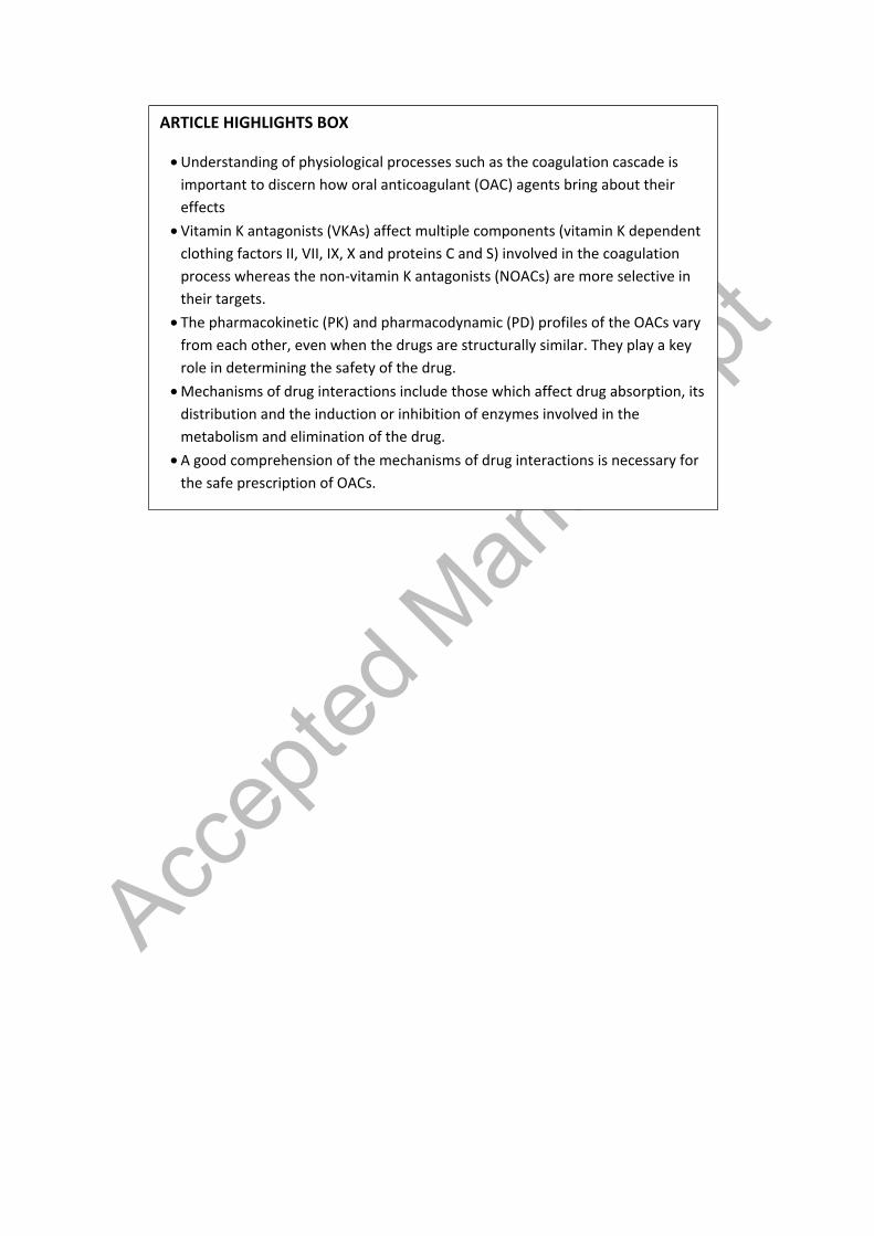
ARTICLE HIGHLIGHTS BOX
• Understanding of physiological processes such as the coagulation cascade is important to discern how oral anticoagulant (OAC) agents bring about their effects
• Vitamin K antagonists (VKAs) affect multiple components (vitamin K dependent clothing factors II, VII, IX, X and proteins C and S) involved in the coagulation process whereas the non-vitamin K antagonists (NOACs) are more selective in their targets.
• The pharmacokinetic (PK) and pharmacodynamic (PD) profiles of the OACs vary from each other, even when the drugs are structurally similar. They play a key role in determining the safety of the drug.
• Mechanisms of drug interactions include those which affect drug absorption, its distribution and the induction or inhibition of enzymes involved in the metabolism and elimination of the drug.
• A good comprehension of the mechanisms of drug interactions is necessary for the safe prescription of OACs.
Accep
ted M
anus
cript
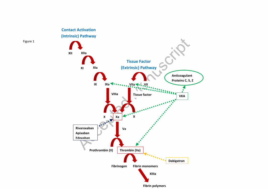
1. Introduction The discovery of oral anticoagulant (OAC) agents in the form of vitamin K antagonists (VKAs) was a boon to mankind and for over 60 years they occupied the limelight as the exclusive choice in the long-term the prevention and treatment of stroke associated with atrial fibrillation (AF) and other thromboembolic disorders. However, their slow onset of action, narrow therapeutic range, titrated dosing, requirement for monitoring, large inter-individual variability and multifarious interactions with other drugs and food are amongst the limitations which have been a source of numerous clinical plights, giving rise to the development of direct or non-vitamin K antagonists (DOACS/ NOACs)[1,2]. Although they have similar effects in terms of their clinical effect (i.e. anticoagulant activity), the pharmacological profile of NOACs varies to that of warfarin, accounting for their superiority in multiple domains. Hence, a good understanding of the physiological processes as well as the pharmacokinetics (PK) [what the body does to the drug] and pharmacodynamics (PD) [what the drug does to the body]) of the drugs is necessary to appreciate the differences, so that this knowledge can be utilised in the decision-making processes regarding the choice of OAC[3,4]. Hence, this review article aims to provide a detailed review of these characteristics of the commonly used VKAs (warfarin (Coumadin®), phenprocoumon (Marcoumar®) and acenocoumarol (Sinthrome®)) as well as NOACs (dabigatran (Pradaxa®), rivaroxaban (Xarelto®), apixaban (Eliquis®) and edoxaban (Lixiana®) . A recent article in this journal by Ingrasciotta and colleagues[5] also discusses the pharmacokinetics of NOACs and its implications on routine clinical care. A literature search was conducted on Pubmed using the keywords ‘warfarin’, ‘phenprocoumon’, ‘acenocoumarol’, ‘vitamin K antagonists’, ‘non-vitamin K antagonists oral anticoagulants’, ‘direct oral anticoagulants’, ‘dabigatran’, ‘rivaroxaban’, ‘apixaban’, ‘edoxaban’, ‘pharmacokinetics’ and ‘pharmacodynamics’ to identify relevant papers. 1.1 The clotting cascade The coagulation cascade comprises of proteolytic enzymes and cofactors, each which catalyse the activation of the next, giving rise to an amplification sequence of events which ultimately result in the formation of a clot or thrombus. It may be triggered by two major pathways; the intrinsic (contact) and extrinsic (tissue factor) pathways (Figure 1). Both these pathways eventually lead to the formation of prothrombin activator (also known as activated factor X) which plays an integral role in the coagulation process[6]. The extrinsic pathway is initiated with trauma to tissue or vascular injury resulting in the release of tissue factor (TF), also known as Factor III. TF catalyses the activation of factor VII

Accep
ted M
anus
cript
to VIIa and the tissue factor-VIIa complex then results in the activation of factor X (FX). Activated factor X (FXa) along with its co-factor activated factor V (FVa), then result in the generation of thrombin (FII) from its precursor prothrombin. Thrombin leads to the activation of soluble fibrinogen into insoluble fibrin (FI) monomers which in the presence of factor XIII, form fibrin polymers, resulting in the formation of a clot[6]. The extrinsic pathway initially results in the production of small amounts of thrombin. This thrombin activates platelets as well as factors V, VII, VIII, XI and XIII, to further amplify the process and lead to formation of more fibrin strands. The intrinsic pathway is usually triggered where there is trauma to the blood cells or exposure of the plasma to artificial surfaces. While it is less significant to haemostasis under normal physiological conditions, it is implicated in thrombotic diseases[7]. The intrinsic pathway is initiated with the activation of factor XII to FXIIa which catalyses the formation of activated factor XI (FXIa). FXIa then acts on factor IX to activate it, so that activated factor IX (FIXa) and its activated co-factor FVIIIa can together result in the formation of FXa. Both the extrinsic and intrinsic pathways therefore converge at the point where there is activation of FX[6] (Figure 1). FXa plays a crucial role in the coagulation processes arising from both pathways, by propagating the fundamental step which is the conversion of prothrombin to thrombin. Further, it is key in the amplification process as one molecule of FXa catalyses the formation of approximately 1000 molecules of thrombin[8], making it an attractive target to hinder the coagulation process[9]. Currently, the majority of NOACs (rivaroxaban, apixaban and edoxaban) specifically target FX, while the remaining NOAC dabigatran inhibits thrombin. All of these are target specific, unlike VKAs which affect multiple components in the cascade, as demonstrated in figure 1. This is amongst the reasons why NOACs have a favourable safety profile (Table 3), particularly in their reduction of the risk of intracerebral haemorrhage (ICH) when compared to VKAs[10]. In particular, VKAs affect factor VII which plays a crucial part in brain haemostasis[11]. Fibrinolysis begins once the structural integrity of the vessels starts to get restored. Endothelial cells secrete tissue plasminogen activator which activates the protease plasmin to degrade fibrin. Thrombin also gives rise to plasmin from plasminogen, which acts directly on the mesh networks of fibrin to break them apart[12]. Moreover, it stimulates production of anti-thrombin through a negative feedback loop, which in turn decreases the amount of thrombin generated from prothrombin as well as the activated FXa. Another natural anti-thrombotic mechanism of the body is the protein C/S system which is also influenced by VKAs due to their vitamin K dependence[13].
Accep
ted M
anus
cript
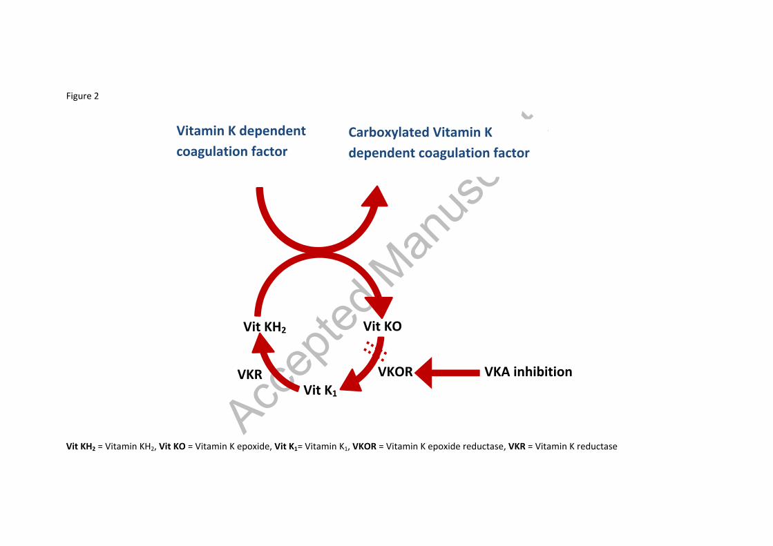
2. Vitamin K antagonists The discovery of VKAs was inadvertent, starting with the perplexing deaths of cattle which, on post-mortem were found to have extensive bleeding and bruising. The source of this was identified as sweet clover and the condition was subsequently dubbed ‘haemorrhagic sweet clover disease’. It was soon discovered that this was associated with a deficiency of prothrombin and the role of vitamin K in preventing bleeding was established. Although first used commercially as a rodent poison, warfarin was approved for medical use in 1954 and has been in the market since[14]. Phenprocoumon and acenocoumarol, followed shortly afterwards and are used in many countries. Other vitamin K antagonists include dicoumarol, fluindione, phenindione etc which are not as extensively used[15,16] Table 1 summarises the pharmacokinetic properties of the commonly used warfarin, phenprocoumon and acenocoumarol. 2.1 Mechanism of action The coagulation factors II, VII, IX and X require γ-carboxylation for their biological activity, with vitamin K acting as a co-factor for this process; hence why they are regarded as the vitamin K dependent clotting factors. Under normal circumstances, γ-carboxylation induces a conformational change in these coagulation proteins that promotes their binding to co-factors on phospholipid surfaces. The carboxylation reaction also involves the simultaneous oxidation of vitamin KH2 (reduced form of vitamin K) to vitamin K epoxide (Figure 2). As vitamin K is recycled, the vitamin K epoxide is reduced back to vitamin KH2 via a 2-step process. It is first converted to vitamin K1, the natural food form of vitamin K, and then vitamin KH2.. Both of these are reductase processes but only the first is sensitive to VKAs. VKAs non-competitively inhibit the vitamin K epoxide reductase complex subunit 1 (VKORC1), preventing the enzyme from catalysing the first reaction. Although VKAs interfere with the coagulation process by limiting the recycling of vitamin K, this can be counteracted by replacement of vitamin K1 through oral supplementation with food or therapeutic administration due to the second step being unaffected by these drugs. Vitamin K1 can accumulate in the liver and can result in warfarin resistance for up to a week, if present within the system in large amounts[17,18]. In addition to this, VKAs also inhibit the carboxylation process of the proteins C and S that are part of the body’s natural anticoagulant system, and may potentially induce a pro-coagulant effect. However, their anticoagulant effect is dominant in most circumstances[13,17]. The protective action of VKAs arises from their effect on prothrombin which is prerequisite to thrombus formation. Although an anticoagulant effect is initiated within 2 days, the full
Accep
ted M
anus
cript
antithrombotic effect requires a longer period of 6 days due to the long half-life of factor II (prothrombin) which is about 60 to 72 hours[19]. The concentration-effect of VKAs is variable. In a study by Penner and colleagues, treatment with warfarin for 14 days resulted in a reduction in FVII and X levels compared to the control by 30% and 20% respectively. Warfarin concentrations at this point were 1.2mg/L. In contrast, Porter et al demonstrated FX activities which were 60% of that of the control after warfarin intake for a similar period. Plasma concentrations of warfarin were 2.5mg/L[20]. 2.2 Measuring VKA effects The international normalised ratio (INR) is universally used as a standardised measure of the anticoagulant activity of VKAs. It is defined as the ratio of prothrombin time (PT) measured in a patient to that of a control, measured as per the World Health Organisation primary standard. PT is the time required for coagulation to occur in vitro and is initially reflective of the reduction in factor VII which has a short half-life of 6 hours[21]. Thereafter, factors X and prothrombin which have longer half-lives contribute to it[22]. 2.3 Warfarin Warfarin is the most widely used VKA. It is a highly water soluble compound made up of roughly equal proportions of both its R and S isomers. All VKAs exist as roughly equimolar racemic mixtures of their two, optically active R and S enantiomers, each which have different chemical properties. In a study that involved administration of single doses of both R and S warfarin to healthy volunteers, O’Reilly and colleagues demonstrated that the area under the plasma concentration time curve (AUC) was 1.9 times higher for R-warfarin than S-warfarin warfarin, indicating a lower clearance rate for the R-form. Further, they also showed that the inhibitory effect of S-warfarin on prothrombin was 1.8 times higher than R-warfarin, suggesting that the S-form is about two to five-fold more potent than the R-form. This is also true for phenprocoumon and acenocumarol[23–26]. Absorption: Warfarin is rapidly absorbed from the gastro-intestinal (GI) tract following oral administration, with the stomach and proximal GI tract being the main sites. Maximal plasma concentrations (Cmax) are achieved within 3-9 hours of oral administration[27]. Warfarin absorption is dissolution-rate controlled but there are several other factors that can influence this. Significant inter-individual variations also exist[27]. Warfarin has an essentially complete oral bioavailability[28]. Distribution: Over 99% of warfarin found in the plasma is protein-bound with virtually all of it attached to albumin[27]. Consequently, only a small amount of free drug is available to

Accep
ted M
anus
cript
exert its biological effects. The volume of distribution ranges from 8-12% of the total body weight and this does not differ significantly between the enantiomers[28]. Metabolism: The metabolism of warfarin is stereoselective and the two enantiomers undergo hepatic transformation via different pathways[29] by the hepatic cytochrome P450 (CYP450) enzymes. The CYP450 enzymes involved include CYP 2C9, 2C19, 2C8, 2C18, 1A2, and 3A4. The more potent S-warfarin is metabolised by CYPC29 while CYP1A2 and CYP3A4 are predominantly responsible for the metabolism of R-warfarin[30]. These enzymes catalyse the hydroxylation reactions which result in the formation of inactive, hydroxylated metabolites as well as cis- and trans-dehydro-warfarins. Five different hydroxylated metabolites of warfarin have been identified. Furthermore, two diastereomeric alcohols with minimal antiacoagulant activity are also formed via reduction processes which occur in the endoplasmic reticulum and cytosol[31,32]. Limited data are available on the phase II metabolism of warfarin which includes the glucuronidation and sulfation of hydroxylated metabolites. This has been mainly observed in rats in which the R-warfarin metabolites had undergone either glucuronidation, sulfation or both[33]. In humans however, these reactions appear to be insignificant.[32] CYP2C9 is the primary enzyme responsible for the metabolism of warfarin and patients with polymorphisms of these enzymes which have a reduced function require lower doses of warfarin due to their risk of over-anticoagulation[30,34,35]. Excretion: The elimination of warfarin is almost exclusively dependent on metabolism, with hydroxylation and reduction processes encompassing 80-85% and 15-20% of metabolic clearance respectively[29]. As a result, only miniscule amounts of unchanged drug are excreted. Approximately 80% of the metabolites are recovered in urine while the other 20% is excreted via the faecal route[36]. The half-life of R-warfarin is 45 hours and S-warfarin is 29 hours, leading to an overall half-life which ranges from 36-42 hours[22]. The effects of warfarin can last from 2-5 days[37]. Interactions: A myriad of interactions occur between warfarin and other drugs, as well as food, owing to a number of mechanisms (Table 4). These include agents that affect the absorption of warfarin (e.g. cholestyramine and sucralfate), its metabolism (e.g. CYP450 inhibitors or inducers such as rifampicin and phenobarbital) or displacement from the protein binding sites (e.g. quinidine) which can increase the amount of free drug available to exert anti-coagulant effects[38,39]. In addition, interactions may also depend on which of the isomers are affected as their potency and clearance differs from one another[23]. For example, phenylbutazone when co-administered with warfarin, results in effects which are stereo-selective. Like warfarin it has a high affinity for plasma proteins, leading to warfarin’s displacement from its albumin binding sites and increased plasma concentrations of active warfarin. As phenylbutazone is a selective inhibitor of CYP2C9, metabolism of the potent S-enantiomer of warfarin and therefore its clearance, is reduced[29]. Appropriate dose

Accep
ted M
anus
cript
adjustments or complete avoidance of an interacting drug may be necessary to avoid adverse reactions. 2.4 Phenprocoumon Phenprocoumon is another vitamin K antagonist and coumarin derivative which like the others. It is more commonly used in European countries such as Germany. In healthy volunteers who took 12mg of phenprocoumon, there was an earlier prolongation of PT, than there was a reduction in prothrombin.[40] Absorption: phenprocoumon is rapidly absorbed after oral intake and has an oral bioavailability of 100%[32]. Distribution: over 99% of phenprocoumon is protein bound and it has a volume of distribution between 0.11-0.14 L/kg[40]. Metabolism: phenprocoumon undergoes phase I metabolism by CYPC29 enzymes predominantly, resulting in the formation of its 4 inactive, hydroxylated metabolites[41]. These then undergo phase II metabolism via glucuronidation[41,42]. Other enzymes implicated in the metabolic processes include CYP 2C8 and 3A4 classes which have been identified using recombinant CYP enzymes within prepared human liver microsomes[43]. Excretion: unlike warfarin and acenocoumarol whose elimination is almost entirely dependent on metabolism, only 60% of phenprocoumon is excreted as metabolites. The remainder is removed in an unchanged form. The majority (about 65%) is eliminated by the kidneys and the other 35% is[41] excreted via the faeces. The terminal half-life of phenprocoumon is much longer in comparison to the others. This is thought to be secondary to the lower intrinsic clearance of the enzymes involved in the hydroxylation reactions as well as the extensive entero-hepatic recycling of the conjugated phenprocoumon[32]. Interactions: given the involvement of the CYP C29, 3A44 and 2C8 enzymes in phenprocoumon metabolism, any drugs which can affect their function can result in adverse reactions, when given together with phenprocoumon. However, phenprocoumon is much less susceptible to interactions compared to warfarin and acenocoumarol, owing to the different pathways of metabolism and excretion. For example, cimetidine when given together with warfarin results in its increased plasma levels and prolongation of PT but these parameters were unaffected when administered with phenprocoumon[44,45]. This is explained by its effects on the enzymes CYP1A2 and CYP 2C19 which are involved in the metabolism of warfarin but play no role in that of phenprocoumon.

Accep
ted M
anus
cript
2.4 Acenocoumarol Acenocoumarol also belongs to the family of VKAs and is used considerably in France and Italy. The clinical activity of acenocoumarol is mainly dependent on R-acenocoumarol despite being the less potent isoform, due to its longer half-life.[46] Absorption: Acenocoumarol undergoes rapid absorption after oral intake with Cmax being achieved within 3hours[47]. In contrast to the other VKAs, the S-enantiomer of this drug undergoes first pass metabolism, making its oral bioavailability relatively lower, though still high (96%)[15]. Distribution: Approximately 98% of acenocoumarol is protein-bound within the plasma[47]. Its volume of distribution ranges from 0.22-0.52 L/kg[32]. Metabolism: The metabolism of acenocoumarol takes place in the liver with CYP2CP playing the major role. It exclusively hydroxylates S-acenocoumarol and is also the main enzyme involved in the hydroxylation of R-acenocoumarol, which undergoes hydroxylation reactions by two other enzymes, CYP1A2 and CYP2C19[48]. Acenocoumarol also undergoes reduction to form amino and acetamido metabolites as well as alcoholic forms but the enzymes accountable for these processes are undiscovered[32,48]. Excretion: Similar to warfarin, nearly all of the absorbed acenocoumarol is eliminated as metabolites with <1% present in urine or faeces in its unchanged form. The proportions excreted in urine and faeces are 65% and 35% respectively[47]. Interactions: drugs affecting the CYP enzymes 1A2, 2C19 and particularly 2C9 can attenuate or potentiate the anticoagulant activity of acenocoumarol. When co-administered with phenylbutazone which selectively inhibits CYP2C9, the clearance of S-acenocoumarol in rats was two times lower[49]. Concurrent administration therefore requires careful consideration.
Accep
ted M
anus
cript
3. The Non-vitamin K antagonist oral anticoagulants In contrast to VKAs, NOACs are specific in their mode of action, have fixed dose regimens without the need for routine monitoring, a rapid onset of action, relatively low inter-individual variability with consistency seen across age, gender and ethnicity and fewer interactions with food and drugs, making them a more convenient and attractive choice (Table 2)[50]The four large phase III trials which have led to the licensing of dabigatran, rivaroxaban, apixaban and edoxaban in patients with atrial fibrillation are the Randomized Evaluation of Long-Term Anticoagulation Therapy (RE-LY) trial[51], Rivaroxaban Once Daily Oral Direct Factor Xa Inhibition Compared with Vitamin K Antagonism for Prevention of Stroke and Embolism Trial in Atrial Fibrillation (ROCKET-AF)[52], Apixaban for Reduction of Stroke and Other Thromboembolic Evens in Atrial Fibrillation (ARISTOTLE)[53] and Effective Anticoagulation with Factor Xa Next Generation in Atrial Fibrillation–Thrombolysis in Myocardial Infarction 48 (ENGAGE AF-TIMI 48) trials respectively[54]. These drugs have also received approval for use in the treatment and prevention of venous thromboembolism (VTE).Additionally, in light of recent evidence indicating a reduced risk of cardiovascular mortality, myocardial infarction (MI) and stroke, low-dose rivaroxaban is approved for use in patients with acute coronary syndromes.[55–57] Table 3 summarises the main safety outcomes of these drugs in the respective trials. 3.1 Measuring NOAC effects Although the inter-and intra-subject variability in the PK of NOACs have been reported as low, notable variations in drug blood levels have been observed in phase III trials of NVAF patients[58,59]. Further, these levels have been shown to correspond with efficacy and safety outcomes. In a pre-specified PK analysis of the RE-LY trial, the risk of ischemic events inversely correlated with dabigatran trough levels (p=0.045), while the major bleeding risk increased with dabigatran exposure (p<0.0001). Significant variations were observed in plasma concentration and renal function was identified as the predominant characteristic determining this, with age, weight and female sex also acting contributing factors. The median trough levels were 55% higher in subjects who had a bleeding event, compared to those who did not[60]. Similarly, a further analysis from the ENGAGE-TIMI-AF trial also demonstrated a dose-response relationship with higher rates of ischemic and bleeding events occurring in patients who had dose reductions, although the efficacy of edoxaban compared to warfarin was reportedly preserved. These findings question the lack of need for routine monitoring of plasma concentrations, at least for a subset of patients whose clinical characteristics may influence concentrations or who may be at an increased risk of developing ischaemic/ bleeding complications despite being on the indicated dose. The PK analysis of the RE-LY trial well-demonstrated the wide variation in plasma drug concentrations and the wide therapeutic range of dabigatran etexilate. Hence, determining an arbitrary cut-off level to guide dose changes can be challenging. At the same time, a dose reduction based on a

Accep
ted M
anus
cript
certain clinical characteristic alone, intended to reduce risks of bleeding, may occur at the expense of a much higher risk of stroke. In a study by Testa et al.[61] attempting to determine a relationship between DOAC trough levels and thromboembolic events, these complications were observed only in patients who had very low trough levels. 40.5% of the 565 patients in the study were treated with lower doses, as per recommendations from the guidelines due to presence of renal dysfunction and other indications. The mean CHA2DS2-VASc score was notably higher in this group compared to the total patient population (5.3 vs. 3.0) but thromboembolic complications were not reported in high cardiovascular risk group that had higher anti-coagulant levels. The study’s findings also demonstrated a high inter-individual variability in drug trough levels at steady state; suggesting older age, multiple co-morbidities and increasing use of prescription drugs as possible factors contributing to this and thus the anti-coagulant effect. The authors highlighted that the fixed dose of DOACs may therefore not be applicable to the ‘real-word patient’ whose profile may not fit that of a trial subject on whom the dose calculation would have been based on. While these are points to consider, the study population was small and subject to effects of confounders such as non-compliance. Further large scale studies are needed to confirm these findings.[61] Moreover, dose-adjustment studies in the future may enable individualisation of DOAC therapy for better outcomes, particularly for those patients whose drug levels fall outside the desired range. Current guidelines do not mandate regular monitoring of NOAC levels due to their wide therapeutic range and predictable PK[62]. That said, the measure of anticoagulant effect may still be useful in certain situations where treatment decisions can be influenced (e.g. in determining whether an urgent procedure can be performed safely) or in the identification of treatment adherence or overdose[63]. Although NOACs affect PT, INR and aPTT to varying degrees, these changes do not accurately correlate to their anticoagulant activity,[64] only providing qualitative evidence for the anticoagulant activity. Even then, the effects are inconsistent with the sensitivity of PT particularly poor for apixaban, while aPTT which can only be used in the qualitative assessment of dabigatran is also inconsistent in its sensitivity[65]. Hence, these coagulation assays cannot be used as accurate measures[66,67]. At present, anti-FXa assays are the only widely available specialised coagulation assay that allows a quantitative assessment of the FXa inhibiting NOACs (apixaban, rivaroxaban and edoxaban). This is reliable as plasma concentrations of NOACs closely correspond to their anticoagulant activity[68]. Anti FIIa assays (including ecarin-based anti FIIa assays) and the HEMOCLOT assay where the diluted thrombin time (TT= time taken for the formation of a stable clot) is measured in a 1:16 dilution of patient plasma compared with normal pooled

Accep
ted M
anus
cript
plasma, and calibrated; can be used as quantitative measures of dabigatran activity. Unfortunately, these are not widely available yet[65,66,69]. 3.2 Dabigatran etexilate Dabigatran is a synthetic, non-peptidic small-molecule direct thrombin inhibitor (DTI) which affects both clot-bound and free thrombin in a competitive, reversible and concentration-dependent manner[70]. It inactivates thrombin by binding to its active site and as this is a reversible interaction, a small amount of free, active thrombin is still available for control of haemostasis[71,72]. Dabigatran etexilate is a prodrug that was created owing to the low oral bioavailability of dabigatran (6-7%), due to its high polarity[73]. It is rapidly converted to its active form dabigatran, once absorbed. While it may be consumed with or without food, modification of the capsule by crushing or chewing it is discouraged as the oral bioavailability of dabigatran etexilate increases by 75% once the capsule shell is broken[74]. Absorption: in order to enhance absorption, the chemical composition of dabigatran etexilate is less basic and less hydrophilic, compared to its active form[75]. The drug formulation constitutes tartaric acid along with the active ingredient, which provides an acidic micro-environment that facilitates drug dissolution and absorption in the stomach and small intestine, regardless of the individual gastric pH[76].[77] In pharmacokinetic analyses of dabigatran etexilate in healthy males, peak plasma concentrations from the time of administration of 2 hours was observed, with steady state concentrations achieved within 72 hours with multiple dosing[73]. Distribution: Dabigatran has a volume of distribution of approximately 60-70L, indicative of moderate tissue distribution[72]. Only about 35% of the drug is bound to human plasma proteins[78]. The distribution phase is rapid, with plasma concentrations declining to <30% from peak levels, within 4-6 hours[79]. Metabolism: Once absorbed, nearly all of the dabigatran etexilate is hydrolysed to its active form. This reaction is catalysed by esterases within the enterocytes and continues along its transit to the portal vein and liver.[77,78] Dabigatran does not undergo significant oxidative metabolism reactions[78]. Up to 20% of it undergoes glucuronidation in a hepatic phase II reaction and these conjugated products have similar properties to and are as pharmacologically active as unconjugated dabigatran[78]. Excretion: While the 20% of the dabigatran that undergoes hepatic biotransformation gets excreted in bile, the remaining 80% is excreted by the kidneys in an unchanged form[78]. Given that dabigatran is predominantly cleared by the kidneys, dose adjustments are
Accep
ted M
anus
cript
required in patients with renal impairment to prevent its accumulation of drug levels within the plasma once creatinine clearance (CrCl) is ≤30ml/min[55,80]Regular monitoring of renal function is therefore necessary for patients not just on dabigatran but all NOACs as deteriorations leading to changes in the estimated glomerular filtration rate (eGFR) have been associated with an increase in the risk of major bleeding[81]. The exposure of dabigatran depicted by the area under the curve (AUC) is 2.7 and 6 fold higher in those with moderate (CrCl 30-50ml/min) and severe (CrCl <30ml/min) renal impairment respectively[82]. Interactions: The risk of food-drug and drug-drug interactions is low with dabigatran due to the lack of involvement of cytochrome p450 and similar oxidoreductase enzymes[72,78]. However, drug interactions may occur during the absorption phase as dabigatran etexilate is a substrate for permeability glycoprotein (P-gp) transporter present in the entire instestine[83]. P-gp is involved in the active secretion of drugs specific to it from the cells, back into the intestinal lumen, resulting in reduced absorption and bioavailability[84]. Hence, co-administration with P-gp inducers and inhibitors is not recommended. In a population pharmacokinetic analysis from the RE-LY trial, proton-pump inhibitors, verapamil and amiodarone affected dabigatran bioavailability significantly but only to moderate amounts[80]. Dose adjustments are only required with concurrent administration of P-gp inhibitors such as dronedarone and ketoconazole, in the presence of moderate renal impairment while complete avoidance is recommended with severe renal impairment[85]. 3.3 Rivaroxaban Rivaroxaban is a selective inhibitor of factor Xa affecting both free and prothrombinase complex bound levels[86]. It also affects clot-bound factor Xa, inhibiting further generation of thrombin within the clot and thereby, the extension of the thrombus[87]. Rivaroxaban also results in prolongation of PT and aPTT, with maximum inhibition of factor Xa activity occurring within 3 hours after dosing[87].
Rivaroxaban is available as a film-coated tablet and can be safely taken crushed or suspended in water, if within 4 hours[88].
Absorption: Rivaroxaban is primarily absorbed in the stomach, in a process which is unaffected by medications that alter the gastric pH.[89] Absorption is reduced in the proximal part of the small intestine (resulting in a drop in the AUC for plasma concentration and Cmax by 29% and 56% respectively), and even further in the distal small bowel and ascending colon[90]. It has a bio-availability that is dose dependent. For low doses of rivaroxaban (10mg or below), the bio-availability is high (80-100%) regardless of whether it is taken with or without food, while this is reduced at higher doses (15mg and 20mg), particularly in fasted states due to absorption limited by the dissolution rate. Hence,
Accep
ted M
anus
cript
administration with food is recommended as this increases bioavailability by up to 80% due to an increased residence time within the stomach thought to enhance solubility and dissolution[75,91].
Distribution: The majority of rivaroxaban (92-95%) is bound to plasma proteins; predominantly albumin. Its volume of distribution is 50L, indicative of tissue distribution that is moderate. However, dose adjustments are not required in the extremes of body weight. In a phase I study involving administration of rivaroxaban to healthy individuals weighing ≤50kg, 70-80kg and >120kg, Cmax was largely unaffected except in those weighing ≤50kg, where only a marginal increase was observed[92].
Metabolism: rivaroxaban is predominantly metabolised in the liver, with two-thirds of the drug undergoing oxidative degradation and hydrolysis by cytochrome 3A4 (CYP3A4) and cytochrome 2J2 (CYP2J2) enzymes as well as CYP independent pathways[55,93].
Excretion: About half of the drug that undergoes hepatic biotransformation is excreted by the kidneys while the other half takes the hepatobiliary route. The third which does not undergo metabolic degradation in the liver, is excreted in the urine an unchanged, active form[93].
In a study evaluating the effects of renal impairment on the PK and PD of rivaroxaban, renal clearance of rivaroxaban was reduced with the decline in renal function, resulting in increased plasma exposure and PD effects. AUC for plasma concentrations were 1.44-fold, 1.52-fold and 1.64-fold for those with mild (CrCl 50-79ml/min), moderate (30-49ml/min) and severe renal impairment (CrCl <30ml/min) respectively, when compared to those with CrCl ≥80ml/min[94].
Similarly, in another study evaluating the effects of hepatic impairment, significant increases in plasma concentrations of rivaroxaban were observed in those with moderate hepatic impairment (AUC = 2.27-fold compared to healthy subjects) resulting in significant increases in PD effects[95]. This has not been studied in patients with severe hepatic impairment. Hence, dose adjustments are required when CrCl is ≤50ml/min and in patients with hepatic impairment associated with coagulopathy. Caution is warranted in its use in those with moderate hepatic impairment that is not associated with coagulopathy[55].
Interactions: in addition to being a substrate of the CYP3A4 and CYP2J2 enzymes, rivaroxaban is also a substrate for the transporters P-gp and BCRP (breast cancer resistant gene protein). The latter is found abundantly within the epithelial cells of organs such as the liver, gut and kidney and can influence PK processes of the drug[96,97]. However, the clinical impact of this transporter is not known at present.
Concurrent administration of rivaroxaban with potent inhibitors of P-gp and/ or CYP3A4 such as azole-antimycotics and HIV protease inhibitors is not recommended while caution is advised with use of agents that affect P-gp and/or CYP3A4 to a lesser extent[55,98]. Care is

Accep
ted M
anus
cript
also warranted when used alongside potent CYP3A4 inducers which can decrease rivaroxaban exposure[55].
Antiplatelet agents such as aspirin and NSAIDs such as naproxen do not affect the PK of rivaroxaban but have been shown to independently increase the bleeding time when concurrently given.
3.4 Apixaban
Apixaban propagates its anticoagulant activity by inhibiting factor Xa in a highly selective and reversible fashion. Not only does it affect factor Xa which is free, clot-bound and found in the prothrombinase complex but its subsequent attenuating effect on thrombin generation leads to the indirect inhibition platelet aggregation induced by thrombin[99]. Hence, it can prolong prothrombin time (PT), international normalised ratio and activated partial thromboplastin time (aPTT)[86,100]. Absorption: Apixaban has an oral bioavailability of about 50% regardless of whether taken with or without food. It is primarily absorbed in the distal small bowel and ascending colon, while some absorption also takes place in the stomach[101]. In healthy individuals, Cmax is achieved within 3hrs of oral administration[102]. Distribution: Approximately 87% of apixaban is bound to plasma proteins, namely albumin[103]. Unlike the other NOACs, apixaban has a relatively small volume of distribution of 21L, meaning distribution is limited within tissues and mainly confined to the blood[103]. In a study looking into the effects of extremes of body weight on the PK and PD of apixaban, only a modest increase in drug levels was noted with the Cmax increasing by 27%. Dose adjustments are therefore not required based on body weight alone but may be indicated in the presence of other factors such as renal impairment which may have an additive effect on Cmax levels[55,104]. Metabolism: Apixaban is largely unchanged within the plasma but a quarter of the orally administered dose undergoes metabolism in the liver and is recovered in the urine and faeces as metabolites. It undergoes O-demethylation, hydroxylation and sulfation reactions which are principally catalysed by the CYP3A4 enzymes, although there is minor involvement of cytochromes CYP1A2, 2C8, 2C9, 2C19 and 2J2[105]. O-demethyl apixaban sulfate is the significant metabolite found in the plasma[105]. Excretion: Of the NOACs, apixaban is least dependent on the kidneys for elimination. Renal clearance accounts for about a third (12-29%) of the absorbed dose while the remainder is removed via the faecal route either through biliary or direct intestinal excretion (55%)[105]. In a study looking into the effects of renal impairment, increases in plasma exposure of apixaban were observed as the renal function declined. The AUC for apixaban was 16%, 29%

Accep
ted M
anus
cript
and 38% higher in those with mild, moderate and severe renal impairment respectively, compared to those with normal renal function (24h CrCl =100ml/min). Cmax, anti-FXa activity and INR were unaffected across all levels of renal impairment. Hence, dose adjustments are not required based on renal function alone[106]. In the case of hepatic impairment, the AUC for apixaban following a dose of 5mg of apixaban increased by 1.03-fold and 1.09-fold in patients with mild (Child-Pugh A) and moderate hepatic impairment (Child-Pugh B) respectively, when compared with normal subjects. Protein binding and therefore the fraction of free, unbound drug remained similar across the three groups. Further, the percentage change from the baseline INR was also comparable between them, although the baseline INR values were higher for those with hepatic impairment[107]. Apixaban may therefore be used with caution in those with mild to moderate hepatic impairment or in those with liver function enzymes (aspartate amino transferase and alanine aminotransferase) which are over 2-fold the upper limit of normal but is contra-indicated in those with severe liver disease or that which is associated with coagulopathy or high risk of bleeding[55]. Interactions: The numbers of drug-drug interactions with apixaban are low due to the low concentrations of drug required within the plasma, largely unchanged form and the various pathways of clearance[108]. However, like rivaroxaban, as it is a substrate for CYP3A4 as well as the P-gp and BRCP transporter proteins, concomitant administration with potent systemic inhibitors and inducers of these is not recommended[55]. Co-administration of apixaban and ketoconazole, a strong dual inhibitor of CYP3A4 and P-gp, led to an increase in the Cmax and AUC of apixaban by 62% and 99% respectively. However, this effect was less pronounced (Cmax of 31% and AUC of 40%) when apixaban was given together with diltiazem, which is a moderate inhibitor of CYP3A4 and P-gp[109]. In another study looking into the effects of rifampicin which is a strong inducer of CYP3A4 and P-gp, Cmax and AUC were significantly decreased and the mean apparent clearance of apixaban increased by 2.1-fold[110]. Although not contra-indicated, plasma concentrations of apixaban can be affected to varying degrees even when given together with less potent CYP3A4 and P-gp inhibitors/ inducers. Therefore, careful consideration is required before concurrent use of these medications[55]. 3.5 Edoxaban Edoxaban is the newest of the NOACs to receive approval for use. It is a selective, small molecule inhibitor of factor Xa which binds to it in a competitive and dose-dependent manner[111]. This in turn inhibits the generation of thrombin, and the subsequent processes that follow. Edoxaban affects free FXa, prothrombinase and also inhibits thrombin-induced platelet aggregation[112]. Like the other factor Xa inhibitors, edoxaban causes modest increases in INR, PT and aPTT[113].
Accep
ted M
anus
cript
Absorption: Edoxaban is available as a film coated tablet and may be taken with or without food[114]. It has an absolute oral bioavailability of 62%[115]. Absorption in the colon is limited and predominantly occurs in the proximal small intestine. Cmax is established rapidly within 1-2 hours of taking the medication and steady state is achieved after 72 hours[116]. Distribution: Approximately 55% of edoxaban is protein-bound in the plasma, which is lesser than the other FXa inhibitors. It is extensively distributed throughout the body with a volume of distribution of 107L[117]. Metabolism: Unchanged edoxaban is most prevalent in the plasma but a small amount is present as metabolites. The major metabolic pathway which results in 25% of the total dose undergoing hydrolysis, is catalysed by the enzyme carboxyl esterase-1 present in the cytosol and liver microsomes. This gives rise to the major metabolite of edoxaban M-4, which is active but accounts for less than 10% of the total edoxaban plasma exposure[118]. Minimal metabolism also takes place via the phase I processes of oxidation and conjugation, mediated by the CYP3A4 enzymes[119]. Edoxaban also undergoes phase 2 metabolism through glucuronidation. In total, six phase 1 metabolites and a glucuronide have been detected in human plasma. However, in the absence of severe renal impairment and CYP inducers, these do not contribute substantially to edoxaban’s anticoagulant activity[118]. Excretion: 50% of edoxaban is excreted via the kidneys in an unchanged form while biliary excretion and metabolism make up for the other half[120]. In the study by Bathala and colleagues which involved administration of radiolabelled edoxaban to six, healthy male subjects, over 97% of the administered dose was recovered on average, with 35.4% eliminated in the urine and 65.2% in faeces[118]. As renal function declines, plasma levels of edoxaban rise but only to a certain extent, as it is just one of the elimination pathways for edoxaban. In a pharmacokinetic study, the total exposure of edoxaban increased by 32%, 74% and 72% in individuals with mild (CrCl 50-80 ml/min), moderate (CrCl 30-50ml/min) and severe (CrCl <30ml/min) respectively compared to those with a renal function. There was no significant difference between moderate and severe renal insufficiency. Metabolic clearance plays a more dominant role in these patients[117]. The effects of hepatic impairment on edoxaban PK and PD have also been studied. Administration of 15mg of edoxaban to patients with mild and moderate hepatic impairment resulted in plasma exposure AUCs of 95.9% and 95.2% compared to their respective controls, indicating a minimal effect. Furthermore, PT was only slightly prolonged in these subjects compared to the healthy subjects, while aPTT profiles were similar across all groups[121]. Nonetheless, edoxaban is contra-indicated in patients with severe hepatic impairment and should be used with caution in those with moderate hepatic impairment due to the risk of coagulopathy and bleeding[122].
Accep
ted M
anus
cript
Interactions: much like the other FXa inhibitors, edoxaban is a substrate for the P-gp transporter protein and CYP3A4 enzymes. However, as the CYP3A4 enzymes only play a minor role in its clearance, plasma exposures of edoxaban are not significantly different to each other when administered with drugs that are dual inhibitors of P-gp and CYP3A4, whether the latter is mild, moderate or strong[117]. However, significant increases in total exposure are still seen due to inhibition of P-gp and dose reductions are required when edoxaban is given together with these drugs [122,123]. When administered with rifampicin, edoxaban clearance increased by 33% with a reduction in half-life by 50% while PT and aPTT did not change significantly. Concurrent use is therefore not contra-indicated but requires careful consideration[124]. In addition to this, concomitant administration with naproxen and low dose aspirin did not alter the PK of edoxaban but high dose aspirin increased edoxaban exposure by about 30%. All three, approximately doubled the bleeding time even though INR, PT and APTT were unaffected[125]. 4. Conclusion The pharmacokinetic profiles of the OACs vary from each other to different degrees and partly explain the differences in their characteristics and mode of action. The clinical effect of the drug is determined not just by one factor, but by a combination of them including PK/PD profiles, patient demographics such as age and gender, genetics and environmental aspects. Indeed, mechanisms which influence the metabolism and elimination pathways can solitarily compromise drug safety. Ultimately, it is not a question of which drug has the more superior pharmacological profile, but a matter of which pharmacological profile is most suited to the patient’s profile. 5. Expert Opinion Although OAC agents belonging to the same class of drugs are similar in structure and function, there are major differences in their pharmacological profiles. In particular, it is the distinctions in the metabolic and elimination pathways are responsible for the heterogeneity in drug-drug and food-drug interactions which either attenuate or potentiate the anticoagulant activity. Hence, insight to the pharmacokinetics and pharmacodynamics is necessary in order to achieve the maximal therapeutic benefit and minimise the possibility of adverse reactions. It is also of relevance so that an informed choice can be made between the drugs. That said, other processes such as factors influencing absorption of the drugs and pharmacodynamic interactions also play a role. Warfarin and VKAs, though expected to be replaced by NOACS, are likely to remain in play due to the breadth of experience with their use and because they
Accep
ted M
anus
cript
are the only OACs currently licensed for use in special populations such as those with mechanical prosthetic valves and rheumatic atrial fibrillation[15]. As such, the focus should not be shifted away from VKAs just yet and further research is needed to better understand its behaviour. The use of warfarin remains problematic due to the diversity of its interactions and the multiple patient factors such as age, gender, diet and smoking status which influence its response. The inter-individual variations in the dose requirements for warfarin can be up to 10-fold[126]. Genetic polymorphisms of some of the key enzymes such as VKORC and CYPC29 have been attributed to this, where the CYPC29*2 and CYPC29*3 alleles are associated with decreased enzyme activity and subsequent increased warfarin levels requiring a reduced dose, while single nucleotide polymorphisms in VKORC1 are associated with warfarin doses across the normal range[127]. It is estimated that only 60% of these differences are accounted for, meaning that factors which are yet to be discovered are silently at play[126]. In this regard, drug transporter science aiming to identify and characterise different transporter proteins as well as their interplay with the pharmacokinetics and subsequent clinical response is an emerging field[128]. Another is pharmacogenomics, with the increasing recognition that it is not just the pharmacokinetics and pharmacodynamics profile which influences the drug effect. Since its acknowledgement, pharmacogenomics ha advanced such that guidelines which incorporate pharmacogenetics algorithms considering VKORC1 and CYPC29 genotypes now exist, in addition to the clinical factors, for warfarin dose prediction[129]. A step ahead of this is the implementation of pharmacogenomic diagnostics which involves screening for genotype variants prior to initiating anti-coagulation therapy. In a randomised controlled trial involving patients with AF and VTE by Pirmohamed and colleagues[130], warfarin was prescribed after genotyping, based on pharmaco-genetic algorithms in the treatment group, and standard dosing was utilised for the control group. Results indicated that that the time in therapeutic range (TTR) was significantly better in the genotype-guided group, compared to the control group. Results from another study by Kimmel and colleagues[131] however, showed no significant differences in the TTR between the genotype-guided and control groups. The widespread uptake of pharmacogenomics diagnostics is yet to take place. Routine use may be implicated by factors such as cost and pharmaco-economic evaluations may be necessary prior to this. However, for a small sub-group of patients who are difficult to manage, or at risk of bleeding with labile INRs whose only choice of treatment is VKAs, genotyping may be of utmost value to help guide anti-coagulation treatment. Less emphasis has been made on NOACs in this section, not due to a lesser importance but because their PK/PD profiles confer a lesser degree of interactions. Indeed, they too have

Accep
ted M
anus
cript
some important interactions and certain variations in their PK/PD processes are likely to be due to genetic polymorphisms, some which are known about (e.g. CYPC29) and some which are yet to be discovered. The exact pathophysiology of reported adverse effects such as GI haemorrhage also requires further study, while breakthroughs are being made in certain avenues such as the development of potential antidotes.[132–134] Lastly, a key area of NOAC-based research should focus on patients with chronic and end stage kidney disease; a prevalent and complex population. The complication rates in terms of major bleeding and ICH as well as the challenges in maintaining INR within the desired range with VKAs in this group of patients have resulted in a ‘now more than ever’ need for alternative drug agents such as DOACs. Unfortunately, all DOAC RCTs excluded patients with CrCl <25-30 ml/min precluding their use in these patients.[51–54]Despite this, DOAC usage has been extended beyond the RCT criteria in certain countries on the basis of limited pharmacokinetic data. This includes the lower dose of dabigatran in patients with renal impairment (CrCl 15-30ml/min) and the use of apixaban in renal patients requiring dialysis in the United States, following approval from the Food and Drug Administration.[135,136] The discrepancies in the international guidelines concerning renal dysfunction reflect our incomplete knowledge of the pharmacology of NOACs in this group of patients and therefore warrant caution, especially as data on the long-term effects is scarce. While data from post-approval studies have been informative and a RCT comparing apixaban to phenprocoumon in patients undergoing chronic haemodialysis is underway, further studies are needed to shed light on the matter [137–139].
Funding
This paper was not funded.
Declaration of Interest
G Lip is a consultant for Bayer/Janssen, BMS/Pfizer, Medtronic, Boehringer Ingelheim, Novartis, Verseon and Daiichi-Sankyo, and a speaker for Bayer, BMS/Pfizer, Medtronic, Boehringer Ingelheim, and Daiichi-Sankyo. No fees are directly received personally. The authors have no other relevant affiliations or financial involvement with any organization or entity with a financial interest in or financial conflict with the subject matter or materials discussed in the manuscript apart from those disclosed.
Reviewer Disclosures
Peer reviewers on this manuscript have no relevant financial or other relationships to disclose.
Accep
ted M
anus
cript
List of figures
Figure 1 – An overview of the clotting cascade and the OAC targets within the cascade
Figure 2 – A diagrammatic representation of the vitamin K cycle and the mechanism of action of VKAs
List of Tables
Table 1 – Summary of pharmacokinetic characteristics of VKAs [23,24,33–37,25–32], [32,40–44], [15,46–50]
Table 2 – Summary of pharmacokinetic characteristics of NOACs [55,72,81,82,73–80], [55,87,96,97,88–95], [55,100,109,110,101–108], [111,112,121–125,113–120]
Table 3 – Results of safety outcomes for bleeding in NOAC phase III trials[51,52,54,140–142]
Table 4 – Summary of interactions of warfarin with drugs and food[38,143–145]
Accep
ted M
anus
cript
VII
XII XIIa
XI XIa
IX IXa
VIIIa
X Xa
VIIa
Tissue factor
X
Prothrombin (II) Thrombin (IIa)
Fibrinogen Fibrin monomers
Fibrin polymers
Contact Activation (Intrinsic) Pathway
Tissue Factor (Extrinsic) Pathway
Va
XIIIa
Rivaroxaban Apixaban Edoxaban
Dabigatran
VKA
Anticoagulant Proteins C, S, Z
Figure 1
Accep
ted M
anus
cript
Carboxylated Vitamin K dependent coagulation factor
VKR
Vit KOVit KH2
Vitamin K dependent coagulation factor
Vit K1
VKA inhibitionVKOR
Figure 2
Vit KH2 = Vitamin KH2, Vit KO = Vitamin K epoxide, Vit K1= Vitamin K1, VKOR = Vitamin K epoxide reductase, VKR = Vitamin K reductase
Accep
ted M
anus
cript
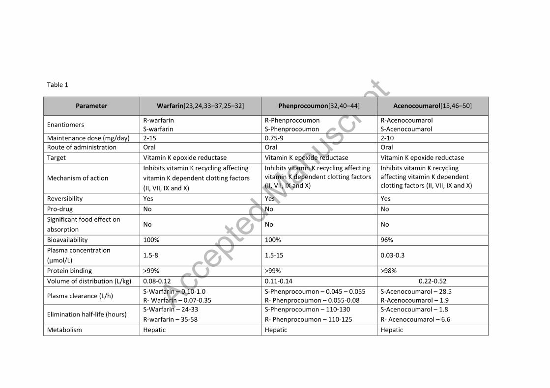
Table 1
Parameter Warfarin[23,24,33–37,25–32] Phenprocoumon[32,40–44] Acenocoumarol[15,46–50]
Enantiomers R-warfarin S-warfarin
R-Phenprocoumon S-Phenprocoumon
R-Acenocoumarol S-Acenocoumarol
Maintenance dose (mg/day) 2-15 0.75-9 2-10 Route of administration Oral Oral Oral Target Vitamin K epoxide reductase Vitamin K epoxide reductase Vitamin K epoxide reductase
Mechanism of action Inhibits vitamin K recycling affecting vitamin K dependent clotting factors (II, VII, IX and X)
Inhibits vitamin K recycling affecting vitamin K dependent clotting factors (II, VII, IX and X)
Inhibits vitamin K recycling affecting vitamin K dependent clotting factors (II, VII, IX and X)
Reversibility Yes Yes Yes Pro-drug No No No Significant food effect on absorption
No No No
Bioavailability 100% 100% 96% Plasma concentration (µmol/L)
1.5-8 1.5-15 0.03-0.3
Protein binding >99% >99% >98% Volume of distribution (L/kg) 0.08-0.12 0.11-0.14 0.22-0.52
Plasma clearance (L/h) S-Warfarin – 0.10-1.0 R- Warfarin – 0.07-0.35
S-Phenprocoumon – 0.045 – 0.055 R- Phenprocoumon – 0.055-0.08
S-Acenocoumarol – 28.5 R-Acenocoumarol – 1.9
Elimination half-life (hours) S-Warfarin – 24-33 R-warfarin – 35-58
S-Phenprocoumon – 110-130 R- Phenprocoumon – 110-125
S-Acenocoumarol – 1.8 R- Acenocoumarol – 6.6
Metabolism Hepatic Hepatic Hepatic
Accep
ted M
anus
cript
Metabolic enzymes involved
S-Warfarin - CYPC29 (major) R-Warfarin - CYP1A2 and CYP3A4 (major) Other: 2C19, 2C8, 2C18 and 1A2 (minor)
S-Phenprocoumon – CYP2C8, CYP2C9 and CYP3A4 R-Phenprocoumon – CYPC29 and CYP3A4
S-Acenocoumarol – CYP2C9 R-Acenocoumarol – CYP2C9, CYP1A2 and CYP2C19
Elimination Urine (80%) Faeces (20%)
Urine (65%) Faeces (35%)
Urine (65%) Faeces (35%)
Routine monitoring Yes Yes Yes Interactions with food Multiple Multiple Multiple Drug-drug interactions Multiple Multiple Multiple
Adverse effects (non-bleeding)
Hypersensitivity reactions (urticaria, anaphylaxis), calciphylaxis, tissue necrosis, systemic atheroemboli, cholesterol microemboli, limb ischaemia, gangrene, acute kidney injury, gastro-intestinal disorders such as nausea, vomiting and abdominal discomfort.
Rare – hepatitis, skin and tissue necrosis[146]
Rare – nausea, vomiting and alopecia, skin necrosis, liver injury, vasculitis
Accep
ted M
anus
cript
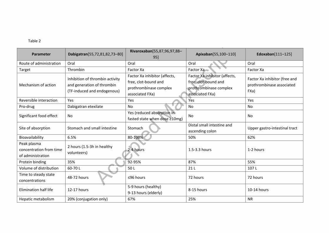
Table 2
Parameter Dabigatran[55,72,81,82,73–80] Rivaroxaban[55,87,96,97,88–
95] Apixaban[55,100–110] Edoxaban[111–125]
Route of administration Oral Oral Oral Oral Target Thrombin Factor Xa Factor Xa Factor Xa
Mechanism of action Inhibition of thrombin activity and generation of thrombin (TF-induced and endogenous)
Factor Xa inhibitor (affects, free, clot-bound and prothrombinase complex associated FXa)
Factor Xa inhibitor (affects, free, clot-bound and prothrombinase complex associated FXa)
Factor Xa inhibitor (free and prothrombinase associated FXa)
Reversible interaction Yes Yes Yes Yes Pro-drug Dabigatran etexilate No No No
Significant food effect No Yes (reduced absorption in fasted state when dose ≥10mg)
No No
Site of absorption Stomach and small intestine Stomach Distal small intestine and ascending colon
Upper gastro-intestinal tract
Bioavailability 6.5% 80-100% 50% 62% Peak plasma concentration from time of administration
2 hours (1.5-3h in healthy volunteers)
2-4 hours 1.5-3.3 hours 1-2 hours
Protein binding 35% 92-95% 87% 55% Volume of distribution 60-70 L 50 L 21 L 107 L Time to steady state concentrations
48-72 hours ≤96 hours 72 hours 72 hours
Elimination half life 12-17 hours 5-9 hours (healthy) 9-13 hours (elderly)
8-15 hours 10-14 hours
Hepatic metabolism 20% (conjugation only) 67% 25% NR

Accep
ted M
anus
cript
Metabolic enzymes involved
None CYP3A4 and CYP 2J2 (major) CYP-independent mechanisms
CYP3A4 (major) CYP1A2, 2C8, 2C9, 2C19 and 2J2 (minor)
carboxyl esterase-1 (major) CYP 3A4 (minor)
Excretion (as a % of total dose)
Renal (80%)- unchanged form Biliary (20%)
Renal (67%) – 33% in unchanged form Hepatobiliary (33%)
Renal (27-30%) Hepatobiliary (70%) – direct intestinal (major) and biliary (minor)
Renal (50%) – unchanged form Hepatobiliary (50%)
Routine monitoring No No No No Interactions with food No No No No
Drug-drug interactions
Few P-gp inducers (e.g. rifampicin) P-gp inhibitors in the presence of moderate-severe rena impairment
Combined P-gp and strong inhibitors of CYP3A4 (e.g. ketoconazole, ritonavir) Combined P-gp and strong inducers of CYP3A4 (e.g. rifampicin)
Combined P-gp and strong inhibitors of CYP3A4 (e.g. ketoconazole, ritonavir) Combined P-gp and strong inducers of CYP3A4 (e.g. rifampicin)
Combined P-gp and strong inhibitors of CYP3A4 (e.g. ketoconazole, ritonavir) High dose aspirin Digoxin
Dose adjustment with hepatic impairment
Avoid if risk of coagulopathy No but caution with use if moderate impairment Avoid if risk of coagulopathy
No but caution with use if moderate impairment Avoid if risk of coagulopathy
No but caution with use if moderate impairment Avoid if risk of coagulopathy
Dose adjustment with renal impairment
Yes Yes Yes (if present with weight ≤50kg and/or age ≥80 years)
Yes
Adverse effects (non-bleeding)
Gastritis like symptoms (15%), Dyspepsia
Rash (≥1%) Skin rash (<1%) Rash, abnormal liver function tests (≥1%)
Reversal Agent Idarucizumab Andexanet Alfa Andexanet Alfa Andexanet Alfa
NR = Not reported
Accep
ted M
anus
cript
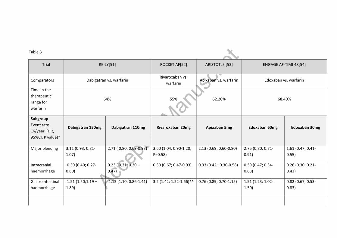
Table 3
Trial RE-LY[51] ROCKET AF[52] ARISTOTLE [53] ENGAGE AF-TIMI 48[54]
Comparators Dabigatran vs. warfarin Rivaroxaban vs.
warfarin Apixaban vs. warfarin Edoxaban vs. warfarin
Time in the therapeutic range for warfarin
64% 55% 62.20% 68.40%
Subgroup Event rate ,%/year (HR, 95%CI, P value)*
Dabigatran 150mg
Dabigatran 110mg
Rivaroxaban 20mg
Apixaban 5mg
Edoxaban 60mg
Edoxaban 30mg
Major bleeding 3.11 (0.93; 0.81-1.07)
2.71 ( 0.80; 0.69-0.93) 3.60 (1.04, 0.90-1.20; P=0.58)
2.13 (0.69; 0.60-0.80) 2.75 (0.80; 0.71-0.91)
1.61 (0.47; 0.41-0.55)
Intracranial haemorrhage
0.30 (0.40; 0.27-0.60)
0.23 ( 0.31; 0.20 – 0.47)
0.50 (0.67; 0.47-0.93) 0.33 (0.42; 0.30-0.58) 0.39 (0.47; 0.34-0.63)
0.26 (0.30; 0.21-0.43)
Gastrointestinal haemorrhage
1.51 (1.50;1.19 – 1.89)
1.12 (1.10; 0.86-1.41) 3.2 (1.42; 1.22-1.66)** 0.76 (0.89; 0.70-1.15) 1.51 (1.23; 1.02-1.50)
0.82 (0.67; 0.53-0.83)
Accep
ted M
anus
cript
All-cause death 3.64 (0.88; 0.77 – 1.00)
3.75 (0.91; 0.80 – 1.03)
1.9 (0.85; 0.70 – 1.02) 3.94 (0.89; 0.80 – 0.998)
3.99 (0.92; 0.83-1.01)
3.80 (0.87; 0.79-0.96)
*The HRs for safety outcomes presented for RE-LY and ARISTOTLE trials are following intention-to-treat analyses, while HRs for ROCKET-AF and ENGAGE-TIMI-AF trials are following per-protocol per-treatment and modified intention-to-treat analyses respectively.
**Major or NMCR GI bleeds
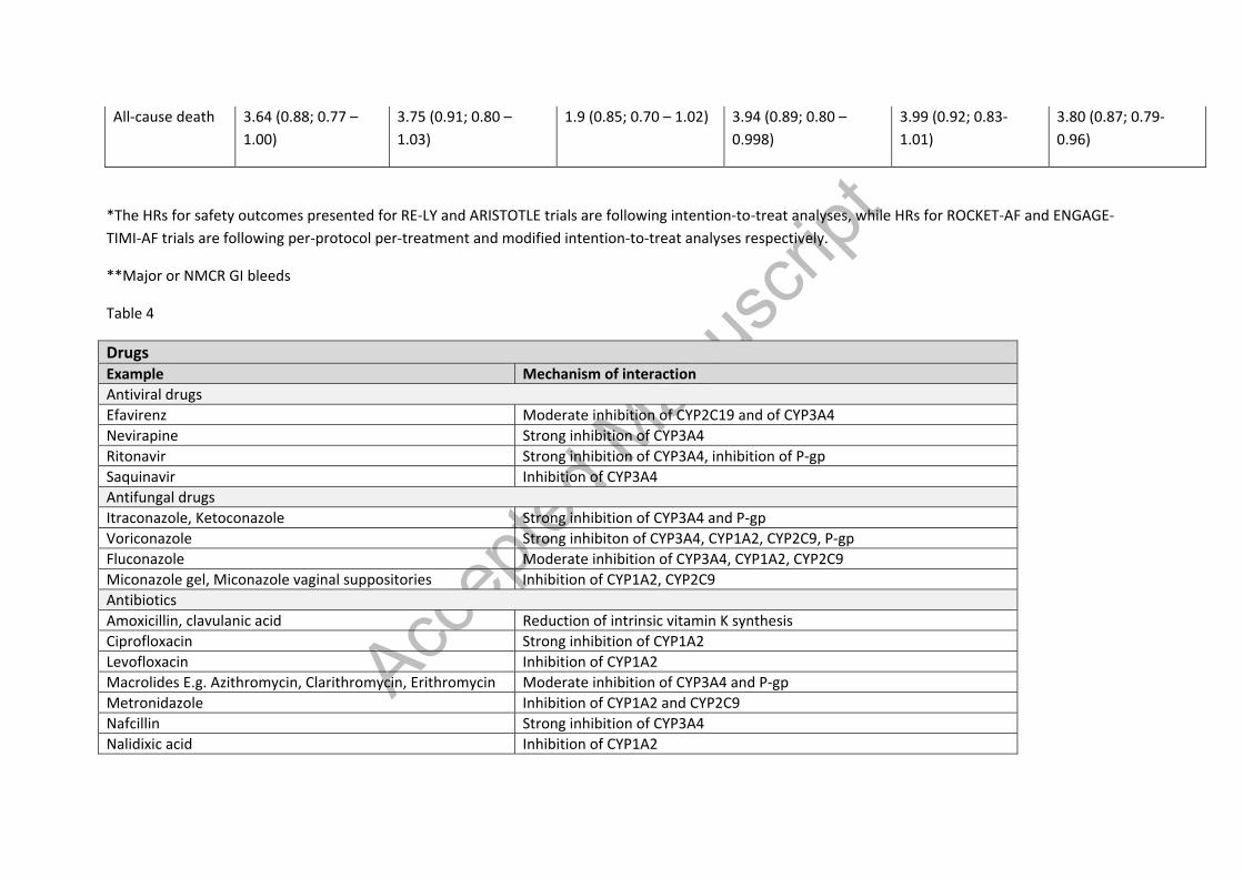
Table 4
Drugs Example Mechanism of interaction Antiviral drugs Efavirenz Moderate inhibition of CYP2C19 and of CYP3A4 Nevirapine Strong inhibition of CYP3A4 Ritonavir Strong inhibition of CYP3A4, inhibition of P-gp Saquinavir Inhibition of CYP3A4 Antifungal drugs Itraconazole, Ketoconazole Strong inhibition of CYP3A4 and P-gp Voriconazole Strong inhibiton of CYP3A4, CYP1A2, CYP2C9, P-gp Fluconazole Moderate inhibition of CYP3A4, CYP1A2, CYP2C9 Miconazole gel, Miconazole vaginal suppositories Inhibition of CYP1A2, CYP2C9 Antibiotics Amoxicillin, clavulanic acid Reduction of intrinsic vitamin K synthesis Ciprofloxacin Strong inhibition of CYP1A2 Levofloxacin Inhibition of CYP1A2 Macrolides E.g. Azithromycin, Clarithromycin, Erithromycin Moderate inhibition of CYP3A4 and P-gp Metronidazole Inhibition of CYP1A2 and CYP2C9 Nafcillin Strong inhibition of CYP3A4 Nalidixic acid Inhibition of CYP1A2

Accep
ted M
anus
cript
Norfloxacin Inhibition of CYP3A4 Isoniazid Inhibition of CYP2C9 Chloramphenicol Inhibition of CYP450 Rifampicin Induction of CYP3A4, CYP2C9 and p-gp Tetracyclines Inhibition of CYP3A4 Trimethoprim Inhibition of CYP3A4 Anti-arrhythmic drugs Amiodarone Moderate inhibition of CYP3A4, CYP1A2, CYP2C9 Dronedarone Moderate inhibition of CYP3A4, inhibitor of P-gp Disopyramide Inhibition of CYP3A4 Propranolol Inhibition of CYP1A2 Propafenone Inhibition of CYP3A4 Quinidine Inhibition of CYP3A4 Other cardiac drugs Diltiazem Inhibition of CYP3A4 Statins (E.g. atorvastatin,lovastatin, rosuvastatin, simvastatin)
Inhibition of CYP3A4
Telmisartan Inhibition of CYP3A4 Verapamil Weak inhibition of CYP3A4; inhibition of P-gp Anti-epileptic drugs Barbiturates (e.g. phenobarbital) Induction of CYP3A44, CYP2J and P-gp Carbamazepine Induction of CYP3A44, CYP2J and P-gp Phenytoin Induction of CYP3A44, CYP2J and P-gp Other drugs Fibrates (E.g. clofibrate, fenofibrate) Inhibition of CYP3A4 Glucagon Inhibition of CYP3A4 Proton pump inhibitors (e.g.) Inhibition of CYP2C9 and increased warfarin absorption Food Examples Mechanism of interaction Bishop’s weed, Carum Ajowan, Citrus aurantium (orange), Uncaria (Cat’s claw), Dehydroepiandrosterone, Echinacea, Goldenseal, Hydrastis Canadensis, Liquorice, Lime, Valerian,
Inhibition of CYP3A4

Accep
ted M
anus
cript
Wild cherry Chamomile Inhibition of CYP1A2, CYP2C9, CYP3A4 Cranberry, Milk thistle Inhibition of CYP3A4 and CYP2C9 Devil’s claw, Garlic, Ginsenoside Rd (found in Ginseng) Inhibition of CYP2C9, CYP2C19, CYP3A4 Eucalyptus, Feverfew, Fo-Ti, Peppermint, Red clover Inhibition of CYP1A2, CYP2C9, CYP2C19, CYP3A4 Gingko, Lycium (Chinese Wolfberry) Inhibition of CYP2C9 Grapefruit juice, Silybum marianum, Resveratrol Inhibition of CYP1A2 and CYP3A4 Mango Inhibition of CYPC219 Diindolylmethane, Grapes (Vitis Vinifera), Indole-3-carbinol Induction of CYP1A2 Ginsenoside Re and Rf (found in Ginseng) Induction of CYP3A4 and CYP2C9 Guggul Induction of CYP3A4 Limonene Induction of CYP2C9 St. John’s Wort Induction of CYP1A2, CYP2C9, CYP3A4 and P-gp

Accep
ted M
anus
cript
Accep
ted M
anus
cript
References
1. Weitz JI, Linkins L-A. Beyond heparin and warfarin: the new generation of anticoagulants. Expert Opin Investig Drugs. 2007 Mar 16;16(3):271–82.
2. Lip GYH, Freedman B, De Caterina R, Potpara TS. Stroke prevention in atrial fibrillation: Past, present and future. Thromb Haemost. 2017 Nov 8;117(07):1230–9.
3. Waller DG, Sampson AP, Waller DG, Sampson AP. Pharmacokinetics. Med Pharmacol Ther. 2018 Jan 1;33–62.
4. Hallworth M. Therapeutic drug monitoring. Clin Biochem Metab Clin Asp. 2014 Jan 1;767–86.
5. Ingrasciotta Y, Crisafulli S, Pizzimenti V, Marcianò I, Mancuso A, Andò G, et al. Pharmacokinetics of new oral anticoagulants: implications for use in routine care. Expert Opin Drug Metab Toxicol. 2018 Oct 3;14(10):1057–69.
6. Smith SA, Travers RJ, Morrissey JH. How it all starts: Initiation of the clotting cascade. Crit Rev Biochem Mol Biol. 2015;50(4):326–36.
7. Marder VJ. Hemostasis and thrombosis : basic principles and clinical practice. [Internet]. Wolters Kluwer/Lippincott Williams & Wilkins Health; 2012. 1566 p.
8. Mann KG, Brummel K, Butenas S. What is all that thrombin for? J Thromb Haemost. 2003 Jul;1(7):1504–14.
9. Ansell J. Factor Xa or thrombin: is factor Xa a better target? J Thromb Haemost. 2007 Jul 9;5:60–4.
10. Wolfe Z, Khan SU, Nasir F, Raghu Subramanian C, Lash B. A systematic review and Bayesian network meta-analysis of risk of intracranial hemorrhage with direct oral anticoagulants. J Thromb Haemost. 2018 Jul 1;16(7):1296–306.
11. Mackman N. The role of tissue factor and factor VIIa in hemostasis. Anesth Analg. 2009 May;108(5):1447–52.
12. Lijnen HR. Elements of the fibrinolytic system. Ann N Y Acad Sci. 2001;936:226–36.
13. Esmon CT, Vigano-D’Angelo S, D’Angelo A, Comp PC. Anticoagulation proteins C and S. Adv Exp Med Biol. 1987;214:47–54.
14. Wardrop D, Keeling D. The story of the discovery of heparin and warfarin.
15. Caterina R, Husted S, Wallentin L, Andreotti F, Arnesen H, Bachmann F, et al. Vitamin K antagonists in heart disease: Current status and perspectives (Section III). Thromb Haemost. 2013 Nov 30;110(12):1087–107.
16. Potpara T. Comparing Non-Vitamin K Antagonist Oral Anticoagulants (NOACs) to Different Coumadins: The Win-Win Scenarios. Thromb Haemost. 2018 May 4;118(05):803–5.

Accep
ted M
anus
cript
17. de Boer-van den Berg MA, Thijssen HH, Vermeer C. The in vivo effects of acenocoumarol, phenprocoumon and warfarin on vitamin K epoxide reductase and vitamin K-dependent carboxylase in various tissues of the rat. Biochim Biophys Acta. 1986 Oct 29;884(1):150–7.
18. Hirsh J, Dalen JE, Deykin D, Poller L, Bussey H. Oral Anticoagulants. Chest. 1995 Oct;108(4):231S–246S.
19. Flier JS, Underhill LH, Wessler S, Gitel SN. Warfarin: From bedside to bench. N Engl J Med. 1984 Sep 6;311(10):645–52.
20. Holford NHG. Clinical Pharmacokinetics and Pharmacodynamics of Warfarin. Clin Pharmacokinet. 1986;11(6):483–504.
21. Kim S, Gaweda AE, Wu D, Li L, Rai SN, Brier ME. Simplified Warfarin Dose-response Pharmacodynamic Models. Biomed Eng Appl basis, Commun. 2015 Feb;27(1).
22. Ansell J, Hirsh J, Hylek E, Jacobson A, Crowther M, Palareti G. Pharmacology and Management of the Vitamin K Antagonists. Chest. 2008 Jun;133(6):160S–198S.
23. Breckenridge A, L’E Orme M. The plasma half lives and the pharmacological effect of the enantiomers of warfarin in rats. Life Sci. 1972 Apr 8;11(7):337–45.
24. Jähnchen E, Meinertz T, Gilfrich HJ, Groth U, Martini A. The enantiomers of phenprocoumon: pharmacodynamic and pharmacokinetic studies. Clin Pharmacol Ther. 1976 Sep;20(3):342–9.
25. Meinertz T, Kasper W, Kahl C, Jähnchen E. Anticoagulant activity of the enantiomers of acenocoumarol. Br J Clin Pharmacol. 1978 Feb;5(2):187–8.
26. O’Reilly RA. Studies on the optical enantiomorphs of warfarin in man. Clin Pharmacol Ther. 1974 Aug;16(2):348–54.
27. Kelly JG, OʼMalley K. Clinical Pharmacokine cs of Oral An coagulants. Clin Pharmacokinet. 1979;4(1):1–15.
28. Hewick DS, McEwen J. Plasma half-lives, plasma metabolites and anticoagulant efficacies of the enantiomers of warfarin in man. J Pharm Pharmacol. 1973 Jun;25(6):458–65.
29. Lewis RJ, Trager WF, Chan KK, Breckenridge A, Orme M, Roland M, et al. Warfarin. Stereochemical aspects of its metabolism and the interaction with phenylbutazone. J Clin Invest. 1974 Jun 1;53(6):1607–17.
30. Kaminsky LS, Zhang Z-Y. Human P450 metabolism of warfarin. Pharmacol Ther. 1997 Jan 1;73(1):67–74.
31. Trager WF, Lewis RJ, Garland WA. Mass spectral analysis in the identification of human metabolites of warfarin. J Med Chem. 1970 Nov;13(6):1196–204.
32. Ufer M. Comparative Pharmacokinetics of Vitamin K Antagonists. Clin Pharmacokinet. 2005;44(12):1227–46.
Accep
ted M
anus
cript
33. Jansing RL, Chao ES, Kaminsky LS. Phase II metabolism of warfarin in primary culture of adult rat hepatocytes. Mol Pharmacol. 1992 Jan;41(1):209–15.
34. Higashi MK, Veenstra DL, Kondo LM, Wittkowsky AK, Srinouanprachanh SL, Farin FM, et al. Association between CYP2C9 genetic variants and anticoagulation-related outcomes during warfarin therapy. JAMA. 2002 Apr 3;287(13):1690–8.
35. Lane S, Al-Zubiedi S, Hatch E, Matthews I, Jorgensen AL, Deloukas P, et al. The population pharmacokinetics of R- and S-warfarin: effect of genetic and clinical factors. Br J Clin Pharmacol. 2012 Jan;73(1):66–76.
36. Toon S, Low LK, Gibaldi M, Trager WF, O’Reilly RA, Motley CH, et al. The warfarin-sulfinpyrazone interaction: stereochemical considerations. Clin Pharmacol Ther. 1986 Jan;39(1):15–24.
37. Baker RI, Coughlin PB, Gallus AS, Harper PL, Salem HH, Wood EM, et al. Warfarin reversal: consensus guidelines, on behalf of the Australasian Society of Thrombosis and Haemostasis. Med J Aust. 2004 Nov 1;181(9):492–7.
38. Wells PS, Holbrook AM, Crowther NR, Hirsh J. Interactions of warfarin with drugs and food. Ann Intern Med. 1994 Nov 1;121(9):676–83.
39. Martín-Pérez M, Gaist D, de Abajo F, Rodríguez L. Population Impact of Drug Interactions with Warfarin: A Real-World Data Approach. Thromb Haemost. 2018 Mar 12;118(03):461–70.
40. Haustein K-O. Pharmacokinetic and Pharmacodynamic Properties of Oral Anticoagulants, Especially Phenprocoumon. Semin Thromb Hemost. 1999 Feb 6;25(01):5–11.
41. Toon S, Heimark LD, Trager WF, O’Reilly RA. Metabolic fate of phenprocoumon in humans. J Pharm Sci. 1985 Oct;74(10):1037–40.
42. He M, Korzekwa KR, Jones JP, Rettie AE, Trager WF. Structural Forms of Phenprocoumon and Warfarin That Are Metabolized at the Active Site of CYP2C9. Arch Biochem Biophys. 1999 Dec;372(1):16–28.
43. Ufer M, Svensson JO, Krausz KW, Gelboin H V., Rane A, Tybring G. Identification of cytochromes P 450 2C9 and 3A4 as the major catalysts of phenprocoumon hydroxylation in vitro. Eur J Clin Pharmacol. 2004 May 1;60(3):173–82.
44. Serlin MJ, Sibeon RG, Mossman S, Breckenridge AM, Williams JR, Atwood JL, et al. Cimetidine: interaction with oral anticoagulants in man. Lancet (London, England). 1979 Aug 18;2(8138):317–9.
45. Harenberg J, Staiger C, de Vries JX, Walter E, Weber E, Zimmermann R. Cimetidine does not increase the anticoagulant effect of phenprocoumon. Br J Clin Pharmacol. 1982 Aug;14(2):292–3.
46. Thijssen H. Altered pharmacokinetics of R - and S -acenocoumarol in a subject heterozygous for CYP2C9*3. Clin Pharmacol Ther. 2001 Sep;70(3):292–8.
Accep
ted M
anus
cript
47. Dieterle W, Faigle JW, Montigel C, Sulc M, Theobald W. Biotransformation and pharmacokinetics of acenocoumarol (Sintrom) in man. Eur J Clin Pharmacol. 1977;11(5):367–75.
48. Thijssen HH, Flinois JP, Beaune PH. Cytochrome P4502C9 is the principal catalyst of racemic acenocoumarol hydroxylation reactions in human liver microsomes. Drug Metab Dispos. 2000 Nov;28(11):1284–90.
49. Thijssen HH, Baars LG, Janssen GM. Phenylbutazone-hydroxycoumarol interactions. Effects on steady state disposition, hepatocellular distribution, and biliary excretion of (S)-acenocoumarol in rats. Drug Metab Dispos. 16(5):744–8.
50. Scaglione F. New Oral Anticoagulants: Comparative Pharmacology with Vitamin K Antagonists. Clin Pharmacokinet. 2013 Feb 5;52(2):69–82.
51. Connolly SJ, Ezekowitz MD, Yusuf S, Eikelboom J, Oldgren J, Parekh A, et al. Dabigatran versus Warfarin in Patients with Atrial Fibrillation. N Engl J Med. 2009 Sep 17;361(12):1139–51.
52. Patel MR, Mahaffey KW, Garg J, Pan G, Singer DE, Hacke W, et al. Rivaroxaban versus Warfarin in Nonvalvular Atrial Fibrillation. N Engl J Med. 2011 Sep 8;365(10):883–91.
53. Granger CB, Alexander JH, McMurray JJV, Lopes RD, Hylek EM, Hanna M, et al. Apixaban versus Warfarin in Patients with Atrial Fibrillation. N Engl J Med. 2011 Sep 15;365(11):981–92.
54. Giugliano RP, Ruff CT, Braunwald E, Murphy SA, Wiviott SD, Halperin JL, et al. Edoxaban versus Warfarin in Patients with Atrial Fibrillation. N Engl J Med. 2013 Nov 28;369(22):2093–104.
55. De Caterina R, Husted S, Wallentin L, De Caterina R, Husted S, Wallentin L, et al. New Oral Anticoagulants in Atrial Fibrillation and Acute Coronary Syndromes. J Am Coll Cardiol. 2012 Apr 17;59(16):1413–25.
56. Ahmed Z, Hassan S, Salzman GA. Novel Oral Anticoagulants for Venous Thromboembolism with Special Emphasis on Risk of Hemorrhagic Complications and Reversal Agents. Curr Drug ther. 2016 Apr;11(1):3–20.
57. Mega JL, Braunwald E, Wiviott SD, Bassand J-P, Bhatt DL, Bode C, et al. Rivaroxaban in Patients with a Recent Acute Coronary Syndrome. N Engl J Med. 2012 Jan 5;366(1):9–19.
58. Kaserer A, Schedler A, Jetter A, Seifert B, Spahn D, Stein P, et al. Risk Factors for Higher-than-Expected Residual Rivaroxaban Plasma Concentrations in Real-Life Patients. Thromb Haemost. 2018 May 3;118(05):808–17.
59. Sacco M, Lancellotti S, Berruti F, Arcovito A, Bellelli A, Ricciardelli T, et al. Apixaban Interacts with Haemoglobin: Effects on Its Plasma Levels. Thromb Haemost. 2018 Oct 20;118(10):1701–12.
60. Reilly PA, Lehr T, Haertter S, Connolly SJ, Yusuf S, Eikelboom JW, et al. The Effect of
Accep
ted M
anus
cript
Dabigatran Plasma Concentrations and Patient Characteristics on the Frequency of Ischemic Stroke and Major Bleeding in Atrial Fibrillation Patients. J Am Coll Cardiol. 2014 Feb 4;63(4):321–8.
61. Testa S, Paoletti O, Legnani C, Dellanoce C, Antonucci E, Cosmi B, et al. Low drug levels and thrombotic complications in high-risk atrial fibrillation patients treated with direct oral anticoagulants. J Thromb Haemost. 2018 May 1;16(5):842–8.
62. Mismetti P, Laporte S. New oral antithrombotics: a need for laboratory monitoring. For. J Thromb Haemost. 2010 Apr 1;8(4):621–6.
63. Boissier E, Genebrier S, Lakhal K, Nedelec-Gac F, Trossaërt M, Ternisien C, et al. Rivaroxaban and Apixaban Anti-Xa Measurements: Impact of Plasma Storage for 7 Days at Room Temperature. Thromb Haemost. 2018 Aug 30;118(08):1488–90.
64. Douxfils J, Ageno W, Samama C-M, Lessire S, ten Cate H, Verhamme P, et al. Laboratory testing in patients treated with direct oral anticoagulants: a practical guide for clinicians. J Thromb Haemost. 2018 Feb 1;16(2):209–19.
65. Cuker A, Siegal DM, Crowther MA, Garcia DA. Laboratory measurement of the anticoagulant activity of the non-vitamin K oral anticoagulants. J Am Coll Cardiol. 2014 Sep 16;64(11):1128–39.
66. Garcia D, Barrett YC, Ramacciotti E, Weitz JI. Laboratory assessment of the anticoagulant effects of the next generation of oral anticoagulants. J Thromb Haemost. 2013 Feb 1;11(2):245–52.
67. Yetman R, Barrett Y, Wang Z, Adamczyk R, Wang J, Ramacciotti E, et al. Apixaban pharmacodynamic activity in umbilical cord, paediatric, and adult plasma. Thromb Haemost. 2017 Nov 22;117(08):1518–27.
68. Studt J-D, Alberio L, Angelillo-Scherrer A, Asmis LM, Fontana P, Korte W, et al. Accuracy and consistency of anti-Xa activity measurement for determination of rivaroxaban plasma levels. J Thromb Haemost. 2017 Aug 1;15(8):1576–83.
69. Ignjatovic V. Thrombin Clotting Time. In: Methods in molecular biology (Clifton, NJ). 2013. p. 131–8.
70. Wienen W, Stassen J-M, Priepke H, Ries UJ, Hauel N. In-vitro profile and ex-vivo anticoagulant activity of the direct thrombin inhibitor dabigatran and its orally active prodrug, dabigatran etexilate. Thromb Haemost. 2007 Jul;98(1):155–62.
71. Gustafsson D, Elg M. The pharmacodynamics and pharmacokinetics of the oral direct thrombin inhibitor ximelagatran and its active metabolite melagatran: a mini-review. Thromb Res. 2003 Jul 15;109 Suppl 1:S9-15.
72. Stangier J, Clemens A. Pharmacology, Pharmacokinetics, and Pharmacodynamics of Dabigatran Etexilate, an Oral Direct Thrombin Inhibitor. Clin Appl Thromb. 2009 Feb 19;15(1_suppl):9S–16S.
73. Stangier J, Rathgen K, Stähle H, Gansser D, Roth W. The pharmacokinetics,
Accep
ted M
anus
cript
pharmacodynamics and tolerability of dabigatran etexilate, a new oral direct thrombin inhibitor, in healthy male subjects. Br J Clin Pharmacol. 2007 Sep;64(3):292–303.
74. Ingelheim B. Relative bioavailability of dabigatran after administration of different application forms of a single oral dose of 150mg of dabigatran etixilate [Internet].
75. Golub AL, Frost RW, Betlach CJ, Gonzalez MA. Physiologic considerations in drug absorption from the gastrointestinal tract. J Allergy Clin Immunol. 1986 Oct 1;78(4 Pt 2):689–94.
76. Stangier J, Eriksson BI, Dahl OE, Ahnfelt L, Nehmiz G, Stähle H, et al. Pharmacokinetic Profile of the Oral Direct Thrombin Inhibitor Dabigatran Etexilate in Healthy Volunteers and Patients Undergoing Total Hip Replacement. J Clin Pharmacol. 2005 May;45(5):555–65.
77. Brighton T. Experimental and clinical pharmacology: New oral anticoagulant drugs - mechanisms of action. Aust Prescr. 2010 Apr 1;33(2):38–41.
78. Blech S, Ebner T, Ludwig-Schwellinger E, Stangier J, Roth W. The Metabolism and Disposition of the Oral Direct Thrombin Inhibitor, Dabigatran, in Humans. Drug Metab Dispos. 2008 Feb 1;36(2):386–99.
79. Stangier J. Clinical Pharmacokinetics and Pharmacodynamics of the Oral Direct Thrombin Inhibitor Dabigatran Etexilate. Clin Pharmacokinet. 2008;47(5):285–95.
80. Liesenfeld K-H, Lehr T, Dansirikul C, Reilly PA, Connolly SJ, Ezekowitz MD, et al. Population pharmacokinetic analysis of the oral thrombin inhibitor dabigatran etexilate in patients with non-valvular atrial fibrillation from the RE-LY trial. J Thromb Haemost. 2011 Nov 1;9(11):2168–75.
81. Becattini C, Giustozzi M, Ranalli MG, Bogliari G, Cianella F, Verso M, et al. Variation of renal function over time is associated with major bleeding in patients treated with direct oral anticoagulants for atrial fibrillation. J Thromb Haemost. 2018 May 1;16(5):833–41.
82. European Medicines Agency. CHMP assessment report for Pradaxa [Internet]. 2008.
83. DeWald TA, Becker RC. The pharmacology of novel oral anticoagulants. J Thromb Thrombolysis. 2014 Feb 17;37(2):217–33.
84. Terao T, Hisanaga E, Sai Y, Tamai I, Tsuji A. Active secretion of drugs from the small intestinal epithelium in rats by P-glycoprotein functioning as an absorption barrier. J Pharm Pharmacol. 1996 Oct;48(10):1083–9.
85. Avgil-Tsadok M, Jackevicius C, Essebag V, Eisenberg MJ, Rahme E, Behlouli H, et al. Dabigatran use in elderly patients with atrial fibrillation. Thromb Haemost. 2016 Jan 22;115(01):152–60.
86. Perzborn E, Strassburger J, Wilmen A, Pohlmann J, Roehrig S, Schlemmer K-H, et al. In vitro and in vivo studies of the novel antithrombotic agent BAY 59-7939-an oral,
Accep
ted M
anus
cript
direct Factor Xa inhibitor. J Thromb Haemost. 2005 Mar;3(3):514–21.
87. Gerotziafas, GT; Elalamy, I; Depasse, F; Perzborn, E; Samama M. In vitro inhibition of thrombin generation, after tissue factor pathway activation, by the oral, direct factor Xa inhibitor rivaroxaban. J Thromb Haemost. 2007;(5):886–8.
88. Moore KT, Krook MA, Vaidyanathan S, Sarich TC, Damaraju C V, Fields LE. Rivaroxaban Crushed Tablet Suspension Characteristics and Relative Bioavailability in Healthy Adults When Administered Orally or Via Nasogastric Tube. Clin Pharmacol Drug Dev. 2014;3(4):321–7.
89. Kubitza D, Becka M, Zuehlsdorf M, Mueck W. Effect of Food, an Antacid, and the H 2 Antagonist Ranitidine on the Absorption of BAY 59-7939 (Rivaroxaban), an Oral, Direct Factor Xa Inhibitor, in Healthy Subjects. J Clin Pharmacol. 2006 May;46(5):549–58.
90. Martin KA, Lee CR, Farrell TM, Moll S. Oral Anticoagulant Use After Bariatric Surgery: A Literature Review and Clinical Guidance. Am J Med. 2017 May;130(5):517–24.
91. Singh BN. Effects of Food on Clinical Pharmacokinetics. Clin Pharmacokinet. 1999 Sep;37(3):213–55.
92. Kubitza D, Becka M, Zuehlsdorf M, Mueck W. Body Weight Has Limited Influence on the Safety, Tolerability, Pharmacokinetics, or Pharmacodynamics of Rivaroxaban (BAY 59-7939) in Healthy Subjects. J Clin Pharmacol. 2007 Feb;47(2):218–26.
93. Weinz C, Schwarz T, Kubitza D, Mueck W, Lang D. Metabolism and Excretion of Rivaroxaban, an Oral, Direct Factor Xa Inhibitor, in Rats, Dogs, and Humans. Drug Metab Dispos. 2009 May 1;37(5):1056–64.
94. Kubitza D, Becka M, Mueck W, Halabi A, Maatouk H, Klause N, et al. Effects of renal impairment on the pharmacokinetics, pharmacodynamics and safety of rivaroxaban, an oral, direct Factor Xa inhibitor. Br J Clin Pharmacol. 2010 Nov;70(5):703–12.
95. Kubitza D, Roth A, Becka M, Alatrach A, Halabi A, Hinrichsen H, et al. Effect of hepatic impairment on the pharmacokinetics and pharmacodynamics of a single dose of rivaroxaban, an oral, direct Factor Xa inhibitor. Br J Clin Pharmacol. 2013 Jul;76(1):89–98.
96. Gnoth MJ, Buetehorn U, Muenster U, Schwarz T, Sandmann S. In Vitro and In Vivo P-Glycoprotein Transport Characteristics of Rivaroxaban. J Pharmacol Exp Ther. 2011 Jul 1;338(1):372–80.
97. Leslie EM, Deeley RG, Cole SPC. Multidrug resistance proteins: role of P-glycoprotein, MRP1, MRP2, and BCRP (ABCG2) in tissue defense. Toxicol Appl Pharmacol. 2005 May 1;204(3):216–37.
98. Mueck W, Kubitza D, Becka M. Co-administration of rivaroxaban with drugs that share its elimination pathways: pharmacokinetic effects in healthy subjects. Br J Clin Pharmacol. 2013 Sep;76(3):455–66.
Accep
ted M
anus
cript
99. DeHaas KA. The Direct Oral Anticoagulants Apixaban, Rivaroxaban, and Edoxaban. Am Soc Clin Lab Sci. 2017 Jan 1;30(1):2–6.
100. Bonar R, Favaloro EJ, Mohammed S, Ahuja M, Pasalic L, Sioufi J, et al. The effect of the direct factor Xa inhibitors apixaban and rivaroxaban on haemostasis tests: a comprehensive assessment using in vitro and ex vivo samples. Pathology. 2016 Jan;48(1):60–71.
101. Song Y, Chang M, Suzuki A, Frost RJA, Kelly A, LaCreta F, et al. Evaluation of Crushed Tablet for Oral Administration and the Effect of Food on Apixaban Pharmacokinetics in Healthy Adults. Clin Ther. 2016 Jul;38(7):1674–1685.e1.
102. Frost C, Nepal S, Wang J, Schuster A, Byon W, Boyd RA, et al. Safety, pharmacokinetics and pharmacodynamics of multiple oral doses of apixaban, a factor Xa inhibitor, in healthy subjects. Br J Clin Pharmacol. 2013 Nov;76(5):776–86.
103. Wong PC, Pinto DJP, Zhang D. Preclinical discovery of apixaban, a direct and orally bioavailable factor Xa inhibitor. J Thromb Thrombolysis. 2011 May;31(4):478–92.
104. Upreti V V, Wang J, Barrett YC, Byon W, Boyd RA, Pursley J, et al. Effect of extremes of body weight on the pharmacokinetics, pharmacodynamics, safety and tolerability of apixaban in healthy subjects. Br J Clin Pharmacol. 2013 Dec;76(6):908–16.
105. Raghavan N, Frost CE, Yu Z, He K, Zhang H, Humphreys WG, et al. Apixaban Metabolism and Pharmacokinetics after Oral Administration to Humans. Drug Metab Dispos. 2009 Jan 1;37(1):74–81.
106. Chang M, Yu Z, Shenker A, Wang J, Pursley J, Byon W, et al. Effect of renal impairment on the pharmacokinetics, pharmacodynamics, and safety of apixaban. J Clin Pharmacol. 2016 May;56(5):637–45.
107. Graff J, Harder S. Anticoagulant Therapy with the Oral Direct Factor Xa Inhibitors Rivaroxaban, Apixaban and Edoxaban and the Thrombin Inhibitor Dabigatran Etexilate in Patients with Hepatic Impairment. Clin Pharmacokinet. 2013 Apr 7;52(4):243–54.
108. Kubisz P, Stanciakova L, Dobrotova M, Samos M, Mokan M, Stasko J. Apixaban - Metabolism, Pharmacologic Properties and Drug Interactions. Curr Drug Metab. 2017 Sep 6;18(7):609–21.
109. Frost CE, Byon W, Song Y, Wang J, Schuster AE, Boyd RA, et al. Effect of ketoconazole and diltiazem on the pharmacokinetics of apixaban, an oral direct factor Xa inhibitor. Br J Clin Pharmacol. 2015 May;79(5):838–46.
110. Vakkalagadda B, Frost C, Byon W, Boyd RA, Wang J, Zhang D, et al. Effect of Rifampin on the Pharmacokinetics of Apixaban, an Oral Direct Inhibitor of Factor Xa. Am J Cardiovasc Drugs. 2016 Apr 9;16(2):119–27.
111. Furugohri T, Isobe K, Honda Y, Kamisato-Matsumoto C, Sugiyama N, Nagahara T, et al. DU-176b, a potent and orally active factor Xa inhibitor: in vitro and in vivo pharmacological profiles. J Thromb Haemost. 2008 Jun;6(9):1542–9.
Accep
ted M
anus
cript
112. Stacy ZA, Call WB, Hartmann AP, Peters GL, Richter SK. Edoxaban: A Comprehensive Review of the Pharmacology and Clinical Data for the Management of Atrial Fibrillation and Venous Thromboembolism. Cardiol Ther. 2016 Jun;5(1):1–18.
113. Chen X, Liu D, Wu Y, Song H, Liu Y, Jiang J, et al. A single-dose study investigating the pharmacokinetics and pharmacodynamics of edoxaban at 30–90 mg in healthy Chinese volunteers. Xenobiotica. 2017 Jul 3;47(7):592–9.
114. Mendell J, Tachibana M, Shi M, Kunitada S. Effects of Food on the Pharmacokinetics of Edoxaban, an Oral Direct Factor Xa Inhibitor, in Healthy Volunteers. J Clin Pharmacol. 2011 May;51(5):687–94.
115. Matsushima N, Lee F, Sato T, Weiss D, Mendell J. Bioavailability and Safety of the Factor Xa Inhibitor Edoxaban and the Effects of Quinidine in Healthy Subjects. Clin Pharmacol Drug Dev. 2013 Oct;2(4):358–66.
116. Parasrampuria DA, Kanamaru T, Connor A, Wilding I, Ogata K, Shimoto Y, et al. Evaluation of regional gastrointestinal absorption of edoxaban using the enterion capsule. J Clin Pharmacol. 2015 Nov;55(11):1286–92.
117. Parasrampuria DA, Truitt KE. Pharmacokinetics and Pharmacodynamics of Edoxaban, a Non-Vitamin K Antagonist Oral Anticoagulant that Inhibits Clotting Factor Xa. Clin Pharmacokinet. 2016;55(6):641–55.
118. Bathala MS, Masumoto H, Oguma T, He L, Lowrie C, Mendell J. Pharmacokinetics, Biotransformation, and Mass Balance of Edoxaban, a Selective, Direct Factor Xa Inhibitor, in Humans. Drug Metab Dispos. 2012 Dec 1;40(12):2250–5.
119. Cada DJ, Baker DE, Ingram K. Edoxaban. Hosp Pharm. 2015 Jul;50(7):619–34.
120. Plitt A, Giugliano RP. Edoxaban: Review of Pharmacology and Key Phase I to III Clinical Trials. J Cardiovasc Pharmacol Ther. 2014 Sep 7;19(5):409–16.
121. Mendell J, Johnson L, Chen S. An open-label, phase 1 study to evaluate the effects of hepatic impairment on edoxaban pharmacokinetics and pharmacodynamics. J Clin Pharmacol. 2015 Dec;55(12):1395–405.
122. Steffel J, Verhamme P, Potpara TS, Albaladejo P, Antz M, Desteghe L, et al. The 2018 European Heart Rhythm Association Practical Guide on the use of non-vitamin K antagonist oral anticoagulants in patients with atrial fibrillation.
123. Voukalis C, Lip GYH, Shantsila E. Drug-drug interactions of non-vitamin K oral anticoagulants. Expert Opin Drug Metab Toxicol. 2016 Dec 26;12(12):1445–61.
124. Mendell J, Chen S, He L, Desai M, Parasramupria DA. The Effect of Rifampin on the Pharmacokinetics of Edoxaban in Healthy Adults. Clin Drug Investig. 2015 Jul 12;35(7):447–53.
125. Mendell J, Lee F, Chen S, Worland V, Shi M, Samama MM. The Effects of the Antiplatelet Agents, Aspirin and Naproxen, on Pharmacokinetics and Pharmacodynamics of the Anticoagulant Edoxaban, a Direct Factor Xa Inhibitor. J
Accep
ted M
anus
cript
Cardiovasc Pharmacol. 2013 Aug;62(2):212–21.
126. Maddison J, Somogyi AA, Jensen BP, James HM, Gentgall M, Rolan PE. The pharmacokinetics and pharmacodynamics of single dose (R)-and (S)-warfarin administered separately and together: relationship to VKORC1 genotype. 2012;
127. Yin T, Miyata T. Warfarin dose and the pharmacogenomics of CYP2C9 and VKORC1 — Rationale and perspectives. Thromb Res. 2007 Jan;120(1):1–10.
128. Ayrton A, Morgan P. Role of transport proteins in drug absorption, distribution and excretion. Xenobiotica. 2001 Jan 22;31(8–9):469–97.
129. Johnson JA, Gong L, Whirl-Carrillo M, Gage BF, Scott SA, Stein CM, et al. Clinical Pharmacogenetics Implementation Consortium Guidelines for CYP2C9 and VKORC1 genotypes and warfarin dosing. Clin Pharmacol Ther. 2011 Oct;90(4):625–9.
130. Pirmohamed M, Burnside G, Eriksson N, Jorgensen AL, Toh CH, Nicholson T, et al. A Randomized Trial of Genotype-Guided Dosing of Warfarin. N Engl J Med. 2013 Dec 12;369(24):2294–303.
131. Kimmel SE, French B, Kasner SE, Johnson JA, Anderson JL, Gage BF, et al. A Pharmacogenetic versus a Clinical Algorithm for Warfarin Dosing. N Engl J Med. 2013 Dec 12;369(24):2283–93.
132. Lu G, Hollenbach SJ, Baker DC, Tan S, Hutchaleelaha A, Curnutte JT, et al. Preclinical safety and efficacy of andexanet alfa in animal models. J Thromb Haemost. 2017 Sep 1;15(9):1747–56.
133. Heo Y-A. Andexanet Alfa: First Global Approval. Drugs. 2018 Jul 20;78(10):1049–55.
134. Weitz JI, Harenberg J. New developments in anticoagulants: Past, present and future. Thromb Haemost. 2017 Nov 11;117(07):1283–8.
135. Steffel J, Verhamme P, Potpara TS, Albaladejo P, Antz M, Desteghe L, et al. The 2018 European Heart Rhythm Association Practical Guide on the use of non-vitamin K antagonist oral anticoagulants in patients with atrial fibrillation. Eur Heart J. 2018 Apr 21;39(16):1330–93.
136. Bristol-Myers. Squibb. Eliquis (apixaban) 2.5 and 5 mg Tablets. [Internet]. 2014.
137. Reinecke H, Jürgensmeyer S, Engelbertz C, Gerss J, Kirchhof P, Breithardt G, et al. Design and rationale of a randomised controlled trial comparing apixaban to phenprocoumon in patients with atrial fibrillation on chronic haemodialysis: the AXADIA-AFNET 8 study. BMJ Open. 2018 Sep 10;8(9):e022690.
138. Stanton BE, Barasch NS, Tellor KB. Comparison of the Safety and Effectiveness of Apixaban versus Warfarin in Patients with Severe Renal Impairment. Pharmacother J Hum Pharmacol Drug Ther. 2017 Apr;37(4):412–9.
139. Martin JL, Esmaeili H, Manuel RC, Petrini M, Wiebe S, Maas H. Pharmacokinetics/Pharmacodynamics of Dabigatran 75 mg Twice Daily in Patients With Nonvalvular Atrial Fibrillation and Severely Impaired Renal Function. J
Accep
ted M
anus
cript
Cardiovasc Pharmacol Ther. 2018 Sep 25;23(5):399–406.
140. Sherwood MW, Nessel CC, Hellkamp AS, Mahaffey KW, Piccini JP, Suh E-Y, et al. Gastrointestinal Bleeding in Patients With Atrial Fibrillation Treated With Rivaroxaban or Warfarin. J Am Coll Cardiol. 2015 Dec 1;66(21):2271–81.
141. Hellenbart E, Faulkenberg K, Finks S. Evaluation of bleeding in patients receiving direct oral anticoagulants. Vasc Health Risk Manag. 2017 Aug;Volume 13:325–42.
142. Granger CB, Alexander JH, McMurray JJV, Lopes RD, Hylek EM, Hanna M, et al. Apixaban versus Warfarin in Patients with Atrial Fibrillation. N Engl J Med. 2011 Sep 15;365(11):981–92.
143. Di Minno A, Frigerio B, Spadarella G, Ravani A, Sansaro D, Amato M, et al. Old and new oral anticoagulants: Food, herbal medicines and drug interactions. Blood Rev. 2017 Jul;31(4):193–203.
144. Nutescu EA, Shapiro NL, Ibrahim S, West P. Warfarin and its interactions with foods, herbs and other dietary supplements. Expert Opin Drug Saf. 2006 May 13;5(3):433–51.
145. Vranckx P, Valgimigli M, Heidbuchel H. The Significance of Drug-Drug and Drug-Food Interactions of Oral Anticoagulation. Arrhythmia Electrophysiol Rev. 2018 Mar;7(1):55–61.
146. Tiaden JD, Wenzel E, Berthold HK, Müller-Oerlinghausen B. Adverse Reactions to Anticoagulants and to Antiplatelet Drugs Recorded by the German Spontaneous Reporting System. Semin Thromb Hemost. 2005 Aug;31(04):371–80.