pharmacokinetics, biodistribution, and pharmacodynamics of...
TRANSCRIPT
JPET # 257113
1
Title: Pharmacokinetic and Pharmacodynamic Properties of Drug Delivery Systems
Authors: Patrick M. Glassman, Vladimir R. Muzykantov
Affiliation: Department of Systems Pharmacology and Translational Therapeutics, Perelman
School of Medicine, University of Pennsylvania
Address: 3400 Civic Center Boulevard, Bldg 421, Philadelphia, Pennsylvania 19104-5158,
United States
This article has not been copyedited and formatted. The final version may differ from this version.JPET Fast Forward. Published on March 5, 2019 as DOI: 10.1124/jpet.119.257113
at ASPE
T Journals on M
arch 15, 2020jpet.aspetjournals.org
Dow
nloaded from
JPET # 257113
2
Running Title: PK/PD Properties of Drug Delivery Systems
Corresponding Authors: Vladimir R. Muzykantov ([email protected], (215)
898-9823) and Patrick M. Glassman ([email protected])
# of Text Pages: 22
# of Tables: 2
# of Figures: 4
Word Count – Abstract: 144
Word Count – Introduction: 350
Word Count – Discussion: N/A
Non-Standard Abbreviations:
Absorption, Distribution, Metabolism, and Elimination (ADME)
Biodistribution (BD)
Drug Delivery Systems (DDSs)
Enhanced Permeability & Retention (EPR)
Gastrointestinal (GI)
Intravenously (IV)
Neonatal Fc Receptor (FcRn)
Monoclonal Antibody (mAb)
Reticuloendothelial System (RES)
Pharmacodynamics (PD)
Pharmacokinetics (PK)
Physiologically-Based Pharmacokinetic (PBPK)
Subcutaneously (SC)
Target-Mediated Drug Disposition (TMDD)
Recommended Section: Drug Discovery and Translational Medicine
This article has not been copyedited and formatted. The final version may differ from this version.JPET Fast Forward. Published on March 5, 2019 as DOI: 10.1124/jpet.119.257113
at ASPE
T Journals on M
arch 15, 2020jpet.aspetjournals.org
Dow
nloaded from
JPET # 257113
3
ABSTRACT
The use of drug delivery systems (DDSs) is an attractive approach to facilitate uptake of
therapeutic agents at the desired site of action, particularly when free drug has poor
pharmacokinetics/biodistribution (PK/BD) or significant off-site toxicities. Successful
translation of DDSs into the clinic is dependent on a thorough understanding of the in vivo
behavior of the carrier, which has for the most part been an elusive goal. This is, at least in part,
due to significant differences in the mechanisms controlling pharmacokinetics for classical drugs
and DDSs. In this review, we summarize the key physiological mechanisms controlling the in
vivo behavior of DDSs, comparing and contrasting this with classical drugs, and describing
engineering strategies designed to improve DDS PK/BD. In addition, we describe quantitative
approaches that could be useful for describing PK/BD of DDSs, as well as critical steps between
tissue uptake and pharmacologic effect.
This article has not been copyedited and formatted. The final version may differ from this version.JPET Fast Forward. Published on March 5, 2019 as DOI: 10.1124/jpet.119.257113
at ASPE
T Journals on M
arch 15, 2020jpet.aspetjournals.org
Dow
nloaded from

JPET # 257113
4
INTRODUCTION
Modern pharmacotherapy employs an expanded roster of distinct classes of therapeutic,
prophylactic, imaging and other agents, ranging in size and complexity from diatomic gases,
oxygen and nitric oxide, to cellular fragments and cells themselves – natural or modified
chemically or genetically. In between these extremes, therapeutics can be divided into classical
small drugs and biologicals or biotherapeutics such as proteins, nucleic acids and other
biomolecules.
Both small molecules and biologicals have issues with delivery in the organism of a patient, from
administration site to the desirable site of action. Accordingly, diverse drug delivery systems
(DDSs: liposomes, nanocarriers, affinity drug conjugates and so on) are devised to enable or
improve delivery of some of these agents. In addition, in some cases DDSs themselves have
additional functions and even therapeutic action. In this review, we highlight critical factors that
affect the behavior of DDS following injection into an organism. The majority of the work
discussed focuses on liposomes, as these have been the most extensively studied DDS to date;
however, the critical parameters affecting in vivo behavior are likely relevant to many types of
DDS.
Each type of these agents - small drugs, biologicals and DDSs - has advantages and challenges,
some of which outlined in Table 1. Here we attempt a comparative review of the main
parameters of their behavior in the body, that we colloquially call pharmacokinetics (PK). PK is
often defined simply as ‘what the body does to the drug’, and is typically described using four
critical processes: absorption (A), distribution (D), metabolism (M), and elimination (E), or
This article has not been copyedited and formatted. The final version may differ from this version.JPET Fast Forward. Published on March 5, 2019 as DOI: 10.1124/jpet.119.257113
at ASPE
T Journals on M
arch 15, 2020jpet.aspetjournals.org
Dow
nloaded from
JPET # 257113
5
ADME. The interactions between the drug molecule (or drug delivery system) and the body
control the relative rates and efficiencies of each of these processes and body compartments
involved.
While these processes are well understood and described for small molecule drugs and for many
protein therapeutics, a thorough understanding of PK (and underlying mechanisms) is often
lacking for DDSs. This is likely due to several reasons, including, but not limited to assay
limitations, interspecies differences in processes controlling PK, and a smaller overall body of
work on PK of drug delivery systems (DDS), particularly in the clinic. In this review, we discuss
differences in ADME processes for small molecule drugs, protein biotherapeutics, and DDS. In
addition, the key features of DDS that can be tuned to modulate PK and analysis of DDS PK will
be discussed in detail.
ADME PROCESSES
One challenge in characterization of the in vivo behavior of DDS is the differences in
mechanisms controlling PK and biodistribution compared with small molecule drugs and
biologics. As the purpose of this review is not to provide a detailed description of the ADME of
small molecules and biologics, but rather to highlight their differences with DDS, only a brief
overview of mechanisms controlling their in vivo behavior will be provided (Figure 1, Table 2).
Absorption
For drugs administered via an extravascular route, the first barrier to reaching the site of action is
absorption into the bloodstream, which can be controlled both by properties of the drug and of
This article has not been copyedited and formatted. The final version may differ from this version.JPET Fast Forward. Published on March 5, 2019 as DOI: 10.1124/jpet.119.257113
at ASPE
T Journals on M
arch 15, 2020jpet.aspetjournals.org
Dow
nloaded from
JPET # 257113
6
the site of administration. For small molecule drugs, absorption most frequently occurs from the
gastrointestinal (GI) tract following oral administration. Briefly, following dosing, the dosage
form must disintegrate and the drug has to dissolve and permeate across the GI wall. The rate
and extent of this process can vary widely between drugs, although predictions can often be
made based on physicochemical properties of the drug molecule (Palm et al., 1997; Lipinski et
al., 2001). It should be noted; however, that interactions with transporters (Estudante et al.,
2013) and drug-metabolizing enzymes (Peters et al., 2016) in the GI tract can significantly
modulate the passive absorption profile that would be predicted using molecular descriptors.
On the other hand, in general, biologics are poorly absorbed following oral absorption, and as
such, are often administered intravenously (IV); however, subcutaneous (SC) dosing of protein
therapeutics has become more popular in recent years. Absorption from this space is generally a
slow process (hours – days), due to the pathway through the lymphatic system that most proteins
follow following SC dosing (Supersaxo et al., 1990; Bittner et al., 2018). While determinants of
the efficiency of SC administration for protein therapeutics are not as well-understood as oral
absorption of small molecules, it is appreciated that molecular properties of the protein (e.g. size,
charge), affinity for the neonatal Fc receptor (FcRn) (Deng et al., 2012; Zheng et al., 2012;
Richter et al., 2018), and addition of absorption enhancers to the formulation (e.g. buffer
components, hyaluronidase) can impact bioavailability (Fathallah et al., 2015; Bittner et al.,
2018).
Finally, for DDS, absorption is not typically a process that is considered, as the efficiency of
uptake into the systemic circulation after extravascular delivery is very low. There have been
This article has not been copyedited and formatted. The final version may differ from this version.JPET Fast Forward. Published on March 5, 2019 as DOI: 10.1124/jpet.119.257113
at ASPE
T Journals on M
arch 15, 2020jpet.aspetjournals.org
Dow
nloaded from
JPET # 257113
7
many preclinical investigations of oral delivery of nanoparticles; however, absorption is often
low due to poor permeation across the GI wall. Following extravascular injection (e.g. SC or
intramuscular) of DDS, bioavailability would likely be very low due to efficient uptake by
resident immune cells in the lymph nodes collecting fluid draining from the injection site;
however, this may be an efficient route of administration for local delivery (Kaledin et al., 1982).
Distribution
Following entry into the systemic circulation, the movement of drugs between blood and tissues
is a critical factor controlling the efficacy and toxicities associated with therapy. As with
absorption, distribution varies widely between drug classes both in kinetics and in mechanism.
The distribution of small molecule drugs, in particular, may range from being confined to the
plasma space to being distributed throughout the entire body. This variability can, in part be
described using molecular descriptors and binding to plasma proteins (Poulin and Theil, 2002b;
Poulin and Theil, 2002a). Distribution of small molecule drugs can be modulated by interactions
with uptake and/or efflux transporters expressed in certain tissues (Giacomini et al., 2010).
The efficiency of distribution of protein therapeutics into tissues is highly dependent on the
molecular weight of the protein, with smaller proteins entering tissues more efficiently than
larger proteins, due to enhanced diffusion and improved permeation through paracellular pores
(convective uptake) (Sarin, 2010). Additionally, tissue uptake can be increased via receptor-
mediated transcytosis for proteins with high affinity for receptors such as the transferrin receptor
(TfR) (Friden et al., 1991; Pardridge et al., 1991).
This article has not been copyedited and formatted. The final version may differ from this version.JPET Fast Forward. Published on March 5, 2019 as DOI: 10.1124/jpet.119.257113
at ASPE
T Journals on M
arch 15, 2020jpet.aspetjournals.org
Dow
nloaded from
JPET # 257113
8
As most DDS are much larger than typical pores between endothelial cells, distribution is often
limited to the vascular space (Allen et al., 1989), in the absence of specific pathologies or affinity
for receptors. However, in tissues with larger endothelial pores (e.g. fenestrations in liver and
spleen), tissue uptake via bulk fluid flow (convection) may be favorable. In a similar manner to
biologics, DDS with affinity for receptors that undergo transcytosis may have enhanced tissue
uptake at sites of target expression (Cerletti et al., 2000; Hatakeyama et al., 2004).
Metabolism/Elimination
As with the previous processes, elimination of drugs from the system occurs via different
mechanisms and at different rates for various types of molecules. For small molecules, there are
two primary routes of elimination. Renal clearance is controlled by the relative efficiencies of
glomerular filtration, active secretion into the urine, and reabsorption (active and passive) from
the tubules (Dave and Morris, 2015). Metabolic clearance, occurring primarily in the liver for
most drugs, is dependent on recognition of the drug molecule by a drug-metabolizing enzyme
(e.g Cytochrome P450). Following metabolism, the metabolite can be further metabolized,
cleared via the bile ducts into the feces, or eliminated in the urine.
For peptides and small protein therapeutics, renal clearance may be significant when molecular
weight is smaller than the glomerular filtration threshold (~ 60 kDa). However, for proteins that
are not eliminated in the urine, catabolic breakdown can occur throughout the body, typically
following uptake into the endo-lysosomal pathway. The efficiency of this breakdown can be
enhanced if a protein with high affinity for an internalizing receptor is taken up via receptor-
mediated endocytosis, in a process often referred to as target-mediated drug disposition (TMDD)
This article has not been copyedited and formatted. The final version may differ from this version.JPET Fast Forward. Published on March 5, 2019 as DOI: 10.1124/jpet.119.257113
at ASPE
T Journals on M
arch 15, 2020jpet.aspetjournals.org
Dow
nloaded from
JPET # 257113
9
(Levy, 1994; Mager and Jusko, 2001a). For proteins containing an Fc region (e.g. mAbs and Fc
fusion proteins), elimination may be blunted via interactions with the neonatal Fc receptor
(FcRn), which protects IgG and albumin from degradation, allowing them to have long
circulating half-lifes (~ 3 weeks in man) (Ghetie et al., 1996; Israel et al., 1996; Junghans and
Anderson, 1996).
For drug delivery systems, the primary route of elimination is via tissues of the
reticuloendothelial system (RES), such as the liver, spleen, bone marrow, and lung. These
tissues contain large amounts of phagocytic cells (e.g. macrophages) that recognize nanoparticles
as foreign bodies and efficiently remove them from the circulation. The efficiency of this
pathway can be enhanced by opsonization of the nanoparticle by serum proteins (e.g.
immunoglobulins and complement proteins), which cause more efficient recognition by
phagocytes (Devine and Marjan, 1997). On the contrary, this clearance mechanism can be
slowed by enhancing the ‘stealthiness’ of nanoparticles via approaches such as conjugation of
polyethylene glycol (PEG) (Klibanov et al., 1990) (see DDS Design Parameters). Similar to
targeted protein therapeutics, specific interactions with the receptors (TMDD) can be a
significant route of elimination for targeted DDS.
PHYSIOLOGICAL FACTORS AFFECTING DDS PHARMACOKINETICS
In order to mechanistically describe the in vivo behavior of any drug (or drug carrier),
understanding of how physiology may control disposition is critical. In this section, we will
provide a high level overview of physiological processes that contribute to the ADME of DDS.
This article has not been copyedited and formatted. The final version may differ from this version.JPET Fast Forward. Published on March 5, 2019 as DOI: 10.1124/jpet.119.257113
at ASPE
T Journals on M
arch 15, 2020jpet.aspetjournals.org
Dow
nloaded from
JPET # 257113
10
Cardiovascular System
Following systemic injection, drugs are immediately present in the bloodstream. While often
described as a simple, well-mixed space in quantitative representations of pharmacokinetics, the
cardiovascular system is in reality a dynamic space that significantly impacts PK. Almost
immediately following injection, nanomaterials are typically coated with a layer of plasma
proteins in a process referred to opsonization, or protein corona formation. While the exact
determinant of the protein corona is highly complex, and likely specific to a given nanoparticle,
species, and individual, it typically will include complement proteins and immunoglobulins,
which lead to more efficient elimination of the particle by immune cells (Devine and Marjan,
1997; Yan et al., 2005).
In addition to the coating of nanoparticles by proteins, there is the potential for dynamic
interactions between particles and blood cells (e.g. erythrocytes, platelets, leukocytes). While
this is not an area that has been studied extensively, flow cytometry has been utilized to
demonstrate rapid association of liposomes with erythrocytes and platelets in mice following IV
injection (Constantinescu et al., 2003).
DDDs aggregation (or initially large size, usually >200-300 nm) leads to rapid mechanical and
charge mediated entrapment in the microvasculature and clearing compartments. This may either
impede delivery (Shuvaev et al., 2011b), or enable rather fortuitous accumulation in the
vasculature of organs of interest (Myerson et al., 2016).
Reticuloendothelial System
This article has not been copyedited and formatted. The final version may differ from this version.JPET Fast Forward. Published on March 5, 2019 as DOI: 10.1124/jpet.119.257113
at ASPE
T Journals on M
arch 15, 2020jpet.aspetjournals.org
Dow
nloaded from
JPET # 257113
11
It has been appreciated since the earliest studies of the in vivo disposition of liposomes that
injected particles are rapidly taken up by the liver (Gregoriadis and Ryman, 1971; Gregoriadis
and Ryman, 1972). The mechanism for this efficient clearance pathway in liver and other tissues
of the RES (e.g. spleen, bone marrow, lung) is via phagocytic uptake of particles by cells
accessible from the vascular space (e.g. hepatic Kupffer cells). This clearance pathway is
saturable at doses of 0.1 – 10 mg lipid, and saturation of the primary RES organs by increasing
doses of liposomes has been shown to lead to decreased uptake in liver, and shifting uptake to
spleen (lower doses) and lung (higher doses) (Abra and Hunt, 1981; Souhami et al., 1981). In
fact, pre-blocking of the RES with empty liposomes has been investigated as a strategy to
improve circulation time (Ellens et al., 1982; Dave and Patel, 1986) and enhance uptake in target
tissues (Sun et al., 2017; Liu et al., 2018). Additionally, Chow and colleagues have
demonstrated that the saturability of the RES not only leads to redistribution to other tissues, but
also allows for altered distribution within the liver, shifting uptake from Kupffer cells to
hepatocytes (Chow et al., 1989).
Target Epitope Properties
Uptake of DDS at the desired site is often obtained via either active targeting or by taking
advantage of pathological alterations in the target tissue and must out-compete elimination via
the RES to achieve efficient uptake. In many cases, passive targeting of nanoparticles is carried
out using pathological changes that lead to advantageous distribution in the site of injury. For
example, in conditions such as inflammation and solid tumors, vascular leakiness is increased,
which may lead to improved uptake into target tissues via bulk fluid flow. In the case of solid
tumors, many studies have utilized this enhanced permeability and retention (EPR) effect in
This article has not been copyedited and formatted. The final version may differ from this version.JPET Fast Forward. Published on March 5, 2019 as DOI: 10.1124/jpet.119.257113
at ASPE
T Journals on M
arch 15, 2020jpet.aspetjournals.org
Dow
nloaded from
JPET # 257113
12
mouse models to obtain delivery of drug into the tumor (Maeda et al., 2013); however, it should
be noted that the magnitude of the EPR effect is likely highly variable and may not exist in all
tumors (Wilhelm et al., 2016).
In the case of active targeting, selection of the target epitope can be critical in obtaining optimal
delivery to the desired site. While many targets are selectively upregulated in pathologies,
expression is still likely to occur in healthy tissues. The relative target expression in diseased
and healthy tissues is a critical parameter that defines drug targeting (Scherpereel et al., 2002;
Shuvaev et al., 2011b).
Additionally, a critical parameter in active targeting is the accessibility of the target, as this will
lead to drastically different concentrations of targeting ligand available to interact with target.
For example, for a target expressed constitutively on the surface of the vascular endothelium, the
entire concentration of affinity ligand in the bloodstream will be able to bind; however, if the
target is located at an extravascular site, then the relevant concentration will be that which has
extravasated into the tissue. This concentration will likely be folds lower than the concentration
within the bloodstream, due to generally poor uptake of particles into tissues, and the limiting
step in targeting may be tissue uptake, rather than target binding (Chacko et al., 2011; Howard et
al., 2014).
Finally, following binding of DDS to target molecules, it is possible that the DDS-target complex
will be internalized. In some cases, the features of DDS induce internalization even though the
DDS is anchored on cellular receptor normally not involved in internalization (Muzykantov,
This article has not been copyedited and formatted. The final version may differ from this version.JPET Fast Forward. Published on March 5, 2019 as DOI: 10.1124/jpet.119.257113
at ASPE
T Journals on M
arch 15, 2020jpet.aspetjournals.org
Dow
nloaded from
JPET # 257113
13
2013; Han et al., 2015). In general, internalization of DDS is desirable, as most DDS release
drugs within the endo-lysosomal space. However, for chronic administration of DDS,
internalization of complex may lead to reduced target available on subsequent doses, leading to
diminished targeting and efficacy on later doses. While not demonstrated to date for
nanomedicines, this principle has previously been shown for mAbs (Meijer et al., 2002).
DDS DESIGN PARAMETERS
In order to reach the desired site of action, DDS must evade major clearance mechanisms (e.g.
RES uptake) and bypass distributional barriers to reach the desired site of action. The use of
DDS dates back nearly 50 years to early publications using liposomes as delivery vehicles
(Gregoriadis et al., 1971). Over this nearly half-century, a myriad of approaches has been
proposed to modulate the in vivo behavior of DDS, with varying degrees of success. In this
section, we will highlight some of the most commonly studied strategies for design of DDS,
mainly focusing on liposomes as a model DDS.
‘Classical’ Design Parameters
From the early days of liposome research, it has been appreciated that modulating the liposome
properties can lead to alterations in blood clearance (Juliano and Stamp, 1975). One parameter
that has been studied in detail for liposomes is the effect of size. Liu and colleagues performed a
detailed characterization of the PK and biodistribution of liposomes, and found that maximal
blood concentrations and minimal liver concentrations were observed for liposomes with in the
size range of 100 – 200 nm (Liu et al., 1992). This ‘sweet spot’ of liposome size has been
hypothesized to be due to efficient extravasation of small (diameter < 100 nm) liposomes in the
This article has not been copyedited and formatted. The final version may differ from this version.JPET Fast Forward. Published on March 5, 2019 as DOI: 10.1124/jpet.119.257113
at ASPE
T Journals on M
arch 15, 2020jpet.aspetjournals.org
Dow
nloaded from
JPET # 257113
14
liver, allowing for hepatocyte uptake, and rapid clearance of large (diameter ~ 500 nm)
liposomes by Kupffer cells and splenic macrophages (Rahman et al., 1982). In addition to size,
the impact of liposome charge has also received a great deal of investigation for its impacts on
PK and distribution. In their early work, Juliano and Stamp observed that cationic liposomes
were cleared more rapidly than anionic or neutral liposomes (Juliano and Stamp, 1975).
Litzinger and colleagues demonstrated that cationic liposomes were rapidly taken up in the liver
(60% ID at 5 minutes), mainly in Kupffer cells (Litzinger et al., 1996). However, much like with
size, it has been hypothesized that there is a ‘sweet spot’ for cationic charge. In rats, it was
shown that liposomes with a zeta potential of ~15 mV had enhanced PK relative to those with
zeta potentials of -5 – +10 mV and > +25 mV. These results were hypothesized to be due to
balanced electrostatic interactions with erythrocytes (favoring circulation) and Kupffer cells
(favoring clearance) (Aoki et al., 1997).
Due to the observation that liposomes were primarily cleared by cells of the innate immune
system, several approaches were put forward to create ‘stealth’ liposomes, with natural abilities
to evade uptake by phagocytic cells. An early method proposed to extend liposome circulation
was to mimic the outer surface of a naturally long-circulating particle, erythrocytes, by including
sphingomyelin and ganglioside, GM1 in the liposome. This approach led to large increases in
blood and tumor uptake, with significant decreases in RES clearance (Gabizon and
Papahadjopoulos, 1988; Allen et al., 1989).
In the early 1990s, multiple groups observed that modifying lipids with PEG provided similar
evasion of RES clearance and extended circulation time (Klibanov et al., 1990; Allen et al.,
This article has not been copyedited and formatted. The final version may differ from this version.JPET Fast Forward. Published on March 5, 2019 as DOI: 10.1124/jpet.119.257113
at ASPE
T Journals on M
arch 15, 2020jpet.aspetjournals.org
Dow
nloaded from
JPET # 257113
15
1991). This approach, termed PEGylation, was utilized in the development of the first approved
liposomal product, liposomal doxorubicin (Doxil). However, it has been observed that following
repeated injections of PEGylated liposomes, clearance and RES uptake were significantly
increased (Dams et al., 2000), which was shown to be due to formation of an antibody response
against PEG (Ishida et al., 2006; Wang et al., 2007).
‘Modern’ Design Parameters
In recent years, as the field has gained tighter control over the ability to reproducibly manipulate
nanomaterials, more intricate design features have been utilized to alter the pharmacokinetics of
DDS. Within the last 15 years, there have been several investigations of the impact of
nanoparticle shape on biodistribution and pharmacokinetics, dating to the observation that long,
worm-like, filomicelles have extended circulation time relative to spherical carriers (Geng et al.,
2007; Shuvaev et al., 2011a). Similarly, it has been shown for mesoporous silica nanoparticles
(MSN) (Huang et al., 2011) and for gold nanoparticles (Arnida et al., 2011) that an extended
(rod-like) configuration leads to extended blood circulation and reduced RES uptake. For
filomicelles, it was suggested that their hydrodynamic properties allowed them to better align
with blood flow and remain in circulation (Geng et al., 2007). While not exhaustive, these
examples highlight the potential for engineering of nanoparticle shape to modulate interactions
with clearance organs and prolong circulation.
With increasing interest in polymeric nanoparticles, there has been an increased ability to tune
not only size and shape, but also mechanical properties, creating ‘soft’ and ‘hard’ nanoparticles.
Anselmo and colleagues have used polymeric hydrogels to demonstrate that increased
This article has not been copyedited and formatted. The final version may differ from this version.JPET Fast Forward. Published on March 5, 2019 as DOI: 10.1124/jpet.119.257113
at ASPE
T Journals on M
arch 15, 2020jpet.aspetjournals.org
Dow
nloaded from
JPET # 257113
16
nanoparticle flexibility led to extended circulation time in mice, and decreased uptake in several
types of cells (macrophage, endothelial, tumor) (Anselmo et al., 2015). Similarly, Guo et al have
shown that by tuning the elasticity of nanolipogels, uptake into tumor and RES organs could be
controlled (Guo et al., 2018). Our group has also demonstrated that lysozyme-dextran nanogels
were highly deformable and allowed for targeting of caveolar targets that were otherwise
inaccessible to rigid particles of a similar size (Myerson et al., 2018), demonstrating the impact
that particle flexibility could have not only on pharmacokinetics, but also on active targeting.
Targeted DDS Design Parameters
Instead of merely relying on passive uptake to guide delivery of DDS to their intended sites,
active targeting using mAbs, antibody fragments, peptides, and small molecules has been
extensively studied. By coating the surface of a particle with a targeting ligand, very high
affinity and avidity for target epitopes can be achieved. It is possible that by modulating
targeting ligand properties, the degree of uptake in the desired site of action can be controlled.
The most straightforward approach to modulating targeting properties would be to modify the
density of targeting ligand coating on the nanoparticle. In the simplest scenario, it would be
expected that by maximizing coating density, targeting to the desired site would be enhanced,
which does appear to hold true in certain cases (Calderon et al., 2011). However, increased
targeting ligand density could also lead to delivery to less desirable (e.g. off-target) sites or
reduced sensitivity to changes in target expression (Zern et al., 2013).
This article has not been copyedited and formatted. The final version may differ from this version.JPET Fast Forward. Published on March 5, 2019 as DOI: 10.1124/jpet.119.257113
at ASPE
T Journals on M
arch 15, 2020jpet.aspetjournals.org
Dow
nloaded from
JPET # 257113
17
Additionally, in the specific scenario where RMT is the desired outcocme, high avidity
nanoparticles have been shown to have reduced transcytosis, due to poor release from the
endothelial surface following exocytosis (Wiley et al., 2013). In general, caution should be
applied when tuning nanoparticle avidity, and in vivo experiments to assess the impact of
changes in avidity on targeting should be performed.
When selecting targeting ligands, the potential impact of the properties of the ligand on
pharmacokinetics and biodistribution should also be considered. Classically, mAbs have been
used to target nanoparticles, but with recent advances in molecular biology, the ability to make
antibody fragments (e.g. Fab, scFv, etc.) that can be conjugated to the surface of particles is
enhanced. By coupling full length mAbs to the surface of nanoparticles, the potential for
significant exposure of Fc fragments is present, potentially leading to increased immune-
mediated clearance (Koning et al., 2001). The clearance of liposomes displaying a high density
of Fc fragments was inhibited in mice by injection of an anti-Fc receptor (FcR) mAb,
demonstrating the potential role of FcR in the PK of immunoliposomes (Aragnol and Leserman,
1986). By using antibody fragments that do not contain an Fc fragment, enhanced delivery of
nanoparticle cargo to tumor was obtained in lymphoma (Cheng and Allen, 2008) and breast
cancer (Duan et al., 2018) models, which was hypothesized to be due to decreased Fc-dependent
clearance.
DESIGN OF IN VIVO STUDIES
Quantitatively accurate, objective, and methodologically reliable characterization of carrier
behavior in vivo (both PK and biodistribution (BD)) is necessary. Non-specific PK/BD
This article has not been copyedited and formatted. The final version may differ from this version.JPET Fast Forward. Published on March 5, 2019 as DOI: 10.1124/jpet.119.257113
at ASPE
T Journals on M
arch 15, 2020jpet.aspetjournals.org
Dow
nloaded from
JPET # 257113
18
influence DDS in many ways, and may ultimately override a proposed targeting mechanism.
Without knowledge of PK/PD, data obtained from animal models are of limited translation
value, as lack of knowledge of these parameters may lead to erroneous interpretation of the
mechanism(s) of delivery and effect. Therefore, it is critical to define the relative contributions
of the designed targeting mechanism and other factors in delivery and effects of DDS.
Interaction with components of the blood may lead to uptake by blood cells, aggregation,
opsonization, degradation, or other alterations to DDS, which may alter PK/PD differentially in
normal vs. diseased organisms. Additionally, drugs and biologically active components of the
DDS may affect PK/PD. To account for all of these scenarios, the following formulations should
be tested in vivo:
(A) Targeted vs. untargeted (coated by inactive ligand) carriers. Pristine characters are
not a proper comparison group, as they may have different size, charge, and surface
properties.
(B) Naïve animals vs. animal model(s) of disease.
(C) Empty vs. drug-loaded DDS.
Available methodologies to study PK vary, and no single method is sufficient to address all
potential questions related to in vivo behavior. By tracing DDS labeled with optical probes,
localization within the tissue at the microscopic level at post-mortem and macroscopically in real
time in sufficiently transparent sites is feasible (Pollinger et al., 2013). However, optical
methods are subjective, relatively low throughput, and difficult to analyze quantitatively.
This article has not been copyedited and formatted. The final version may differ from this version.JPET Fast Forward. Published on March 5, 2019 as DOI: 10.1124/jpet.119.257113
at ASPE
T Journals on M
arch 15, 2020jpet.aspetjournals.org
Dow
nloaded from
JPET # 257113
19
The use of molecular imaging approaches such as positron emission tomography (PET), single-
photon emission computed tomography (SPECT), and magnetic resonance imaging (MRI) is
insufficient to analyze sub-tissue localization, but these clinically useful technologies allow for
real-time imaging of isotope-labeled components of DDS (Danilov et al., 1989; Rossin et al.,
2008; Brinkhuis et al., 2012; Zern et al., 2013) at a macroscopic level, with the ability for
quantitative approximation of the intensity of a signal from a region of interest (partially
subjective).
Labeling of a DDS may alter PK/PD features and lead to artifacts due to dissociation of the
labeled component from the DDS. To mitigate this, ideally, both the drug cargo and carrier (but
not targeting moiety) should be stably traced by conjugated labels (Simone et al., 2012). Direct
measurement of the isotope level in drawn blood samples and tissue specimens post-mortem is
arguably the most reliable approach for PK studies (Danilov et al., 1991; Muzykantov et al.,
1991; Muzykantov et al., 1996; Muzykantov et al., 1999; Shuvaev et al., 2011a; Pan et al., 2013).
It allows for accurate, quantitative analysis of key parameters of PK, targeting, and
biodistribution, including: percent of injected dose (%ID) in tissues, localization ratio (LR, or
ratio of %ID per gram of tissue to that in blood), and immunospecificity index (ISI, or ratio of
LR for targeted vs. untargeted formulations) (Muzykantov et al., 1995; Muzykantov et al., 1996).
It is critical that PK/BD data be normalized to the injected dose of DDS. Using the concentration
in the first blood draw as 100 %ID is not acceptable, as a significant fraction of DDS may be
eliminated within seconds. This can lead to artifacts in blood and tissue concentrations when
analyzing PK/BD data (Figure 2).
This article has not been copyedited and formatted. The final version may differ from this version.JPET Fast Forward. Published on March 5, 2019 as DOI: 10.1124/jpet.119.257113
at ASPE
T Journals on M
arch 15, 2020jpet.aspetjournals.org
Dow
nloaded from
JPET # 257113
20
A useful approach to increase the throughput of PK/BD studies would be to inject in the same
animal a mixture of both targeted and untargeted formulations labeled by different isotopes.
This can help to minimize individual variability and significantly reduce efforts. However,
caution should be taken to not administer a cumulative dose of DDS that would lead to saturation
of non-specific clearance processes (e.g. RES uptake).
QUANTITATIVE DESCRIPTIONS OF DDS PHARMACOKINETICS
‘Non-Mechanistic’ Approaches
For simple comparison of the blood kinetics of DDS formulations, simple, non-mechanism-
based approaches are often sufficient. The simplest of these, termed non-compartmental analysis
(NCA), simply utilizes values that can be extracted from the concentration vs. time curve to
characterize the PK of drugs (Figure 3a). Common parameters that are obtained from NCA
include the terminal half-life (t1/2), volume of distribution (Vss, Vd), clearance (CL), area under
the curve (AUC), and mean residence time (MRT). This approach is useful for obtaining
estimates of parameters related to drug exposure and distribution. In order to obtain further
description of the concentration vs. time curve, simple mammillary models can be used (Figure
3b). Briefly, these models link compartments representing volumes in rapid and slow
equilibrium with the blood stream via distributional clearance terms (CLD) and assume all
elimination occurs from the central compartment (in rapid equilibrium with blood). These
models can be used with either linear or non-linear (saturable) clearance kinetics. While there
have been many models proposed for liposomes, many of them are used to describe the kinetics
This article has not been copyedited and formatted. The final version may differ from this version.JPET Fast Forward. Published on March 5, 2019 as DOI: 10.1124/jpet.119.257113
at ASPE
T Journals on M
arch 15, 2020jpet.aspetjournals.org
Dow
nloaded from
JPET # 257113
21
of the loaded and free cargo, as opposed to the particle (Harashima et al., 1999; Hempel et al.,
2003; Fetterly et al., 2008). However, there are several examples of models proposed to describe
the PK of the particle in rodents (Kume et al., 1991; Palatini et al., 1991; Decker et al., 2013),
suggesting the potential utility of simple, mammillary models in describing the PK of DDS.
Mechanism-Based Modeling
In order to make meaningful extrapolations from modeling analyses, some degree of mechanism
should be included in the model. Simple TMDD models can be developed via inclusion of
parameters related to target binding, expression, and turnover in a mammillary model structure is
a common approach used to describe non-linear PK of targeted therapeutics (e.g. mAbs) (Mager
and Jusko, 2001a). To date, there have been no descriptions of the use of TMDD models for
DDS in a mammillary model; however, with the large number of studies of targeted liposomes,
this model structure could potentially be useful for those seeking to characterize target-specific
parameters without needing to build a physiologically-based model.
A more elegant, and possibly predictive, approach to describe the in vivo behavior of DDS
would be to build pharmacokinetic models including some degree of physiological relevance.
One such example, semi-physiologically pharmacokinetic modeling, adds a tissue of interest
onto a mammillary model (Figure 3b). This tissue is described using physiologically relevant
volumes and flow rates, and is used to describe the tissue concentration vs. time profile of drug.
This approach was used previously to describe the blood, liver, and tumor PK of radiolabeled
liposomes, detected by positron emission tomography (PET) imaging (Qin et al., 2010), and
more recently to describe the processes controlling tumor exposure to nanoparticle-encapsulated
This article has not been copyedited and formatted. The final version may differ from this version.JPET Fast Forward. Published on March 5, 2019 as DOI: 10.1124/jpet.119.257113
at ASPE
T Journals on M
arch 15, 2020jpet.aspetjournals.org
Dow
nloaded from
JPET # 257113
22
drugs (Benchimol et al., 2019). In addition, we have recently used a semi-physiologic model to
describe the pharmacokinetics of vascular targeted nanocarriers in a mouse model of acute-
respiratory distress syndrome (ARDS). Using this model, we were able to predict the
heterogeneous distribution of nanocarriers across the lung and support experimental hypotheses
regarding the mechanisms controlling lung distribution (Brenner et al., 2017).
The ‘gold standard’ for prediction of drug behavior in an in vivo setting is the full
physiologically-based pharmacokinetic (PBPK) model, which has been widely applied both for
small molecules (Jones et al., 2015; Sager et al., 2015) and for biologics (Wong and Chow, 2017;
Glassman and Balthasar, 2018). Briefly, these models include all tissues of the body, and are
parameterized with physiologically-relevant values (e.g. blood flow, tissue volume, receptor
expression, etc.). To date, there have been several reviews describing the potential utility of
PBPK in nanomedicine; however, there are relatively few examples of applications of this
approach (Li et al., 2010; Yang et al., 2010; Moss and Siccardi, 2014; Li et al., 2017; Yuan et al.,
2019). Kagan and colleagues were among the first to demonstrate the use of PBPK for DDS. In
their paper, they considered the blood and tissue PK of AmBisome (liposomal amphotericin) in
mice, rats, and man, and ultimately used their model to predict the clinical PK of AmBisome
over a multiple-dosing regimen. Key features of their model include: (1) dual-level modeling of
encapsulated and released drug, (2) consideration of saturable uptake by phagocytic cells of the
RES, and (3) interspecies scaling to predict the clinical behavior of liposomal drug (Kagan et al.,
2014). More recently, Carlander and colleagues have proposed an extension to the model
developed by Li for PEGylated polyacrylamide nanoparticles (Li et al., 2014) in order to
consider several types of nanomaterials (polyacrylamide, gold, TiO2). In this model, the authors
This article has not been copyedited and formatted. The final version may differ from this version.JPET Fast Forward. Published on March 5, 2019 as DOI: 10.1124/jpet.119.257113
at ASPE
T Journals on M
arch 15, 2020jpet.aspetjournals.org
Dow
nloaded from
JPET # 257113
23
considered saturable uptake by phagocytic cells in all tissues of the body, potentially providing a
platform that could be used to describe the redistribution of nanoparticles from liver and spleen
at doses that would saturate RES clearance (Carlander et al., 2016). Further development of
PBPK models incorporating critical determinants of DDS disposition would be desirable for
prediction of the behavior of DDS in pathologies or for optimization of dosing regimens.
PHARMACODYNAMICS OF DDS
Beyond merely understanding what the body does to the DDS (e.g. pharmacokinetics), it is just
as important to characterize what the DDS does to the body (e.g. pharmacodynamics). This
generally is a less well-understood process; however, by delineating the key steps required to
move from uptake into tissues into therapeutic effect, one can gain an appreciation for the
complexity of the underlying mechanisms, and potentially gain insights into the kinetics of each
individual step (Figure 4).
Following uptake into the tissue of interest, the journey of a DDS (and its cargo) is not complete.
While merely understanding total tissue concentrations, or concentrations in a pathologically
altered region of tissue, may be sufficient to generate a dose-response relationship, the
pharmacologically relevant concentration is likely to be within a subset of that space. For most
DDS, the site of action is within the intracellular space of a target cell (e.g. tumor cell).
Therefore, following extravasation into the target tissue, the first critical processes are binding
(generally rapid for highly avid particles) to and internalization by target cells (dependent on
target epitope). For the therapeutic payload (cargo) to reach its intracellular destination, release
This article has not been copyedited and formatted. The final version may differ from this version.JPET Fast Forward. Published on March 5, 2019 as DOI: 10.1124/jpet.119.257113
at ASPE
T Journals on M
arch 15, 2020jpet.aspetjournals.org
Dow
nloaded from
JPET # 257113
24
of drug should occur from the DDS within the endo-lysosomal route, often via breakdown of the
particle, allowing the payload to diffuse to its target organelle and elicit a pharmacologic effect.
From this simplified schematic of DDS processing and drug release, it becomes apparent that a
critical step in the pharmacodynamics of drugs loaded into DDS is the release from the particle.
For most delivery systems, drug release is optimally slow in the circulation, and rapid inside of
target cells. In general, burst release from the particle within the endo-lysosomal space is ideal
for molecules that are stable within this harsh environment, while for macromolecules (e.g
proteins and nucleic acids), release into the cytoplasm would be desirable.
In order to tune release within intracellular compartments, several strategies have been proposed,
including: (1) incorporation of pH-sensitive lipids into the bilayer, which destabilize the
liposome at acidic pH and/or induce fusion with the endosomal membrane (Connor and Huang,
1985; Straubinger et al., 1985; Connor and Huang, 1986), (2) incorporation of endosomal escape
peptides (Parente et al., 1988; Mandal and Lee, 2002; Kakimoto et al., 2009) or lipids (Du et al.,
2014; Sabnis et al., 2018) into the nanoparticle to facilitate cytoplasmic release, or (3) reliance on
natural breakdown of the liposome in the harsh lysosomal environment. Each of these methods
may provide different kinetics and efficiencies of release of therapeutic payload into the cell,
potentially leading to differential kinetics of pharmacologic effect.
In general, these transduction steps between delivery to target cells and pharmacologic effect are
hidden away in a ‘black box’, due to poor understanding of the kinetics of each individual step.
With this level of knowledge, the best case scenario for describing pharmacodynamics would be
This article has not been copyedited and formatted. The final version may differ from this version.JPET Fast Forward. Published on March 5, 2019 as DOI: 10.1124/jpet.119.257113
at ASPE
T Journals on M
arch 15, 2020jpet.aspetjournals.org
Dow
nloaded from
JPET # 257113
25
to link an estimated total or receptor bound target tissue concentration to a therapeutic outcome
using a signal transduction model (Sun and Jusko, 1998; Mager and Jusko, 2001b; Lobo and
Balthasar, 2002). However, in order to open this ‘black box’ of transduction compartments,
recent developments in cellular pharmacokinetic/pharmacodynamic models could be repurposed
in nanomedicine, leveraging in vitro cellular processing data to predict in vivo effects following
receptor binding. In particular, models developed for antibody-drug conjugates (ADC) could be
of particular utility, as they consider similar processes as would be required for nanoparticle-
based DDS (Cilliers et al., 2016; Singh et al., 2016; Singh and Shah, 2017).
CONCLUSIONS
Successful use of drug delivery systems in clinical medicine has been hampered by poor
understanding of the mechanisms controlling pharmacokinetics and biodistribution, as well as
the kinetics of each of these processes. In this review, we have provided an overview of critical
differences in ADME processes for small molecule drugs, protein therapeutics, and DDS,
focusing in on the physiological mechanisms relevant for DDS. By understanding the interplay
between the organism and the DDS, engineering strategies can be applied to the drug carrier to
modulate the efficiency of various ADME processes. Well-designed PK/BD studies for DDS
coupled with quantitative approaches for describing PK can be useful in predicting the
pharmacologic effect (pharmacodynamics), and ultimately allow for design of better drug
delivery systems.
This article has not been copyedited and formatted. The final version may differ from this version.JPET Fast Forward. Published on March 5, 2019 as DOI: 10.1124/jpet.119.257113
at ASPE
T Journals on M
arch 15, 2020jpet.aspetjournals.org
Dow
nloaded from
JPET # 257113
26
Authorship Contributions
P.M.G. and V.R.M. contributed to the writing and editing of the manuscript.
This article has not been copyedited and formatted. The final version may differ from this version.JPET Fast Forward. Published on March 5, 2019 as DOI: 10.1124/jpet.119.257113
at ASPE
T Journals on M
arch 15, 2020jpet.aspetjournals.org
Dow
nloaded from
JPET # 257113
27
REFERENCES
Abra RM and Hunt CA (1981) Liposome disposition in vivo. III. Dose and vesicle-size effects.
Biochim Biophys Acta 666:493-503.
Allen TM, Hansen C, Martin F, Redemann C and Yau-Young A (1991) Liposomes containing
synthetic lipid derivatives of poly(ethylene glycol) show prolonged circulation half-lives
in vivo. Biochim Biophys Acta 1066:29-36.
Allen TM, Hansen C and Rutledge J (1989) Liposomes with prolonged circulation times: factors
affecting uptake by reticuloendothelial and other tissues. Biochim Biophys Acta 981:27-
35.
Anselmo AC, Zhang M, Kumar S, Vogus DR, Menegatti S, Helgeson ME and Mitragotri S
(2015) Elasticity of nanoparticles influences their blood circulation, phagocytosis,
endocytosis, and targeting. ACS Nano 9:3169-3177.
Aoki H, Tottori T, Sakurai F, Fuji K and Miyajima K (1997) Effects of positive charge density
on the liposomal surface on disposition kinetics of liposomes in rats. International
Journal of Pharmaceutics 156:163-174.
Aragnol D and Leserman LD (1986) Immune clearance of liposomes inhibited by an anti-Fc
receptor antibody in vivo. Proc Natl Acad Sci U S A 83:2699-2703.
Arnida, Janat-Amsbury MM, Ray A, Peterson CM and Ghandehari H (2011) Geometry and
surface characteristics of gold nanoparticles influence their biodistribution and uptake by
macrophages. Eur J Pharm Biopharm 77:417-423.
This article has not been copyedited and formatted. The final version may differ from this version.JPET Fast Forward. Published on March 5, 2019 as DOI: 10.1124/jpet.119.257113
at ASPE
T Journals on M
arch 15, 2020jpet.aspetjournals.org
Dow
nloaded from
JPET # 257113
28
Benchimol MJ, Bourne D, Moghimi SM and Simberg D (2019) Pharmacokinetic analysis reveals
limitations and opportunities for nanomedicine targeting of endothelial and extravascular
compartments of tumors. J Drug Target:1-25.
Bittner B, Richter W and Schmidt J (2018) Subcutaneous Administration of Biotherapeutics: An
Overview of Current Challenges and Opportunities. BioDrugs 32:425-440.
Brenner JS, Bhamidipati K, Glassman PM, Ramakrishnan N, Jiang D, Paris AJ, Myerson JW,
Pan DC, Shuvaev VV, Villa CH, Hood ED, Kiseleva R, Greineder CF, Radhakrishnan R
and Muzykantov VR (2017) Mechanisms that determine nanocarrier targeting to healthy
versus inflamed lung regions. Nanomedicine 13:1495-1506.
Brinkhuis RP, Stojanov K, Laverman P, Eilander J, Zuhorn IS, Rutjes FP and van Hest JC
(2012) Size dependent biodistribution and SPECT imaging of (111)In-labeled
polymersomes. Bioconjug Chem 23:958-965.
Calderon AJ, Bhowmick T, Leferovich J, Burman B, Pichette B, Muzykantov V, Eckmann DM
and Muro S (2011) Optimizing endothelial targeting by modulating the antibody density
and particle concentration of anti-ICAM coated carriers. J Control Release 150:37-44.
Carlander U, Li D, Jolliet O, Emond C and Johanson G (2016) Toward a general
physiologically-based pharmacokinetic model for intravenously injected nanoparticles.
Int J Nanomedicine 11:625-640.
Cerletti A, Drewe J, Fricker G, Eberle AN and Huwyler J (2000) Endocytosis and transcytosis of
an immunoliposome-based brain drug delivery system. J Drug Target 8:435-446.
Chacko AM, Hood ED, Zern BJ and Muzykantov VR (2011) Targeted Nanocarriers for Imaging
and Therapy of Vascular Inflammation. Curr Opin Colloid Interface Sci 16:215-227.
This article has not been copyedited and formatted. The final version may differ from this version.JPET Fast Forward. Published on March 5, 2019 as DOI: 10.1124/jpet.119.257113
at ASPE
T Journals on M
arch 15, 2020jpet.aspetjournals.org
Dow
nloaded from
JPET # 257113
29
Cheng WW and Allen TM (2008) Targeted delivery of anti-CD19 liposomal doxorubicin in B-
cell lymphoma: a comparison of whole monoclonal antibody, Fab' fragments and single
chain Fv. J Control Release 126:50-58.
Chow DD, Essien HE, Padki MM and Hwang KJ (1989) Targeting small unilamellar liposomes
to hepatic parenchymal cells by dose effect. J Pharmacol Exp Ther 248:506-513.
Cilliers C, Guo H, Liao J, Christodolu N and Thurber GM (2016) Multiscale Modeling of
Antibody-Drug Conjugates: Connecting Tissue and Cellular Distribution to Whole
Animal Pharmacokinetics and Potential Implications for Efficacy. AAPS J 18:1117-1130.
Connor J and Huang L (1985) Efficient cytoplasmic delivery of a fluorescent dye by pH-
sensitive immunoliposomes. J Cell Biol 101:582-589.
Connor J and Huang L (1986) pH-sensitive immunoliposomes as an efficient and target-specific
carrier for antitumor drugs. Cancer Res 46:3431-3435.
Constantinescu I, Levin E and Gyongyossy-Issa M (2003) Liposomes and blood cells: a flow
cytometric study. Artif Cells Blood Substit Immobil Biotechnol 31:395-424.
Dams ET, Laverman P, Oyen WJ, Storm G, Scherphof GL, van Der Meer JW, Corstens FH and
Boerman OC (2000) Accelerated blood clearance and altered biodistribution of repeated
injections of sterically stabilized liposomes. J Pharmacol Exp Ther 292:1071-1079.
Danilov SM, Martynov AV, Klibanov AL, Slinkin MA, Sakharov I, Malov AG, Sergienko VB,
Vedernikov A, Muzykantov VR and Torchilin VP (1989) Radioimmunoimaging of lung
vessels: an approach using indium-111-labeled monoclonal antibody to angiotensin-
converting enzyme. J Nucl Med 30:1686-1692.
This article has not been copyedited and formatted. The final version may differ from this version.JPET Fast Forward. Published on March 5, 2019 as DOI: 10.1124/jpet.119.257113
at ASPE
T Journals on M
arch 15, 2020jpet.aspetjournals.org
Dow
nloaded from
JPET # 257113
30
Danilov SM, Muzykantov VR, Martynov AV, Atochina EN, Sakharov I, Trakht IN and Smirnov
VN (1991) Lung is the target organ for a monoclonal antibody to angiotensin-converting
enzyme. Lab Invest 64:118-124.
Dave J and Patel HM (1986) Differentiation in hepatic and splenic phagocytic activity during
reticuloendothelial blockade with cholesterol-free and cholesterol-rich liposomes.
Biochim Biophys Acta 888:184-190.
Dave RA and Morris ME (2015) Quantitative structure-pharmacokinetic relationships for the
prediction of renal clearance in humans. Drug Metab Dispos 43:73-81.
Decker C, Schubert H, May S and Fahr A (2013) Pharmacokinetics of temoporfin-loaded
liposome formulations: correlation of liposome and temoporfin blood concentration. J
Control Release 166:277-285.
Deng R, Meng YG, Hoyte K, Lutman J, Lu Y, Iyer S, DeForge LE, Theil FP, Fielder PJ and
Prabhu S (2012) Subcutaneous bioavailability of therapeutic antibodies as a function of
FcRn binding affinity in mice. MAbs 4:101-109.
Devine DV and Marjan JM (1997) The role of immunoproteins in the survival of liposomes in
the circulation. Crit Rev Ther Drug Carrier Syst 14:105-131.
Du Z, Munye MM, Tagalakis AD, Manunta MD and Hart SL (2014) The role of the helper lipid
on the DNA transfection efficiency of lipopolyplex formulations. Sci Rep 4:7107.
Duan D, Wang A, Ni L, Zhang L, Yan X, Jiang Y, Mu H, Wu Z, Sun K and Li Y (2018)
Trastuzumab- and Fab' fragment-modified curcumin PEG-PLGA nanoparticles:
preparation and evaluation in vitro and in vivo. Int J Nanomedicine 13:1831-1840.
Ellens H, Mayhew E and Rustum YM (1982) Reversible depression of the reticuloendothelial
system by liposomes. Biochim Biophys Acta 714:479-485.
This article has not been copyedited and formatted. The final version may differ from this version.JPET Fast Forward. Published on March 5, 2019 as DOI: 10.1124/jpet.119.257113
at ASPE
T Journals on M
arch 15, 2020jpet.aspetjournals.org
Dow
nloaded from
JPET # 257113
31
Estudante M, Morais JG, Soveral G and Benet LZ (2013) Intestinal drug transporters: an
overview. Adv Drug Deliv Rev 65:1340-1356.
Fathallah AM, Turner MR, Mager DE and Balu-Iyer SV (2015) Effects of hypertonic buffer
composition on lymph node uptake and bioavailability of rituximab, after subcutaneous
administration. Biopharm Drug Dispos 36:115-125.
Fetterly GJ, Grasela TH, Sherman JW, Dul JL, Grahn A, Lecomte D, Fiedler-Kelly J, Damjanov
N, Fishman M, Kane MP, Rubin EH and Tan AR (2008)
Pharmacokinetic/pharmacodynamic modeling and simulation of neutropenia during
phase I development of liposome-entrapped paclitaxel. Clin Cancer Res 14:5856-5863.
Friden PM, Walus LR, Musso GF, Taylor MA, Malfroy B and Starzyk RM (1991) Anti-
transferrin receptor antibody and antibody-drug conjugates cross the blood-brain barrier.
Proc Natl Acad Sci U S A 88:4771-4775.
Gabizon A and Papahadjopoulos D (1988) Liposome formulations with prolonged circulation
time in blood and enhanced uptake by tumors. Proc Natl Acad Sci U S A 85:6949-6953.
Geng Y, Dalhaimer P, Cai S, Tsai R, Tewari M, Minko T and Discher DE (2007) Shape effects
of filaments versus spherical particles in flow and drug delivery. Nat Nanotechnol 2:249-
255.
Ghetie V, Hubbard JG, Kim JK, Tsen MF, Lee Y and Ward ES (1996) Abnormally short serum
half-lives of IgG in beta 2-microglobulin-deficient mice. Eur J Immunol 26:690-696.
Giacomini KM, Huang SM, Tweedie DJ, Benet LZ, Brouwer KL, Chu X, Dahlin A, Evers R,
Fischer V, Hillgren KM, Hoffmaster KA, Ishikawa T, Keppler D, Kim RB, Lee CA,
Niemi M, Polli JW, Sugiyama Y, Swaan PW, Ware JA, Wright SH, Yee SW, Zamek-
This article has not been copyedited and formatted. The final version may differ from this version.JPET Fast Forward. Published on March 5, 2019 as DOI: 10.1124/jpet.119.257113
at ASPE
T Journals on M
arch 15, 2020jpet.aspetjournals.org
Dow
nloaded from
JPET # 257113
32
Gliszczynski MJ and Zhang L (2010) Membrane transporters in drug development. Nat
Rev Drug Discov 9:215-236.
Glassman PM and Balthasar JP (2018) Physiologically-based modeling of monoclonal antibody
pharmacokinetics in drug discovery and development. Drug Metab Pharmacokinet.
Gregoriadis G, Leathwood PD and Ryman BE (1971) Enzyme entrapment in liposomes. FEBS
Lett 14:95-99.
Gregoriadis G and Ryman BE (1971) Liposomes as carriers of enzymes or drugs: a new
approach to the treatment of storage diseases. Biochem J 124:58P.
Gregoriadis G and Ryman BE (1972) Fate of protein-containing liposomes injected into rats. An
approach to the treatment of storage diseases. Eur J Biochem 24:485-491.
Guo P, Liu D, Subramanyam K, Wang B, Yang J, Huang J, Auguste DT and Moses MA (2018)
Nanoparticle elasticity directs tumor uptake. Nat Commun 9:130.
Han J, Shuvaev VV, Davies PF, Eckmann DM, Muro S and Muzykantov VR (2015) Flow shear
stress differentially regulates endothelial uptake of nanocarriers targeted to distinct
epitopes of PECAM-1. J Control Release 210:39-47.
Harashima H, Tsuchihashi M, Iida S, Doi H and Kiwada H (1999)
Pharmacokinetic/pharmacodynamic modeling of antitumor agents encapsulated into
liposomes. Adv Drug Deliv Rev 40:39-61.
Hatakeyama H, Akita H, Maruyama K, Suhara T and Harashima H (2004) Factors governing the
in vivo tissue uptake of transferrin-coupled polyethylene glycol liposomes in vivo. Int J
Pharm 281:25-33.
Hempel G, Reinhardt D, Creutzig U and Boos J (2003) Population pharmacokinetics of
liposomal daunorubicin in children. Br J Clin Pharmacol 56:370-377.
This article has not been copyedited and formatted. The final version may differ from this version.JPET Fast Forward. Published on March 5, 2019 as DOI: 10.1124/jpet.119.257113
at ASPE
T Journals on M
arch 15, 2020jpet.aspetjournals.org
Dow
nloaded from
JPET # 257113
33
Howard MD, Hood ED, Zern B, Shuvaev VV, Grosser T and Muzykantov VR (2014)
Nanocarriers for vascular delivery of anti-inflammatory agents. Annu Rev Pharmacol
Toxicol 54:205-226.
Huang X, Li L, Liu T, Hao N, Liu H, Chen D and Tang F (2011) The shape effect of mesoporous
silica nanoparticles on biodistribution, clearance, and biocompatibility in vivo. ACS Nano
5:5390-5399.
Ishida T, Ichihara M, Wang X, Yamamoto K, Kimura J, Majima E and Kiwada H (2006)
Injection of PEGylated liposomes in rats elicits PEG-specific IgM, which is responsible
for rapid elimination of a second dose of PEGylated liposomes. J Control Release
112:15-25.
Israel EJ, Wilsker DF, Hayes KC, Schoenfeld D and Simister NE (1996) Increased clearance of
IgG in mice that lack beta 2-microglobulin: possible protective role of FcRn. Immunology
89:573-578.
Jones HM, Chen Y, Gibson C, Heimbach T, Parrott N, Peters SA, Snoeys J, Upreti VV, Zheng
M and Hall SD (2015) Physiologically based pharmacokinetic modeling in drug
discovery and development: a pharmaceutical industry perspective. Clin Pharmacol Ther
97:247-262.
Juliano RL and Stamp D (1975) The effect of particle size and charge on the clearance rates of
liposomes and liposome encapsulated drugs. Biochem Biophys Res Commun 63:651-658.
Junghans RP and Anderson CL (1996) The protection receptor for IgG catabolism is the beta2-
microglobulin-containing neonatal intestinal transport receptor. Proc Natl Acad Sci U S A
93:5512-5516.
This article has not been copyedited and formatted. The final version may differ from this version.JPET Fast Forward. Published on March 5, 2019 as DOI: 10.1124/jpet.119.257113
at ASPE
T Journals on M
arch 15, 2020jpet.aspetjournals.org
Dow
nloaded from
JPET # 257113
34
Kagan L, Gershkovich P, Wasan KM and Mager DE (2014) Dual physiologically based
pharmacokinetic model of liposomal and nonliposomal amphotericin B disposition.
Pharm Res 31:35-45.
Kakimoto S, Hamada T, Komatsu Y, Takagi M, Tanabe T, Azuma H, Shinkai S and Nagasaki T
(2009) The conjugation of diphtheria toxin T domain to poly(ethylenimine) based vectors
for enhanced endosomal escape during gene transfection. Biomaterials 30:402-408.
Kaledin VI, Matienko NA, Nikolin VP, Gruntenko YV, Budker VG and Vakhrusheva TE (1982)
Subcutaneously injected radiolabeled liposomes: transport to the lymph nodes in mice. J
Natl Cancer Inst 69:67-71.
Klibanov AL, Maruyama K, Torchilin VP and Huang L (1990) Amphipathic
polyethyleneglycols effectively prolong the circulation time of liposomes. FEBS Lett
268:235-237.
Koning GA, Morselt HW, Gorter A, Allen TM, Zalipsky S, Kamps JA and Scherphof GL (2001)
Pharmacokinetics of differently designed immunoliposome formulations in rats with or
without hepatic colon cancer metastases. Pharm Res 18:1291-1298.
Kume Y, Maeda F, Harashima H and Kiwada H (1991) Saturable, non-Michaelis-Menten uptake
of liposomes by the reticuloendothelial system. J Pharm Pharmacol 43:162-166.
Levy G (1994) Pharmacologic target-mediated drug disposition. Clin Pharmacol Ther 56:248-
252.
Li D, Johanson G, Emond C, Carlander U, Philbert M and Jolliet O (2014) Physiologically based
pharmacokinetic modeling of polyethylene glycol-coated polyacrylamide nanoparticles in
rats. Nanotoxicology 8 Suppl 1:128-137.
This article has not been copyedited and formatted. The final version may differ from this version.JPET Fast Forward. Published on March 5, 2019 as DOI: 10.1124/jpet.119.257113
at ASPE
T Journals on M
arch 15, 2020jpet.aspetjournals.org
Dow
nloaded from
JPET # 257113
35
Li M, Al-Jamal KT, Kostarelos K and Reineke J (2010) Physiologically based pharmacokinetic
modeling of nanoparticles. ACS Nano 4:6303-6317.
Li M, Zou P, Tyner K and Lee S (2017) Physiologically Based Pharmacokinetic (PBPK)
Modeling of Pharmaceutical Nanoparticles. AAPS J 19:26-42.
Lipinski CA, Lombardo F, Dominy BW and Feeney PJ (2001) Experimental and computational
approaches to estimate solubility and permeability in drug discovery and development
settings. Adv Drug Deliv Rev 46:3-26.
Litzinger DC, Brown JM, Wala I, Kaufman SA, Van GY, Farrell CL and Collins D (1996) Fate
of cationic liposomes and their complex with oligonucleotide in vivo. Biochim Biophys
Acta 1281:139-149.
Liu D, Mori A and Huang L (1992) Role of liposome size and RES blockade in controlling
biodistribution and tumor uptake of GM1-containing liposomes. Biochim Biophys Acta
1104:95-101.
Liu F, Han L, Huang X, Sang M, Liu B, Li C, Ma C, Liu W, Feng F and Qu W (2018)
Reticuloendothelial System Pre-Block Strategy to Improve Tumor Targeting Efficacy for
Hyaluronic Acid Related Drug Delivery System. J Biomed Nanotechnol 14:1731-1743.
Lobo ED and Balthasar JP (2002) Pharmacodynamic modeling of chemotherapeutic effects:
application of a transit compartment model to characterize methotrexate effects in vitro.
AAPS PharmSci 4:E42.
Maeda H, Nakamura H and Fang J (2013) The EPR effect for macromolecular drug delivery to
solid tumors: Improvement of tumor uptake, lowering of systemic toxicity, and distinct
tumor imaging in vivo. Adv Drug Deliv Rev 65:71-79.
This article has not been copyedited and formatted. The final version may differ from this version.JPET Fast Forward. Published on March 5, 2019 as DOI: 10.1124/jpet.119.257113
at ASPE
T Journals on M
arch 15, 2020jpet.aspetjournals.org
Dow
nloaded from
JPET # 257113
36
Mager DE and Jusko WJ (2001a) General pharmacokinetic model for drugs exhibiting target-
mediated drug disposition. J Pharmacokinet Pharmacodyn 28:507-532.
Mager DE and Jusko WJ (2001b) Pharmacodynamic modeling of time-dependent transduction
systems. Clin Pharmacol Ther 70:210-216.
Mandal M and Lee KD (2002) Listeriolysin O-liposome-mediated cytosolic delivery of
macromolecule antigen in vivo: enhancement of antigen-specific cytotoxic T lymphocyte
frequency, activity, and tumor protection. Biochim Biophys Acta 1563:7-17.
Meijer RT, Koopmans RP, ten Berge IJ and Schellekens PT (2002) Pharmacokinetics of murine
anti-human CD3 antibodies in man are determined by the disappearance of target antigen.
J Pharmacol Exp Ther 300:346-353.
Moss DM and Siccardi M (2014) Optimizing nanomedicine pharmacokinetics using
physiologically based pharmacokinetics modelling. Br J Pharmacol 171:3963-3979.
Muzykantov VR (2013) Targeted Drug Delivery to Endothelial Adhesion Molecules. ISRN
Vascular Medicine 2013:27.
Muzykantov VR, Atochina EN, Ischiropoulos H, Danilov SM and Fisher AB (1996)
Immunotargeting of antioxidant enzyme to the pulmonary endothelium. Proc Natl Acad
Sci U S A 93:5213-5218.
Muzykantov VR, Christofidou-Solomidou M, Balyasnikova I, Harshaw DW, Schultz L, Fisher
AB and Albelda SM (1999) Streptavidin facilitates internalization and pulmonary
targeting of an anti-endothelial cell antibody (platelet-endothelial cell adhesion molecule
1): a strategy for vascular immunotargeting of drugs. Proc Natl Acad Sci U S A 96:2379-
2384.
This article has not been copyedited and formatted. The final version may differ from this version.JPET Fast Forward. Published on March 5, 2019 as DOI: 10.1124/jpet.119.257113
at ASPE
T Journals on M
arch 15, 2020jpet.aspetjournals.org
Dow
nloaded from
JPET # 257113
37
Muzykantov VR, Gavriluk VD, Reinecke A, Atochina EN, Kuo A, Barnathan ES and Fisher AB
(1995) The functional effects of biotinylation of anti-angiotensin-converting enzyme
monoclonal antibody in terms of targeting in vivo. Anal Biochem 226:279-287.
Muzykantov VR, Puchnina EA, Atochina EN, Hiemish H, Slinkin MA, Meertsuk FE and
Danilov SM (1991) Endotoxin reduces specific pulmonary uptake of radiolabeled
monoclonal antibody to angiotensin-converting enzyme. J Nucl Med 32:453-460.
Myerson JW, Anselmo AC, Liu Y, Mitragotri S, Eckmann DM and Muzykantov VR (2016)
Non-affinity factors modulating vascular targeting of nano- and microcarriers. Adv Drug
Deliv Rev 99:97-112.
Myerson JW, Braender B, McPherson O, Glassman PM, Kiseleva RY, Shuvaev VV, Marcos-
Contreras O, Grady ME, Lee HS, Greineder CF, Stan RV, Composto RJ, Eckmann DM
and Muzykantov VR (2018) Flexible Nanoparticles Reach Sterically Obscured
Endothelial Targets Inaccessible to Rigid Nanoparticles. Adv Mater 30:e1802373.
Palatini P, Viola G, Bigon E, Menegus AM and Bruni A (1991) Pharmacokinetic
characterization of phosphatidylserine liposomes in the rat. Br J Pharmacol 102:345-350.
Palm K, Stenberg P, Luthman K and Artursson P (1997) Polar molecular surface properties
predict the intestinal absorption of drugs in humans. Pharm Res 14:568-571.
Pan H, Myerson JW, Hu L, Marsh JN, Hou K, Scott MJ, Allen JS, Hu G, San Roman S, Lanza
GM, Schreiber RD, Schlesinger PH and Wickline SA (2013) Programmable nanoparticle
functionalization for in vivo targeting. FASEB J 27:255-264.
Pardridge WM, Buciak JL and Friden PM (1991) Selective transport of an anti-transferrin
receptor antibody through the blood-brain barrier in vivo. J Pharmacol Exp Ther 259:66-
70.
This article has not been copyedited and formatted. The final version may differ from this version.JPET Fast Forward. Published on March 5, 2019 as DOI: 10.1124/jpet.119.257113
at ASPE
T Journals on M
arch 15, 2020jpet.aspetjournals.org
Dow
nloaded from
JPET # 257113
38
Parente RA, Nir S and Szoka FC, Jr. (1988) pH-dependent fusion of phosphatidylcholine small
vesicles. Induction by a synthetic amphipathic peptide. J Biol Chem 263:4724-4730.
Peters SA, Jones CR, Ungell AL and Hatley OJ (2016) Predicting Drug Extraction in the Human
Gut Wall: Assessing Contributions from Drug Metabolizing Enzymes and Transporter
Proteins using Preclinical Models. Clin Pharmacokinet 55:673-696.
Pollinger K, Hennig R, Ohlmann A, Fuchshofer R, Wenzel R, Breunig M, Tessmar J, Tamm ER
and Goepferich A (2013) Ligand-functionalized nanoparticles target endothelial cells in
retinal capillaries after systemic application. Proc Natl Acad Sci U S A 110:6115-6120.
Poulin P and Theil FP (2002a) Prediction of pharmacokinetics prior to in vivo studies. 1.
Mechanism-based prediction of volume of distribution. J Pharm Sci 91:129-156.
Poulin P and Theil FP (2002b) Prediction of pharmacokinetics prior to in vivo studies. II.
Generic physiologically based pharmacokinetic models of drug disposition. J Pharm Sci
91:1358-1370.
Qin S, Seo JW, Zhang H, Qi J, Curry FR and Ferrara KW (2010) An imaging-driven model for
liposomal stability and circulation. Mol Pharm 7:12-21.
Rahman YE, Cerny EA, Patel KR, Lau EH and Wright BJ (1982) Differential uptake of
liposomes varying in size and lipid composition by parenchymal and kupffer cells of
mouse liver. Life Sci 31:2061-2071.
Richter WF, Christianson GJ, Frances N, Grimm HP, Proetzel G and Roopenian DC (2018)
Hematopoietic cells as site of first-pass catabolism after subcutaneous dosing and
contributors to systemic clearance of a monoclonal antibody in mice. MAbs 10:803-813.
This article has not been copyedited and formatted. The final version may differ from this version.JPET Fast Forward. Published on March 5, 2019 as DOI: 10.1124/jpet.119.257113
at ASPE
T Journals on M
arch 15, 2020jpet.aspetjournals.org
Dow
nloaded from
JPET # 257113
39
Rossin R, Muro S, Welch MJ, Muzykantov VR and Schuster DP (2008) In vivo imaging of
64Cu-labeled polymer nanoparticles targeted to the lung endothelium. J Nucl Med
49:103-111.
Sabnis S, Kumarasinghe ES, Salerno T, Mihai C, Ketova T, Senn JJ, Lynn A, Bulychev A,
McFadyen I, Chan J, Almarsson O, Stanton MG and Benenato KE (2018) A Novel
Amino Lipid Series for mRNA Delivery: Improved Endosomal Escape and Sustained
Pharmacology and Safety in Non-human Primates. Mol Ther 26:1509-1519.
Sager JE, Yu J, Ragueneau-Majlessi I and Isoherranen N (2015) Physiologically Based
Pharmacokinetic (PBPK) Modeling and Simulation Approaches: A Systematic Review of
Published Models, Applications, and Model Verification. Drug Metab Dispos 43:1823-
1837.
Sarin H (2010) Physiologic upper limits of pore size of different blood capillary types and
another perspective on the dual pore theory of microvascular permeability. J Angiogenes
Res 2:14.
Scherpereel A, Rome JJ, Wiewrodt R, Watkins SC, Harshaw DW, Alder S, Christofidou-
Solomidou M, Haut E, Murciano JC, Nakada M, Albelda SM and Muzykantov VR
(2002) Platelet-endothelial cell adhesion molecule-1-directed immunotargeting to
cardiopulmonary vasculature. J Pharmacol Exp Ther 300:777-786.
Shuvaev VV, Ilies MA, Simone E, Zaitsev S, Kim Y, Cai S, Mahmud A, Dziubla T, Muro S,
Discher DE and Muzykantov VR (2011a) Endothelial targeting of antibody-decorated
polymeric filomicelles. ACS Nano 5:6991-6999.
This article has not been copyedited and formatted. The final version may differ from this version.JPET Fast Forward. Published on March 5, 2019 as DOI: 10.1124/jpet.119.257113
at ASPE
T Journals on M
arch 15, 2020jpet.aspetjournals.org
Dow
nloaded from
JPET # 257113
40
Shuvaev VV, Tliba S, Pick J, Arguiri E, Christofidou-Solomidou M, Albelda SM and
Muzykantov VR (2011b) Modulation of endothelial targeting by size of antibody-
antioxidant enzyme conjugates. J Control Release 149:236-241.
Simone EA, Zern BJ, Chacko AM, Mikitsh JL, Blankemeyer ER, Muro S, Stan RV and
Muzykantov VR (2012) Endothelial targeting of polymeric nanoparticles stably labeled
with the PET imaging radioisotope iodine-124. Biomaterials 33:5406-5413.
Singh AP, Maass KF, Betts AM, Wittrup KD, Kulkarni C, King LE, Khot A and Shah DK
(2016) Evolution of Antibody-Drug Conjugate Tumor Disposition Model to Predict
Preclinical Tumor Pharmacokinetics of Trastuzumab-Emtansine (T-DM1). AAPS J
18:861-875.
Singh AP and Shah DK (2017) Measurement and Mathematical Characterization of Cell-Level
Pharmacokinetics of Antibody-Drug Conjugates: A Case Study with Trastuzumab-vc-
MMAE. Drug Metab Dispos 45:1120-1132.
Souhami RL, Patel HM and Ryman BE (1981) The effect of reticuloendothelial blockade on the
blood clearance and tissue distribution of liposomes. Biochim Biophys Acta 674:354-371.
Straubinger RM, Duzgunes N and Papahadjopoulos D (1985) pH-sensitive liposomes mediate
cytoplasmic delivery of encapsulated macromolecules. FEBS Lett 179:148-154.
Sun X, Yan X, Jacobson O, Sun W, Wang Z, Tong X, Xia Y, Ling D and Chen X (2017)
Improved Tumor Uptake by Optimizing Liposome Based RES Blockade Strategy.
Theranostics 7:319-328.
Sun YN and Jusko WJ (1998) Transit compartments versus gamma distribution function to
model signal transduction processes in pharmacodynamics. J Pharm Sci 87:732-737.
This article has not been copyedited and formatted. The final version may differ from this version.JPET Fast Forward. Published on March 5, 2019 as DOI: 10.1124/jpet.119.257113
at ASPE
T Journals on M
arch 15, 2020jpet.aspetjournals.org
Dow
nloaded from
JPET # 257113
41
Supersaxo A, Hein WR and Steffen H (1990) Effect of molecular weight on the lymphatic
absorption of water-soluble compounds following subcutaneous administration. Pharm
Res 7:167-169.
Wang X, Ishida T and Kiwada H (2007) Anti-PEG IgM elicited by injection of liposomes is
involved in the enhanced blood clearance of a subsequent dose of PEGylated liposomes.
J Control Release 119:236-244.
Wiley DT, Webster P, Gale A and Davis ME (2013) Transcytosis and brain uptake of
transferrin-containing nanoparticles by tuning avidity to transferrin receptor. Proc Natl
Acad Sci U S A 110:8662-8667.
Wilhelm S, Tavares AJ, Dai Q, Ohta S, Audet J, Dvorak HF and Chan WCW (2016) Analysis of
nanoparticle delivery to tumours. Nature Reviews Materials 1:16014.
Wong H and Chow TW (2017) Physiologically Based Pharmacokinetic Modeling of Therapeutic
Proteins. J Pharm Sci 106:2270-2275.
Yan X, Scherphof GL and Kamps JA (2005) Liposome opsonization. J Liposome Res 15:109-
139.
Yang RS, Chang LW, Yang CS and Lin P (2010) Pharmacokinetics and physiologically-based
pharmacokinetic modeling of nanoparticles. J Nanosci Nanotechnol 10:8482-8490.
Yuan D, He H, Wu Y, Fan J and Cao Y (2019) Physiologically Based Pharmacokinetic
Modeling of Nanoparticles. J Pharm Sci 108:58-72.
Zern BJ, Chacko AM, Liu J, Greineder CF, Blankemeyer ER, Radhakrishnan R and Muzykantov
V (2013) Reduction of nanoparticle avidity enhances the selectivity of vascular targeting
and PET detection of pulmonary inflammation. ACS Nano 7:2461-2469.
This article has not been copyedited and formatted. The final version may differ from this version.JPET Fast Forward. Published on March 5, 2019 as DOI: 10.1124/jpet.119.257113
at ASPE
T Journals on M
arch 15, 2020jpet.aspetjournals.org
Dow
nloaded from
JPET # 257113
42
Zheng Y, Tesar DB, Benincosa L, Birnbock H, Boswell CA, Bumbaca D, Cowan KJ, Danilenko
DM, Daugherty AL, Fielder PJ, Grimm HP, Joshi A, Justies N, Kolaitis G, Lewin-Koh N,
Li J, McVay S, O'Mahony J, Otteneder M, Pantze M, Putnam WS, Qiu ZJ, Ruppel J,
Singer T, Stauch O, Theil FP, Visich J, Yang J, Ying Y, Khawli LA and Richter WF
(2012) Minipig as a potential translatable model for monoclonal antibody
pharmacokinetics after intravenous and subcutaneous administration. MAbs 4:243-255.
This article has not been copyedited and formatted. The final version may differ from this version.JPET Fast Forward. Published on March 5, 2019 as DOI: 10.1124/jpet.119.257113
at ASPE
T Journals on M
arch 15, 2020jpet.aspetjournals.org
Dow
nloaded from
JPET # 257113
43
FOOTNOTES
This work was supported by the National Institutes of Health National Heart, Lung, and Blood
Institute (R01HL126874, R01HL125462, and T32HL007971).
This article has not been copyedited and formatted. The final version may differ from this version.JPET Fast Forward. Published on March 5, 2019 as DOI: 10.1124/jpet.119.257113
at ASPE
T Journals on M
arch 15, 2020jpet.aspetjournals.org
Dow
nloaded from
JPET # 257113
44
FIGURE LEGENDS
Figure 1: Mechanisms controlling the behavior of small molecule drugs (left), protein
therapeutics (center), and drug delivery systems (right) in blood (top) and in eliminating organs
(bottom).
Figure 2: Apparent pharmacokinetics for a theoretical DDS with a terminal half-life of 6 hours
when normalizing to injected dose (blue), blood concentration 1 (orange), 5 (grey), or 10
(yellow) minutes post-injection for a DDS with 50% of injected dose eliminated in 1 (a), 5 (b),
10 (c), or 30 (d) minutes.
Figure 3: Pharmacokinetic data analysis. (a) Sample concentration vs. time curve showing
calculations for select parameters derived using non-compartmental analysis. (b)
Pharmacokinetic model structure for a simple 2-compartment mammillary (grey) and a semi-
physiologic (blue + grey) model.
Figure 4: Transduction steps between DDS arrival in system and pharmacologic effect. (1a)
Extravasation via endothelial pores into tissue interstitium, (1b) Transendothelial uptake into the
interstitium, (2) Diffusion within the interstitial space, (3) Binding to target epitope, (4)
Internalization into endosomes and sub-cellular sorting, and (5) Drug release into cell allowing
for pharmacologic activity.
This article has not been copyedited and formatted. The final version may differ from this version.JPET Fast Forward. Published on March 5, 2019 as DOI: 10.1124/jpet.119.257113
at ASPE
T Journals on M
arch 15, 2020jpet.aspetjournals.org
Dow
nloaded from
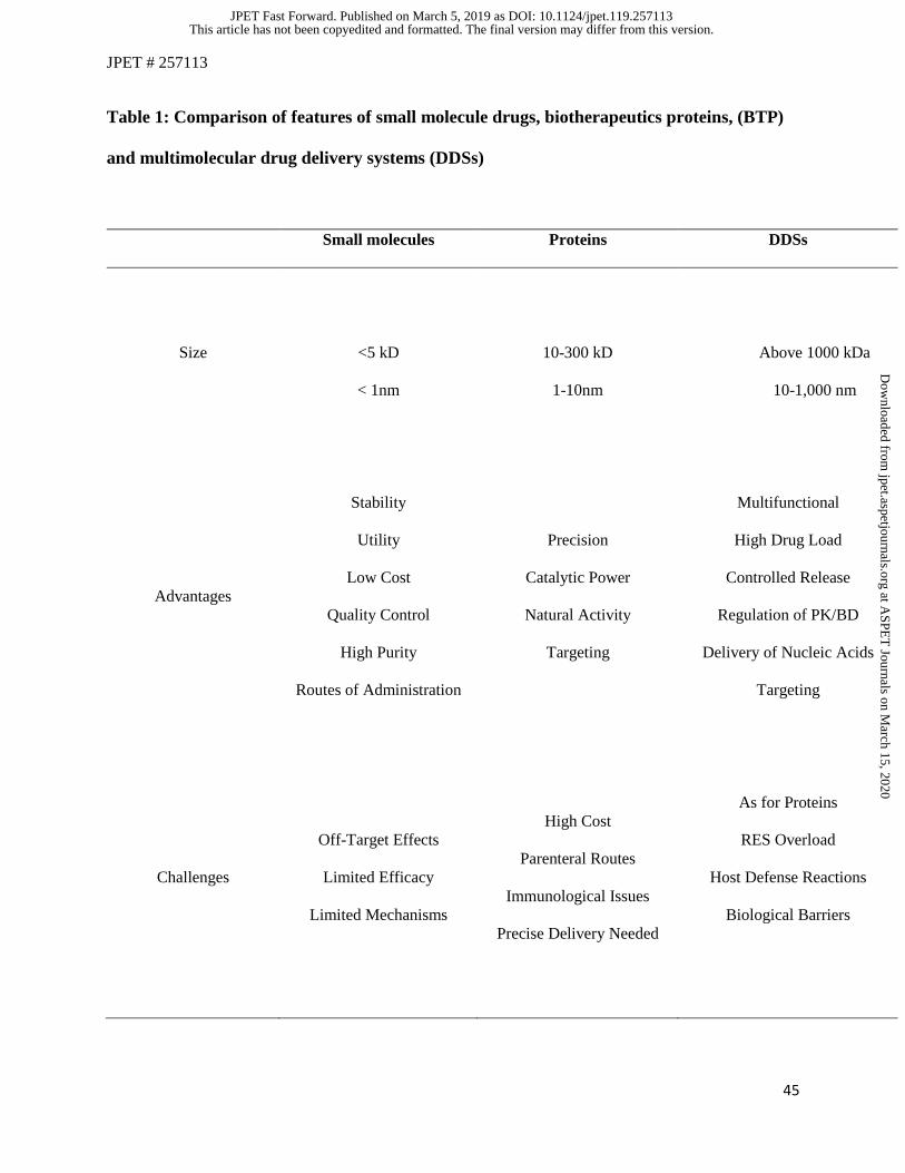
JPET # 257113
45
Table 1: Comparison of features of small molecule drugs, biotherapeutics proteins, (BTP)
and multimolecular drug delivery systems (DDSs)
Small molecules Proteins DDSs
Size
<5 kD
< 1nm
10-300 kD
1-10nm
Above 1000 kDa
10-1,000 nm
Advantages
Stability
Utility
Low Cost
Quality Control
High Purity
Routes of Administration
Precision
Catalytic Power
Natural Activity
Targeting
Multifunctional
High Drug Load
Controlled Release
Regulation of PK/BD
Delivery of Nucleic Acids
Targeting
Challenges
Off-Target Effects
Limited Efficacy
Limited Mechanisms
High Cost
Parenteral Routes
Immunological Issues
Precise Delivery Needed
As for Proteins
RES Overload
Host Defense Reactions
Biological Barriers
This article has not been copyedited and formatted. The final version may differ from this version.JPET Fast Forward. Published on March 5, 2019 as DOI: 10.1124/jpet.119.257113
at ASPE
T Journals on M
arch 15, 2020jpet.aspetjournals.org
Dow
nloaded from
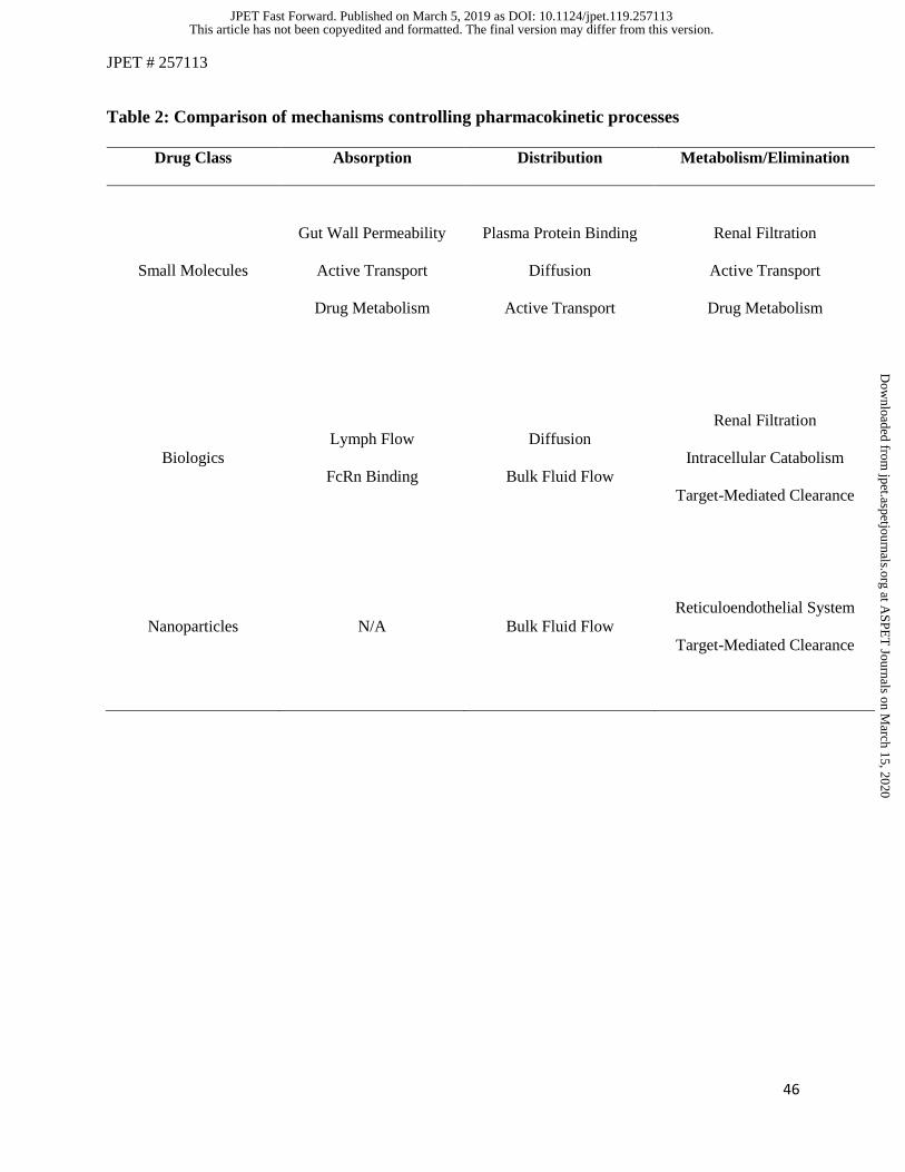
JPET # 257113
46
Table 2: Comparison of mechanisms controlling pharmacokinetic processes
Drug Class Absorption Distribution Metabolism/Elimination
Small Molecules
Gut Wall Permeability
Active Transport
Drug Metabolism
Plasma Protein Binding
Diffusion
Active Transport
Renal Filtration
Active Transport
Drug Metabolism
Biologics
Lymph Flow
FcRn Binding
Diffusion
Bulk Fluid Flow
Renal Filtration
Intracellular Catabolism
Target-Mediated Clearance
Nanoparticles
N/A
Bulk Fluid Flow
Reticuloendothelial System
Target-Mediated Clearance
This article has not been copyedited and formatted. The final version may differ from this version.JPET Fast Forward. Published on March 5, 2019 as DOI: 10.1124/jpet.119.257113
at ASPE
T Journals on M
arch 15, 2020jpet.aspetjournals.org
Dow
nloaded from
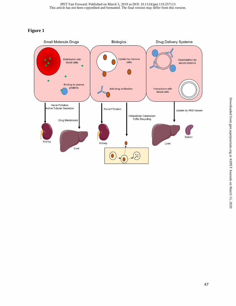
47
Figure 1
This article has not been copyedited and formatted. The final version may differ from this version.JPET Fast Forward. Published on March 5, 2019 as DOI: 10.1124/jpet.119.257113
at ASPE
T Journals on M
arch 15, 2020jpet.aspetjournals.org
Dow
nloaded from
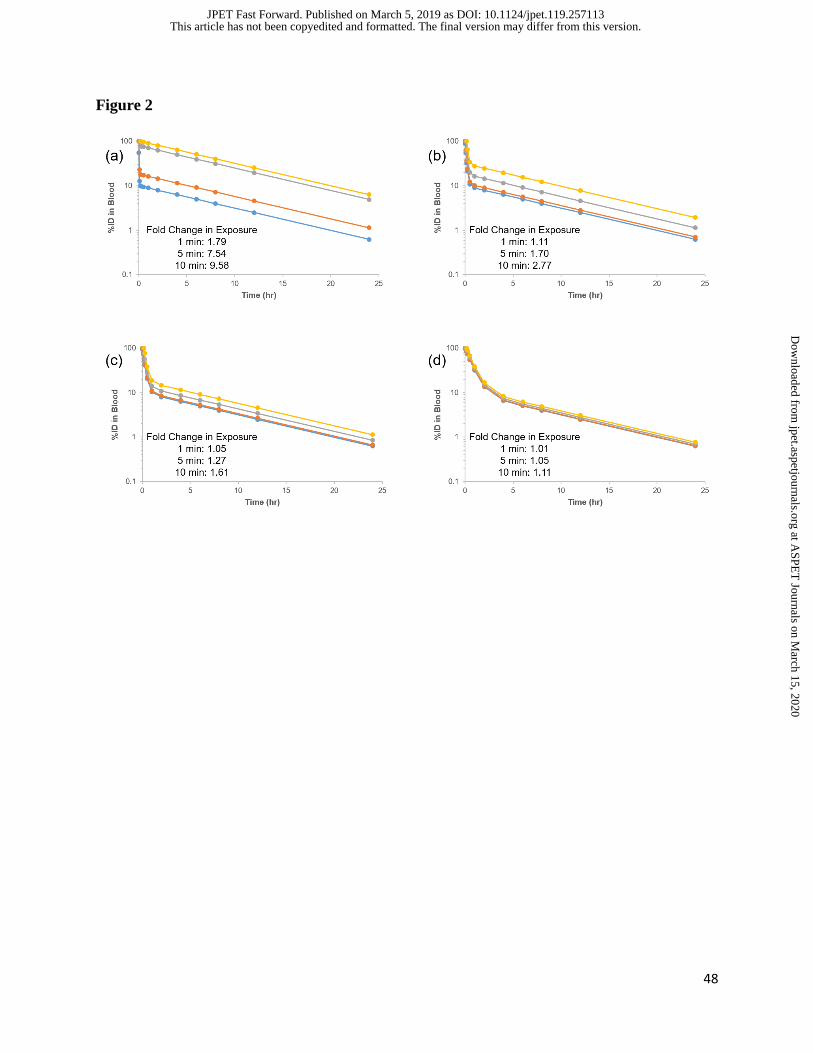
48
Figure 2
This article has not been copyedited and formatted. The final version may differ from this version.JPET Fast Forward. Published on March 5, 2019 as DOI: 10.1124/jpet.119.257113
at ASPE
T Journals on M
arch 15, 2020jpet.aspetjournals.org
Dow
nloaded from
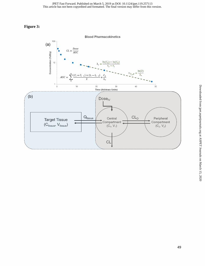
49
Figure 3:
This article has not been copyedited and formatted. The final version may differ from this version.JPET Fast Forward. Published on March 5, 2019 as DOI: 10.1124/jpet.119.257113
at ASPE
T Journals on M
arch 15, 2020jpet.aspetjournals.org
Dow
nloaded from
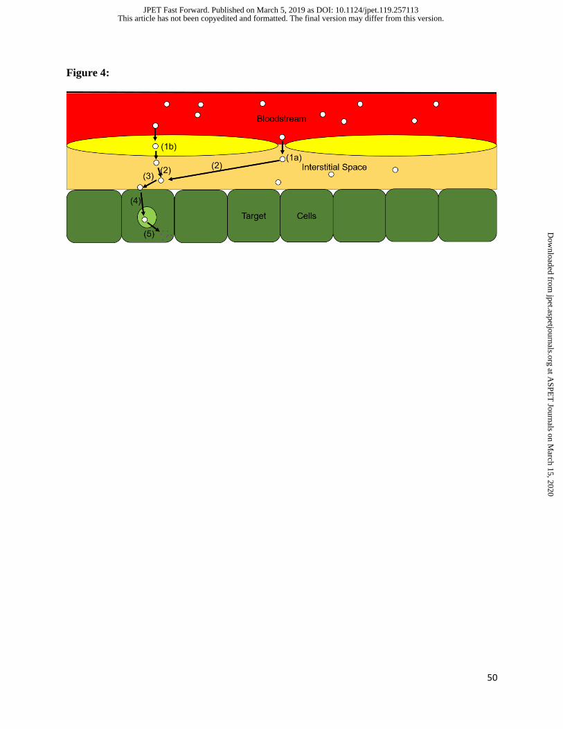
50
Figure 4:
This article has not been copyedited and formatted. The final version may differ from this version.JPET Fast Forward. Published on March 5, 2019 as DOI: 10.1124/jpet.119.257113
at ASPE
T Journals on M
arch 15, 2020jpet.aspetjournals.org
Dow
nloaded from
51
This article has not been copyedited and formatted. The final version may differ from this version.JPET Fast Forward. Published on March 5, 2019 as DOI: 10.1124/jpet.119.257113
at ASPE
T Journals on M
arch 15, 2020jpet.aspetjournals.org
Dow
nloaded from