pharmacological explanation for the medicinal use of juniperus excelsa in hyperactive...
TRANSCRIPT
ORIGINAL PAPER
Pharmacological explanation for the medicinal use of Juniperusexcelsa in hyperactive gastrointestinal and respiratory disorders
Munasib Khan • Arif-ullah Khan • Najeeb-ur-Rehman •
Anwarul-Hassan Gilani
Received: 8 July 2011 / Accepted: 2 September 2011 / Published online: 3 December 2011
� The Japanese Society of Pharmacognosy and Springer 2011
Abstract Crude extract of Juniperus excelsa (JeExt),
which tested positive for the presence of anthraquinone,
flavonoids, saponins, sterols, terpenes and tannin, exhibited
a protective effect against castor oil-induced diarrhoea in
mice at 100–1000 mg/kg. In rabbit jejunum preparations,
JeExt (0.01–1.0 mg/mL) caused relaxation of spontaneous
and K? (80 mM)-induced contractions at similar concen-
trations to papaverine, whereas verapamil was relatively
more potent against K?. JeExt (0.03–0.3 mg/mL) shifted
Ca2? concentration–response curves to the right, like
papaverine or verapamil. JeExt (0.003–0.01 mg/mL)
caused a leftward shift of isoprenaline-induced inhibitory
concentration–response curves, similar to papaverine.
JeExt (1.0–30 mg/kg) caused suppression of carbachol
(CCh, 100 lg/kg)-induced increase in inspiratory pressure
of anaesthetized rats. In guinea-pig trachea, JeExt
(0.001–3.0 mg/mL) relaxed CCh (1 lM)- and high K?-
induced contractions and shifted isoprenaline-induced
inhibitory curves to the left. This study suggests that Ju-
niperus excelsa possibly exhibits a combination of Ca2?
antagonist and phosphodiesterase inhibitory effects, which
provides a pharmacological basis for its traditional use in
disorders of gut and airways hyperactivity, such as diar-
rhoea, colic and asthma.
Keywords Juniperus excelsa � Ca2? channel blocker �PDE inhibitor � Gut and airways disorders
Introduction
Juniperus excelsa Bieb. (Cupressaceae/Coniferae), com-
monly known as ‘‘pencil cedar/Juniper’’ and locally as
‘‘Dhup Guggal’’ is found throughout the eastern Mediter-
ranean from northeastern Greece and southern Bulgaria
across Turkey to Syria and the Caucasus mountains at an
altitude of 2000–4000 m. It also occurs in Alborz and other
mountains of Iran, east to northwestern Pakistan and Oman
[1, 2]. Juniperus excelsa is used in folk medicine to treat
diarrhoea, abdominal spasm, asthma [3], fever, gonorrhoea,
headache and leucorrhoea [4, 5] as well as being consid-
ered useful as an antihypertensive, diuretic, appetizer,
carminative, stimulant, anticonvulsant and flavouring agent
[6]. Phytochemical studies on the plant revealed the pres-
ence of (?)-cedrol, (?)-sabinene, (?)-limonene, menthene,
terpinene-4-ol, a-cedrene, b-cedrene, p-cymene, b-phel-
landrene, a-copaene, muurolene, b-guaiene, guaiazulene
[7], a-thujene, a-fenchene, camphene, a-phellandrene, c-3-
carene, a-terpinene, trans-ocimene, c-terpinene, terpino-
lene, endo-fenchol, cis-pinene hydrate, a-campholenal,
trans-pinocarveol, camphor, borneol, c-terpineol, naph-
thalene, a-terpineaol, myrtenol, verbenone, trans-carveol,
endo-fenchyl acetate, piperitone, bornyl acetate, carvacrol,
b-cubebene, thujopsene a-cadinene, a-humulene, b-aco-
radiene, b-cadinene, c-muurolene [8], toluene, tricyclene,
M. Khan was on leave from University of Malakand for the PhD
study.
M. Khan � Najeeb-ur-Rehman � A.-H. Gilani (&)
Natural Products Research Unit, Department of Biological
and Biomedical Sciences, Aga Khan University Medical
College, Karachi 74800, Pakistan
e-mail: [email protected]
M. Khan
Department of Pharmacology, Faculty of Pharmacy,
University of Karachi, Karachi 75270, Pakistan
A. Khan
Institute of Pharmaceutical Sciences, Kohat University
of Science and Technology, Kohat 26000, KPK, Pakistan
123
J Nat Med (2012) 66:292–301
DOI 10.1007/s11418-011-0605-z
thujene, pinene, camphene, triene cycloheptane 1,3,5-
trismethylene, b-myrcene, o-allyl toluene, m-cymene, d,l-
limonene, a-pinene oxide, a-terpinolene, 3-thujanone and
a-campholene aldehyde [9].
Despite the fact that extensive phytochemical research
has been carried out on Juniperus excelsa, reports related
to pharmacological investigation are limited, only citing its
antibacterial [10] and antifungal [11] activities. In the
present research, we provide evidence that Juniperus
excelsa exhibits antidiarrheal, antispasmodic and bron-
chodilatory activities, occurring via a combination of Ca2?
channel blockade and phosphodiesterase (PDE) inhibitory
pathways, which explains the medicinal use of Juniperus
excelsa in hyperactive gut and airways disorders such as
diarrhea, colic and asthma.
Materials and methods
Plant material and extraction
The aerial parts (stem ? leaves) of Juniperus excelsa were
collected from northern areas of Pakistan (Chitral) in
September 2007. The plant was identified with the help of a
taxonomist, Dr. Ilyas Iqbal, Department of Botany, Uni-
versity of Malakand, KPK, Pakistan. A voucher specimen
(UOM/BGH/150) has been submitted to the herbarium of
the same university. Plant material was cleaned, shade-
dried and coarsely ground. The powdered material
(580.27 g) was soaked in aqueous methanol (70%) at room
temperature (25 ± 2.0�C) for 3 days with occasional
shaking. It was filtered through muslin cloth and then
through Whatman qualitative grade 1 filter paper [12]. The
procedure of maceration and filtration was repeated twice
more. All the filtrates were combined and evaporated
to dryness in a rotary evaporator under reduced pressure
(-760 mmHg) at 35–40�C to obtain crude extract of
Juniperus excelsa (JeExt), yielding approx. 24%. JeExt
was dissolved in normal saline/distilled water for use in
in-vivo and in-vitro experiments.
Chemicals
Acetylcholine chloride (ACh), carbachol (CCh), isoprena-
line, loperamide, papaverine and verapamil were purchased
from Sigma Chemical Co., St Louis, MO, USA. Ami-
nophylline, pentothal sodium (thiopental) and castor oil
were obtained from GlaxoSmithKline, Abbott Laboratories
and KCL Pharma, Karachi, Pakistan, respectively. Chem-
icals used for making physiological salt solutions were:
potassium chloride (Sigma), calcium chloride, glucose,
magnesium chloride, magnesium sulfate, potassium dihy-
drogen phosphate, sodium bicarbonate, sodium dihydrogen
phosphate (Merck, Darmstadt, Germany) and sodium
chloride (BDH Laboratory Supplies, Poole, UK). The
chemicals used in phytochemical analysis include: acetic
anhydride, aluminum chloride, ammonium hydroxide,
ferric chloride (Sigma), benzene, chloroform, hydrochloric
acid and petroleum ether (BDH). All chemicals used were
of analytical grade and dissolved in distilled H2O/saline.
Phytochemical screening
Preliminary investigation of the plant extracts for the
presence of various phytochemical classes, such as sapo-
nins, coumarins, sterols, terpenes, flavonoids, anthraqui-
nones and tannins was done according to reported methods
[13]. The presence of saponins was detected based on the
appearance of froth upon vigorous shaking of diluted
samples. The observation of yellow fluorescence under
ultraviolet light on examination of filter paper previously
exposed to the vapours from boiling plant material indi-
cated coumarins. For the detection of sterols and terpenes,
plant material was treated with petroleum ether and sub-
sequently extracted with chloroform. The appearance of
green to pink (for sterols) and pink to purple colours (for
terpenes) was then noted after treatment of the chloroform
layer with acetic anhydride and concentrated HCl in suc-
cession. Plant material was noted as positive for flavonoids
when it gave a yellow colour with aluminum chloride
reagent, and for tannins when green or black colour was
produced with aqueous ferric chloride. Lastly, for detecting
anthraquinones, the extract was dissolved in 1% HCl, then
in benzene, and observed if the extract showed a pink,
violet or red colour with ammonium hydroxide.
Experimental animals
Rabbits (1–1.2 kg), guinea pigs (500–550 g), Sprague–
Dawley rats (200–250 g) and BALB/c mice (20–25 g) of
local breed and either sex were used for this study and were
housed at the Animal House of the Aga Khan University,
maintained at 23–25�C and given a standard diet and tap
water. Rabbits starved for 24 h were killed by a blow to the
back of the head, and guinea pigs by cervical dislocation.
The experiments complied with the rules of the Institute of
Laboratory Animal Resources, Commission on Life Sci-
ences, National Research Council [14] and were approved
by the Ethical Committee of the Aga Khan University.
Castor oil-induced diarrhea
Mice were fasted for 24 h before the experiment. Animals
were housed in individual cages and divided in five groups,
each containing 10 mice. The first group received saline
(10 mL/kg, orally) and served as a negative control. The
J Nat Med (2012) 66:292–301 293
123
doses of the test extract were selected on a trial basis
and three increasing doses of extract, 100, 300 and
1000 mg/kg, were given orally to three different groups.
One group of mice was treated with loperamide (10 mg/kg,
orally) as a positive control. One hour after treatment, each
animal received 10 mL/kg of castor oil orally through a
feeding needle. Afterward, the cages were inspected for the
presence of diarrhoea droppings; their absence was noted
as a positive result, indicating protection from diarrhoea at
that time [15].
Rabbit jejunum
The rabbit abdomen was opened and the jejunum was
dissected out, kept in normal Tyrode’s solution and cleaned
of mesenteries [16]. Each segment of about 2 cm length
was suspended in a 10 mL tissue bath containing Tyrode’s
solution (pH 7.4), maintained at 37�C and aerated with a
mixture of 95% O2 and 5% CO2 (carbogen). The compo-
sition of Tyrode’s solution was (in mM): NaCl: 136.9, KCl:
2.7, MgCl2�6H2O: 0.5, NaHCO3: 11.9, NaH2PO4�2H2O:
0.32, CaCl2: 1.8, and glucose: 5.05. One end of the seg-
ment was attached to a metal tissue hook and the other was
attached by a cotton thread to an isotonic Bioscience
transducer, connected to a Student oscillograph (Harvard
Apparatus, Holliston, MA, USA). An initial load of 1 g
was applied to each tissue and was allowed to equilibrate
for 30 min before the addition of any drug. Following the
equilibration period, each preparation was then stabilized
with a sub-maximal concentration of ACh (0.3 lM) at
3 min intervals until constant responses were recorded. The
inhibitory effects of test substances were measured as the
percent change in spontaneous contractions of the jejunum.
For the determination of the Ca2? channel blockade effect,
high K? (80 mM) was used to depolarize the preparations,
as described by Farre et al. [17]. High K? ([30 mM) is
known to cause smooth muscle contractions through the
opening of voltage-dependent Ca2? channels, thus allow-
ing influx of extracellular Ca2? causing a contractile effect;
a substance causing inhibition of high K?-induced con-
traction is considered to be a blocker of Ca2? influx
through L-type Ca2? channels [18]. Once the induced
contraction achieved a plateau (usually within 7–10 min),
the test material was then added in a cumulative fashion to
obtain concentration-dependent inhibitory responses. To
confirm the Ca2? antagonist action of the test substance,
the tissue was allowed to stabilize in normal Tyrode’s
solution, which was then replaced with Ca2?-free Tyrode’s
solution containing EDTA (0.1 mM) for 30 min in order to
remove Ca2? from the tissues. This solution was further
replaced with K?-rich and Ca2?-free Tyrode’s solution,
having the composition (in mM): NaCl: 91.03, KCl: 50,
MgCl2�6H2O: 0.50, NaHCO3: 11.9, NaH2PO4�2H2O: 0.32,
glucose: 5.05, and EDTA-Na2�2H2O: 0.1. Following an
incubation period of 30 min, control concentration–
response curves of Ca2? were obtained. When the control
Ca2? concentration–response curves were found to be
superimposable (usually after two cycles), the tissue was
pretreated with the test drug for 1 h. The concentration–
response curves of Ca2? were reconstructed in the presence
of different concentrations of the test material to observe
the Ca2? antagonist effect. The PDE inhibitory effect was
studied indirectly by constructing isoprenaline-induced
inhibitory concentration–response curves against CCh-
induced contractions in the absence and presence of the test
substance, as PDE-inhibitors are known to potentiate the
effect of isoprenaline [19, 20].
Bronchodilatory activity
Rats were anaesthetized with sodium thiopental (Pento-
thal, 80–100 mg/kg, intraperitoneally), then incubated
with a tracheal tube and ventilated with a volume venti-
lator (Miniature Ideal Pump, Bioscience, UK) adjusted at
a rate of 70–80 strokes/min to deliver 7–10 mL/kg of
room air [15]. A polyethylene catheter was inserted into
the jugular vein for drug administration. Changes in air-
ways resistance (mmHg) were measured by a pressure
transducer (MLT-1199) connected to the side arm of
tracheal cannula and recorded by PowerLab 4/25 with
running chart software via a Quad bridge amplifier
(ADInstruments, Bella Vista, NSW, Australia). Broncho-
constriction was induced with CCh (100 lg/kg), which
was reversed within 7–10 min. The test drug was given to
the animals 5–8 min prior to administration of CCh. The
responses were expressed as the percent reduction of the
CCh-induced bronchospasm.
Guinea pig trachea
Trachea was dissected from guinea pigs and kept in
normal Kreb’s solution. The tracheal tube was cut into
rings, 2–3 mm wide, each containing about two cartilages.
Each ring was opened by a longitudinal cut on the ventral
side opposite the smooth muscle, forming a strip with
smooth muscle in the centre and cartilaginous portions on
the edges [21]. The preparation was mounted in a 20 mL
tissue bath containing Kreb’s solution (pH 7.4) at 37�C
and aerated with carbogen. The composition of Kreb’s
solution was (in mM): NaCl: 118.2, NaHCO3: 25.0,
CaCl2: 2.5, KCl: 4.7, KH2PO4: 1.2, MgSO4�7H2O: 1.2,
and glucose: 11.7. A tension of 1 g was applied to tra-
cheal strips continuously throughout the experiment. The
tissues were allowed to equilibrate for 1 h before the
addition of any drug. CCh (1 lM) was used for the sta-
bilization. When sustained contractions were obtained in
294 J Nat Med (2012) 66:292–301
123
the preparations using the spasmogens, such as CCh and/
or K?, the relaxant effect of the test substance was
assessed by adding it in a cumulative fashion. As in the
jejunum, isoprenaline concentration–response curves were
constructed in trachea as described previously [22]. Iso-
metric responses were recorded on a Grass model 7
Polygraph (Grass Instrument Company, Quincy, MA,
USA).
Acute toxicity test
Mice were divided into groups of five mice each. The test
was performed using increasing doses of the plant extract,
given orally in 10 mL/kg volume to different test groups.
Another group of mice was administered saline (10 mL/kg,
orally) as negative control. The mice were allowed food
ad libitum and kept under regular observation for lethality
recorded after 24 h.
Statistical analysis
The data expressed are mean ± standard error of mean
(SEM, n = number of experiment) and median effective
concentrations (EC50) with 95% confidence intervals. The
statistical parameters applied were the chi-squared test for
antidiarrhoeal assay, one-way analysis of variance fol-
lowed by Dunnett’s test for bronchodilatory activity, and
Student’s t test to compare the curves of the in-vitro
experiment results. P \ 0.05 was considered statistically
significant. Concentration–response curves were analyzed
by non-linear regression using GraphPad software
(GraphPAD, San Diego, CA, USA).
Results
Phytochemical analysis
JeExt was found to contain anthraquinone, flavonoids,
saponins, sterols, terpenes and tannin.
Effect on castor oil-induced diarrhea
JeExt exhibited a dose-dependent (100–1000 mg/kg) pro-
tective effect against castor oil-induced diarrhoea in mice.
The negative control group (saline-treated) did not show
any protection against castor oil-induced diarrhea. Pre-
treatment of animals with the plant extract showed 50%
protection from diarrhoea at 100, 70% at 300 and 80%
protection at 1000 mg/kg (P \ 0.05 vs. saline group at
each dose). Loperamide (10 mg/kg) showed 100% pro-
tection from diarrhoea (P \ 0.01 vs. saline group) in the
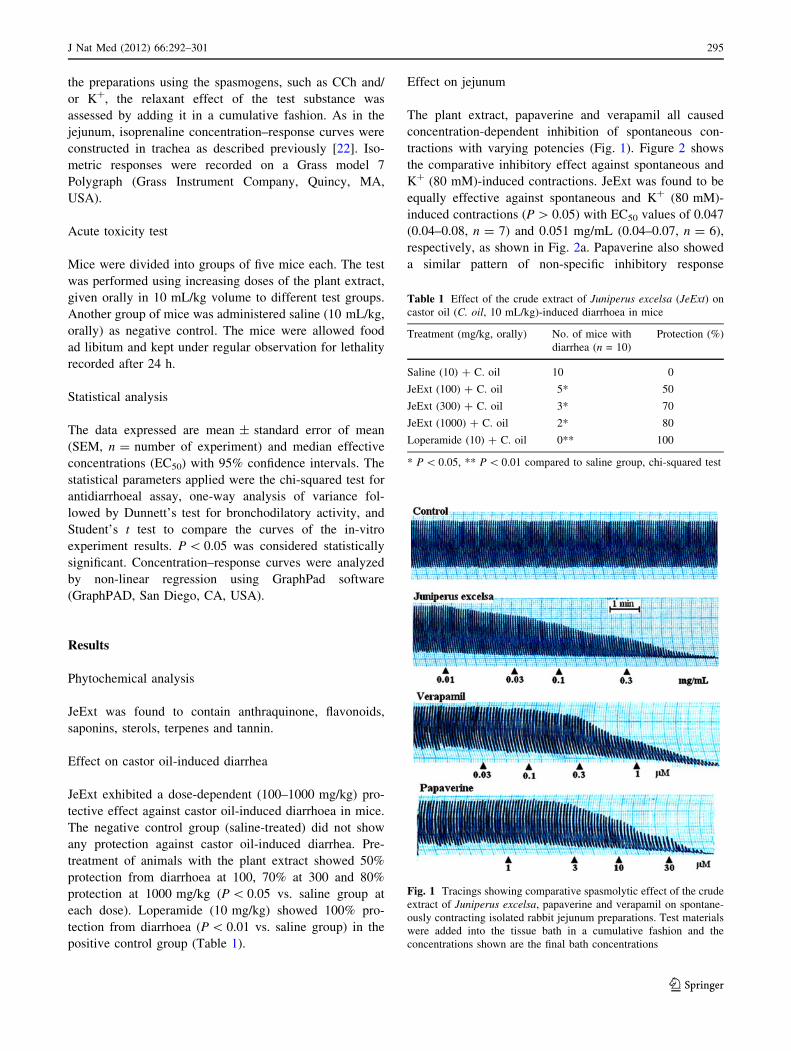
positive control group (Table 1).
Effect on jejunum
The plant extract, papaverine and verapamil all caused
concentration-dependent inhibition of spontaneous con-
tractions with varying potencies (Fig. 1). Figure 2 shows
the comparative inhibitory effect against spontaneous and
K? (80 mM)-induced contractions. JeExt was found to be
equally effective against spontaneous and K? (80 mM)-
induced contractions (P [ 0.05) with EC50 values of 0.047
(0.04–0.08, n = 7) and 0.051 mg/mL (0.04–0.07, n = 6),
respectively, as shown in Fig. 2a. Papaverine also showed
a similar pattern of non-specific inhibitory response
Table 1 Effect of the crude extract of Juniperus excelsa (JeExt) on
castor oil (C. oil, 10 mL/kg)-induced diarrhoea in mice
Treatment (mg/kg, orally) No. of mice with
diarrhea (n = 10)
Protection (%)
Saline (10) ? C. oil 10 0
JeExt (100) ? C. oil 5* 50
JeExt (300) ? C. oil 3* 70
JeExt (1000) ? C. oil 2* 80
Loperamide (10) ? C. oil 0** 100
* P \ 0.05, ** P \ 0.01 compared to saline group, chi-squared test
Fig. 1 Tracings showing comparative spasmolytic effect of the crude
extract of Juniperus excelsa, papaverine and verapamil on spontane-
ously contracting isolated rabbit jejunum preparations. Test materials
were added into the tissue bath in a cumulative fashion and the
concentrations shown are the final bath concentrations
J Nat Med (2012) 66:292–301 295
123
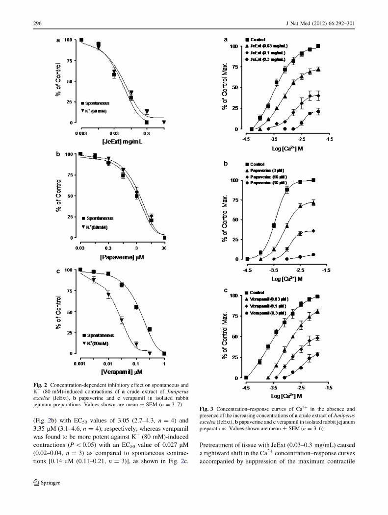
(Fig. 2b) with EC50 values of 3.05 (2.7–4.3, n = 4) and
3.35 lM (3.1–4.6, n = 4), respectively, whereas verapamil
was found to be more potent against K? (80 mM)-induced
contractions (P \ 0.05) with an EC50 value of 0.027 lM
(0.02–0.04, n = 3) as compared to spontaneous contrac-
tions [0.14 lM (0.11–0.21, n = 3)], as shown in Fig. 2c.
Pretreatment of tissue with JeExt (0.03–0.3 mg/mL) caused
a rightward shift in the Ca2? concentration–response curves
accompanied by suppression of the maximum contractile
Fig. 2 Concentration-dependent inhibitory effect on spontaneous and
K? (80 mM)-induced contractions of a crude extract of Juniperusexcelsa (JeExt), b papaverine and c verapamil in isolated rabbit
jejunum preparations. Values shown are mean ± SEM (n = 3–7)Fig. 3 Concentration–response curves of Ca2? in the absence and
presence of the increasing concentrations of a crude extract of Juniperusexcelsa (JeExt), b papaverine and c verapamil in isolated rabbit jejunum
preparations. Values shown are mean ± SEM (n = 3–6)
296 J Nat Med (2012) 66:292–301
123
effect (Fig. 3a), similar to that caused by papaverine
(3–30 lM; Fig. 3b) and verapamil (0.03–0.3 lM), as shown
in Fig. 3c. When tested for possible interaction with iso-
prenaline, pretreatment of the tissues with JeExt
(0.003–0.01 mg/mL) shifted the isoprenaline-induced
inhibitory concentration–response curves to the left, show-
ing a potentiating effect (Fig. 4a). Papaverine caused a
similar concentration-dependent (0.3–1.0 lM) leftward
shift in the concentration–response curves of isoprenaline
(Fig. 4b), while verapamil (0.3–1.0 lM) did not alter the
inhibitory response to isoprenaline (Fig. 4c).
Effect on carbachol-induced bronchoconstriction
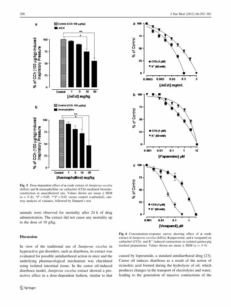
JeExt at doses of 1, 3, 10 and 30 mg/kg caused 3.4 ± 1.2,
10.2 ± 1.1, 25.7 ± 5.9 and 45.14 ± 6.5% (n = 5–7),
respectively, suppression of CCh (100 lg/kg)-induced
increase in inspiratory pressure of anaesthetized rats
(Fig. 5a). Aminophylline was used as a positive control,
and inhibited the CCh (100 lg/kg)-mediated bronchocon-
striction at 1, 3, 10 and 30 mg/kg by 8.0 ± 4.4, 19.3 ± 7.4,
28.0 ± 9.2 and 53.2 ± 11.8% (n = 8), respectively
(Fig. 5b).
Effect on trachea
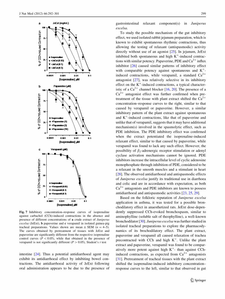
JeExt, papaverine and verapamil were all found devoid of
any stimulant action when screened on the tracheal resting
baseline. When tested against CCh (1 lM)- and K?
(80 mM)-induced contractions, JeExt caused concentra-
tion-dependent inhibition with EC50 values of 0.04
(0.03–0.14, n = 4) and 0.21 mg/mL (0.18–0.30, n = 4),
respectively, as shown in Fig. 6a. Similarly, papaverine
had an inhibitory effect against CCh (1 lM)- and K?
(80 mM)-induced contractions (Fig. 6b) with EC50 values
of 0.82 (0.61–0.93, n = 5) and 2.4 lM (1.7–3.4, n = 5),
respectively. Verapamil was found to be more potent in its
inhibitory effect against K? (80 mM)-induced contractions
with an EC50 value of 0.03 lM (0.02–0.04, n = 4) com-
pared to CCh-induced contractions [0.87 lM (0.53–1.44,
n = 3)], as shown in Fig. 6c. Pretreatment of tracheal
preparations with JeExt shifted the isoprenaline-induced
inhibitory concentration–response curves to the left
(Fig. 7a) in a concentration-dependent manner
(0.03–0.3 mg/mL), similar to that caused by papaverine
(1.0–10 lM), showing a potentiating effect (Fig. 7b), while
verapamil (0.03–0.3 lM) did not alter the inhibitory
response to isoprenaline (Fig. 7c).
Acute toxicity
The three different groups of mice were given JeExt in the
graded doses of 1, 5 and 10 g/kg, respectively, and the
Fig. 4 Inhibitory concentration–response curves of isoprenaline
against carbachol (CCh)-induced contractions in the absence and
presence of different concentrations of a crude extract of Juniperusexcelsa (JeExt), b papaverine and c verapamil in isolated rabbit
jejunum preparations. Values shown are mean ± SEM (n = 3–4).
The curves obtained by pretreatment of tissues with JeExt and
papaverine are significantly different from the respective isoprenaline
control curves (P \ 0.05), while that obtained in the presence of
verapamil is not significantly different (P [ 0.05), Student’s t test
J Nat Med (2012) 66:292–301 297
123
animals were observed for mortality after 24 h of drug
administration. The extract did not cause any mortality up
to the dose of 10 g/kg.
Discussion
In view of the traditional use of Juniperus excelsa in
hyperactive gut disorders, such as diarrhoea, its extract was
evaluated for possible antidiarrhoeal action in mice and the
underlying pharmacological mechanism was elucidated
using isolated intestinal tissue. In the castor oil-induced
diarrhoea model, Juniperus excelsa extract showed a pro-
tective effect in a dose-dependent fashion, similar to that
caused by loperamide, a standard antidiarrhoeal drug [23].
Castor oil induces diarrhoea as a result of the action of
ricinoleic acid formed during the hydrolysis of oil, which
produces changes in the transport of electrolytes and water,
leading to the generation of massive contractions of the
Fig. 5 Dose-dependent effect of a crude extract of Juniperus excelsa(JeExt) and b aminophylline on carbachol (CCh)-mediated broncho-
constriction in anaesthetized rats. Values shown are mean ± SEM
(n = 5–8), *P \ 0.05, **P \ 0.01 versus control (carbachol), one-
way analysis of variance, followed by Dunnett’s test
Fig. 6 Concentration–response curves showing effect of a crude
extract of Juniperus excelsa (JeExt), b papaverine, and c verapamil on
carbachol (CCh)- and K?-induced contractions in isolated guinea-pig
tracheal preparations. Values shown are mean ± SEM (n = 3–5)
298 J Nat Med (2012) 66:292–301
123
intestine [24]. Thus a potential antidiarrhoeal agent may
exhibit its antidiarrhoeal effect by inhibiting bowel con-
tractions. The antidiarrhoeal activity of JeExt following
oral administration appears to be due to the presence of
gastrointestinal relaxant component(s) in Juniperus
excelsa.
To study the possible mechanism of the gut inhibitory
effect, we used isolated rabbit jejunum preparation, which is
known to exhibit spontaneous rhythmic contractions, thus
allowing the testing of relaxant (antispasmodic) activity
directly without use of an agonist [25]. In jejunum, JeExt
inhibited both spontaneous and high K?-induced contrac-
tions with similar potency. Papaverine, PDE and Ca2? influx
inhibitor [26] caused similar patterns of inhibitory effect
with comparable potency against spontaneous and K?-
induced contractions, while verapamil, a standard Ca2?
antagonist [27], was relatively selective in its inhibitory
effect on the K?-induced contractions, a typical character-
istic of a Ca2? channel blocker [16, 20]. The presence of a
Ca2? antagonist effect was further confirmed when pre-
treatment of the tissue with plant extract shifted the Ca2?
concentration–response curves to the right, similar to that
caused by verapamil or papaverine. However, a similar
inhibitory pattern of the plant extract against spontaneous
and K?-induced contractions, like that of papaverine and
unlike that of verapamil, suggests that it may have additional
mechanism(s) involved in the spasmolytic effect, such as
PDE inhibition. The PDE inhibitory effect was confirmed
when the extract potentiated the isoprenaline-induced
relaxant effect, similar to that caused by papaverine, while
verapamil was found to lack any such effect. However, the
possibility of b2-adrenergic receptor stimulation or adenyl
cyclase activation mechanisms cannot be ignored. PDE
inhibitors increase the intracellular level of cyclic adenosine
monophosphate through inhibition of PDE, considered to be
a relaxant in the smooth muscles and a stimulant in heart
[28]. The observed antidiarrhoeal and antispasmodic effects
of Juniperus excelsa justify its traditional use in diarrhoea
and colic and are in accordance with expectation, as both
Ca2? antagonists and PDE inhibitors are known to possess
antidiarrhoeal and antispasmodic activities [23, 25, 29].
Based on the folkloric reputation of Juniperus excelsa
application in asthma, it was tested for a possible bron-
chodilatory effect in anaesthetized rats. JeExt dose-depen-
dently suppressed CCh-evoked bronchospasm, similar to
aminophylline (soluble salt of theophylline), a well-known
bronchodilator [30]. Juniperus excelsa was further studied in
isolated tracheal preparations to explore the pharmacody-
namics of its brochodilatory effect. The plant extract,
papaverine and verapamil all caused relaxation of trachea
precontracted with CCh and high K?. Unlike the plant
extract and papaverine, verapamil was found to be compar-
atively more potent against high K?- than against CCh-
induced contractions, as expected from Ca2? antagonists
[31]. Pretreatment of tracheal tissues with the plant extract
shifted the isoprenaline-induced inhibitory concentration–
response curves to the left, similar to that observed in gut
Fig. 7 Inhibitory concentration–response curves of isoprenaline
against carbachol (CCh)-induced contractions in the absence and
presence of different concentrations of a crude extract of Juniperusexcelsa (JeExt), b papaverine and c verapamil in isolated guinea-pig
tracheal preparations. Values shown are mean ± SEM (n = 4–5).
The curves obtained by pretreatment of tissues with JeExt and
papaverine are significantly different from the respective isoprenaline
control curves (P \ 0.05), while that obtained in the presence of
verapamil is not significantly different (P [ 0.05), Student’s t test
J Nat Med (2012) 66:292–301 299
123
preparations, indicating the presence of additional PDE
inhibitory bronchodilatory substance(s) in Juniperus
excelsa. Interestingly, the PDE inhibitory effect of the plant
extract in jejunum was observed at lower concentrations to
that in tracheal preparation. The possible explanation could
be localization of different PDE subfamilies in the two
physiological systems [32–34], though species difference
cannot be ruled out. The usefulness of PDE inhibitors in
asthma is well established [35, 36], though the major limi-
tation is cardiac stimulation as a side-effect [37]. Interest-
ingly, Ca2? antagonists have also been shown to be useful in
bronchoconstriction [38] and are known to exhibit a cardio-
suppressant effect [39]. The co-existence of Ca2? channel
blocker constituents with PDE inhibitor(s) in Juniperus
excelsa is perhaps meant by nature to offset the tachycardia
associated with PDE inhibitors when used alone. This find-
ing strengthens the concept that natural remedies known to
possess synergistic and/or side-effect-neutralizing potential
in addition to cost-effectiveness offer merit in evidence-
based studies [40]. Thus the presence of the combined
inhibitory effect on PDE and Ca2? channels might account
for the medicinal use of Juniperus excelsa in airways
hyperactivity and asthma.
The presence of phyotchemicals, anthraquinone, flavo-
noids, saponins, sterols, terpenes and tannins may be partly
responsible for the reported pharmacological effects of
Juniperus excelsa. However, further in-depth studies are
required to probe the nature of its chemical constituents
and the molecular basis of its biological activities. In the
acute toxicity study, JeExt was found to be safe up to
the dose of 10 mg/kg, which indicate a safety profile of the
plant at a wide dose range.
In conclusion, these results reveals that the crude extract
of Juniperus excelsa possesses antidiarrhoeal, antispas-
modic and bronchodilatory effects, mediated through a
combination of spasmolytic mechanisms, such as inhibition
of Ca2? influx and PDE enzyme(s). Thus, this study pro-
vides a sound mechanistic background to the medicinal use
of Juniperus excelsa in hyperactive gut and airways dis-
orders, like diarrhoea, abdominal spasm and asthma.
Moreover, its in-vivo antidiarrhoeal and bronchodilatory
activities prove the effectiveness of the plant in such con-
ditions, which is a step forward towards the evidence-based
medicinal use of phytomedicine.
Acknowledgments This study was partially supported by Higher
Education Commission of Pakistan. Munasib Khan was on leave from
University of Malakand for the PhD study.
References
1. Shanjani PS (2003) Nitrogen effect of callus induction and plant
regeneration of Juneiperus excels. Int J Agr Biol 4–5:419–422
2. Emami SA, Asili J, Mohagheghi Z, Hassanzadeh MK (2007)
Antioxidant activity of leaves and fruits of Iranian conifers.
Evidence Based Complem Altern Med 4:313–319
3. Kaul MK (1997) Medicinal plants of Kashmir and Ladakh:
temperate and cold acrid Himalaya. Indus Publishing Company,
New Delhi, p 173
4. Nadkarni KM (1976) Indian materia medica, 3rd edn. Popular
Prakashan, Bombay, p 713
5. Baquar SR (1989) Medicinal and poisonous plants of Pakistan.
Printas, Karachi, pp 248–249
6. Usmanghani K, Saeed A, Alam MT (1997) Indusyunic medicine.
University of Karachi Press, Karachi, pp 468–469
7. Thappa RK, Aggarwal SG, Kapahi BK, Sarin YK (1987) Juni-perus excelsa leaf oil, a new source of cedrol. J Nat Prod
50:323–324
8. Adam RP (1990) The chemical composition of leaf oils of
Juniperus excelsa M. Bieb. J Essent Oil Res 2:45–48
9. Unlu M, Unlu GV, Vural N, Donmez E, Akmak O (2008)
Composition and antibacterial activity of juniperus excelsaessential oil. Chem Nat Comp 44:129–131
10. Muhammad I, Mossa JS, Al-Yahya MA, Ramadan AF, El-Feraly
FS (2006) Further antibacterial diterpenes from the bark and leaves
of Juniperus procera Hochst. ex Endl. Phytother Res 9:584–588
11. Marina D, Sokovi J, Risti M, Grubi A (2004) Chemical compo-
sition and antifungal activity of the essential oil from Juniperusexcelsa berries. Pharm Biol 42:328–334
12. Williamson EM, Okpako DT, Evans FJ (1998) Selection prepa-
ration and pharmacological evaluation of plant material. Wiley,
Chichester, pp 15–23
13. Edeoga HO, Okwu DE, Mbaebie BO (2005) Phytochemical
constituents of some Nigerian medicinal plants. Afr J Biotechnol
4:685–688
14. National Research Council (1996) Guide for the care and use of
laboratory animals. National Academy Press, Washington, pp 1–7
15. Khan A, Gilani AH (2011) Antidiarrheal and bronchodilatory
activities of olive extract. Lat Am J Pharm 30:5–9
16. Gilani AH, Shah AJ, Ghayur MN, Majeed K (2005) Pharmaco-
logical basis for the use of turmeric in gastrointestinal and
respiratory disorders. Life Sci 76:3089–3105
17. Farre AJ, Columbo M, Fort M, Gutierrez B (1991) Differential
effects of various Ca?? antagonists. Gen Pharmacol 22:177–181
18. Godfraind T, Miller R, Wibo M (1986) Calcium antagonism and
calcium entry blockade. Pharmacol Rev 38:321–416
19. Lorenz KL, Wells JN (1983) Potentiation of the effects of sodium
nitroprusside and isoproterenol by selective phosphodiesterase
inhibitors. Mol Pharmacol 23:424–430
20. Gilani AH, Khan A, Subhan F, Khan M (2005) Antispasmodic
and bronchodilator activities of St. John’s wort are putatively
mediated through dual inhibition of calcium influx and phos-
phodiesterase. Fundam Clin Pharmacol 19:695–705
21. Gilani AH, Khan A, Ali T, Ajmal S (2008) Mechanisms under-
lying the antispasmodic and bronchodilatory properties of Ter-minalia bellerica fruit. J Ethnopharmacol 116:528–538
22. Shah AJ, Gilani AH (2010) Bronchodilatory effect of Acoruscalamus is mediated through multiple pathways. J Ethnopharma-
col 131:471–477
23. Reynolds IJ, Gould RJ, Snyder SH (1984) Loperamide: blockade
of calcium channels as a mechanism for antidiarrhoeal effects.
J Pharmacol Exp Ther 231:628–632
24. Croci T, Landi M, Elmonds-Alt X, Le-Fur G, Maffrand JP,
Manara L (1997) Role of tachykinins in castor oil-induced diar-
rhoea in rats. Br J Pharmacol 121:375–380
25. Bashir S, Memon R, Gilani AH (2011) Antispasmodic and anti-
diarrheal activities of Valeriana hardwickii rhizome are puta-
tively mediated through calcium channel blockade. Evidence
Based Complem Altern Med 1:6
300 J Nat Med (2012) 66:292–301
123
26. Rang HP, Dale MM, Ritter JM (1999) Pharmacology, 4th edn.
Churchill Livingstone, New York, pp 289–290
27. Fleckenstein A (1977) Specific pharmacology of calcium in
myocardium, cardiac pacemakers, and vascular smooth muscle.
Annu Rev Pharmacol Toxicol 17:149–166
28. Smith BV, Spina D, Page CP (2006) Phosphodiesterase inhibi-
tors. Br J Pharmacol 47:252–257
29. Sopory S, Kaur T, Visweswariah SS (2004) The cGMP-binding,
cGMP-specific phosphodiesterase (PDE5): intestinal cell
expression, regulation and role in fluid secretion. Cell Signal
16:681–692
30. Evans WV, Monie RD, Crimmins J, Seton A (1980) Aminoph-
ylline, salbutamol and combined intravenous infusions in acute
severe asthma. Br J Dis Chest 74:385–389
31. Nielsen-Kudsk JE, Karlsson JA, Persson CGA (1986) Relaxant
effects of xanthines, a b2-receptor agonist and Ca?? antagonists
in guinea-pig tracheal preparations contracted by potassium or
carbachol. Eur J Pharmacol 128:33–40
32. Rabe KF, Magnussen H, Dent G (1995) Theophylline and
selective PDE inhibitors as bronchodilators and smooth muscle
relaxants. Eur Respir J 8:637–642
33. Murthy KS (2006) Signaling for contractions and relaxation in
smooth muscle of the gut. Annu Rev Physiol 68:345–374
34. Schwarz ER, Kapur V, Rodriguez J, Rastogi S, Rosanio S (2007)
The effects of chronic phosphodiesterase-5 inhibitor use on dif-
ferent organ systems. Int J Impot Res 19:139–148
35. Lipworth BJ (2005) Phosphodiesterase-4 inhibitors for asthma
and chronic obstructive pulmonary disease. Lancet 365:167–175
36. Chung KF (2006) Phosphodiesterase inhibitors in airways dis-
ease. Eur J Pharmacol 533:110–117
37. Nawarth H (1981) Action potential, membrane currents and force
of contraction in cat ventricular heart muscle treated with
papaverine. J Pharmacol Exp Ther 218:544–549
38. Twiss MA, Harman E, Chesrown S, Handeles L (2002) Efficacy
of calcium channel blockers as maintenance therapy for asthma.
Br J Clin Pharmacol 53:243–249
39. Billman GE (1992) The antiarrhythmic effects of the calcium
antagonists. In: Epstein M (ed) Calcium antagonists in clinical
medicine. Hanley and Belfus, Philadelphia, pp 183–212
40. Gilani AH, Atta-ur-Rahman (2005) Trends in ethnopharmacol-
ogy. J Ethnopharmacol 100:43–49
J Nat Med (2012) 66:292–301 301
123