pharmacological propertie of excitators y neuromuscular...
TRANSCRIPT
J. Exp. Bwl. (1968), 49, 341-361 3 4 IWith 1 plate and 9 text-figures
Printed in Great Britain
PHARMACOLOGICAL PROPERTIES OF EXCITATORYNEUROMUSCULAR SYNAPSES IN THE LOCUST
BY P. N. R. USHERWOOD AND P. MACHILI
Department of Zoology, University of Glasgow
(Received 6 March 1968)
INTRODUCTION
Although insect nervous tissue is known to contain concentrations of acetylcholinefar in excess of those found in the central nervous tissue of vertebrates (Long, 1961)and there is some evidence for cholinergic transmission at insect central synapses(Treherne & Smith, 1965), it is now generally accepted that chemical transmissionat the insect neuromuscular junction is not cholinergic (e.g. Hill & Usherwood, 1961;Usherwood, 1963a; Usherwood & Grundfest, 1964, 1965). A large number of sub-stances have been tested on insect nerve-muscle preparations in attempts to identifythe chemical mediator or mediators involved in neuromuscular synaptic transmissionin insects. Of these substances the most promising results have been obtained withcertain amino acids, notably L-glutamic acid (e.g. Usherwood & Grundfest, 1965;Kerkut, Shapira & Walker, 1965; Usherwood & Machili, 1966) and y-aminobutyricacid (GAJBA) (Usherwood & Grundfest, 1964, 1965). Usherwood & Grundfest (1964,1965) have demonstrated that GABA mimics the transmitter at the inhibitory neuro-muscular synapses in the locust and the cockroach, while Usherwood & Grundfest(1965) and Usherwood & Machili (1966) have suggested a role for L-glutamate at theexcitatory neuromuscular synapse in the locust. L-Glutamate is also thought to beinvolved in transmission at the excitatory neuromuscular synapse in the cockroach(Kerkut, Shapira & Walker, 1965). In view of the close phylogenetic relationshipsbetween crustaceans and insects it is perhaps not very surprising that the neuromuscularsystems of these two animal groups have strikingly similar properties. Indeed many ofthe studies of the physiology and pharmacology of insect neuromuscular systems havebeen inspired by earlier investigations of crustacean nerve-muscle preparations (e.g.Robbins, 1958, 1959; van Harreveld, 1959; van Harreveld & Mendelson, 1959;Takeuchi & Takeuchi, 1963, 1964). The present study was undertaken to clarify thepharmacological properties of the excitatory neuromuscular synapse in the locust,the grasshopper and the cockroach. Many different amino acids and related compoundshave been tested on a large number of nerve-muscle preparations from these insectsand the results substantiate earlier claims for a role for L-glutamate as an excitatorytransmitter in these insects. These results also provide a basis for speculation on thestructural configuration of the chemical receptors on the excitatory postsynapticmembranes of insect muscle fibres.
342 P . N . R. USHERWOOD AND P. MACHILI
METHODS
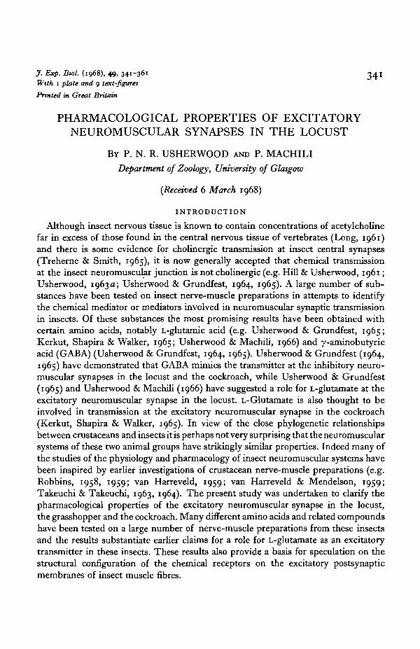
The metathoracic retractor unguis muscles of the locusts Schistocerca gregaria andLocusta migratoria, the grasshopper Romalea microptera, and the cockroach Blaberusgiganticus were used. These small muscles can be isolated from the insect with theirinnervating axons and therefore have many advantages over the larger, complex in situnerve-muscle preparations used in previous investigations of insect neuromuscular
Exoskeleton of trochanterattached to perfusionchamber with Tackiwax
Glass microelectrodes
—Glass shaft ofstrain gauge
Perspexblock
Text-fig, i. Diagrammatic representation of perfusion bath and isolated retractor unguis nerve-muscle preparation. Muscle not drawn to scale. The bath was constructed out of a block of clearPerspex. The over-all design of the perfusion system is illustrated in (b) while details of thearrangement of stimulating and recording electrodes and attachment of muscle to the per-fusion chamber and transducer are illustrated in (a). (Drawings by the late S. L. Hill.)
physiology and pharmacology (e.g. Hill & Usherwood, 1961; Usherwood & Grund-fest, 1964, 1965). The retractor unguis muscle is about 1 cm long and 300 fi in dia-meter in the locust and cockroach (slightly larger in the grasshopper) and rarelycontains more than 18 fibres. The fibres are arranged in parallel and are intimatelyconnected together by branches of the tracheolar respiratory system. The muscle isattached proximally to the lateral wall of the femoral segment of the hind leg and ends
Excitatory neuromuscular synapses in the locust 343
in a long filamentous apodeme (tendon) which runs through the femoral and tibialsegments to the tibial ('toe') joint.
Hind legs were removed from adult insects and the femoral segments were fixedhorizontally in a perfusion bath by attaching the trochanteral segment to the wall of thebath with Tackiwax (Text-fig. 1). The flexor and extensor tibiae muscles were thenremoved from the femur, together with most of the exoskeleton, leaving the retractorunguis muscle and its innervating nerve. The muscle apodeme was then attached to aGrass FT 10 strain gauge with a short strand of Terylene, the muscle being stretchedto maximal body length. The total volume of the bath, including inlet and outletreservoirs, was about 2-2 ml. and the contents could be replaced within 1 sec. Mechani-cal recording artifacts, resulting from the flow of saline over the preparation and pastthe strain-gauge shaft, were reduced to a minimum by covering the part of the bathcontaining the muscle preparation with thin Polythene sheeting. This did not seriouslyaffect the rate of replacement of saline around the nerve-muscle preparation. Withpractice, it was possible to dissect out the nerve-muscle preparation and arrange it inthe perfusion bath in less than 10 min. The dissection and setting-up procedure wereperformed in continuously flowing saline. These isolated nerve-muscle preparationscan obtain sufficient oxygen from the saline medium provided they are perfused slowly,but continuously, throughout the experimental period.
The muscles were stimulated indirectly through fine (40-80 /i) copper or platinumwire electrodes insulated to their tips and placed on the retractor unguis nerve.Intracellular recordings from the muscle fibres were made using 3 M-KC1 glass micro-electrodes. The microelectrodes were connected to a negative-capacitance d.c. pre-amplifier with an agar/AgCl bridge. A second agar/AgCl bridge was used to earth theperfusion bath. High-resistance (> 20 M£2) glass microelectrodes (tip diam. less than1 fi) filled with molar sodium L-glutamate (pH 8-o) were used for iontophoreticapplication of glutamate.
In most experiments drugs were applied to the nerve-muscle preparation by dis-solving them in locust (10 K) saline of the following composition: NaCl, 140; KC1, 10;CaCl2 2; NaH2PO4, 4; NajjHPO ,̂ 6 mM. All the salines were buffered at pH 6-8 andall experiments were carried out at 18-190 C.
RESULTS
Structure and physiology of the retractor unguis nerve-muscle preparation
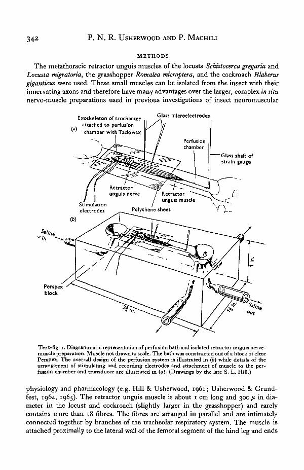
The retractor unguis muscle is differentiated structurally and functionally into twounits, although outwardly it has the appearance of a compact single bundle of fibres(Usherwood, 19676). Part of the muscle is made up of a group of large white fibres(c. 70 fi diam.), with sarcomere lengths of about 4 fi when the muscle is at maximalbody length. The rest of the muscle contains red fibres of about 40 ft diameter. Thesefibres are characterized by 7 /i sarcomeres at maximal body length and an abundanceof mitochondria near the Z-lines. Significantly, the smaller fibres fatigue more slowlyunder sustained tetanic stimulation (Text-fig. 2 A, B). The two bundles of fibres areinnervated separately, each bundle receiving a single motor axon. In all fibres theelectrical response to neural stimulation consists of a large excitatory postsynapticpotential (EPSP) plus graded electrically excited response (Text-fig. 2 C). The mecnani-
344 P. N. R. USHERWOOD AND P. MACHILI
cal responses of both sets of fibres consist of twitch contractions at low frequenciesand tetanic contractions at high frequencies of nerve stimulation, but the white fibresfatigue more quickly than the red fibres during sustained tetanic stimulation. Themuscle undergoes a single brief (c. 4 sec.) phasic contraction during potassiumdepolarization. The retractor unguis muscle fibres are multiterminally innervatedwith synapses distributed along the surface of the fibres. The structure of the neuro-muscular synapse is illustrated in Plate 1 and is characteristic of most insect neuro-muscular synapses in the absence of any visible structural differentiation of the post-synaptic membrane. Other properties of the retractor unguis muscle have been de-scribed in three recent publications (Cochrane & Elder, 1967; Usherwood, 1967a, b).
I I
10 sec.
(b)
- »-U - J L UText-fig. 2. (A-B) Mechanical responses of an isolated metathoracic retractor unguis nerve-muscle preparation from the locust. (A) Responses of pink fibres to neural stimulation atdifferent frequencies. Note slow rate of fatigue of these fibres even during tetanic stimulation.(B) Combined responses of pink and white fibres to neural stimulation at different frequencies.The white fibres fatigued much more quickly than the pink fibres. Stimulation frequencieswere: (a), o^/sec., (6) o-s/sec., (c) i/sec, (<f) 3/sec, (e) 6/sec., (/) 15/sec. (C) Electricalresponses from (a) a pink fibre and (6) a white fibre during neural stimulation. Each responseconsists of a large EPSP plus a graded electrically excited response. Calibration pulse atbeginning of each trace was 10 mV. 20 msec.
Pharmacological properties
The presence of short-chained dicarboxylic amino acids, such as aspartate andglutamate, in insect tissues and blood in concentrations as high as io"3 M (Frontalis,1961) is suggestive of the important role of these amino acids in physiological andmetabolic processes in insects.
Table 1 summarizes the effects of various amino acids and related compounds onthe retractor unguis preparation. With few exceptions modifications of the structureor position of the distal carboxyl grouping of the a-amino-dicarboxylic acids appear tocause profound changes in the activities of the compounds. Of the dicarboxylic acids
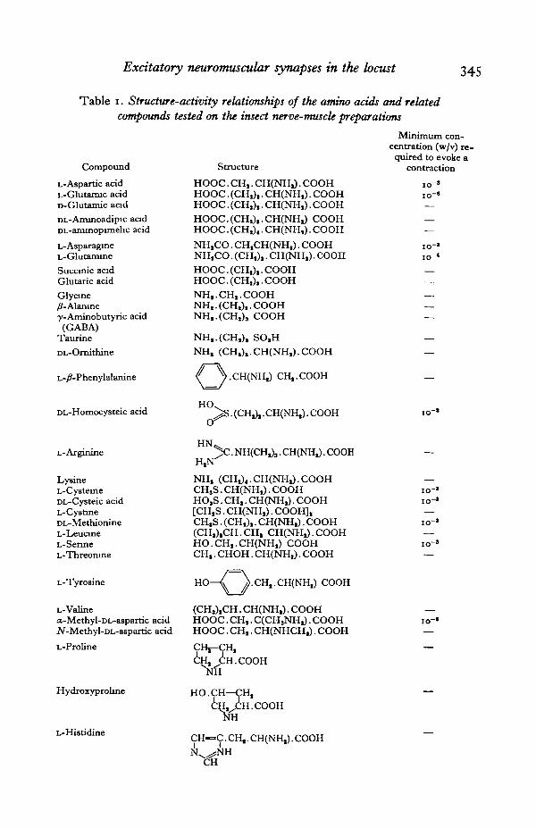
Excitatory neuromuscular synapses in the locust 345
Table i. Structure-activity relationships of the amino acids and related
compounds tested on the insect nerve-muscle preparations
Compound
L-Aspartic acidL-Glutamic acidD-Glutamic acidDL-Aminoadipic acidDL-aminopimelic acid
L-AsparagineL-GlutamineSuccinic acidGlutaric acidGlycine/?-Alaniney-Aminobutyric acid(GABA)
Taurine
DL-Orni thine
L-/?-Phenylalanine
Structure
HOOC. CH,. CH(NH,). COOHHOOC. (CH,),. CH(NH.). COOHHOOC. (CH,),. CH(NH.). COOH
HOOC.(CH,),.CH(NH,) COOHHOOC. (CH,),. CH(NH.). COOH
NH.CO. CH,CH(NH.). COOHNH.CO. (CH,),. CH(NH,). COOH
HOOC. (CH,),. COOHHOOC. (CH,),. COOH
NH,.CH,.COOHNH,.(CH,),.COOHNH,.(CH,)S COOH
NH,.(CH,), SO.HNH, (CH,),.CH(NH,).COOH
« VcH(NHJ CH..COOH
Minimum con-centration (w/v) re-quired to evoke a
contraction
DL-Homocysteic acid
L-Arginine
LysineL-CysteineDL-Cysteic acidL-CyatineDL-MethionineL-LeucineL-SenneL-Threonine
HO.,^S . (CH,),. CH(NHJ. COOH
. COOHH N ^
H,N^
NH, (CH,)4.CH(NH,).COOHCHjS. CH(NH.). COOHHO.S. CH,. CH(NH.). COOH[CH,S. CH(NH.). COOH],CH.S. (CH,),. CH(NH,). COOH(CH,),CH.CH, CH(NH,).COOHHO.CH,.CH(NH.) COOHCH,. CHOH. CH(NH,). COOH
L-Tyrosine
L-Valinea-Methyl-DL-aspartic acidJV-Methyl-DL-aspartic acid
L-Proline
Hydroxyproline
L-Histidine
)>.CH,.CH(NH,) COOH
(CH,),CH. CH(NH.). COOHHOOC. CH,. C(CH,NH,). COOHHOOC. CH,. CH(NHCH,). COOH
CHj-CH,CH, CH.COOH
^NH
HO.CH—CH,CHyCH.COOH
CH—C. CH,. CH(NH,). COOHN N H
346 P. N. R. USHERWOOD AND P. MACHILI
tested in the present investigations, L-glutamic acid was the most active. Whenapplied to the preparation in concentrations higher than io~7 (w/v) this substanceevoked a single phasic contraction followed by a decline in the neurally evoked con-tractions. Maximal contractions were obtained with concentrations of L-glutamic acidof about 10-6 (w/v). When preparations were perfused with L-glutamic acid in con-centrations > 5 x io"6 (w/v) the neurally evoked responses were completely abolished.The neurally evoked contractions were enhanced or potentiated during perfusion ofthe muscle with saline containing L-glutamic acid in concentrations between io~7 (w/v)and io^12 (w/v). The effects of L-glutamic acid were fully reversible and could bereadily repeated whilst the preparation remained viable (about 30 hr. at 180 C) . TheD-enantiomorph of glutamic acid was about 100 times less active than the naturallyoccurring L-form. L-Aspartic acid was even less active, having no effect at concentra-tions less than io~* (w/v). The amide derivatives of L-aspartic acid and L-glutamicacid were surprisingly active, although much less so than L-glutamic acid. Asparagineat a concentration of io-5 (w/v) evoked a phasic contraction accompanied by abolitionof the neurally evoked contractions, while the latter were enhanced by io"8 (w/v)asparagine.
The a-decarboxylation products of the short-chained amino acids could be ofspecial significance in insects in view of the possible implication of GABA at inhibi-tory neuromuscular synapses (Usherwood & Grundfest, 1964,1965). However, since themuscles used in these studies do not receive an inhibitory innervation it is perhaps notvery surprising that glycine, GABA, /?-alanine and taurine have only minimal effects.
The effects of substitution in the carbon chain by methyl or ring groupings on theactivity of amino acids on the retractor unguis preparation were examined by perfusingthe preparation with iV-methyl-DL-aspartate, L-proline, L-hydroxyproline andL-histidine. The effects of these substances were minimal, even at concentrations> io~s (w/v). Some of the compounds, e.g. DL-cysteic acid, in which the distalcarboxyl groupings of dicarboxylic amino acids are replaced by different configura-tions, at high concentrations were active, but most were inactive (i.e. > io"1 w/v).Succinic acid, which lacks the amino group characteristic of L-glutamic acid and otheramino acids, was completely inactive.
Site of action of h-glutamate
There is little doubt that L-glutamic acid is by far the most active of all the aminoacids and closely related compounds on insect (at least orthopteran) excitatory nerve-muscle preparations. In view of this the action of this substance on the retractor unguispreparation has been studied in some detail in an attempt to locate the specific site orsites of action of this substance.
In locust saline containing 10 mM potassium the normal resting membrane potentialof the retractor unguis muscle fibre is about —60 mV. L-Glutamic acid in concentra-tions > io~7 (w/v) depolarizes these muscle fibres, the magnitude of the depolariza-tion being directly proportional to the concentration of amino acid. Graded electricallyexcited responses accompany the larger glutamate depolarizations. The effective mem-brane resistance of the retractor unguis muscle fibre in 10 K saline is usually between300-500 Kfi, but declines to about 30 % of this value during treatment with io^6 (w/v)L-glutamic acid. However, changes in membrane properties during prolonged expo-
Excitatory neuromuscular synapses in the locust 347
sure to high concentrations of L-glutamic acid were only transitory since the mem-brane potential and effective resistance slowly returned to normal. Concomitant re-covery of the neurally evoked contractions did not occur, however, yet followingrecovery of the membrane potential and effective resistance the muscle fibres con-tracted in response to direct electrical stimulation. Apparently L-glutamate has noeffect on the electrically excitable membrane and contractile properties of the musclefibres. It also has no effect on axonal conduction. Therefore the decline in magnitudeof the neurally evoked contractions is indicative of failure of synaptic transmission.
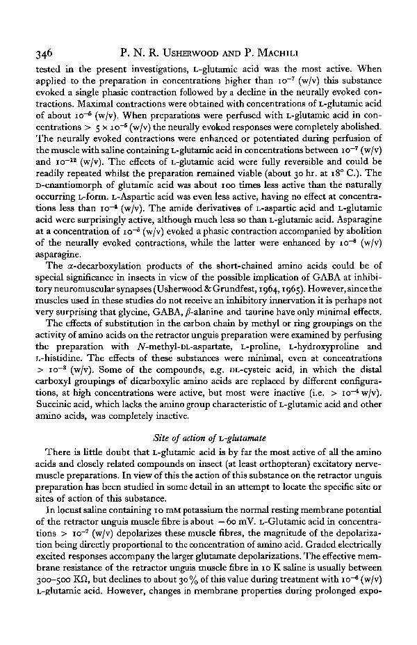
0 5 g
1 mm.
\\\\\..
(c)
10-1 (w/v) 5-HT 10"J (w/v) tryptamine
1/
Text-fig. 3. Abolition of glutamate contractions of a locust retractor unguis muscle by trypta-mine and 5-hydroxytryptamine. (a, d) Phasic contractions evoked by io~* (w/v) L-glutamicacid. Neurally evoked twitch contractions are shown at the beginning and end of each record;neural stimulation was discontinued for a short period before, during and after treatment withL-glutamic acid, (b, e) Absence of phasic contractions in response to L-glutamic acid (io~§
w/v) when preparation was perfused with (b) io~' (w/v) 5-HT and (e) io~' (w/v) tryptamine.These indolalkylamines are thought to block excitation of the excitatory postsynaptic mem-brane by competing with the excitatory transmitter for the postsynaptic receptor sites (Hill &Usherwood, 1961). After removal of the indolalkylamines phasic contractions could onceagain be evoked from the preparations by application of L-glutamic acid (io~5, w/v) (c./.).Brief applications of L-glutamic acid indicated by arrows above each trace. Time and tensioncalibrations same for (a—f).
High concentrations of 5-hydroxytryptamine (5-HT), tryptamine and a number oftryptamine analogues block neuromuscular transmission in locusts possibly by com-peting with the transmitter at the excitatory synapses for receptor sites on the excita-tory postsynaptic membrane (Hill & Usherwood, 1961). These substances have noeffect on either the axonal membrane or the electrically excitable membrane of themuscle fibre. Therefore if glutamate mimics the excitatory transmitter it should haveno effect on preparations treated with 5-HT or tryptamine. To test this, preparationswere perfused with either io~3 (w/v) 5-HT or io~3 (w/v) tryptamine. The neurallyevoked contractions were abolished in the presence of these indolalkylamines, andapplication of L-glutamic acid in concentrations as high as io"3 (w/v) now had noeffect on these preparations (Text-fig. 3). After removing the 5-HT or tryptamine the

348 P. N. R. USHERWOOD AND P. MACHILI
neurally evoked contractions reappeared and the muscle now responded normally toglutamate. It would appear therefore that the action of glutamate on the retractorunguis nerve-muscle preparation is limited mainly to the excitatory postsynapticmembrane of the muscle fibre, although the possibility of an additional presynapticaction cannot be ruled out.
Possibly the best way to pinpoint the site of action of glutamate is to apply itlocally to the surface of the muscle fibre. Preliminary experiments using the ionto-
10"7A
10"7A.
10-'A.
5mV.
1 sec.
Text-fig. 4. Intracellular recordings (lower traces) of membrane depolarizations in response tolontophoretic application of L-glutamate. Upper traces, monitored ejection currents, (a) Largedepolarization with the glutamate electrode close by a synaptic site. (6) Graded responses toincreasing ejection currents, (c—d) Glutamate potentials followed by EPSPs. Muscle treatedwith saline containing 40 mM magnesium to reduce height of EPSPs. In (d) the duration of theejection current was increased and a second glutamate potential was observed. Presumably thedrug was reaching two synaptic sites. (<) De-sensitization of glutamate receptors by repetitiveejection of glutamate from a focally sited glutamate electrode. The intracellular recordings in(a-d) are preceded by a calibration pulse: (a) iomV., 10 msec., (6) 2mV., joomse2 mV., 100 msec.
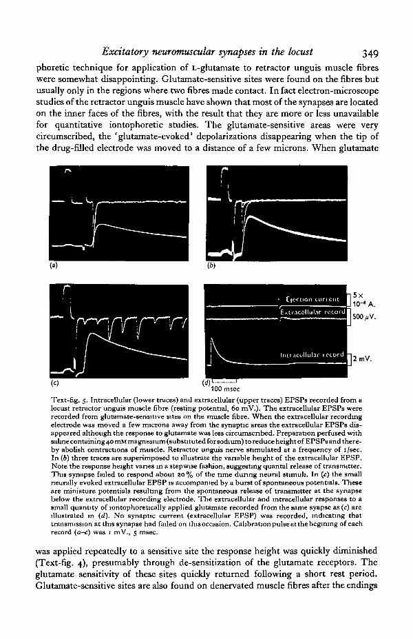
Excitatory neuromuscular synapses in the locust 349
phoretic technique for application of L-glutamate to retractor unguis muscle fibreswere somewhat disappointing. Glutamate-sensitive sites were found on the fibres butusually only in the regions where two fibres made contact. In fact electron-microscopestudies of the retractor unguis muscle have shown that most of the synapses are locatedon the inner faces of the fibres, with the result that they are more or less unavailablefor quantitative iontophoretic studies. The glutamate-sensitive areas were verycircumscribed, the ' glutamate-evoked' depolarizations disappearing when the tip ofthe drug-filled electrode was moved to a distance of a few microns. When glutamate
2mV.
100 msec
Text-fig. 5. Intracellular (lower traces) and extracellular (upper traces) EPSPs recorded from alocust retractor unguis muscle fibre (resting potential, 60 mV.). The extracellular EPSPs wererecorded from glutamate-sensitive sites on the muscle fibre. When the extracellular recordingelectrode was moved a few microns away from the synaptic areas the extracellular EPSPs dis-appeared although the response to glutamate was less circumscribed. Preparation perfused withsaline containing 40 mM magnesium (substituted for sodium) to reduce height of EPSPs and there-by abolish contractions of muscle. Retractor unguis nerve stimulated at a frequency of i/sec.In (6) three traces are superimposed to illustrate the variable height of the extracellular EPSP.Note the response height vanes in a stepwise fashion, suggesting quantal release of transmitter.This synapse failed to respond about 20% of the time during neural stimuli. In (c) the smallneurally evoked extracellular EPSP is accompanied by a burst of spontaneous potentials. Theseare miniature potentials resulting from the spontaneous release of transmitter at the synapsebelow the extracellular recording electrode. The extracellular and intracellular responses to asmall quantity of lontophoretically applied glutamate recorded from the same syapse as (c) areillustrated in (d). No synaptic current (extracellular EPSP) was recorded, indicating thattransmission at this synapse had failed on thisoccasion. Calibration pulse at the begirung of eachrecord (a-c) was 1 mV., 5 msec.
was applied repeatedly to a sensitive site the response height was quickly diminished(Text-fig. 4), presumably through de-sensitization of the glutamate receptors. Theglutamate sensitivity of these sites quickly returned following a short rest period.Glutamate-sensitive sites are also found on denervated muscle fibres after the endings
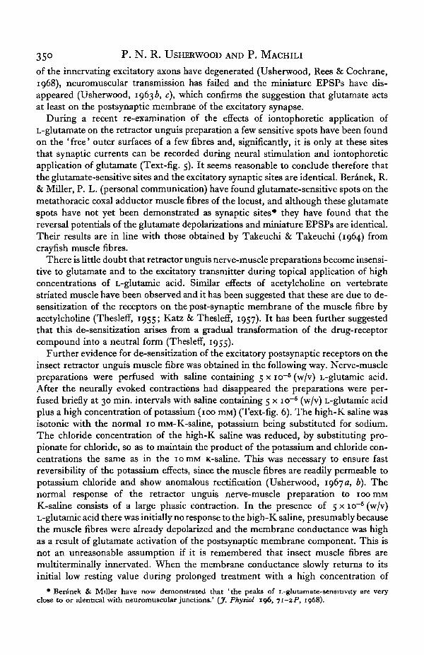
350 P. N. R. USHERWOOD AND P. MACHILI
of the innervating excitatory axons have degenerated (Usherwood, Rees & Cochrane,1968), neuromuscular transmission has failed and the miniature EPSPs have dis-appeared (Usherwood, 19636, c), which confirms the suggestion that glutamate actsat least on the postsynaptic membrane of the excitatory synapse.
During a recent re-examination of the effects of iontophoretic application ofL-glutamate on the retractor unguis preparation a few sensitive spots have been foundon the ' free' outer surfaces of a few fibres and, significantly, it is only at these sitesthat synaptic currents can be recorded during neural stimulation and iontophoreticapplication of glutamate (Text-fig. 5). It seems reasonable to conclude therefore thatthe glutamate-sensitive sites and the excitatory synaptic sites are identical. Beranek, R.& Miller, P. L. (personal communication) have found glutamate-sensitive spots on themetathoracic coxal adductor muscle fibres of the locust, and although these glutamatespots have not yet been demonstrated as synaptic sites* they have found that thereversal potentials of the glutamate depolarizations and miniature EPSPs are identical.Their results are in line with those obtained by Takeuchi & Takeuchi (1964) fromcrayfish muscle fibres.
There is little doubt that retractor unguis nerve-muscle preparations become insensi-tive to glutamate and to the excitatory transmitter during topical application of highconcentrations of L-glutamic acid. Similar effects of acetylcholine on vertebratestriated muscle have been observed and it has been suggested that these are due to de-8ensitization of the receptors on the post-synaptic membrane of the muscle fibre byacetylcholine (Thesleff, 1955; Katz & Thesleff, 1957). It has been further suggestedthat this de-sensitization arises from a gradual transformation of the drug-receptorcompound into a neutral form (TheslefT, 1955).
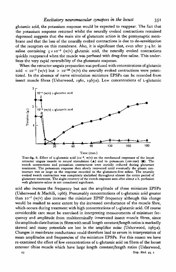
Further evidence for de-sensitization of the excitatory postsynaptic receptors on theinsect retractor unguis muscle fibre was obtained in the following way. Nerve-musclepreparations were perfused with saline containing 5 x io~6 (w/v) L-glutamic acid.After the neurally evoked contractions had disappeared the preparations were per-fused briefly at 30 min. intervals with saline containing 5 x io"6 (w/v) L-glutamic acidplus a high concentration of potassium (100 IEM) (Text-fig. 6). The high-K saline wasisotonic with the normal 10 mM-K-saline, potassium being substituted for sodium.The chloride concentration of the high-K saline was reduced, by substituting pro-pionate for chloride, so as to maintain the product of the potassium and chloride con-centrations the same as in the 1 o IBM K-saline. This was necessary to ensure fastreversibility of the potassium effects, since the muscle fibres are readily permeable topotassium chloride and show anomalous rectification (Usherwood, 1967 a, b). Thenormal response of the retractor unguis nerve-muscle preparation to 100 mMK-saline consists of a large phasic contraction. In the presence of 5 x io"5 (w/v)L-glutamic acid there was initially no response to the high-K saline, presumably becausethe muscle fibres were already depolarized and the membrane conductance was highas a result of glutamate activation of the postsynaptic membrane component. This isnot an unreasonable assumption if it is remembered that insect muscle fibres aremultiterminally innervated. When the membrane conductance slowly returns to itsinitial low resting value during prolonged treatment with a high concentration of
• Beranek & Miller have now demonstrated that 'the peaks of L-glutamate-sensitiwty are veryclose to or identical with neuromuscular junctions.' (J. Phytiol 196, 71-2P, 1968).
Excitatory neuromuscular synapses in the locust 351
glutamic acid, the potassium response would be expected to reappear. The fact thatthe potassium response returned whilst the neurally evoked contractions remaineddepressed suggests that the main site of glutamate action is the postsynaptic mem-brane and that the loss of the neurally evoked contractions is due to de-sensitizationof the receptors on this membrane. Also, it is significant that, even after 3-4 hr. insaline containing 5 x io"6 (w/v) glutamic acid, the neurally evoked contractionsquickly reappeared when the muscle was perfused with drug-free saline. This under-lines the very rapid reversibility of the glutamate response.
When the retractor unguis preparation was perfused with concentrations of glutamicacid < io~7 (w/v) but > io~12 (w/v) the neurally evoked contractions were poten-tiated. In the absence of nerve stimulation miniature EPSPs can be recorded frominsect muscle fibres (Usherwood, 1961, 1963a). Low concentrations of L-glutamic
120 150 180
Time (min.)Text-fig. 6. Effect of L-glutamic acid (io~*, w/v) on the mechanical responses of the locustretractor unguis muscle to neural stimulation (A) and to potassium (ioomM) (•). Thetwitch contractions and potassium contractures were initially reduced during glutamatetreatment. The potassium response then slowly recovered until eventually the phasic con-tracture was as large as the response recorded in the glutamate-free saline. The neurallyevoked twitch contraction was completely abolished throughout almost the entire period ofglutamate treatment. The slight recovery of the twitch response seen after about 2 h. perfusionwith glutamate-salme is not considered significant.
acid also increase the frequency but not the amplitude of these miniature EPSPs(Usherwood & Machili, 1966). Presumably concentrations of L-glutamic acid greaterthan io~7 (w/v) also increase the miniature EPSP frequency although this changewould be masked to some extent by the increased conductance of the muscle fibre,which occurs during treatment with high concentrations of L-glutamic acid. Of courseconsiderable care must be exercised in interpreting measurements of miniature fre-quency and amplitude from multiterminally innervated insect muscle fibres, sincethe amplitude distribution in fibres with small length constant/length ratios is markedlyskewed and many potentials are lost in the amplifier noise (Usherwood, 1963 a).Changes in membrane conductance could therefore lead to errors in interpretation ofmean amplitudes and frequencies of the miniature EPSPs. For this reason we havere-examined the effect of low concentrations of L-glutamic acid on fibres of the locustextensor tibiae muscle which have large length constant/length ratios (Usherwood,
23 Exp. Biol. 49, 2
352 P. N . R. USHERWOOD AND P. MACHILI
1963 a). The results from these studies confirmed those obtained from our previousexperiments of the retractor unguis muscle, i.e. low concentrations of L-glutamic acidincrease miniature frequency but not miniature amplitude. It has been proposed (e.g.del Castillo & Katz, 1954a, b) that changes in miniature EPSP frequency reflectedsome alteration in presynaptic rather than postsynaptic properties. Possibly glutamatefacilitates the release of transmitter from the excitatory nerve terminals. This couldaccount for potentiation of the neurally evoked contractions in low concentrations ofL-glutamate (Usherwood & Machili, 1966).
Enzymes and enzyme inhibitors
At vertebrate neuromuscular junctions there is clear evidence for the presence ofhigh concentrations of a powerful enzyme, cholinesterase, which is thought to hy-drolyse the transmitter released from the motor nerve terminals and acetylcholineapplied either topically or iontophoretically. Administration of potent cholinesteraseinhibitors like prostigmine, eserine and edrophonium leads to increases in amplitudeof the miniature EPSPs, EPSPs and neurally evoked twitch contractions of the skeletalmuscle fibres (e.g. del Castillo & Katz, 1957; Axelsson & Thesleff, 1958). In view of
Table 2. Effect of enzyme inhibitors*Minimum con-
centration (w/v) re-quired to affect
indirect mechanicalCompound Enzyme responses
Serrucarbazide HC1 Glutanuc-pyruvic io~'transaminase
Glutamic decarboxylaseDiamine oxidase
Hydroxylamine HC1 Glutamic-pyruvic io~*transaminaseGlutamic decarboxylaseDiamine oxidase
Thiosemicarbazide HC1 Glutamic-pyruvic io~*transaminase
Glutamic decarboxylaseDiamine oxidase
Phenylhydrazine HC1 Glutamic-pyruvic io~'transaminase
Glutamic decarboxylaseDiamine oxidase
p-Phenylenediarrune HC1 Glutamic-oxaloacetic io~»transaminase
Glutamic pyruvictransaminase
• Adapted from Curtis, Phillips & Watkins, i960.
the undoubted enzyme-transmitter relationships in vertebrate neuromuscular systemsit seemed worth while to search for parallel systems in the insect, working on theassumption that the chemical mediator at the excitatory nerve-muscle synapse ininsects is L-glutamate. The enzyme glutamic decarboxylase occurs in relatively highconcentrations in some insect tissues (Frontalis, 1961). This enzyme mediates the
Excitatory neuromuscular synapses in the locust 353
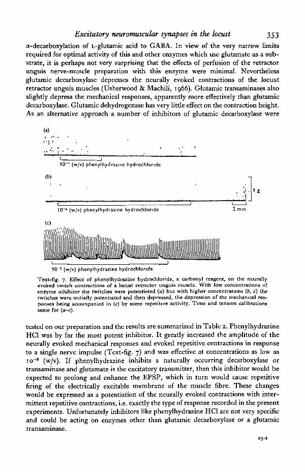
a-decarboxylation of L-glutamic acid to GABA. In view of the very narrow limitsrequired for optimal activity of this and other enzymes which use glutamate as a sub-strate, it is perhaps not very surprising that the effects of perfusion of the retractorunguis nerve-muscle preparation with this enzyme were minimal. Neverthelessglutamic decarboxylase depresses the neurally evoked contractions of the locustretractor unguis muscles (Usherwood & Machili, 1966). Glutamic transaminases alsoslightly depress the mechanical responses, apparently more effectively than glutamicdecarboxylase. Glutamic dehydrogenase has very little effect on the contraction height.As an alternative approach a number of inhibitors of glutamic decarboxylase were
10 ' (w/v) phenylhydrazine hydrochlonde
(b)
1 _
10"* (w/v) phenylhydrazine hydrochlonde 2 mm
10"J (w/v) phenylhydrazine hydrochlonde
Text-fig. 7. Effect of phenylhydrazine hydrochloride, a carbonyl reagent, on the neurallyevoked twitch contractions of a locust retractor unguis muscle. With low concentrations ofenzyme inhibitor the twitches were potentiated (a) but with higher concentrations (6, c) thetwitches were initially potentiated and then depressed, the depression of the mechanical res-ponses being accompanied in (c) by some repetitive activity. Time and tension calibrationssame for (a-c).
tested on our preparation and the results are summarized in Table 2. PhenylhydrazineHC1 was by far the most potent inhibitor. It greatly increased the amplitude of theneurally evoked mechanical responses and evoked repetitive contractions in responseto a single nerve impulse (Text-fig. 7) and was effective at concentrations as low asio~9 (w/v). If phenylhydrazine inhibits a naturally occurring decarboxylase ortransaminase and glutamate is the excitatory transmitter, then this inhibitor would beexpected to prolong and enhance the EPSP, which in turn would cause repetitivefiring of the electrically excitable membrane of the muscle fibre. These changeswould be expressed as a potentiation of the neurally evoked contractions with inter-mittent repetitive contractions, i.e. exactly the type of response recorded in the presentexperiments. Unfortunately inhibitors like phenylhydrazine HC1 are not very specificand could be acting on enzymes other than glutamic decarboxylase or a glutamictransaminase.
23-2
354 P- N. R. USHERWOOD AND P. MACHILI
Analysis of amino acids in locust haemolymph (blood)
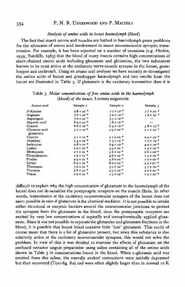
The fact that insect nerves and muscles are bathed in haemolymph poses problemsfor the advocates of amino acid involvement in insect neuromuscular synaptic trans-mission. For example, it has been reported on a number of occasions (e.g. Florkin,1959; Sutcliffe, 1963) that the blood of many insects contains high concentrations ofshort-chained amino acids including glutamate and glutamine, the two substancesknown to be most active at the excitatory nerve-muscle synapse in the locust, grass-hopper and cockroach. Using an amino acid analyser we have recently re-investigatedthe amino acids of locust and grasshopper haemolymph and two results from thelocust are illustrated in Table 3. If glutamate is the excitatory transmitter then it is
Table 3. Molar concentrations of free amino acids in the haemolymph(blood) of the locust, Locusta migratoria
Amino acid/?-AlanineArginineAsparagineAspartic acidCystineGlutamic acidglutamine
GlycineHistidineIsoleucineLysineMethioninePhenylalanineProlineSerineThreonineTyrosineVahne
Sample 19-8 x io-*5-7XIO-*7-0 x io"*84 x io"5
6-6xio-5
3-7x10-*
5-1 x io"»5 4X 10-*28 x 10-*I-OX 10"*
1-7 x io"*1-2 X 10"*2-9 x io"J
6-4 x io~*7-0 x io~*2-8x10"*70 x 10-*
Sample 21-7 x 10-*72 x io"1
3-1 x io-*18 x io-'6 4X io~*5-4x10-*
5 1 x io"1
1-3 x io"'6-4 x io-1
3-3x10-*5 6 x io"*4-6 X 10"'9 8x 10-*6-ox io"6
3-1 x 10-*2-5 x 10-*i-5 x 10"*
Sample 31-7 x 10-'1-2 X IO-'
—
—
3 8 xio-»5-1 x io"1
2 4 x io-1
1-7 x io"1
4-2 x io-*1-4 x io"1
2-6 X I0~*1-5 x io~*1-2 X 1 0 - '59x01-*5-7 x 10-*2-2 X IO~*1-5 x io-'
difficult to explain why the high concentration of glutamate in the haemolymph of thelocust does not de-sensitize the postsynaptic receptors on the muscle fibres. In otherwords, transmission at the excitatory neuromuscular synapses of the locust does notseem possible in vivo if glutamate is the chemical mediator. It is not possible to invokeeither structural or enzymic barriers around the neuromuscular junctions to protectthe synapses from the glutamate in the blood, since the postsynaptic receptors areexcited by very low concentrations of topically and iontophoretically applied gluta-mate. Since it was not possible to separate the glutamine and glutamate fractions in theblood, it is possible that locust blood contains little 'free' glutamate. This could ofcourse mean that there is a lot of glutamine present, but since this substance is alsorelatively active at the excitatory neuromuscular synapses, this would not solve theproblem. In view of this it was decided to examine the effects of glutamate on theperfused retractor unguis preparation using saline containing all of the amino acidsshown in Table 3 in concentrations found in the blood. When L-glutamic acid wasomitted from this saline, the neurally evoked contractions were initially depressedbut then recovered (Text-fig. 8 a) and were often slightly larger than in normal 10 K
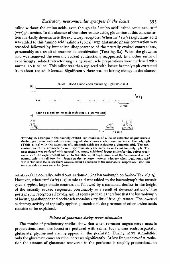
Excitatory neuromuscular synapses in the locust 355
saline without the amino acids, even though the 'amino acid' saline contained io~*(w/v) glutamine. In the absence of the other amino acids, glutamine at this concentra-tion markedly de-sensitizes the excitatory receptors. When io~* (w/v) L-glutamic acidwas added to this ' amino acid' saline a typical large glutamate phasic contraction wasrecorded followed by immediate disappearance of the neurally evoked contractions,presumably as a result of receptor de-sensitization (Text-fig. 86). When the glutamicacid was removed the neurally evoked contractions reappeared. In another series ofexperiments isolated retractor unguis nerve-muscle preparations were perfused withnormal 10 K saline. This saline was then replaced with locust haemolymph extractedfrom about 100 adult locusts. Significantly there was no lasting change in the charac-
Salme +blood amino acids excluding L-glutamic acidI
"•»•
(b)
2 mm
ammo acids including L-glutamic acidd
Text-fig. 8. Changes in the neurally evoked contractions of a locust retractor unguis muscleduring perfusion with saline containing all the amino acids found in locust haemolymph(Table 3): (a) with the exception of L-glutamic acid; (6) including L-glutamic acid. The con-centrations of the amino acids were approximately the same as in locust haemolymph. Thepreparation was perfused with normal (i.e. amino acid-free) locust saline for 3 hr. before treat-ment with the experimental saline. In the absence of L-glutamic acid the ' amino acid saline'caused only a small transient change in the response pattern, whereas when L-glutamic acidwas included in the saline there was a sustained abolition of the mechanical responses. Time andtension calibrations same for (a-b).
teristics of the neurally evoked contractions during haemolymph perfusion (Text-fig. 9).However, when io~* (w/v) L-glutamic acid was added to the haemolymph the musclegave a typical large phasic contraction, followed by a sustained decline in the heightof the neurally evoked responses, presumably as a result of de-sensitization of thepostsynaptic receptors (Text-fig. 96). It seems probable therefore that the haemolymphof locust, grasshopper and cockroach contains very little ' free' glutamate. The loweredexcitatory activity of topically applied glutamine in the presence of other amino acidsremains to be explained.
Release of glutamate during nerve stimulation
The results of preliminary studies show that when retractor unguis nerve-musclepreparations from the locust are perfused with saline, four amino acids, aspartate,glutamate, glycine and alanine appear in the perfusate. During nerve stimulationonly the glutamate concentration increases significantly. At low frequencies of stimula-tion the amount of glutamate recovered in the perfusate is roughly proportional to

356 P. N. R. USHERWOOD AND P. MACHILI
the number of stimuli applied to the retractor unguis nerve. Kerkut, Leake, Shapira,Cowan & Walker (1965) obtained L-glutamate from cockroach leg preparations duringnervous stimulation, the amount recovered being proportional to the number ofstimuli applied to the leg nerves. It seems probable therefore that glutamate is re-leased by motor nerve endings in the grasshopper, locust and cockroach and that thisamino acid is the transmitter at the neuromuacular excitatory synapses in these insects.Significantly the amount of glutamate appearing in the perfusate from the locustretractor unguis nerve-muscle preparation during neural stimulation is increased byblocking the excitatory synapses with 5-hydroxytryptamine. This will be discussedmore fully in a later publication.
(a) Locust haemolymph (blood)
Locust haemolymph (blood) + 10"' (w/v) L-glutamic acid(b) I 1
' 0 2 g
.....\«\V10 min. I
2 mm.
Text-fig. 9. (a) Absence of any permanent change in the magnitude of the neurally evoked con-tractions of a locust retractor unguis muscle when perfused with locust haemolymph (blood).However, when IO"4 (w/v) L-glutamic acid was added to the haemolymph the contractions wereabolished (b), presumably as a result of de-sengitization of the glutamate (excitatory trans-mitter?) receptors. Time and tension calibrations same for (a-b).
DISCUSSION
It is clear from these studies that glutamic acid is the most potent amino acid at theexcitatory synapses in the locust, grasshopper and cockroach. It is perhaps significantin this respect that the a-decarboxylation product of glutamic acid, GABA, mimicsthe transmitter at inhibitory neuromuscular synapses in insects (Usherwood &Grundfest, 1964, 1965). There are striking similarities between the effects of aminoacids on insect neuromuscular preparations, on the one hand, and crustacean neuro-muscular preparations (Robbins, 1959, van Harreveld & Mendelson, 1959; Takeuchi& Takeuchi, 1964) on the other. There are also similarities between insect neuro-muscular preparations and cat spinal neurones (Curtis & Watkins, i960; Curtis,Phillis & Watkins, i960) with respect to their responsiveness to amino acids. Curtis &Watkins (i960) concluded that for optimal excitation of cat spinal neurones the aminoacids must have two acidic groups and one basic group, with two or three carbon atomsbetween the amino group and one of the acidic groups (distal). The other acidic groupis optimally situated a with respect to the amino group. Activity is abolished if theacidic or basic groups are not free and is reduced or abolished by substitution withinthe intermediate carbon chain or within the amino group, depending on the size,number and position of the constituents. In insects the presence of the distal acid
Excitatory neuromuscular synapses.in the locust 357
group in the amino acid chain is absolutely essential for activity. This group, as wellas the amino group, could of course be almost completely ionized in the physiologicalmedium. There is a clear relationship between optimum activity and the position ofthe amino group with respect to the distal acid group. For example, the a-dicarboxylicamino acids aspartate, glutamate, a-aminoadipate and a-amino pimelate carry similarcharges on their molecules (a positive charge on the amino group and a negative chargeon each of the two carboxyl groups) and yet their activities are considerably different.The only structural variation among these amino acids is of course the length of thecarbon chain and therefore the distance of the distal acid group from the positivelycharged amino group. Reduction of the negative charge on the distal acid group, forinstance by substitution of OH~ by an amino group (e.g. glutamine), also reducesresponsiveness, whereas complete removal of the acidic group (e.g. GABA, /?-alanine)or its replacement with neutral groups (e.g. tyrosine, valine and leucine) abolishesactivity altogether. Absence of the amino group, as in succinic acid, abolishes activitycompletely. So also does any substitution within the carbon chain. From these studiesit seems possible that glutamate is the only compound with the necessary chainlength and distribution of the ionized groups suitable for interaction with ioniccharges on the receptor sites. Steric orientation of the transmitter molecule could alsobe decisive for optimal activity, which could account for the fact that D-glutamic acidis much less active than the naturally occurring L-isomer. L-Glutamate is also the mostactive amino acid at crustacean excitatory neuromuscular synapses and here, as inthe insects, activity of the D-enantiomorph is considerably lower (Robbins, 1959;Takeuchi & Takeuchi, 1964).
The effects of L-glutamate on insect neuromuscular preparations raises the questionof whether this substance is the transmitter at the excitatory synapses. There areseveral criteria for identification of a substance as a chemical synaptic transmitter(Paton, 1958; Florey, i960; Werman, 1966) among which the most fundamental beingthat the substance must have the same action on the transmitter and that it should berecoverable in the perfusion fluid during neural stimulation. Additional support forthe role of a substance as a chemical mediator at synapses would be the demonstrationof enzyme systems capable of synthesis and destruction of the substance at the synapticsites. There seems little doubt that L-glutamate mimics the transmitter at excitatoryneuromuscular synapses in the locust, cockroach and grasshopper. By applyingL-glutamate topically and iontophoretically to neuromuscular preparations fromthese insects it has been shown that this substance acts specifically at the excitatorysynapses and that its mode of action is identical to that of the excitatory transmitter.For example, the identification of glutamate-sensitive sites on the muscle fibres withthe excitatory synapses, the excitatory effects of potassium on glutamate-de-sensitizedmuscle fibres where neuromuscular transmission was completely blocked, and theabsence of glutamate responses in preparations treated with the excitatory synapticblocking agents 5-HT and tryptamine point conclusively to a specifically synapticaction for L-glutamate. Furthermore, the positive effects of L-glutamate on dener-vated muscle fibres in which the nerve endings have demonstrably degenerated andthe positive effects of L-glutamic acid on muscle fibres treated with magnesium ions inconcentrations which block synaptic transmission presynaptically (Usherwood, 1963 a),confirm at least the postsynaptic action of this amino acid. In other words, it appears
358 P. N . R. USHERWOOD AND P. MACHILI
from this evidence that the excitatory receptors on the postsynaptic membrane are infact glutamate receptors. This does not necessarily imply that the excitatory trans-mitter is L-glutamate, although the recovery of L-glutamate in the perfusate fromneuromuscular preparations of locust, grasshopper and cockroach following nervestimulation makes this extremely likely. It will of course be necessary to demonstratethe synthesis and/or storage of glutamate in the terminals of these insect excitatorymotoneurones before this can be conclusively established.
The relatively brief depolarizations recorded in response to low levels of ionto-phoretically applied L-glutamate could be due to enzymic removal of this substancealthough it is possible that it simply diffuses away from the synaptic region and entersthe ordinary metabolic pool. Removal of GABA, a possible inhibitory transmitter ininsects, is thought to occur by diffusion only, at least at inhibitory neuromuscularsynapses of cockroach, grasshopper and locust (Usherwood & Grundfest, 1965).Enzymic destruction of glutamic acid could, however, facilitate the removal of gluta-mate in vivo from the excitatory neuromuscular synapses. The reduction of theneurally evoked contractions during treatment with glutamic decarboxylase andglutamic transaminases gives added weight to the argument that glutamate is thetransmitter and the potentiation of these responses in the presence of decarboxylaseand transaminase inhibitors like phenylhydrazine hydrochloride is suggestive of someenzymic role at insect excitatory neuromuscular synapses. In fact the responses ofinsect neuromuscular preparations to these inhibitors are strikingly reminiscent of theeffects of anticholinesterases on nerve-striated muscle preparations in vertebrates. Theeffects of carbonyl reagents on insect neuromuscular preparations must, however, beinterpreted with some caution. An inhibitor completely specific for either glutamicdecarboxylase or transaminases has not yet been tested on our insect preparations.Furthermore, although insect nervous tissue is known to contain high concentrationsof glutamic decarboxylase (Frontalis, 1961), neither this enzyme nor transaminaseshave not been shown to occur at the excitatory neuromuscular synapses.
There remains the problem of the potentiating effects of low concentrations ofL-glutamic acid on the neurally evoked contractions of insect muscles. It has beensuggested that this is due, at least in part, to facilitated release of the excitatory trans-mitter as a result of some action of glutamate on the presynaptic axonal membrane(Usherwood & Machili, 1966; Usherwood, 19676). This suggestion was based on theobservation that in locust muscle fibres the frequency but not the amplitudes of theminiature EPSPs was increased in the presence of low concentrations of L-glutamicacid. The neurally evoked contractions of crustacean muscle fibres are also potentiatedby low concentrations of L-glutamic acid (Robbins, 1958). However, Takeuchi &Takeuchi (1964) have demonstrated that this could be, at least in part, postsynaptic inorigin. They found that the depolarization of crutacean muscle fibres resulting frombrief iontophoretic applications of glutamate to the excitatory synapses were tran-siently potentiated when a small conditioning dose of this amino acid was appliedto the synapse. Possibly, both presynaptic and postsynaptic changes contribute tothe potentiation of the neurally evoked contractions of these arthopodean musclefibres.
Suggestions that the transmitter at insect excitatory neuromuscular synapses isL-glutamate have been viewed in the past with some degree of scepticism. This was
Excitatory neuromuscular synapses in the locust 359
due to the apparent presence of very high (io~*, w/v) concentrations of L-glutamatein the haemolymph of many insects, i.e. glutamate concentrations which should com-pletely de-sensitize the postsynaptic receptors at the excitatory synapses. The resultsof the present investigations should remove some of this uncertainty, since it hasbeen shown that isolated locust neuromuscular preparations function quite well whenperfused with haemolymph and contract phasically when io"6 (w/v) L-glutamic acid isadded to the haemolymph. Apparently locust haemolymph contains very little 'free'glutamate. There seems to be no reason, therefore, why L-glutamate should not be thetransmitter at excitatory neuromuscular synapses in the locust, grasshopper and cock-roach. Indeed, glutamate-sensitive sites have recently been found on muscle fibres ofmealworm (Tenebrio molitor) larvar (Usherwood, unpublished). The mealwormbelongs to the Coleoptera, an insect group far removed from the orthopteroid groupsto which the cockroach, locust and grasshopper belong. Perhaps, therefore, glutamateis the excitatory transmitter at all insect excitatory neuromuscular synapses.
This work was supported by U.S. Public Health Service Grant 5-RO i-NB 05626-03and an Agricultural Research Council Grant to P. N. R. Usherwood. We are in-debted to Dr G. Leaf of the Department of Biochemistry, Glasgow University, for theanalyses of insect blood and nerve-muscle perfusates.
SUMMARY
1. The effects of a wide range of amino acids and related compounds on retractorunguis nerve-muscle preparations from the locust, grasshopper and cockroach havebeen investigated.
2. L-glutamate is the most active excitatory substance. The presence of two acidicgroups and one amino group is essential for excitatory activity while the position of theamino group is of some importance in determining the level of activity.
3. When L-glutamate is applied iontophoretically to the muscle fibres, 'glutamate'depolarizations are recorded only at the synaptic sites. Other evidence that the actionof glutamate is restricted to the synaptic sites is presented.
4. Perfusion of isolated locust retractor unguis nerve-muscle preparations withlocust haemolymph does not markedly affect the neurally evoked mechanical responses.It appears that locust haemolymph contains little ' free' L-glutamate.
5. Four acidic amino aids have been identified in the perfusate from isolatedretractor unguis preparations namely, glycine, alanine, aspartate and L-glutamate.However, only L-glutamate increases in concentration during stimulation of theretractor unguis nerve.
REFERENCES
AXELSSON, J. & THESLEFF, S. (1958). The desensitizing effect of acetylchokne on the mammalian motorend-plate. Acta phyttol. tcand. 43, 15-26.
COCHRANE, D. G. & ELDER, H. Y. (1967). Morphological changes in insect muscle during influx andefflux of potassium ions, chloride ions and water. J. Pkytiol., Lond. 191, 30— 1 P.
CURTIS, D. R., PHILLIS, J. W. & WATKINS, J. C. (i960). The chemical excitation of spinal neurones byacidic amino acids. J. Pkytiol., Lond. 150, 656-82.
CURTIS, D. R. & WATKINS, J. C. (i960). The excitation and depression of spinal neurones bv structurallyrelated amino acids. J. Neurochem. 6, 117-41.
360 P . N . R. USHERWOOD AND P . MACHILI
DEL CASTILLO, J. & KATZ, B. (1954a). The effect of magnesium on the activity of motor nerve endings.J. Physiol., Lond. 129, 553-59-
DEL CASTILLO, J. & KATZ, B. (19546). Changes in end-plate activity produced by pre-synaptic polariza-tion. J. Phytiol., Lond. 124, 586-604.
DEL CASTILLO, J. & KATZ, B. (1957). Interaction at end-plate receptors between different cholinederivatives. Proc. Roy. Soc. B. 146, 369-81.
F ORKIN, M. (1959). The free amino acids of insect haemolymph. 4th Int. Congr. Biochem. XII: Bio-chemistry of Insects. Oxford: Pergamon.
FLOREY, E. (i960). Physiological evidence for naturally occurring inhibitory substances. In Inhibition inthe Nervous System and a-ammo Butyric Acid, ed. E. Roberts. Oxford: Pergamon.
FRONTALIS, N. (1961). Activity of glutamic acid decarboxylase in insect nerve tissue. Nature, Lond. 191,178-9.
VAN HARREVELD, A. (1959). Compounds in brain extracts causing spreading depression of cerebralcortical activity and contraction of crustacean muscle. J. Neurochem. 3, 300-315.
VAN HARREVELD, A. & MENDELSON, M. (1959). Glutamate-induced contractions in crustacean muscle.J. cell comp. Physwl. 54, 85-95.
HILL, R. B. & USHERWOOD, P. N. R. (1961). The action of 5-hydroxytryptamine and related compoundson neuro-muscular transmission in the locust Schistocerca gregana. J. Physwl., Lond. 1ST, 393~401.
KATZ, B. & THESLEFF, S. (1957). A study of the ' desensitization' produced by acetylchokne at themotor end-plate. J. Physiol., Lond. 138, 63-80.
KKRKUT, G. A., LEAKE, L. D., SHAPIRA, A., COWAN, S. & WALKER, R. J. (1965). The presence of glu-tamate in the nerve-muscle perfusates of Helix, Carcinus and Pertplaneta. Comp. Biochem. Physiol.15. 485-5°3-
KERKUT, G. A., SHAPIRA, A. & WALKER, R. J. (1965). The effect of acetylchohne, glutamic acid andGABA on contractions of the perfused cockroach leg. Comp. Biochem. Physiol. 16, 37-48.
LONG, C. (1961) In Biochemist's Handbook. London: Spon.PATON, W. D. M. (1958). Central and synaptic transmission in the nervous system (pharmacological
aspects). A. Rev. Physio!. 30, 431-470.ROBBINS, J. (1958). The effect of amino acids on the crustacean muscle neuromuscular system. Anat.
Rec. 133, 492-3.ROBBINS, J. (1959)- The excitation and inhibition of crustacean muscle by amino acids. J. Physiol.,
Lond. 148, 39-50.SUTCLIFFE, D. W. (1963). The chemical composition of haemolymph in insects and some other arthro-
pods, in relation to their phylogeny. Comp Biochem. Physiol. 9, 121-35.TAKEUCHI, A. & TAKEUCHI, N. (1963). Glutamate-induced depolarization in crustacean muscle. Nature,
Lond. 198, 400-1.TAKEUCHI, A. & TAKEUCHI, N. (1964). The effect on crayfish muscle of lontophoretically applied
glutamate. J. Physiol., Lond. 170, 296-317.THESLEFF, S. (1955). The mode of neuromuscular block caused by acetylcholine, nicotine, decametho-
nium and succinylcholine. Ada physiol. scand. 34, 218-31.TREHERNE, J. E. & SMITH, D. S. (1965). The metabolism of acetylcholine in the intact central nervous
system of an insect, Penplaneta amencana J. exp. Btol. 43, 441-54.USHERWOOD, P. N. R., (1961). Spontaneous miniature potentials from insect muscle fibres. Nature,
Lond. 191, 814-15.USHBRWOOD, P. N. R. (1963a). Spontaneous miniature potentials from insect muscle fibres. J. Physiol.,
Lond. 169, 149-60.USHERWOOD, P. N. R. (19636). Response of insect muscle to denervation. I. Resting potential changes.
J. Insect Physwl. 9, 247-55.USHERWOOD, P. N. R. (1963 c). Response of insect muscle to denenation. II Changes in neuromuscular
transmission. J. Insect. Physiol. 9, 811-25USHERWOOD, P. N. R. (1967a). Permeability of insect muscle fibres to potassium and chloride ions.
J. Physiol., Lond. 191, 29-30P.USHERWOOD, P. N. R. (19676) Insect neuromuscular mechanisms. Am. Zool. 7, 553-82.USHERWOOD, P. N. R. & GRUNDFEST, H. (1964). Inhibitory post-synaptic potentials in grasshopper
muscle. Science, N.Y. 143, 817-18.USHERWOOD, P. N. R. & GRUNDFEST, H. (1965). Peripheral inhibition in skeletal muscle of insects.
J. Neurophysiol. 38, 497-518.USHERWOOD, P. N. R. & MACHILI, P. (1966). Chemical transmission at the insect excitatory neuro-
muscular synapse. Nature, Lond. 210, 634-6.USHERWOOD, P. N. R., COCHRANE, D. G. & REES, D. (1968). Changes in structural, physiological and
pharmacological properties of insect excitatory nerve-muscle synapses after motor nerve section.Nature Lond. 218, 580-91.
WERMAN, R. (1966). Criteria for identification of a central nervous system transmitter. Comp. Biochem.Physiol. 18, 745-66.


Journal of Experimental Biology, Vol. 49, No. 2 Plate 1
P. N. R. USHERWOOD AND P MACHILI (Facing p. 361)
Excitatory neuromuscular synapses in the locust 361
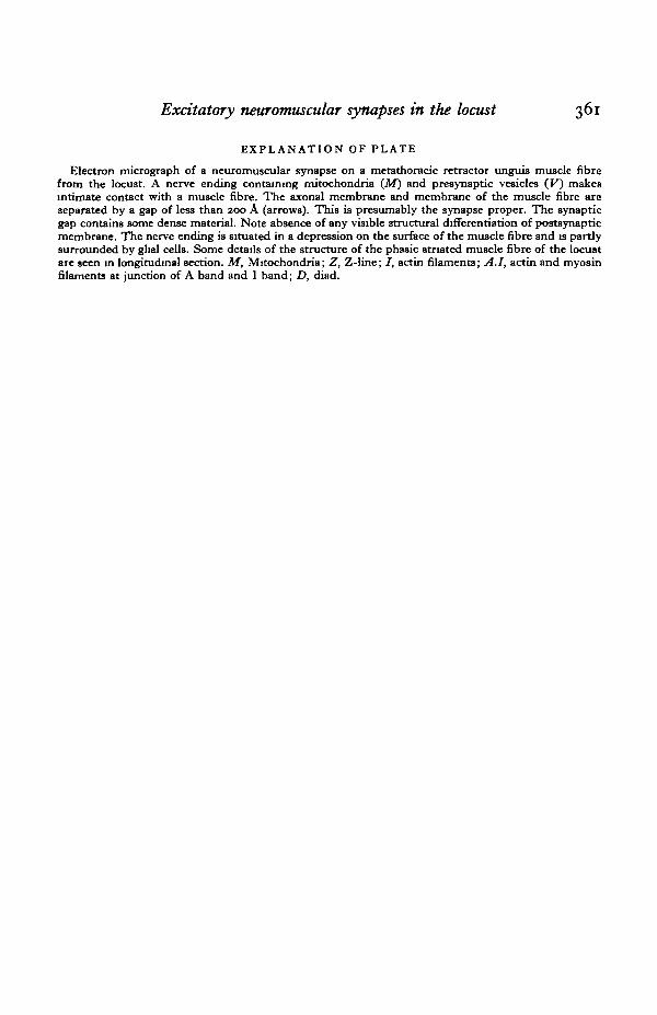
EXPLANATION OF PLATE
Electron micrograph of a neuromuscular synapse on a metathoracic retractor unguis muscle fibrefrom the locust. A nerve ending containing mitochondria (M) and presynaptic vesicles (F) makesintimate contact with a muscle fibre. The axonal membrane and membrane of the muscle fibre areseparated by a gap of less than 200 A (arrows). This is presumably the synapse proper. The synapticgap contains some dense material. Note absence of any visible structural differentiation of postsynapticmembrane. The nerve ending is situated in a depression on the surface of the muscle fibre and is partlysurrounded by ghal cells. Some details of the structure of the phasic striated muscle fibre of the locustare seen in longitudinal section. M, Mitochondria; Z, Z-line; /, actin filaments; A.I, actin and myosinfilaments at junction of A band and I band; D, diad.