pharmacological therapy for acute lung injurycritic15/images/1st...pharmacological therapy for acute...
TRANSCRIPT

The Open Critical Care Medicine Journal, 2010, 3, 45-57 45
1874-8287/10 2010 Bentham Open
Open Access
Pharmacological Therapy for Acute Lung Injury
Rob Mac Sweeney1,2
and Daniel F. McAuley*,1,2
1Respiratory Medicine Research Programme, Centre of Infection and Immunity, Queens University of Belfast,
Grosvenor Road, Belfast BT12 6BN, Northern Ireland
2Regional Intensive Care Unit, Royal Victoria Hospital, Belfast BT12 6BA, Northern Ireland
Abstract: Many pharmacological therapies have been investigated for use in acute lung injury (ALI) and the acute
respiratory distress syndrome (ARDS). These therapies can broadly be classified as being either anti-inflammatory or
physiology based. Despite promising pre-clinical and small clinical studies almost all therapies have been shown to be
unsuccessful in large scale randomized controlled trials. The evidence for pharmacological treatment for ALI/ARDS is
reviewed. Potential future treatments are also presented.
Keywords: Acute lung injury (ALI), acute respiratory distress syndrome (ARDS), pharmacological treatment, anti-inflammatory, ventilation, alveolar function.
INTRODUCTION
Mechanical ventilation, as a supportive treatment for respiratory failure, was popularised by Ibsen in Copenhagen during the polio epidemic in 1952. As polio caused neuromuscular failure the replacement of endogenous ventilation with mechanical ventilation was logical. However, for pulmonary pathologies which cause alveolar failure mechanical ventilation, although currently essential as supportive care, is potentially injurious. Delivering gases to the alveoli is only one component of the respiratory process. Gaseous diffusion and alveolar perfusion still need to occur and a ventilator does not target these mechanisms specifically. The options for alveolar failure are either alveolar replacement, via some form of extracorporeal gas exchange device, or a strategy for maximising endogenous alveolar function. This review will focus on pharmacological methods of maximising endogenous alveolar function, and will review the evidence for past, present and potential future pharmacological therapies for ALI/ARDS.
BASIS FOR PHARMACOLOGICAL TREATMENT STRATEGIES
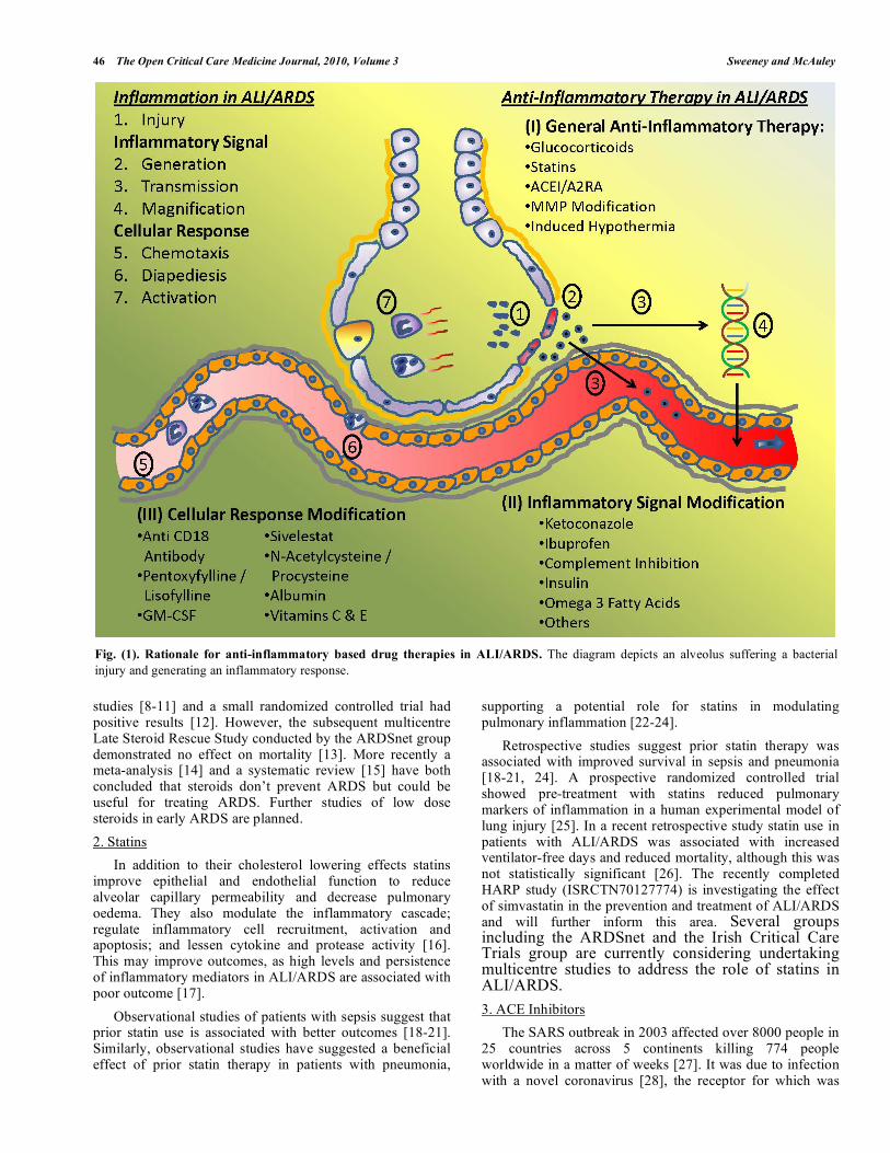
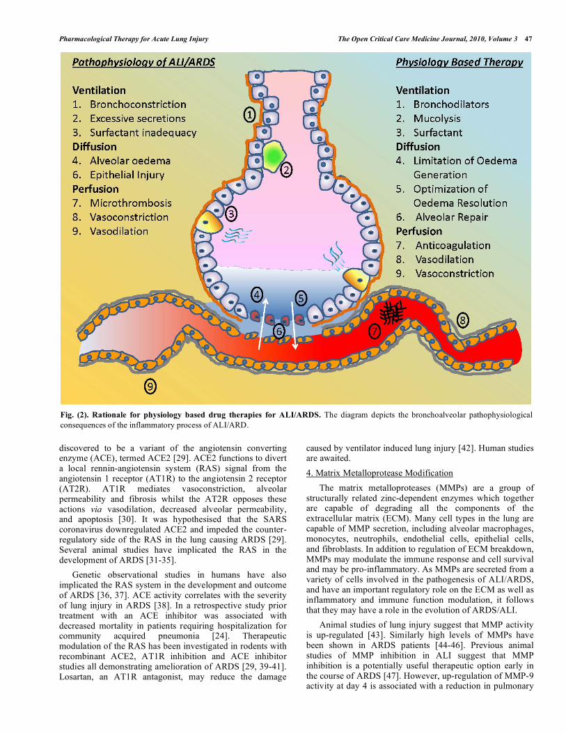
ALI/ARDS are acute inflammatory conditions of the lung. The inflammatory process can be targeted for manipulation anywhere from the genome through to leukocyte activation and reactive oxygen species release (Fig. 1). This inflammation can injure all 3 major components of the alveolus; the airspace (epithelium), the interstitium, and the capillary (endothelium). Structural damage leads to functional impairment of each component and these component dysfunctions combine to result in alveolar failure (Fig. 2). Although an oversimplification, therapy can thus be classified as being either
*Address correspondence to this author at the Respiratory Medicine
Research Programme, Centre of Infection and Immunity, Queens University
of Belfast, Grosvenor Road, Belfast BT12 6BA, Northern Ireland; Tel: 0044
(0) 28 90 632711; Fax: 0044 (0)28 90 632697;
E-mail: [email protected]
anti-inflammatory or physiological in nature. Physiology based therapies seek to optimise individual component processes and can be classified as drugs affecting ventilation, diffusion or perfusion.
Anti-inflammatory therapy dampens the excessively harmful host response and can be classified as general anti-inflammatory therapy, inflammatory signalling modification or cellular response modification. Although it is theoretically attractive to pharmacologically decrease the inflammatory response, the complexity of the inflammatory process, and its interdependency with other homeostatic mechanisms such as coagulation, means this approach could potentially be detrimental. For any individual it is difficult to ascertain which components of inflammation are functional and necessary, and which components are dysfunctional and harmful. Immuno-paralysis, with the consequent development of infection, is a risk and makes the timing of potential anti-inflammatory therapy important.
Many therapies may mechanistically overlap and could potentially act synergistically. This has been comprehensively reviewed elsewhere [1]. Ultimately treatment of the cause is required, with mechanical ventilation being necessary for life saving respiratory support.
Anti-Inflammatory Therapy
(I) General Anti-Inflammatory Therapy
1. Glucocorticoids
Steroids act at many levels throughout the inflammatory process. Several early trials demonstrated that short course, high dose methylprednisolone is ineffective in preventing the development of ARDS in high risk patients [2-5]. Although an initial trial of high dose steroids early in the course of ARDS was negative [6], a recent study using prolonged low dose methylprednisolone showed reduced durations of mechanical ventilation and ICU stay [7].
The anti-fibrotic properties of steroids have been investigated in the later stages of ARDS. Observational
46 The Open Critical Care Medicine Journal, 2010, Volume 3 Sweeney and McAuley
studies [8-11] and a small randomized controlled trial had positive results [12]. However, the subsequent multicentre Late Steroid Rescue Study conducted by the ARDSnet group demonstrated no effect on mortality [13]. More recently a meta-analysis [14] and a systematic review [15] have both concluded that steroids don’t prevent ARDS but could be useful for treating ARDS. Further studies of low dose steroids in early ARDS are planned.
2. Statins
In addition to their cholesterol lowering effects statins improve epithelial and endothelial function to reduce alveolar capillary permeability and decrease pulmonary oedema. They also modulate the inflammatory cascade; regulate inflammatory cell recruitment, activation and apoptosis; and lessen cytokine and protease activity [16]. This may improve outcomes, as high levels and persistence of inflammatory mediators in ALI/ARDS are associated with poor outcome [17].
Observational studies of patients with sepsis suggest that prior statin use is associated with better outcomes [18-21]. Similarly, observational studies have suggested a beneficial effect of prior statin therapy in patients with pneumonia,
supporting a potential role for statins in modulating pulmonary inflammation [22-24].
Retrospective studies suggest prior statin therapy was associated with improved survival in sepsis and pneumonia [18-21, 24]. A prospective randomized controlled trial showed pre-treatment with statins reduced pulmonary markers of inflammation in a human experimental model of lung injury [25]. In a recent retrospective study statin use in patients with ALI/ARDS was associated with increased ventilator-free days and reduced mortality, although this was not statistically significant [26]. The recently completed HARP study (ISRCTN70127774) is investigating the effect of simvastatin in the prevention and treatment of ALI/ARDS and will further inform this area. Several groups including the ARDSnet and the Irish Critical Care Trials group are currently considering undertaking multicentre studies to address the role of statins in ALI/ARDS.
3. ACE Inhibitors
The SARS outbreak in 2003 affected over 8000 people in 25 countries across 5 continents killing 774 people worldwide in a matter of weeks [27]. It was due to infection with a novel coronavirus [28], the receptor for which was
Fig. (1). Rationale for anti-inflammatory based drug therapies in ALI/ARDS. The diagram depicts an alveolus suffering a bacterial
injury and generating an inflammatory response.
Pharmacological Therapy for Acute Lung Injury The Open Critical Care Medicine Journal, 2010, Volume 3 47
discovered to be a variant of the angiotensin converting enzyme (ACE), termed ACE2 [29]. ACE2 functions to divert a local rennin-angiotensin system (RAS) signal from the angiotensin 1 receptor (AT1R) to the angiotensin 2 receptor (AT2R). AT1R mediates vasoconstriction, alveolar permeability and fibrosis whilst the AT2R opposes these actions via vasodilation, decreased alveolar permeability, and apoptosis [30]. It was hypothesised that the SARS coronavirus downregulated ACE2 and impeded the counter-regulatory side of the RAS in the lung causing ARDS [29]. Several animal studies have implicated the RAS in the development of ARDS [31-35].
Genetic observational studies in humans have also implicated the RAS system in the development and outcome of ARDS [36, 37]. ACE activity correlates with the severity of lung injury in ARDS [38]. In a retrospective study prior treatment with an ACE inhibitor was associated with decreased mortality in patients requiring hospitalization for community acquired pneumonia [24]. Therapeutic modulation of the RAS has been investigated in rodents with recombinant ACE2, AT1R inhibition and ACE inhibitor studies all demonstrating amelioration of ARDS [29, 39-41]. Losartan, an AT1R antagonist, may reduce the damage
caused by ventilator induced lung injury [42]. Human studies are awaited.
4. Matrix Metalloprotease Modification
The matrix metalloproteases (MMPs) are a group of structurally related zinc-dependent enzymes which together are capable of degrading all the components of the extracellular matrix (ECM). Many cell types in the lung are capable of MMP secretion, including alveolar macrophages, monocytes, neutrophils, endothelial cells, epithelial cells, and fibroblasts. In addition to regulation of ECM breakdown, MMPs may modulate the immune response and cell survival and may be pro-inflammatory. As MMPs are secreted from a variety of cells involved in the pathogenesis of ALI/ARDS, and have an important regulatory role on the ECM as well as inflammatory and immune function modulation, it follows that they may have a role in the evolution of ARDS/ALI.
Animal studies of lung injury suggest that MMP activity is up-regulated [43]. Similarly high levels of MMPs have been shown in ARDS patients [44-46]. Previous animal studies of MMP inhibition in ALI suggest that MMP inhibition is a potentially useful therapeutic option early in the course of ARDS [47]. However, up-regulation of MMP-9 activity at day 4 is associated with a reduction in pulmonary
Fig. (2). Rationale for physiology based drug therapies for ALI/ARDS. The diagram depicts the bronchoalveolar pathophysiological
consequences of the inflammatory process of ALI/ARD.

48 The Open Critical Care Medicine Journal, 2010, Volume 3 Sweeney and McAuley
oedema [46]. These data caution against broad-spectrum MMP inhibition in ARDS. The timing of the intervention may be important where early intervention may decrease proteolytic damage but later in the course of ALI. MMPs and in particular MMP-9 may be important in repair.
(II) Inflammatory Signalling Modification
1. Ketoconazole
Ketoconazole is an imidazole antifungal agent with anti-inflammatory properties. It blocks the synthesis of pro-inflammatory mediators such as the eicosanoid leukotrienes and thromboxane A2 and also reduces macrophage pro-inflammatory cytokine production [48]. Early small studies were successful in preventing of ARDS in high risk patients [49-51], however a later study by the ARDSnet group of ketoconazole in 234 patients with ARDS was negative [52].
2. Ibuprofen
Ibuprofen is a non-steroidal anti-inflammatory agent which inhibits cyclo-oxygenase. In a large sepsis study of 448 patients Ibuprofen diminished prostanoid production and was associated with trends towards decreased duration of pulmonary dysfunction and ARDS, but this did not reach statistical significance [53]. Modulation of other inflammatory mediators has also been investigated [54].
3. Complement Inhibition
Complement can contribute to ALI/ARDS by both propagating inflammation, via the generation of pro-inflammatory mediators [55], and also causing cellular injury via the production of the membrane attack complex, C5b-9 [56]. Complement receptor 1 is a cell surface receptor on erythrocytes and leukocytes which can inhibit both classical and alternative complement pathways. Initial animal studies [57, 58] and a small human phase 1 study [56] have confirmed that recombinant soluble cytokine receptor 1 is safe and can inhibit the complement cascade. Clinical trials are awaited.
4. Insulin
Insulin has anti-inflammatory effects via inhibition of the pro-inflammatory transcription factor NFkB [59]. A landmark trial of intensive insulin therapy (IIT) in critical care reported a large decrease in mortality by maintaining serum glucose levels between 80 and 110 mg/dL [60]. Subsequent critical care studies have had mixed results [61-63], and a significant risk of hypoglycaemia was apparent upon meta-analysis of intensive insulin therapy studies [64]. In a rat model of endotoxin induced ALI tight glycaemic control to 90-110mg/dl reduced the severity of lung injury [65]. The role of intensive insulin therapy in preventing ALI/ARDS by maintaining tight glycaemic control (80 to 110 mg/dL) is currently being studied.
5. Immunonutrition
Fish oils, which contain the omega 3 poly unsaturated fatty acids eicosapentaenoic acid (EPA), gamma-linolenic acid (GLA) and docosahexaenoic acid (DHA), can lessen the production of pro-inflammatory arachadonic acid metabolites. Clinical studies using fish oils in ALI/ARDS have demonstrated reduced inflammation in the form of decreased pulmonary neutrophil infiltration, improved
physiology in the form of decreased microvascular permeability and pulmonary vascular resistance and improved outcomes in the form of reduced duration of ventilation and ICU stay and improved mortality [66-70]. The beneficial effects of fish oil supplementation in ALI/ARDS have been reiterated in a recent systematic review on immunonutrition [71]. The OMEGA trial, investigating omega 3 fatty acids, GLA and anti-oxidants in ALI has recently been presented at meeting level and reported negative findings. Full publication is awaited. Further studies of fish oils in ALI/ARDS are underway in Spain and America.
Additional uses for immunonutrition are the manipulation of the generation of carbon dioxide, via a low carbohydrate, high fat feed, resulting in decreased ventilator requirements [72]. Feeding enterally, rather than parenterally, can stimulate lung and gut IgA defences [73]. The early use of enteral nutrition by itself may improve outcomes in ALI [74].
6. Others
Interleukin 8 (IL-8) is a chemoattractant for neutrophil migration into the alveolus [75]. In a rat model of gastric aspiration anti-IL-8 antibody significantly reduced neutrophil recruitment to the alveolus and reduced the severity of lung injury [76]. Similarly, a rabbit model of acute pancreatitis induced lung injury was attenuated with anti-IL-8 antibody [77]. As IL-8 levels are elevated in at risk patients who subsequently develop ARDS [78] and in early ARDS [75, 79, 80] this represents a potential therapeutic target.
Other current studies of potential anti-inflammatory treatments include a trial investigating the safety, tolerability and efficacy of recombinant human interferon beta in ALI/ARDS (NCT00789685) and a phase 2 trial of IC14, a recombinant chimeric monoclonal antibody to CD14, to block CD14 medicated cellular activation in patients with sepsis-induced ALI (NCT00233207). This trial has recently been terminated and results are awaited. Based on the evidence to date routine use of anti-inflammatory therapy for ALI/ARDS is not recommended.
(III) Cellular Response Modification
1. Anti-Adhesion Molecule Therapy
Transfer of immune cells from the vascular to the extra-vascular space is vital in the process of tissue inflammation. Blockage of CD18, a neutrophil adhesion molecule necessary for diapedesis, reduces the severity of experimental lung injury [81, 82]. Human data is awaited.
2. Immune Cell Blockade
Pentoxyfylline [83-85] and its derivative lisofylline [86-89] have various inhibitory effects on immune cell function [83-85]. A small study using pentoxyfyline in ARDS failed to show physiological improvements in either gas exchange or haemodynamics [90]. A large multicentre ARDSnet study of lisofylline in 235 patients with ALI/ARDS was negative [91].
Granulocyte-macrophage colony stimulating factor (GM-CSF) plays a role in the control of both alveolar macrophages and epithelial cells [92]. Alveolar macrophage

Pharmacological Therapy for Acute Lung Injury The Open Critical Care Medicine Journal, 2010, Volume 3 49
tumour necrosis factor- initiates alveolar epithelial repair by inducing autocrine epithelial GM-CSF signalling [93]. Higher GM-CSF levels in bronchoalveolar lavage fluid in ARDS patients correlates with improved survival [94]. Recombinant human GM-CSF improved oxygenation in a small placebo controlled study of 18 patients with sepsis related pulmonary dysfunction [95]. Work with this agent is ongoing.
Neutrophil elastase, which is released by activated neutrophils, contributes to alveolar endothelial injury, increased permeability and airspace flooding [96, 97]. Sivelestat, a neutrophil elastase inhibitor, has had mixed results in clinical trials. A Japanese study demonstrated improved pulmonary function and reduced duration of ICU stay with trends towards reduction in both duration of mechanical ventilation and mortality [98]. The subsequent larger international STRIVE study was stopped after an interim analysis due to an increase in 6 month all cause mortality [99]. Additionally, pulmonary function did not improve and 28 day mortality was not reduced.
3. Anti-Oxidants
Free radicals are highly reactive molecules due to the presence of unpaired electrons. Immune cells partly exert their injurious effects via the generation of these substances. Pulmonary levels of glutathione, an antioxidant, are known to be low in ARDS [100]. N-acetylcysteine and procysteine are precursors for this molecule and their administration can replete pulmonary glutathione levels in ARDS [101]. N-acetylcysteine has had mixed success in small ALI/ARDS studies [101-104], while a study of procysteine in lung injury was stopped early due to increased mortality (unpublished data). Additionally, N-acetylcysteine can also downregulate NFkB, a pro-inflammatory transcription factor, with ensuing reductions in inflammatory markers.
Albumin exerts antioxidant effects via its thiol group. Non-survivors of ARDS have reduced thiol values [105]. The infusion of albumin is associated with increased plasma thiol levels in sepsis [106] and ARDS [107] and decreased markers of oxidant injury
Vitamins C & E may reduce the duration of mechanical ventilation and ICU stay, but not prevent the development of ARDS [108].
Physiological Derangement
(I) Ventilation
1. Surfactant
Surfactant is a multifunctional substance secreted by alveolar type 2 cells. It decreases alveolar surface tension preventing alveolar collapse during expiration, and has anti-inflammatory and antimicrobial properties. Local ventilation is hindered by surfactant deficiency during ALI/ARDS. This deficiency is both qualitative and quantitative, with reduced amounts of less functional surfactant produced during ALI/ARDS.
Although respiratory distress syndrome, the infantile form of ALI/ARDS, has been successfully treated with exogenous surfactant, adult studies have been disappointing. Small studies have shown physiological improvements [109-115], but subsequent larger studies have demonstrated no
change in mortality [116, 117]. These findings have been confirmed in a recent meta-analysis, with improved oxygenation without improved duration of ventilation or mortality being the conclusion [118]. Further studies using different formulations, methods of delivery, timing of initiation of therapy, and duration of therapy are underway in response to criticisms of the earlier studies.
2. Bronchodilators
Three small studies of inhaled beta-agonists demonstrated beneficial effects on lung mechanics by reducing airflow resistance, peak and plateau airway pressure, and improving lung compliance [119-121]. The BALTI trail, a phase 2 trial of the effect of intravenous salbutamol on the clearance of alveolar oedema, also demonstrated reduced peak airway pressures [122].
3. Mucolytics
Dornase alfa reduces sputum viscosity and improves sputum clearance by cleaving extracellular DNA released by degenerating leukocytes. Mucolytics improve lung function in cystic fibrosis [123-125] and have been investigated in other respiratory conditions. Dornase alfa reduced the duration of mechanical ventilation in children after cardiac surgery [126]. The successful use of Dornase alfa in patients undergoing mechanical ventilation for asthma [127] and ARDS [128] has been published only as case reports.
(II) Gas Diffusion
ALI/ARDS are forms of increased permeability pulmonary oedema. Strategies to minimise alveolar oedema include measures to limit the generation of alveolar flooding and measures to increases the resolution of this oedema.
1. Limitation of Generation of Alveolar Oedema
Alveolar flooding is primarily dependent on three factors, as described by Starlings law: capillary hydrostatic pressure, oncotic pressure and permeability. Capillary permeability is increased in ALI/ARDS, with the reflection coefficient being reduced from the normal 0.7-0.9 to about 0.5 [129] In this setting potentially reducing hydrostatic pressure and/or increasing oncotic pressure may ameliorate the development of pulmonary oedema [130].
Hydrostatic pressure may be manipulated in a number of ways. Fluid intake can be restricted or fluid output increased, either with diuretics or renal replacement therapy. Vasomotor tone can be decreased with vasodilators. Cardiac filling pressures can be used to guide the above measures.
The ARDSnet FACTT study showed improved duration of ventilation and ICU stay with a restrictive fluid strategy [131]. Fluid management was governed by a complex protocol of diuretic therapy based on filling pressures. Patients in the fluid restrictive arm of the study averaged approximately a net fluid balance over 7 days of 0mls. Those in the liberal fluid arm averaged approximately plus 7000mls. Of note there was no increase in renal failure or organ hypoperfusion with the fluid restrictive strategy. Mortality was unchanged between the groups.
The reduction of both central venous [131] and pulmonary capillary wedge [132] pressures may be associated with improved outcomes in ALI/ARDS. However

50 The Open Critical Care Medicine Journal, 2010, Volume 3 Sweeney and McAuley
the use of a pulmonary artery catheter is not superior to the use of a central venous catheter for managing ALI/ARDS [133]. A positive fluid balance [134-137] and increased extravascular lung water (EVLW) [138] are both associated with poor outcomes. Using EVLW measurements with a PiCCO device to direct fluid management may be better than pulmonary artery wedge pressure based management [139]. As lung size is dependent on height rather than weight the use of EVLW indexed to predicted body weight is superior to EVLW indexed to actual body weight (Craig - in press Critical Care Medicine, ISRCTN70127774)
Renal replacement therapy has been shown to reduce pulmonary oedema via reductions in pulmonary vascular pressures and permeability in experimental models of lung injury. Human experience has been limited to two small observational studies. 10 children with ALI/ARDS after bone marrow transplantation or chemotherapy treated with RRT had an 80% survival rate in contrast to a historical survival of 15% [140]. Thirty seven adults with renal failure and ALI/ARDS treated with RRT and a zero fluid balance had no pulmonary improvements within the first 24 hours of treatment [141]. The role of RRT in the management of ALI/ARDS remains uncertain.
The choice of fluid for resuscitation in ALI/ARDS remains unclear. Theoretically a colloid with higher oncotic pressure would be more suitable than a crystalloid, but this has not been borne out in a large comparative study of saline versus albumin for fluid resuscitation in critical illness [142]. Hypoproteinaemia is associated with the development of lung injury and is a marker of weight gain and death [143, 144]. Two small studies have investigated the use of furosemide with albumin infusions in hypoproteinaemic patients with acute lung injury. Both showed increases in total serum protein and more negative fluid balances with furosemide and albumin administration. This was associated with improvements in oxygenation, but without improving mortality [145, 146].
An Oregon based study is presently investigating whether minimising EVLW, as measured by PiCCO and directed by the FACTT diuretic algorithm, is superior to central venous pressure guided therapy (NCT00624650). A phase 2 study investigating the role of recombinant human atrial natriuretic peptide (Carperitide) in minimising pulmonary oedema in ARDS has recently completed and its report is awaited (NCT00030121).
Lung injury is often heralded by a rise in pulmonary vascular resistance, with an imbalance between pulmonary vasoconstrictors and vasodilators being seen in animal endotoxin shock models. Intravenous adenosine reduces EVLW, whilst intravenous nitroprusside and nitroglycerin also reduce pulmonary oedema generation, but at the expense of increasing V/Q mismatch. To date there is no clear evidence to support the role of vasodilator treatment targeted to decrease hydrostatic pressure in ALI/ARDS.
2. Maximising Clearance of Alveolar Oedema
Alveolar fluid clearance (AFC) is impaired in over 50% of those with ALI/ARDS, with this group having higher mortality rates [147]. Beta-agonists upregulate AFC via an effect on sodium ion movement [148]. Aerosolized salbutamol has been shown to accelerate the resolution of
pulmonary oedema after lung resection [149]. A clinical trial of intravenous salbutamol in ARDS demonstrated reduced extravascular lung water and a trend towards increased survival [150]. A retrospective study of salbutamol exposure in ALI suggested an association between higher exposure and improved outcome [151]. Beta 2 agonists may exert several other beneficial effects in ALI/ARDS including increased surfactant secretion, decreased lung endothelial permeability, decreased airway resistance and decreased airway pressures [150]. A large phase 3 UK multicentre study is currently in progress examining the effects of intravenous salbutamol on outcome in ARDS (ISRCTN38366450). The ARDSnet group have recently terminated a multicentre, randomized, placebo controlled study investigating albuterol for the treatment of ALI (ALTA trial) [152]. This study was halted early for futility after studying 279 patients who received the active drug. There was no reduction in duration of ventilation or 60 day mortality with albuterol. It has been speculated that this result may be due to 2 factors [153]. Firstly, this study was performed in a less sick ALI population likely to have retained an ability to clear alveolar fluid without the need for exogenous upregulation, rather than a sicker ARDS population. Additionally, inadequate delivery of the drug to the alveolus may have contributed to this failure.
The alveolar epithelium contains active glucose-sodium co-transporters [154] and alveolar glucose levels correlate with experimental alveolar fluid clearance [155]. In the setting of increased permeability higher alveolar glucose levels could theoretically improve AFC, however higher airway glucose concentrations are associated with increased risk of nosocomial infection in critically ill patients [156]. The effect of tight blood glucose control in critically ill patients on AFC is unclear and requires evaluation.
Gene therapy to increase the expression of the ion channels and pumps needed for AFC is another possible future therapy. An animal study investigating overexpression of the beta-1 subunit of the sodium-potassium ATPase pump demonstrated increased rates of AFC and improved survival [157]. If the alveolar epithelium is badly injured then cellular regeneration may be required before a functioning epithelial layer can be manipulated.
3. Epithelial Repair
Stem cells have the capacity for limitless self-renewal and differentiation. Embryonic stem cells are pleuripotent and have the ability to differentiate into any cell type in the body, whilst adult stem cells are multipotent and have the ability to differentiate into several cell types, including cell types of other organ systems [158].
Stem cells provide 3 therapeutic opportunities [158]. Firstly, endogenous stem cells may be stimulated via exogenously administered growth factors. Hepatocyte growth factor [159] and transforming growth factor (TGF
) [160] have been shown to reduce the effects of acute lung injury in animal models. Epidermal growth factor [161, 162], TGF [163] and KGF [164, 165] can all up regulate AFC. Vascular endothelial growth factor (VEGF) promotes angiogenesis and regulates vascular permeability [166]. Genetic polymorphisms of the VEGF gene are associated with lower levels of VEGF and increased mortality in ARDS

Pharmacological Therapy for Acute Lung Injury The Open Critical Care Medicine Journal, 2010, Volume 3 51
[167]. The administration of VEGF enhances alveolar repair in both cell [168] and animal models [169] and its role in ARDS is currently being studied at Cornell University (NCT00319631). The administration of exogenous growth factors has not yet been directly studied in human trials of ALI/ARDS.
Keratinocyte growth factor (KGF) is a heparin-binding growth factor that is secreted by fibroblasts and is known to act via a receptor specific to epithelial cells [170]. It induces potent proliferative activity in a variety of epithelial cells including alveolar type 2 cells. Other potentially useful effects include cytoprotection, augmented surfactant secretion and an antioxidant effect [171-172]. KGF reduces the severity of lung injury in experimental models [171, 173-174]. A phase 2 study investigating the effects of KGF pre-treatment in adults using an inhaled LPS model of lung injury is currently underway (ISRCTN98813895).
Secondly, administration of exogenous stem cells, either embryonic or adult, can provide repair to an injured alveolus. In an LPS induced ALI model bone marrow progenitor cells localised to the site of injury and differentiated into endothelial and epithelial cells [175]. Embryonic stem cells have the ability to produce a fully differentiated airway epithelium when cultured in an air-liquid interface [176]. Patients with pneumonia [177] and ALI/ARDS [178] have higher levels of endothelial progenitor cells and this higher level correlates with outcome. Autologous transplantation of endothelial progenitor cells preserves endothelial function and maintains the integrity of the pulmonary alveolar-capillary barrier in an animal study [179]. Mesenchymal stem cells were originally thought to act as a source of regenerative cells by differentiating into, and locally replacing, lethally injured cells. However their primary mechanism of action is via the secretion of growth factors, cytokines and other signalling molecules to cause the trophic modulation of inflammation, cell death, fibrosis and tissue repair [180].
The third role of stem cells is their ability to deliver gene therapy to the injured lung. Endothelial progenitor cells have been used to deliver vasodilatory genes to the pulmonary vasculature with resultant decreases in pulmonary artery pressures in experimental pulmonary hypertension [181, 182]. Human studies are awaited.
(III) Perfusion
1. Vasodilators
Nitric oxide (NO) is an endogenous vasodilator produced by the endothelium. When administered by inhalation it vasodilates the circulation of ventilated alveoli thus reducing shunt and pulmonary hypertension. Its short half life and apparent lack of side-effects made it an attractive therapy. Early studies demonstrated physiological improvements with NO in ARDS [183-187], however mortality remained unchanged. Two meta-analyses showed no mortality benefit [188-189] and reported possible harm due to methaemoglobinaemia, toxic nitrogen compounds, increased pulmonary oedema, rebound pulmonary hypertension and renal failure. As NO is expensive, possibly harmful, and without a mortality benefit, its routine use is not recommended although it may have a place as rescue therapy
for severe hypoxaemia given its ability to improve oxygenation [190].
Prostacylins are derivatives of arachadonic acid and have potentially beneficial effects including vasodilation, inhibition of platelet aggregation, reduction of neutrophil adhesion, and inhibition of both macrophage and neutrophil activation [191]. Inhaled PGI2 (prostacyclin) has been compared to inhaled NO in ARDS [192-194]. PGI2 has similar efficacy and some advantages including minimal systemic effects, absence of platelet dysfunction, easy administration, harmless metabolites and no requirement for monitoring [195]. No placebo controlled randomized trial has yet studied PGI2 in ARDS, but a current Pakistani study aims to show that nebulized PGI2 (iloprost) decreases pulmonary hypertension selectively and improves oxygenation in ARDS (NCT00314548).
Intravenous prostacyclin in the form of PGE1 has also been investigated in ARDS. Although vasodilatory effect can cause hypotension and increase pulmonary shunting [196], prostacyclin is anti-inflammatory and can increase both cardiac output and oxygen delivery [197] and improve oxygen extraction during reduced oxygen delivery [198]. Early studies [199-201] in ARDS showed no significant benefit although the dose delivered was questioned [202]. PGE1 was reformulated as liposomal PGE1 in order to increase pulmonary drug delivery and minimise side effects. Again despite a promising preclinical study [203] further studies were negative [204, 205].
Endothelin 1 is a potent vasoconstrictor which has been implicated in the pathophysiology of lung injury [206-208]. Tezosentan, an endothelin receptor antagonist, has been investigated in animal models of lung injury and with mixed results thus far [209, 210].
2. Vasoconstrictors
Almitrine is a pulmonary vasoconstrictor which may increase hypoxic pulmonary vasoconstriction and reduce shunt. In a small ARDS study oxygenation was improved with minimal increase in pulmonary vascular pressures [211]. The combination of intravenous almitrine to decrease blood flow to hypoxic lung units and inhaled NO, to increase blood flow to ventilated lung units, has been investigated in both experimental lung injury, and a small clinical study [212]. Both found the combination superior than either therapy alone at increasing PaO2 with a minimal rise in pulmonary artery pressure. Further research is required.
3. Coagulation
An imbalance between fibrinogenesis and fibrinolysis in ARDS results in widespread fibrin deposition in the alveolar airspace, interstitium and blood vessels [213]. Pulmonary intravascular thrombosis and vasoconstriction can lead to the development of dead space, a known independent predictor of mortality in ARDS [214]. Several anticoagulants have been proposed as potential therapies in ALI/ARDS and have undergone investigation in animal models. Tissue factor pathway inhibitor (TFPI), factor VIIai, heparin, antithrombin III, activated protein C (APC) and thrombomodulin have all been shown to have beneficial effects at this level of investigation [215].

52 The Open Critical Care Medicine Journal, 2010, Volume 3 Sweeney and McAuley
Protein C levels are lower in patients with ARDS than normal controls and the level of protein C correlates with clinical outcome [216]. However, a small randomized controlled trial of APC in ARDS did not reduce either duration of ventilation or mortality. (156) A Dutch study investigating APC in inflammatory and infectious ALI/ARDS is currently in progress (ISRCTN52566874). A phase 2 trial of recombinant TFPI demonstrated improvements in lung dysfunction score and survival [217]. Antithrombin III has had mixed results on pulmonary function in sepsis studies [218-220].
Reasons why Pharmacological Therapy is Ineffective in ALI/ARDS
Despite repeated promising pre-clinical and clinical phase 1 and 2 studies of therapies for ALI/ARDS, no non-ventilatory strategy has yet convincingly improved outcomes. This situation is not unique to lung injury. Over the past 30 years just 3 out of 38 new therapies evaluated for sepsis returned positive results [221]. All three therapies have since been seriously questioned and their place is presently unclear.
There are many reasons for the scientific failure of translation from bench to bedside [222]. Animal studies, which inform higher levels of clinical study, suffer from numerous limitations including the lack of generalization of inter-species physiology, immunology, genetics and host response to injury. The use of young healthy animals and pre-treatment before injury models directly contradicts true clinical practice. Large randomized controlled trials suffer from a heterogeneous study population which usually lacks identification of the phenotype most likely to receive a beneficial effect from the study drug. Allied with a small signal from the study intervention in comparison with the large signal from the multiple co-morbidities suffered by most elderly patients, genuine positive interventional effects may be difficult to recognise. The use of outcome measures such as oxygenation in a condition in which only a small minority die from refractory hypoxemia [223, 224] makes this signal even more difficult to recognise.
The use of pharmacological agents as adjuncts to increase oxygenation allowing the limitation of injurious ventilation may be associated with improved outcomes but this remains to be tested.
CONCLUSION
Despite promising scientific advances, non-ventilatory strategies for ALI/ARDS remain elusive. The best evidence we have is for minimising pulmonary oedema via fluid restriction when appropriate. Other therapies may occasionally be justified as salvage therapy in severe ALI/ARDS, but with the knowledge that their risk benefit ratio remains unclear.
CONFLICT OF INTEREST
The authors have no conflicts of interest to report. This review article is based upon a book chapter entitled “Non-ventilatory strategies for acute lung injury and the acute respiratory distress syndrome” due to be published in “An Evidence Based Practice of Critical Care” (Deutschman CS, Neligan P (Editors), Elsevier (Publisher).
ACKNOWLEDGEMENTS
RMS is funded by a doctoral fellowship by the research and development office, Northern Ireland.
REFERENCES
[1] Raghavendran K, Pryhuber G, Chess P, Davidson B, Knight P, Notter R. Pharmacotherapy of acute lung injury and acute
respiratory distress syndrome. Curr Med Chem 2008; 15: 1911-24. [2] Weigelt JA, Norcross JF, Borman KR, Snyder WH, III. Early
steroid therapy for respiratory failure. Arch Surg 1985; 120: 536-40.
[3] Sprung CL, Caralis PV, Marcial EH, et al. The effects of high-dose corticosteroids in patients with septic shock. A prospective,
controlled study. N Engl J Med 1984; 311: 1137-43. [4] Bone RC, Fisher CJ' Jr., Clemmer TP, Slotman GJ, Metz CA. Early
methylprednisolone treatment for septic syndrome and the adult respiratory distress syndrome [published erratum appears in Chest
1988; 94(2): 448]. Chest 1987; 92(6): 1032-6. [5] Luce JM, Montgomery AB, Marks JD, et al. Ineffectiveness of
high-dose methylprednisolone in preventing parenchymal lung injury and improving mortality in patients with septic shock. Am
Rev Respir Dis 1988; 138: 62-8. [6] Bernard GR, Luce JM, Sprung CL, et al. High-dose corticosteroids
in patients with the adult respiratory distress syndrome. N Engl J Med 1987; 317: 1565-70.
[7] Meduri GU, Golden E, Freire AX, et al. Methylprednisolone Infusion in Early Severe ARDS. Results of a Randomized
Controlled Trial. Chest 2007; 131(4): 954-63. [8] Hooper RG, Kearl RA. Established ARDS treated with a sustained
course of adrenocortical steroids. Chest 1990; 97(1): 138-43. [9] Hooper RG, Kearl RA. Established adult respiratory distress
syndrome successfully treated with corticosteroids. South Med J 1996; 89(4): 359-64.
[10] Meduri GU, Belenchia JM, Estes RJ, Wunderink RG, el Torky M, Leeper KV. Fibroproliferative phase of ARDS. Clinical findings
and effects of corticosteroids. Chest 1991; 100(4): 943-52. [11] Meduri GU, Chinn AJ, Leeper KV, et al. Corticosteroid rescue
treatment of progressive fibroproliferation in late ARDS. Patterns of response and predictors of outcome. Chest 1994; 105(5): 1516-
27. [12] Meduri GU, Headley AS, Golden E, et al. Effect of prolonged
methylprednisolone therapy in unresolving acute respiratory distress syndrome: a randomized controlled trial. JAMA 1998;
280(2): 159-65. [13] The national heart, lung and blood institute acute respiratory
distress syndrome clinical trials network. Efficacy and safety of corticosteroids for persistent acute respiratory distress syndrome. N
Engl J Med 2006; 354(16): 1671-84. [14] Peter JV, John P, Graham PL, Moran JL, George IA, Bersten A.
Corticosteroids in the prevention and treatment of acute respiratory distress syndrome (ARDS) in adults: meta-analysis. BMJ 2008;
336(7651): 1006-9. [15] Deal EN, Holland JM, Schramm GE, Micek ST. Role of
corticosteroids in the management of acute respiratory distress syndrome. Clin Ther 2008; 30: 787-99.
[16] Craig T, O'Kane CM, McAuley DF. Potential mechanisms by which statins modulate pathogenic mechanisms important in the
development of acute lung injury. In 27th yearbook of intensive care and emergency medicine edited by: Vincent JL Berlin,
Germany: Springer-Verlag; 2007: 287-300. [17] Frank JA, Parsons PE, Matthay MA. Pathogenetic significance of
biological markers of ventilator-associated lung injury in experimental and clinical studies. Chest 2006; 130: 1906-14.
[18] Liappis AP, Kan VL, Rochester CG, Simon GL. The effect of statins on mortality in patients with bacteremia. Clin Infect Dis
2001; 33(8): 1352-7. [19] Almog Y, Shefer A, Novack V, et al. Prior statin therapy is
associated with a decreased rate of severe sepsis. Circulation 2004; 110(7): 880-5.
[20] Hackam DG, Mamdani M, Li P, Redelmeier DA. Statins and sepsis in patients with cardiovascular disease: a population-based cohort
analysis. Lancet 2006; 367(9508): 413-8. [21] Kruger P, Fitzsimmons K, Cook D, Jones M, Nimmo G. Statin
therapy is associated with fewer deaths in patients with bacteraemia. Intensive Care Med 2006; 32: 75-9.
Pharmacological Therapy for Acute Lung Injury The Open Critical Care Medicine Journal, 2010, Volume 3 53
[22] Thomsen RW, Riis A, Kornum JB, Christensen S, Johnsen SP,
Sorensen HT. Preadmission use of statins and outcomes after hospitalization with pneumonia: population-based cohort study of
29900 patients. Arch Intern Med 2008; 168(19): 2081-7. [23] Chalmers JD, Singanayagam A, Murray MP, Hill AT. Prior statin
use is associated with improved outcomes in community-acquired pneumonia. Am J Med 2008; 121(11): 1002-7.e1.
[24] Mortensen EM, Pugh MJ, Copeland LA, et al. Impact of statins and angiotensin-converting enzyme inhibitors on mortality of subjects
hospitalised with pneumonia. Eur Respir J 2008; 31(3): 611-7. [25] Shyamsundar M, McKeown STW, O'Kane CM, et al. Simvastatin
decreases lipopolysaccharide-induced pulmonary inflammation in healthy volunteers. Am J Respir Crit Care Med 2009; 179(12):
1107-14. [26] Kor DJ, Iscimen R, Yilmaz M, Brown MJ, Brown DR, Gajic O.
Statin administration did not influence the progression of lung injury or associated organ failures in a cohort of patients with acute
lung injury. Intensive Care Med 2009; 35: 1039-46. [27] Peiris JSM, Guan Y, Yuen KY. Severe acute respiratory syndrome.
Nat Med 2004; 10(12): S88-S97. [28] Drosten C, Gunther S, Preiser W, et al. Identification of a Novel
Coronavirus in Patients with Severe Acute Respiratory Syndrome. N Engl J Med 2003; 348(20): 1967-76.
[29] Kuba K, Imai Y, Rao S, et al. A crucial role of angiotensin converting enzyme 2 (ACE2) in SARS coronavirus-induced lung
injury. Nat Med 2005; 11: 875-9. [30] Imai Y, Kuba K, Penninger JM. The renin-angiotensin system in
acute respiratory distress syndrome. Drug Discovery Today: Disease Mechanisms 2006; 3(2): 225-9.
31] Kiely DG, Cargill RI, Wheeldon NM, Coutie WJ, Lipworth BJ. Haemodynamic and endocrine effects of type 1 angiotensin II
receptor blockade in patients with hypoxaemic cor pulmonale. Cardiovasc Res 1997; 33(1): 201-8.
[32] Xu Z-H, Shimakura T, Wang L-M, Mineshita S. Pulmonary oedema induced by angiotensin I in rats. Jpn J Pharmacol 1998; 76:
51-6. [33] Yamamoto T, Wang L, Shimakura K, Sanaka M, Koike Y,
Mineshita S. Angiotensin II-induced pulmonary edema in a rabbit model. Jpn J Pharmacol 1997; 73: 33-40.
[34] Victorino GP, Newton CR, Curran B. Effect of angiotensin II on microvascular permeability. J Surg Res 2002; 104: 77-81.
[35] Imai Y, Kuba K, Rao S, et al. Angiotensin-converting enzyme 2 protects from severe acute lung failure. Nature 2005; 436(7047):
112-6. [36] Marshall RP, Webb S, Bellingan GJ, et al. Angiotensin converting
enzyme insertion/deletion polymorphism is associated with susceptibility and outcome in acute respiratory distress syndrome.
Am J Respir Crit Care Med 2002; 166(5): 646-50. [37] Jerng JS, Yu CJ, Wang HC, Chen KY, Cheng SL, Yang PC.
Polymorphism of the angiotensin-converting enzyme gene affects the outcome of acute respiratory distress syndrome. Crit Care Med
2006; 34: 1001-6. [38] Orfanos SE, Armaganidis A, Glynos C, et al. Pulmonary capillary
endothelium-bound angiotensin-converting enzyme activity in acute lung injury. Circulation 2000; 102(16): 2011-8.
[39] Imai Y, Kuba K, Penninger JM. The discovery of angiotensin-converting enzyme 2 and its role in acute lung injury in mice. Exp
Physiol 2008; 93(5): 543-8. [40] Raiden S, Nahmod K, Nahmod V, et al. Nonpeptide antagonists of
AT1 receptor for angiotensin ii delay the onset of acute respiratory distress syndrome. J Pharmacol Exp Ther 2002; 303(1): 45-51.
[41] Lukkarinen HP, Laine J, Aho H, Zagariya A, Vidyasagar D, Kääpä PO. Angiotensin II receptor inhibition prevents pneumocyte
apoptosis in surfactant-depleted rat lungs. Pediatr Pulmonol 2005; 39(4): 349-58.
[42] Yao S, Feng D, Wu Q, Li K, Wang L. Losartan attenuates ventilator-induced lung injury. J Surg Res 2008; 145(1): 25-32.
[43] O’Kane CM, Frank J, McAuley DF. Matrix metalloproteinases: A potential role in the pathogenesis of ARDS. In:Yearbook of
Intensive Care and Emergency Medicine Vincent JL (Ed) Springer, Heidelberg, Germany. 2004; 287-300.
[44] Torii K, Iida K, Miyazaki Y, et al. Higher concentrations of matrix metalloproteinases in bronchoalveolar lavage fluid of patients with
adult respiratory distress syndrome. Am J Respir Crit Care Med 1997; 155(1): 43-6.
[45] Lanchou Jrm, Corbel M, Tanguy Ml, et al. Imbalance between
matrix metalloproteinases (MMP-9 and MMP-2) and tissue inhibitors of metalloproteinases (TIMP-1 and TIMP-2) in acute
respiratory distress syndrome patients. Crit Care Med 2003; 31(2): 536-42.
[46] Pugin J, Verghese G, Widmer M-C, Matthay MA. The alveolar space is the site of intense inflammatory and profibrotic reactions
in the early phase of acute respiratory distress syndrome. Crit Care Med 1999; 27(2): 304-12.
[47] Carney DE, Lutz CJ, Picone AL, et al. Matrix Metalloproteinase Inhibitor Prevents Acute Lung Injury After Cardiopulmonary
Bypass. Circulation 1999; 100(4): 400-6. [48] Williams JG, Maier RV. Ketoconazole inhibits alveolar
macrophage production of inflammatory mediators involved in acute lung injury (adult respiratory distress syndrome). Surgery
1992; 112: 270-7. [49] Slotman GJ, Burchard KW, D’Arezzo A, et al. Ketoconazole
prevents acute respiratory failure in critically ill surgical patients. J Trauma 1988; 28: 648-54.
[50] Yu M, Tomasa G. A double blind prospective randomized trial of ketoconazole, a thromboxane synthetase inhibitor, in the
prophylaxis of adult respiratory distress syndrome. Crit Care Med 1993; 21: 1635-42.
[51] Sinuff T, Cook DJ, Peterson JC, Fuller HD. Development, implementation, and evaluation of ketoconazole practice guidelines
for ARDS prophylaxis. J Crit Care 1999; 14: 1-6. [52] The ARDS network authors for the ARDS network. Ketoconazole
for early treatment of acute lung injury and acute respiratory distress syndrome: a randomized controlled trial. JAMA 2000;
283(15): 1995-2002. [53] Bernard GR, Wheeler AP, Russell JA, et al. The Effects of
Ibuprofen on the Physiology and Survival of Patients with Sepsis. N Engl J Med 1997; 336(13): 912-8.
[54] Bulger E, Maier R. Lipid mediators in the pathophysiology of critical illness [Signal transduction in critical care medicine:
scientific reviews]. Crit Care Med 2000; 28(4): N27-N36. [55] Morgan BP. Physiology and pathophysiology of complement:
progress and trends. Crit Rev Clin Lab Sci 1995; 32: 265-98. [56] Zimmerman JL, Dellinger RP, Straube RC, Levin JL. Phase 1 trial
of the recombinant soluble complement receptor 1 in acute lung injury and acute respiratory distress syndrome. Crit Care Med
2000; 28: 3149-54. [57] Mulligan MS, Yeh CG, Rudolph AR, Ward PA. Protective effects
of soluble CR1 in complement- and neutrophil- mediated tissue injury. J Immunol 1992; 148(5): 1479-85.
[58] Rabinovici R, Yeh CG, Hillegass LM, et al. Role of complement in endotoxin/platelet-activating factor-induced lung injury. J Immunol
1992; 149(5): 1744-50. [59] Dandona P, Aljada A, Mohanty P, et al. Insulin inhibits
intranuclear nuclear factor {kappa} B and stimulates I {kappa} B in mononuclear cells in obese subjects: Evidence for an anti-
inflammatory effect? J Clin Endocrinol Metab 2001; 86(7): 3257-65.
[60] Van den Berghe G, Wouters P, Weekers F, et al. Intensive insulin therapy in critically Ill patients. N Engl J Med 2001; 345(19):
1359-67. [61] Van den Berghe G, Wilmer A, Hermans G, et al. Intensive insulin
therapy in medical intensive care patients. N Engl J Med 2006; 354: 449-61.
[62] Krinsley JS. Effect of an intensive glucose management protocol on the mortality of critically ill adult patients. Mayo Clin Proc
2004; 79(8): 992-1000. [63] Arabi YM, Dabbagh OC, Tamim HM, et al. Intensive versus
conventional insulin therapy: A randomized controlled trial in medical and surgical critically ill patients. Crit Care Med 2008;
36(12): 3190-7. [64] Weiner RE, Sasso DE, Gionfriddo MA, et al. Early detection of
oleic acid-induced lung injury in rats using (111)In-labeled anti-rat intercellular adhesion molecule-1. J Nucl Med 2001; 42(7): 1109 -
15. [65] Chen HI, Yeh DY, Liou H-L, Kao S-J. Insulin attenuates
endotoxin-induced acute lung injury in conscious rats. Crit Care Med 2006; 34: 758-64.
[66] Singer P, Theilla M, Fisher H, et al. Benefit of an enteral diet enriched with eicosapentaenoic acid and gamma-linolenic acid in

54 The Open Critical Care Medicine Journal, 2010, Volume 3 Sweeney and McAuley
ventilated patients with acute lung injury. Crit Care Med 2006; 34:
1033-8. [67] Pontes-Arruda A, Aragão AMA, Albuquerque JD. The effects of
enteral feeding with eicosapentaenoic acid, gamma-linolenic acid, and antioxidants in mechanically ventilated patients with severe
sepsis and septic shock. Crit Care Med 2006; 34: 2325-33. [68] Pacht ER, DeMichele SJ, Nelson JL, Hart J, Wennberg AK, Gadek
JE. Enteral nutrition with eicosapentaenoic acid, gamma-linolenic acid, and antioxidants reduces alveolar inflammatory mediators and
protein influx in patients with acute respiratory distress syndrome. Crit Care Med 2003; 31: 491-500.
[69] Gadek JE, DeMichele SJ, Karlstad MD, et al. Effect of enteral feeding with eicosapentaenoic acid, gamma-linolenic acid, and
antioxidants in patients with acute respiratory distress syndrome. Enteral nutrition in ARDS study group. Crit Care Med 1999; 27:
1409-20. [70] Elamin EM, Hughes LF, Drew D. Effect of enteral nutrition with
eicosapentaenoic acid (EPA), gamma-linolenic acid (GLA), and antioxidants reduces alveolar inflammatory mediators and protein
influx in patients with acute respiratory distress syndrome (ARDS). Chest 2005; 128(4): 225S-b-.
[71] Marik PE, Zaloga GP. Immunonutrition in critically ill patients: a systematic review and analysis of the literature. Intensive Care Med
2008; 34: 1980-90. [72] Al-Saady NM, Blackmore CM, Bennett ED. High fat, low
carbohydrate, enteral feeding lowers PaCO2 and reduces the period of ventilation in artificially ventilated patients. Intensive Care Med
1989; 15: 290-5. [73] King BK, Kudsk KA, Li J, et al. Route and type of nutrition
influence mucosal immunity to bacterial pneumonia. Ann Surg 1999; 229: 272-8.
[74] Rice TW, Stapleton RD, Wheeler AP, Bernard GR, Hite RD. Early Enteral Intake Is Associated with Improved Outcomes in Acute
Lung Injury. Am J Respir Crit Care Med 2009; 179: A5097. [75] Donnelly SC, Strieter RM, Kunkel SL, et al. Interleukin-8 and
development of adult respiratory distress syndrome in at-risk patient groups. Lancet 1993; 341: 643-7.
[76] Folkesson HG, Matthay MA, Hebert CA, et al. Acid aspiration induced lung injury in rabbits is mediated by interleukin-8
dependent mechanisms. J Clin Invest 1995; 96: 107-116. [77] Osman MO, Kristensen JU, Jacobsen NO, et al. A monoclonal anti-
interleukin 8 antibody (WS-4) inhibits cytokine response and acute lung injury in experimental severe acute necrotising pancreatitis in
rabbits. Gut 1998; 43(2): 232-9. [78] Donnelly TJ, Meade P, Jagels M, et al. Cytokine, complement, and
endotoxin profiles associated with the development of the adult respiratory distress syndrome after severe injury. Crit Care Med
1994; 22: 768-76. [79] Miller EJ, Cohen AB, Matthay MA. Increased interleukin-8
concentrations in the pulmonary edema fluid of patients with acute respiratory distress syndrome from sepsis. Crit Care Med 1996; 24:
1448-54. [80] Chollet-Martin S, Montravers P, Gibert C, et al. High levels of
interleukin-8 in the blood and alveolar spaces of patients with pneumonia and adult respiratory distress syndrome. Infect Immun
1993; 61: 4553-9. [81] Mileski WJ, Winn RK, Vedder NB, Pohlman TH, Harlan JM, Rice
CL. Inhibition of CD18-dependent neutrophil adherence reduces organ injury after hemorrhagic shock in primates. Surgery 1990;
108: 206-12. [82] Vedder NB, Winn RK, Rice CL, Chi EY, Arfors KE. A
monoclonal antibody to the adherence-promoting leukocyte glycoprotein CD18, reduces organ injury and improves survival
from hemorrhagic shock and resuscitation. J Clin Invest 1988; 81: 939-44.
[83] Mandell GL. ARDS, neutrophils, and pentoxifylline. Am Rev Respir Dis 1988; 138: 1103-5.
[84] Welsh CH, Lien D, Worthen GS, et al. Pentoxifylline decreases endotoxin-induced pulmonary neutrophil sequestration and
extravascular protein accumulation in the dog. Am Rev Respir Dis 1988; 138: 1106-14.
[85] Seear MD, Hannam VL, Kaapa P, et al. Effect of pentoxifylline on hemodynamics, alveolar fluid reabsorption, and pulmonary edema
in a model of acute lung injury. Am Rev Respir Dis 1990; 142: 1083-7.
[86] Guidot DM, Bursten SL, Rice GC, et al. Modulating phosphatidic
acid metabolism decreases oxidative injury in rat lungs. Am J Physiol Lung Cell Mol Physiol 1997; 273(5): L957-66.
[87] Hasegawa N, Oka Y, Nakayama M, et al. The effects of post-treatment with lisofylline, a phosphatidic acid generation inhibitor,
on sepsis-induced acute lung injury in pigs. Am J Respir Crit Care Med 1997; 155(3): 928-36.
[88] Rice GC, Brown PA, Nelson RJ, Bianco JA, Singer JW, Bursten S. Protection from endotoxic shock in mice by pharmacologic
inhibition of phosphatidic acid. Proc Nati Acad Sci USA 1994; 91(9): 3857-61.
[89] George CLS, Fantuzzi G, Bursten S, Leer L, Abraham E. Effects of lisofylline on hyperoxia-induced lung injury. Am J Physiol Lung
Cell Mol Physiol 1999; 276(5): L776-85. [90] Montravers P, Fagon JY, Gilbert C, Blanchet F, Novara A, Chastre
J. Pilot study of cardiopulmonary risk from pentoxifylline in adult respiratory distress syndrome. Chest 1993; 103(4): 1017-22.
[91] The acute respiratory disease syndrome network. Randomized, placebo controlled trial of lisofylline for early treatment of acute
lung injury and acute respiratory distress syndrome. Crit Care Med. 2002;30:1-6.
[92] Lechner AJ, Lamprech KE, Potthoff LH, Tredway TL, Matuschak GM. Recombinant GM-CSF reduces lung injury and mortality
during neutropenic Candida sepsis. Am J Physiol Lung Cell Mol Physiol 1994; 266(5): L561-8.
[93] Cakarova L, Marsh LM, Wilhelm J, et al. Macrophage Tumor Necrosis Factor-{alpha} Induces Epithelial Expression of
Granulocyte-Macrophage Colony-stimulating Factor: Impact on Alveolar Epithelial Repair. Am J Respir Crit Care Med 2009;
180(6): 521-32. [94] Matute-Bello G, Liles WC, Radella F, et al. Modulation of
neutrophil apoptosis by granulocyte colony-stimulating factor and granulocyte/macrophage colony-stimulating factor during the
course of acute respiratory distress syndrome. Crit Care Med 2000; 28(1): 1-7
[95] Presneill JJ, Harris T, Stewart AG, Cade JF, Wilson JW. A randomized phase ii trial of granulocyte-macrophage colony-
stimulating factor therapy in severe sepsis with respiratory dysfunction. Am J Respir Crit Care Med 2002; 166(2): 138-43.
[96] Carden D, Xiao F, Moak C, et al. Neutrophil elastase promotes lung microvascular injury and proteolysis of endothelial cadherins.
Am J Physiol 1998; 275: H385-H92. [97] Moraes T, Chow C, Downey G. Proteases and lung injury. Crit
Care Med. 2003;31:S189-S94. [98] Tamakuma S, Shiba T, Hirasawa H, et al. A phase III clinical study
of neutrophil elastase inhibitor ONO-5046 Na in SIRS patients. J Clin Ther Med (Japan) 1998; 14: 289-318.
[99] Zeiher BG, Artigas A, Vincent JL, et al. Neutrophil elastase inhibition in acute lung injury: results of the strive study. Crit Care
Med 2004; 32: 1695-702. [100] Pacht ER, Timerman AP, Lykens MG, Merola AJ. Deficiency of
alveolar fluid glutathione in patients with sepsis and the adult respiratory distress syndrome. Chest 1991; 100(5): 1397-403.
[101] Bernard GR, Wheeler AP, Arons MM, et al. A Trial of Antioxidants N-acetylcysteine and Procysteine in ARDS. Chest
1997; 112(1): 164-72. [102] Ortolani O, Conti A, De Gaudio AR, Masoni M, Novelli G.
Protective effects of N-acetylcysteine and rutin on the lipid peroxidation of the lung epithelium during the adult respiratory
distress syndrome. Shock 2000; 13(1): 14-8. [103] Jepsen S, Herlevsen P, Knudsen P, et al. Antioxidant treatment
with n-acetylcyesteine during adult respiratory distress syndrome: a prospective randomized placebo controlled study. Crit Care Med
1992; 20: 819-923. [104] Suter PM, Domenighetti G, Schaller MD, Laverrière MC, Ritz R,
Perret C. N-acetylcysteine enhances recovery from acute lung injury in man. A randomized, double-blind, placebo-controlled
clinical study. Chest 1994; 105(1): 190-4. [105] Quinlan GJ, Evans TW, Gutteridge JM. Oxidative damage to
plasma proteins in adult respiratory distress syndrome. Free Radic Res 1994; 20: 289-98.
[106] Quinlan GJ, Mumby S, Martin GS, et al. Albumin influences total plasma antioxidant capacity favorably in patients with acute lung
injury. Crit Care Med 2004; 32: 755-9.

Pharmacological Therapy for Acute Lung Injury The Open Critical Care Medicine Journal, 2010, Volume 3 55
[107] Quinlan GJ, Margarson MP, Mumby S, et al. Administration of
albumin to patients with sepsis syndrome: a possible beneficial role in plasma thiol repletion. Clin Sci (Lond) 1998; 95: 459-65.
[108] Nathens AB, Neff MJ, Jurkovich GJ, et al. Randomized, prospective trial of antioxidant supplemtntation in critically ill
surgical patients. Ann Surg 2002; 236(6): 814-22. [109] Reines HD, Silverman H, Hurst J, et al. Effects of two
concentrations of nebulized surfactant (Exosurf) in sepsis-induced adult respiratory distress syndrome (ARDS). Crit Care Med 1992;
20: S61. [110] Spragg RG, Gilliard N, Richman P, et al. Acute effects of a single
dose of porcine surfactant on patients with the adult respiratory distress syndrome. Chest 1994; 105(1): 195-202.
[111] Weg JG, Balk RA, Tharratt RS, et al. Safety and potential efficacy of an aerosolized surfactant in human sepsis-induced adult
respiratory distress syndrome. JAMA 1994; 272(18): 1433-8. [112] Walmrath D, Gunther A, Ghofrani HA, et al. Bronchoscopic
surfactant administration in patients with severe adult respiratory distress syndrome and sepsis. Am J Respir Crit Care Med 1996;
154(1): 57-62. [113] Walmrath D, Grimminger F, Pappert D, et al. Bronchoscopic
administration of bovine natural surfactant in ARDS and septic shock: impact on gas exchange and haemodynamics. Eur Respir J
2002; 19(5): 805-10. [114] Wiswell TE, Smith RM, Katz LB, et al. Bronchopulmonary
segmental lavage with surfaxin (KL4-surfactant) for acute respiratory distress syndrome. Am J Respir Crit Care Med 1999;
160(4): 1188-95. [115] Gregory TJ, Steinberg KP, Spragg R, et al. Bovine surfactant
therapy for patients with acute respiratory distress syndrome. Am J Respir Crit Care Med 1997; 155(4): 1309-15.
[116] Anzueto A, Baughman RP, Guntupalli KK, et al. Aerosolized surfactant in adults with sepsis-induced acute respiratory distress
syndrome. N Engl J Med 1996; 334(22): 1417-22. [117] Spragg RG, Lewis JF, Wurst W, et al. Treatment of acute
respiratory distress syndrome with recombinant surfactant protein c surfactant. Am J Respir Crit Care Med 2003; 167(11): 1562-6.
[118] Davidson WJ, Dorscheid D, Spragg RG, Schulzer M, Mak E, Ayas NT. Exogenous pulmonary surfactant for the treatment of adult
patients with acute respiratory distress syndrome: results of a meta-analysis. Crit Care 2006; 10(2): R41.
[119] Wright PE, Carmichael LC, Bernard GR. Effect of bronchodilators on lung mechanics in the acute respiratory distress syndrome
(ARDS). Chest 1994; 106(5): 1517-23. [120] Pesenti A, Pelosi P, Rossi N, et al. Respiratory mechanics and
bronchodilator responsiveness in patients with the adult respiratory distress syndrome. Crit Care Med 1993; 21: 78-83.
[121] Morina P, Herrera M, Venegas J, et al. Effects of nebulized salbutamol on respiratory mechanics in adult respiratory distress
syndrome. Intensive Care Med 1997; 23: 58-64. [122] Perkins GD, McAuley DF, Thickett DR, Gao F. The beta-agonist
lung injury trial (BALTI): A randomized placebo-controlled clinical trial. Am J Respir Crit Care Med 2006; 173(3): 281-7.
[123] Rochat T, Pastore FD, Schlegel-Haueter SE, et al. Aerosolized rhDNase in cystic fibrosis: effect on leucocyte proteases in sputum.
Eur Respir J 1996; 9(11): 2200-6. [124] Aitken ML, Burke W, McDonald G, Shak S, Montgomery AB,
Smith A. Recombinant human DNase inhalation in normal subjects and patients with cystic fibrosis. A phase 1 study. JAMA 1992;
267(14): 1947-51. [125] Harms HK, Matouk E, Tournier G, et al. Multicenter, open-label
study of recombinant human DNase in cystic fibrosis patients with moderate lung disease. Pediatric Pulmonology 1998; 26(3): 155-61.
[126] Riethmueller J, Borth-Bruhns T, Kumpf M, et al. Recombinant human deoxyribonuclease shortens ventilation time in young,
mechanically ventilated children. Pediatric Pulmonology 2006; 41(1): 61-6.
[127] Durward A, Forte V, Shemie SD. Resolution of mucus plugging and atelectasis after intratracheal rhDNAase therapy in a
mechanically ventilated child with refractory status athmaticus. Crit Care Med 2000; 28: 560-2.
[128] Morris C, Mullan B. Use of dornase alfa in the management of ARDS. Anaesthesia 2004; 59(12): 1249.
[129] Nakazawa H, Noda H, Noshima S, et al. Pulmonary transvascular fluid flux and cardiovascular function in sheep with chronic sepsis.
J Appl Physiol 1993; 75(6): 2521-8.
[130] Groeneveld ABJ. Vascular pharmacology of acute lung injury and
acute respiratory distress syndrome. Vascul Pharmacol 2003; 39: 247-56.
[131] The national heart lung and blood institute, acute respiratory distress syndrome clinical trials network. Comparison of two fluid-
management strategies in acute lung injury. N Engl J Med 2006; 354(24): 2564-75.
[132] Humphrey H, Hall J, Sznajder I, Silverstein M, Wood L. Improved survival in ARDS patients associated with a reduction in
pulmonary capillary wedge pressure. Chest 1990; 97(5): 1176-80. [133] The national heart lung and blood institute, acute respiratory
distress syndrome clinical trials network. Pulmonary-artery versus central venous catheter to guide treatment of acute lung injury. N
Engl J Med 2006; 354(21): 2213-24. [134] Rosenberg AL, Dechert RE, Park PK, Bartlett RH, NIH NHLBI
ARDS Network. Review of a large clinical series: association of cumulative fluid balance on outcome in acute lung injury: a
retrospective review of the ARDSnet tidal volume study cohort. J Intensive Care Med 2009; 24(1): 35-46.
[135] Schuller D, Mitchell JP, Calandrino FS, Schuster DP. Fluid balance during pulmonary edema. Is fluid gain a marker or a cause of poor
outcome? Chest 1991; 100(4): 1068-75. [136] Sakr Y, Vincent J-L, Reinhart K, et al. High tidal volume and
positive fluid balance are associated with worse outcome in acute lung injury. Chest 2005; 128(5):30 98-108.
[137] Simmons RS, Berdine CC, Seidenfeld JJ, Prihada TJ, Harris GD, Smith JD. Fluid balance and the adult respiratory distress
syndrome. Am Rev Respir Dis 1987; 135: 924-29. [138] Davey-Quinn A, Gedney J, Whiteley S, et al. Extravascular lung
water and acute respiratory distress syndrome: oxygenation and outcome. Anaesth Intensive Care 1999; 27: 357-62.
[139] Mitchell JP, Schuster D, Calandrino F, Schuster DP. Improved outcome based on fluid management in critically ill patients
requiring pulmonary artery catheterization Am Rev Respir Dis 1992; 145: 1229-33.
[140] DiCarlo JV, Alexander SR, Agarwal R, Joshua D. Continuous veno-venous haemofiltration may improve survival from acute
respiratory distress syndrome after bone marrow transplantation or chemotherapy. J Pediatr Hematol Oncol 2003; 25(10): 801-5.
[141] Hoste EAJ, Vanholder RC, Lameire NH, et al. No early respiratory benefit with CVVHDF in patients with acute renal failure and acute
lung injury. Nephrol Dial Transplant 2002; 17(12): 2153-8. [142] The SAFE study investigators. A comparison of albumin and saline
for fluid resuscitation in the intensive care unit. N Engl J Med 2004; 350(22): 2247-56.
[143] Arif SK, Verhij J, Groeneveld ABJ, Raijmakers PGHM. Hypoproteinemia as a marker of acute respiratory distress
syndrome in critically ill patients with pulmonary edema. Intensive Care Med 2002; 28: 310-7.
[144] Mangialardi RJ, Martin GS, Bernard GR, et al. Hypoproteinemia predicts acute respiratory distress syndrome development, weight
gain, and death in patients with sepsis: Ibuprofen in Sepsis Study Group. Crit Care Med 2000; 28: 3137-45.
[145] Martin GS, Mangialardi RJ, Wheeler AP, Dupont WD, Morris JA, Bernard GR. Albumin and furosemide therapy in hypoproteinemic
patients with acute lung injury. Crit Care Med 2002; 30: 2175-82. [146] Martin GS, Moss M, Wheeler AP, et al. A randomized, controlled
trial of furosemide with or without albumin in hypoproteinemic patients with acute lung injury. Crit Care Med 2005; 33: 1681-7.
[147] Ware LB, Matthay MA. Alveolar fluid clearance is impaired in the majority of patients with acute lung injury and the acute respiratory
distress syndrome. Am J Respir Crit Care Med 2001; 163(6): 1376-83.
[148] Sakuma T, Folkesson HG, Suzuki S, Okaniwa G, Fujimura S, Matthay MA. Beta-adrenergic agonist stimulated alveolar fluid
clearance in ex vivo human and rat lungs. Am J Respir Crit Care Med 1997; 155: 506-12.
[149] Licker M, Tschopp J-M, Robert J, Frey J-G, Diaper J, Ellenberger C. aerosolized salbutamol accelerates the resolution of pulmonary
edema after lung resection. Chest 2008; 133(4): 845-52. [150] Perkins GD, McAuley DF, Richter A, Thickett DR, Gao F. Bench-
to-bedside review: beta2-Agonists and the acute respiratory distress syndrome. Crit Care 2004; 8: 25-32.
[151] Manocha S, Gordon A, Salehifar E, Groshaus H, Walley K, Russell J. Inhaled beta-2 agonist salbutamol and acute lung injury: an
56 The Open Critical Care Medicine Journal, 2010, Volume 3 Sweeney and McAuley
association with improvement in acute lung injury. Crit Care 2006;
10(1): R12. [152] Matthay MA, Brower R, Thompson BT, et al. Randomized,
placebo controlled trial of an aerosolized beta-2 adrenergic agonist (albuterol) for the treatment of acute lung injury. Am J Resp Crit
Care Med 2009; 179: A2166. [153] McAuley DF, Matthay MA. A role for beta 2 agonists in ARDS -
the question remains unanswered. J Intensive Care Med 2009; 10(3): 172-3.
[154] de Prost N, Saumon G. Glucose transport in the lung and its role in liquid movement. Respir Physiol Neurobiol 2007; 159(3): 331-7.
[155] Basset G, Crone C, Saumon G. Fluid absorption by rat lung in situ: Pathways for sodium entry in the luminal membrane in the alveolar
epithelium. J Physiol 1987; 384: 325-45. [156] Liu KD, Levitt J, Zhuo H, et al. Randomized clinical trial of
activated protein C for the treatment of acute lung injury. Am J Respir Crit Care Med 2008; 178(6): 618-23.
[157] Factor P, Dumasius V, Saldias F, Brown LAS, Sznajder JI. Adenovirus-mediated transfer of an Na+/K+-ATPase 1 subunit
gene improves alveolar fluid clearance and survival in hyperoxic rats. Hum Gene Ther 2000; 11(16): 2231-42.
[158] Yen CC, Yang SH, Lin CY, Chen CM. Stem cells in the lung parenchyma and prospects for lung injury therapy. Eur J Clin
Invest 2006; 36: 310-9. [159] Ohmichi H, Matsumoto K, Nakamura T. In vivo mitogenic action
of HGF on lung epithelial cells: pulmotrophic role in lung regeneration. Am J Physiol Lung Cell Mol Physiol 1996; 270(6):
L1031-9. [160] Hardie WD, Prows DR, Leikauf GD, Korfhagen TR. Attenuation
of acute lung injury in transgenic mice expressing human transforming growth factor-alpha. Am J Physiol Lung Cell Mol
Physiology 1999; 277(5): L1045-50. [161] Sznajder JI, Ridge KM, Yeates DB, Ilekis J, Olivera W. Epidermal
growth factor increases lung liquid clearance in rat lungs. J Appl Physiol 1998; 85(3): 1004-10.
[162] Borok Z, Hami A, Danto SI, Lubman RL, Kim KJ, Crandall ED. Effects of EGF on alveolar epithelial junctional permeability and
active sodium transport. Am J Physiol Lung Cell Mol Physiol 1996; 270(4): L559-65.
[163] Folkesson HG, Pittet JF, Nitenberg G, Matthay MA. Transforming growth factor-alpha increases alveolar liquid clearance in
anesthetized ventilated rats. Am J Physiol Lung Cell Mol Physiol 1996; 271(2): L236-44.
[164] Wang Y, Folkesson HG, Jayr C, Ware LB, Matthay MA. Alveolar epithelial fluid transport can be simultaneously upregulated by both
KGF and beta-agonist therapy. J Appl Physiol 1999; 87: 1852-60. [165] Borok Z, Danto SI, Dimen LL, Zhang X-L, Lubman RL. Na+-K+-
ATPase expression in alveolar epithelial cells: upregulation of active ion transport by KGF. Am J Physiol Lung Cell Mol Physiol
1998; 274(1): L149-58. [166] Mura M, dos Santos CC, Stewart D, Liu M. Vascular endothelial
growth factor and related molecules in acute lung injury. J Appl Physiol 2004; 97(5): 1605-17.
[167] Zhai R, Gong MN, Zhou W, et al. Genotypes and haplotypes of the VEGF gene are associated with higher mortality and lower VEGF
plasma levels in patients with ARDS. Thorax 2007; 62(8): 718-22. [168] Ohwada A, Yoshioka Y, Iwabuchi K, Nagaoka I, Dambara T,
Fukuchi Y. VEGF regulates the proliferation of acid-exposed alveolar lining epithelial cells. Thorax 2003; 58(4): 328-32.
[169] Kunig AM, Balasubramaniam V, Markham NE, et al. Recombinant human VEGF treatment enhances alveolarization after hyperoxic
lung injury in neonatal rats. Am J Physiol Lung Cell Mol Physiol 2005; 289(4): L529-35.
[170] Miki T, Fleming TP, Bottaro DP, Rubin JS, Ron D, Aaronson SA. Expression cDNA cloning of the KGF receptor by creation of a
transforming autocrine loop. Science 1991; 251: 72-5. [171] Yano T, Deterding RR, Simonet WS, Shannon JM, Mason RJ.
Keratinocyte growth factor reduces lung damage due to acid instillation in rats. Am J Respir Cell Mol Biol 1996; 15(4): 433-42.
[172] Panos RJ, Bak PM, Simone WS, Runin JS, Smith LJ. Intratracheal instillation of keratinocyte growth factor decreases hyperoxia-
induced mortality in rats. J Clin Invest 1995; 96: 2026-33. [173] Ulrich K, Stern M, Goddard ME, et al. Keratinocyte growth factor
therapy in murine oleic acid-induced acute lung injury. Am J Physiol Lung Cell Mol Physiol 2005; 288(6): L1179-92.
[174] Atabai K, Ishigaki M, Geiser T, Ueki I, Matthay MA, Ware LB.
Keratinocyte growth factor can enhance alveolar epithelial repair by nonmitogenic mechanisms. Am J Physiol Lung Cell Mol
Physiol 2002; 283(1): L163-9. [175] Yamada M, Kubo H, Kobayashi S, et al. Bone marrow-derived
progenitor cells are important for lung repair after lipopolysaccharide-induced lung injury. J Immunol 2004; 172(2):
1266-72. [176] Coraux C, Nawrocki-Raby B, Hinnrasky J, et al. Embryonic stem
cells generate airway epithelial tissue. Am J Respir Cell Mol Biol 2005; 32(2): 87-92.
[177] Yamada M, Kubo H, Ishizawa K, Kobayashi S, Shinkawa M, Sasaki H. Increased circulating endothelial progenitor cells in
patients with bacterial pneumonia: evidence that bone marrow derived cells contribute to lung repair. Thorax 2005; 60(5): 410-3.
[178] Burnham EL, Taylor WR, Quyyumi AA, Rojas M, Brigham KL, Moss M. Increased circulating endothelial progenitor cells are
associated with survival in acute lung injury. Am J Respir Crit Care Med 2005; 172(7): 854-60.
[179] Lam C-F, Jiu Y-C, Hsu J-K, et al. Autologous transplantation of endothelial progenitor cells attenuates acute lung injury in rabbits.
Anesthesiology 2008; 108(3): 392-401. [180] van Poll D, Parekkadan B, Borel Rinkes IHM, Tilles AW, Yarmush
ML. Mesenchymal stem cell therapy for protection and repair of injured vital organs. Cell Mol Bioeng 2008; 1(1): 42-50.
[181] Campbell AIM, Kuliszewski MA, Stewart DJ. Cell-based gene transfer to the pulmonary vasculature. endothelial nitric oxide
synthase overexpression inhibits monocrotaline-induced pulmonary hypertension. Am J Respir Cell Mol Biol 1999; 21(5): 567-75.
[182] Campbell AIM, Zhao Y, Sandhu R, Stewart DJ. cell-based gene transfer of vascular growth factor attenuates monocrotaline-induced
pulmonary hypertension. Circulation 2001; 104(18): 2242-8. [183] Dellinger RP, Zimmerman JL, Taylor RW, et al. Effects of inhaled
nitric oxide in patients with acute respiratory distress syndrome: Results of a randomized phase II trial. Crit Care Med 1998; 26(1):
15-23. [184] Michael John R, Barton Richard G, Saffle Jeffrey R, et al. Inhaled
nitric oxide versus conventional therapy. effect on oxygenation in ARDS. Am J Respir Crit Care Med 1998; 157(5): 1372-80.
[185] Troncy E, Collet J-P, Shapiro S, et al. Inhaled nitric oxide in acute respiratory distress syndrome. A pilot randomized controlled study.
Am J Respir Crit Care Med 1998; 157(5): 1483-8. [186] Dobyns EL, Cornfield DN, Anas NG, et al. Multicenter
randomized controlled trial of the effects of inhaled nitric oxide therapy on gas exchange in children with acute hypoxemic
respiratory failure. J Pediatr 1999; 134: 406 -12. [187] Lundin S, Mang H, Smithies M, Stenqvist O, Frostell C, for the
European study group of inhaled nitric oxide. Inhalation of nitric oxide in acute lung injury: results of a European multicenter study.
Intensive Care Med 1999; 25: 911-19. [188] Sokol J, Jacobs SE, Bohn D. Inhaled nitric oxide for acute hypoxic
respiratory failure in children and adults: A meta-analysis. Anesth Analg 2003; 97(4): 989-98.
[189] Adhikari N, Granton JT. Inhaled nitric oxide for acute lung injury: no place for no? JAMA. 2004; 291(13): 1629-31.
[190] Ferguson ND. Inhaled nitric oxide for the acute respiratory distress syndrome. BMJ 2007; 334: 757-8.
[191] Cepkova M, Matthay MA. Pharmacology of acute lung injury and the acute respiratory distress syndrome. J Intensive Care Med 2006;
21: 119-43. [192] Zwissler B, Kemming G, Habler O, et al. Inhaled prostacyclin
(PGI2) versus inhaled nitric oxide in adult respiratory distress syndrome. Am J Respir Crit Care Med 1996; 154(6): 1671-7.
[193] Walmrath D, Schneider T, Schermuly R, Olschewski H, Grimminger F, Seeger W. Direct comparison of inhaled nitric
oxide and aerosolized prostacyclin in acute respiratory distress syndrome. Am J Respir Crit Care Med 1996; 153(3): 991-6.
[194] Van Heerden PV, Blythe D, Webb SA. Inhaled aerosolized prostacyclin and nitric oxide as selective pulmonary vasodilators in
ARDS-a pilot study. Anaesth Intensive Care 1996; 24: 564-8. [195] Cranshaw J, Griffiths MJD, Evans TW. The pulmonary physician
in critical care c 9: Non-ventilatory strategies in ARDS. Thorax 2002; 57(9): 823-9.
[196] Rossaint R, Falke KJ, Lopez F, Slama K, Pison U, Zapol WM. Inhaled nitric oxide for the adult respiratory distress syndrome. N
Engl J Med 1993; 328(6): 399-405.

Pharmacological Therapy for Acute Lung Injury The Open Critical Care Medicine Journal, 2010, Volume 3 57
[197] Silverman HJ, Slotman G, Bone RC, et al. Effects of prostaglandin
E1 on oxygen delivery and consumption in patients with the adult respiratory distress syndrome. Results from the prostaglandin E1
multicenter trial. The Prostaglandin E1 study group. Chest 1990; 98: 405-10.
[198] Zhang H, Benlabed M, Spapen H, et al. Prostaglandin E1 increases oxygen extraction capabilities in experimental sepsis. J Surg Res
1994; 57: 470-9. [199] Holcroft JW, Vassar MJ, Weber CJ. Prostaglandin E1 and survival
in patients with adult respiratory distress syndrome. A prospective trial. Ann Surg 1986; 203(4): 371-8.
[200] Bone RC, Slotman G, Maunder R, et al. Randomized double-blind, multicenter study of prostaglandin E1 in patients with the adult
respiratory distress syndrome. Prostaglandin E1 study group. Chest 1989; 96(1): 114-9.
[201] Slotman GJ, Kerstein MD, Bone RC, et al. The effects of prostaglandin E1 on non-pulmonary organ function during clinical
acute respiratory failure. The Prostaglandin E1 study group. J Trauma 1992; 32: 480-8.
[202] Rossignon MD, Khayat D, Royer C, et al. Functional and metabolic activity of polymorphonuclear leukocytes from patients
with adult respiratory distress syndrome: results of a randomized double-blind placebo-controlled study on the activity of
prostaglandin E1. Anesthesiology 1990; 72: 276-81. [203] Leff JA, Baer JW, Kirkman JM, et al. Liposome-entrapped PGE1
post treatment decreases IL-1 alpha-induced neutrophil accumulation and lung leak in rats. J Appl Physiol 1994; 76: 151-7.
[204] Abraham E, Baughman R, Fletcher E, et al. Liposomal prostaglandin E1 (TLC C-53) in acute respiratory distress
syndrome: a controlled, randomized, double-blind, multicenter clinical trial. Crit Care Med 1999; 27(8): 1478-85.
[205] Vincent JL, Brase R, Santman F, et al. A multi-centre, doubleblind, placebo-controlled study of liposomal prostaglandin E1 (TLC C-
53) in patients with acute respiratory distress syndrome. Intensive Care Med 2001; 27: 1578-83.
[206] Sanai L, Haynes WG, MacKenzie A, Grant IS, Webb DJ. Endothelin production in sepsis and the adult respiratory distress
syndrome. Intensive Care Med 1996; 22(1): 52-6. [207] Langleben D, DeMarchie M, Laporta D, Spanier AH, Schlesinger
RD, Stewart DJ. Endothelin-1 in acute lung injury and the adult respiratory distress syndrome. Am Rev Respir Dis 1993; 148:
1646-50. [208] Berger MM, Rozendal CS, Schieber C, et al. The effect of
endothelin-1 on alveolar fluid clearance and pulmonary edema formation in the rat. Anesth Analg 2009; 108(1): 225-31.
[209] Cox RA, Enkhabaatar P, Burke AS, et al. Effects of a dual endothelin-1 antagonist on airway obstruction and acute lung injury
in sheep following smoke inhalation and burn injury. Clin Sci (Lond) 2005; 108: 265-72.
[210] Kuklin VN, Kirov MY, Evgenov OV, et al. Novel endothelin
receptor antagonist attenuates endotoxin-induced lung injury in sheep. Crit Care Med 2004; 32: 766-73.
[211] Reyes A, Roca J, Rodriquez-Roisin R, Torres A, Ussetti P, Wagner PD. Effect of almitrine on ventilation-perfusion distribution in
adult respiratory distress syndrome. Am Rev Respir Dis 1988; 137(5): 1062-7.
[212] Gallart L, Lu QIN, Puybasset L, et al. Intravenous almitrine combined with inhaled nitric oxide for acute respiratory distress
syndrome. Am J Respir Crit Care Med 1998; 158(6): 1770-7. [213] Idell S. Coagulation, fibrinolysis, and fibrin deposition in acute
lung injury. Crit Care Med 2003; 31: S213-S20. [214] Nuckton TJ, Alonso JA, Kallet RH, et al. Pulmonary dead-space
fraction as a risk factor for death in the acute respiratory distress syndrome. N Engl J Med 2002; 346(17): 1281-6.
[215] Laterre PF, Wittebole X, Dhainaut JF. Anticoagulant therapy in acute lung injury. Crit Care Med 2003; 31: S329-S36.
[216] Ware LB, Fang X, Matthay MA. Protein C and thrombomodulin in human acute lung injury. Am J Physiol Lung Cell Mol Physiol
2003; 285(3): L514-21. [217] Abraham E, Reinhart K, Svoboda P, et al. Assessment of the safety
of recombinant tissue factor pathway inhibitor in patients with severe sepsis: A multicenter, randomized, placebo-controlled,
single-blind, dose escalation study. Crit Care Med 2001; 29: 2081-9.
[218] Fourrier F, Chopin C, Huart JJ, Runge I, Caron C, Goudemand J. Double-blind, placebo-controlled trial of antithrombin III
concentrates in septic shock with disseminated intravascular coagulation. Chest 1993; 104(3): 882-8.
[219] Baudo F, Caimi TM, de Cataldo F, et al. Antithrombin III (ATIII) replacement therapy in patients with sepsis and/or postsurgical
complications: A controlled, doubleblind, randomized, multicenter study. Intensive Care Med 1998; 24: 336-42.
[220] Eisele B, Lamy M, Thijs LG, et al. Antithrombin III in patients with severe sepsis: A randomized, placebo-controlled, double-
blind, multicenter trial plus a meta-analysis on all randomized-placebo-controlled, double blind trials with antithrombin III in
severe sepsis. Intensive Care Med 1998; 24: 663-72. [221] Opal SM, Patrozou E. Translational research in the development of
novel sepsis therapeutics: Logical deductive reasoning or mission impossible? Crit Care Med 2009; 37: S10-S5.
[222] Dellinger RP, Vincent J-L, Marshall J, Reinhart K. Important issues in the design and reporting of clinical trials in severe sepsis
and acute lung injury. J Critical Care 2008; 23(4): 493-9. [223] Montgomery AB, Stager MA, Carrico CJ, Hudson LD. Causes of
mortality in patients with adult respiratory distress syndrome. Am Rev Respir Dis 1985; 132(3): 485-9.
[224] Stapleton RD, Wang BM, Hudson LD, Rubenfeld GD, Caldwell ES, Steinberg KP. Causes and timing of death in patients with
ARDS. Chest 2005; 128(2): 525-32.
Received: October 7, 2009 Revised: October 30, 2009 Accepted: November 16, 2009
© Sweeney and McAuley; Licensee Bentham Open.
This is an open access article licensed under the terms of the Creative Commons Attribution Non-Commercial License (http://creativecommons.org/licenses/
by-nc/3.0/) which permits unrestricted, non-commercial use, distribution and reproduction in any medium, provided the work is properly cited.