pharmacologyonline 3: 886-902 (2010) tiwari and...
TRANSCRIPT
Pharmacologyonline 3: 886-902 (2010) Tiwari and Patel
886
EVALUATION OF CARDIOPROTECTIVE POTENTIAL OF DRAKSHASAVA PREPARED BY TRADITIONAL AND MODERN METHODS ON ISOPROTERENOL
INDUCED MYOCARDIAL INFARCTION IN ALBINO RATS
Preeti Tiwari*1 and Rakesh K. Patel2
*1Department of Pharmacognosy, Shri Sarvajanik Pharmacy College, Mehsana-384001, India
2Head of Department of Pharmacognosy, Shri S. K. Patel College of Pharmaceutical Education and Research, Kherva-382711, India
Corresponding Author: E mail- [email protected]
Summary
The present study was designed to evaluate the cardio-protective potential of Drakshasava-T and Drakshasava-M which were prepared by traditional and modern methods respectively and also of Dabur Drakshasava on the basis of histopathological and biochemical parameters in the Isoproterenol (ISO) induced Myocardial infarction (MI) in albino rats and to compare with Inderal*10 (contains Propranolol hydrochloride 10 mg), a known cardio-protective agent. Wistar albino rats of either sex weighing between 200-220g were divided into six groups as – normal, ISO control, Inderal*10 treated and Drakshasava-T, Drakshasava-M and Dabur Drakshasava treatment groups. Drakshasava-T, Drakshasava-M and Dabur Drakshasava were administered at the dose of 2 ml/kg body weight and Inderal*10 at the dose of 10mg/kg orally for 30 days. On 29th and 30th day, the rats in the ISO control and in all the treatment groups were given ISO (85mg/kg, i.p.) at an interval of 24h. On 31st day , blood was collected by retro-orbital bleeding under mild ether anaesthesia and level of serum marker enzymes viz. Creatine Kinase (CK-MB) , Lactate Dehydrogenase (LDH), Aspartate amino-transeferase (AST) and Alanine amino-transferase ( ALT) were determined and serum lipid profile was also measured. After that, these animals were subsequently sacrificed with a higher dose of ether anaesthesia, hearts were removed, weighed and immediately processed for histopathological and biochemical studies. Relative heart weight to body weight ratio was also studied. A significant decrease (P<0.001) in Glutathione (GSH) and increase (P<0.001) in lipid per-oxidation marker Malonyldialdehyde (MDA) level was observed in the hearts of ISO Control group as compared to the normal group which was significantly (P<0.001) prevented by all these test formulations as Drakshasava-T, M and Dabur Drakshasava. Increased level of serum marker enzymes as CK-MB, LDH, AST and ALT as well as lipid profile in ISO induced myocardial infarction in rats was restored with the pretreatment of all these test formulations.
Furthermore, increased heart weight, relative heart weight to body weight ratio and cardiac architecture damage were also found improved with the pretreatment of all these test formulations. Thus, all these test
Pharmacologyonline 3: 886-902 (2010) Tiwari and Patel
887
formulations were found near by equally effective to that of standard cardio-protective agent Inderal *10. Experimental finding suggests that cardio-protective activity of Drakshasava-T, Drakshasava-M and Dabur Drakshasava may be due to an augmentation of endogenous antioxidants as GSH and inhibition of lipid per-oxidation of cardiac membrane. The presence of enriched phenolic and flavonoid compounds in all these test formulations act as antioxidants and might be helpful in stabilizing the cardiac membrane.
Keywords: Cardioprotective, Myocardial Infarction, Isoproterenol, Drakshasava-T, Drakshasava-M
Introduction
Myocardial infarction (MI) is the most lethal manifestation of cardiovascular diseases and has been the object of intense investigation by clinicians and basic medical Scientists1. It is the necrotic condition that occurs due to imbalance between coronary blood supply and demand2. Currently, there is increasing realization that herbs can influence the course of heart diseases and its treatment by providing an integrated structure of nutritional substances which aid in restoring and maintaining balanced body systems3,4. Use of herbs for the treatment of cardiovascular diseases in Ayurveda, Chinese and Unani systems of medicine has given a new lead to understand the pathophysiology of these diseases. Therefore, it is rational to use the formulations which were prepared by using natural resources for identifying and selecting inexpensive and safer approaches for the management of cardiovascular diseases along with the current therapy.
Drakshasava is a polyherbal hydro-alcoholic ayurvedic formulation and is used to improve digestion, as blood purifier, in the treatment of anaemia and advised as a choice of remedy in respiratory problems. The chief ingredient of Drakshasava is draksha, dried fruits of Vitis vinifera5. The composition and properties of fruits of Vitis vinifera, have been extensively investigated and it was reported that they contain large amount of phenolic compounds as catechins, epicatechin, quercetin, and gallic acid, dimeric, trimeric and tetrameric procyanidins6. These compounds have many favorable effects on human health such as lowering of human low density lipoproteins, reduction of heart disease and cancer7-10.
It has been previously recognized that ß-adrenoceptor stimulation with Isoproterenol (ISO) in high dose results in cardiac hypertrophy as well as myocardial infarction11. Therefore, we undertook the present investigation to evaluate the cardio-protective effect of Drakshasava-T and Drakshasava-M which were prepared by traditional and modern methods respectively, on Isoproterenol-induced myocardial infarction (MI) in wistar albino rats.
Material and Methods
Preparation of Drakshasava
Drakshasava-T
Pharmacologyonline 3: 886-902 (2010) Tiwari and Patel
888
This was prepared by the method as given in Ayurvedic Formulary of India5. The ingredients of Drakshasava were procured from Local market, Jamnagar. Identification of all the individual plant material was done as per Ayurvedic Pharmacopoeia of India. Authentification of all these ingredients was done in the Botany Department of Central Institute of Medicinal and Aromatic Plants (CIMAP), Lucknow. Prepared herbarium has been deposited in the CIMAP for future reference.
According to this method, dried fruits of Vitis vinifera were crushed and then placed in polished vessel of brass along with prescribed quantity of water (16.384L), and allowed to steep overnight. After overnight steeping, this material was warmed at medium flame until the water for decoction reduced to one fourth of the prescribed quantity (4.096L), then the heating was stopped and it was filtered through unstarched muslin cloth in cleaned and fumigated vessel and after that jaggery and honey were added and mixed properly. Then Dhataki flowers (Woodfordia floribunda) and prescribed quantity of coarsely powdered prakshepa dravyas as Myristica fragrans (flowers), Eugenia caryophyllus (flower bud), Cubeba officinalis (fruits), Santalum album (heart wood), Piper nigrum (fruits), Cinnamomum zeyleynicum (stem bark), Eletteria cardamomum (seeds) and Cinnamomum tamala (leaves) were added and this sweet filtered fluid was placed for fermentation in incubator for fifteen days at 33oC±1oC. After fifteen days completion of fermentation was confirmed by standard tests12. The fermented preparation was filtered with unstarched muslin cloth and kept in cleaned covered vessel for further next seven days. Then, it was poured in clean amber colored glass bottles previously rinsed with ethyl alcohol, packed and labelled properly.
Drakshasava-M
Method of preparation was same as followed with Drakshasava-T, only dhataki flowers were replaced with yeast for inducing fermentation13.
Animals:
Adult Wistar albino rats, weighing between 200-220g of either sex were acclimatized to normal environmental conditions in the animal house for one week. The animals were housed in standard polypropylene cages and maintained under controlled room temperature (22 oC±2oC) and humidity (55±5%) with 12:12 hour light and dark cycle. All the animals were given a standard chow diet (Hindustan Lever Limited), and water ad libitum. The guidelines of the Committee for the Purpose of Control and Supervision of Experiments on Animals (CPCSEA) of the Government of India were followed and prior permission was granted from the Institutional Animal Ethics Committee (CPCSEA No. 07/09).
Experimental Procedure
The cardioprotective effect of Drakshasava-T, Drakshasava-M and Dabur Drakshasava was determined on Isoproterenol- induced (ISO- induced) Myocardial Infarction (MI) in albino rats14. All the animals were randomly divided into the 6 groups comprising 6 animals in each group. All the three types of Drakshasava as Drakshasava-T, Drakshasava-M and Dabur Drakshasava were given at a dose of 2 ml/kg body weight and a known cardio-protective agent, Inderal*10 (Piramal
Pharmacologyonline 3: 886-902 (2010) Tiwari and Patel
889
Healthcare Limited, Baddi, India) which contains propranolol hydrochloride 10 mg was given at the dose of 10 mg/kg per os (orally) per day for 30 days to all the ISO treated animals.
Group I: Normal control rats received normal saline as vehicle (2 ml/kg, per os) Group II: ISO Control rats received normal saline as vehicle (2 ml/kg, per os) Group III: ISO treated rats pre-treated with standard cardio-protective agent Inderal*1015 (contains propranolol hydrochloride 10 mg/kg, per os) Group IV: ISO treated rats pre-treated with Drakshasava-T (2 ml/kg, per os) Group V: ISO treated rats pre-treated with Drakshasava-M (2 ml/kg, per os) Group VI: ISO treated rats pre-treated with Dabur Drakshasava (2 ml/kg, per os)
On 29th and 30th day, the rats in the ISO control and in all the treatment groups were given (±) Isoproterenol-hydrochloride (Sigma, St. Louis, U.S.A.) at the dose of 85 mg/kg, intra-peritoneally, at an interval of 24 h. At the end of the experimental period, i.e. 24 h after the last injection of ISO, on 31st day, the blood samples were withdrawn by retro-orbital bleeding under mild ether anaesthesia and were centrifuged at 2000 rpm for 10 minutes for the separation of serum. The animals were subsequently sacrificed with an over dose of ether anaesthesia, hearts were removed, weighed and immediately processed for biochemical and histopathological studies. The ratio of heart weight to body weight (mg/g) was also determined.
Biochemical analysis of Serum
The separated serum was analysed for various serum marker enzymes as Lactate dehydrogenase (LDH)16, Creatine Kinase (CK)17, Alanine amino-transferase (ALT) and Aspartate amino-transferase (AST)18. Serum lipid profile was also measured. Serum was assessed for Cholesterol19, serum HDL and LDL20 and triglyceride (TG)21. Span and Erba diagnostic kits were used for the measurement of all these serum marker enzymes.
Biochemical analysis of myocardial tissue
A 10% homogenate of myocardial tissue was prepared in 50 mM phosphate buffer of pH 7.4. This homogenate was centrifuged at 2000 rpm for 10 min and an aliquot of the supernatant was used for the estimation of malonyldialdehyde (MDA)22 and glutathione (GSH)23.
Histopathological Examinations
The heart was removed from all the animals, washed immediately with physiological saline and then fixed in 10% buffered neutral formalin solution. The hearts were then embedded in paraffin and sections were cut at the thickness of 5µ and stained with Haematoxylin and Eosin
Pharmacologyonline 3: 886-902 (2010) Tiwari and Patel
890
100x. These sections were then examined under a light microscope for histopathological changes and photographs were taken.
Statistical Analysis
The results are expressed as mean±SEM. Statistical analysis of data among the various groups was performed by using one way analysis of variance (ANOVA) followed by the Tukey’s test using Graph Pad Prism software of Statistics.
Results
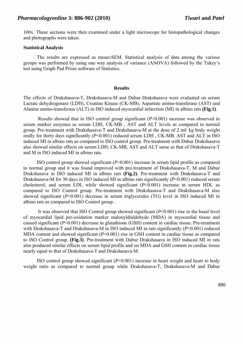
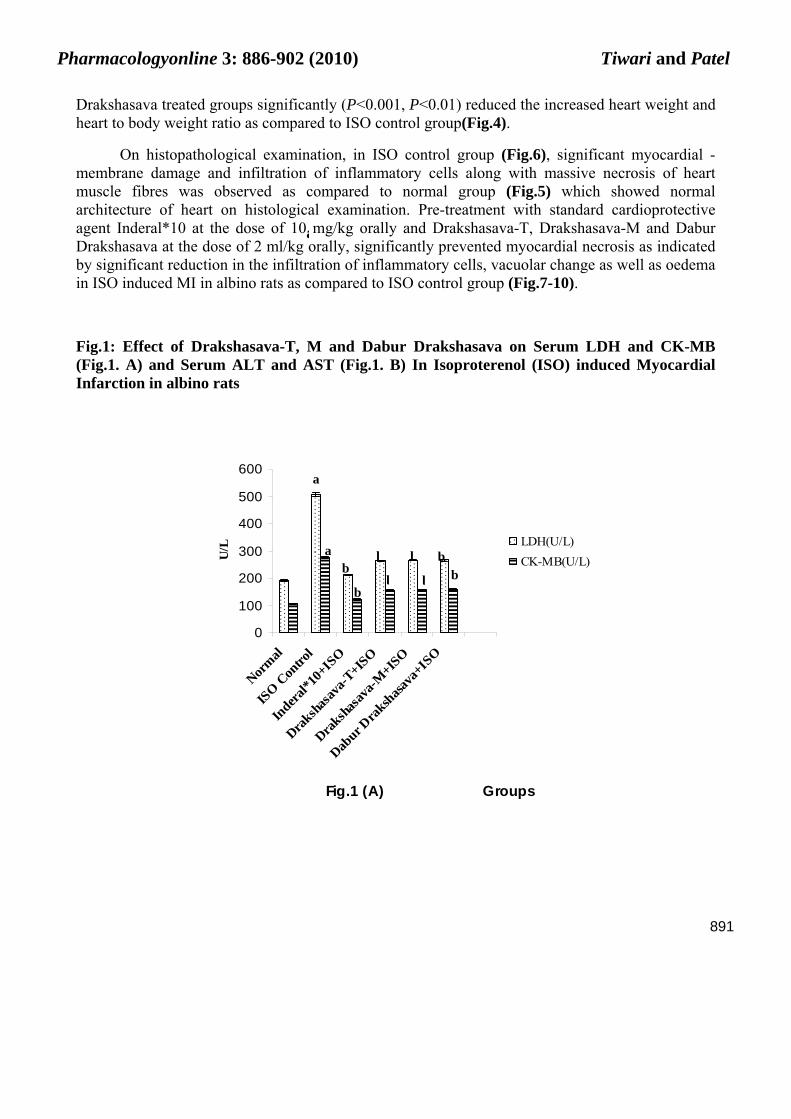
The effects of Drakshasava-T, Drakshasava-M and Dabur Drakshasava were evaluated on serum Lactate dehydrogenase (LDH), Creatine Kinase (CK-MB), Aspartate amino-transferase (AST) and Alanine amino-transferase (ALT) in ISO induced myocardial infarction (MI) in albino rats (Fig.1).
Results showed that in ISO control group significant (P<0.001) increase was observed in serum marker enzymes as serum LDH, CK-MB , AST and ALT levels as compared to normal group. Pre-treatment with Drakshasava-T and Drakshasava-M at the dose of 2 ml/ kg body weight orally for thirty days significantly (P<0.001) reduced serum LDH , CK-MB, AST and ALT in ISO induced MI in albino rats as compared to ISO control group. Pre-treatment with Dabur Drakshasava also showed similar effects on serum LDH, CK-MB, AST and ALT same as that of Drakshasava-T and M in ISO induced MI in albino rats.
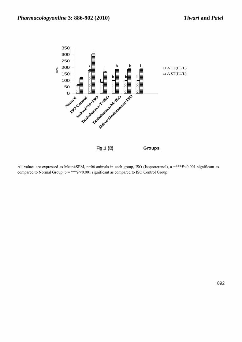
ISO control group showed significant (P<0.001) increase in serum lipid profile as compared to normal group and it was found improved with pre-treatment of Drakshasava-T, M and Dabur Drakshasava in ISO induced MI in albino rats (Fig.2). Pre-treatment with Drakshasava-T and Drakshasava-M for 30 days in ISO induced MI in albino rats significantly (P<0.001) reduced serum cholesterol, and serum LDL while showed significant (P<0.001) increase in serum HDL as compared to ISO Control group. Pre-treatment with Drakshasava-T and Drakshasava-M also showed significant (P<0.001) decrease in serum triglycerides (TG) level in ISO induced MI in albino rats as compared to ISO Control group..
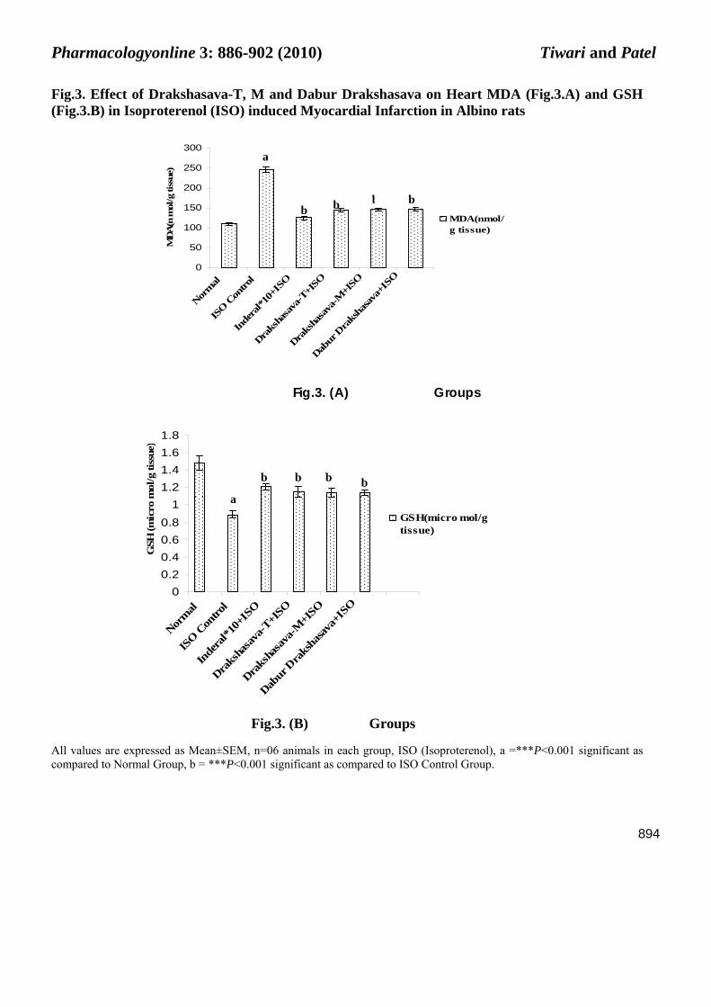
It was observed that ISO Control group showed significant (P<0.001) rise in the basal level of myocardial lipid per-oxidation marker malonyldialdehyde (MDA) in myocardial tissue and caused significant (P<0.001) decrease in glutathione (GSH) content in cardiac tissue. Pre-treatment with Drakshasava-T and Drakshasava-M in ISO induced MI in rats significantly (P<0.001) reduced MDA content and showed significant (P<0.001) rise in GSH content in cardiac tissue as compared to ISO Control group. (Fig.3). Pre-treatment with Dabur Drakshasava in ISO induced MI in rats also produced similar effects on serum lipid profile and on MDA and GSH content in cardiac tissue nearly equal to that of Drakshasava-T and Drakshasava-M.
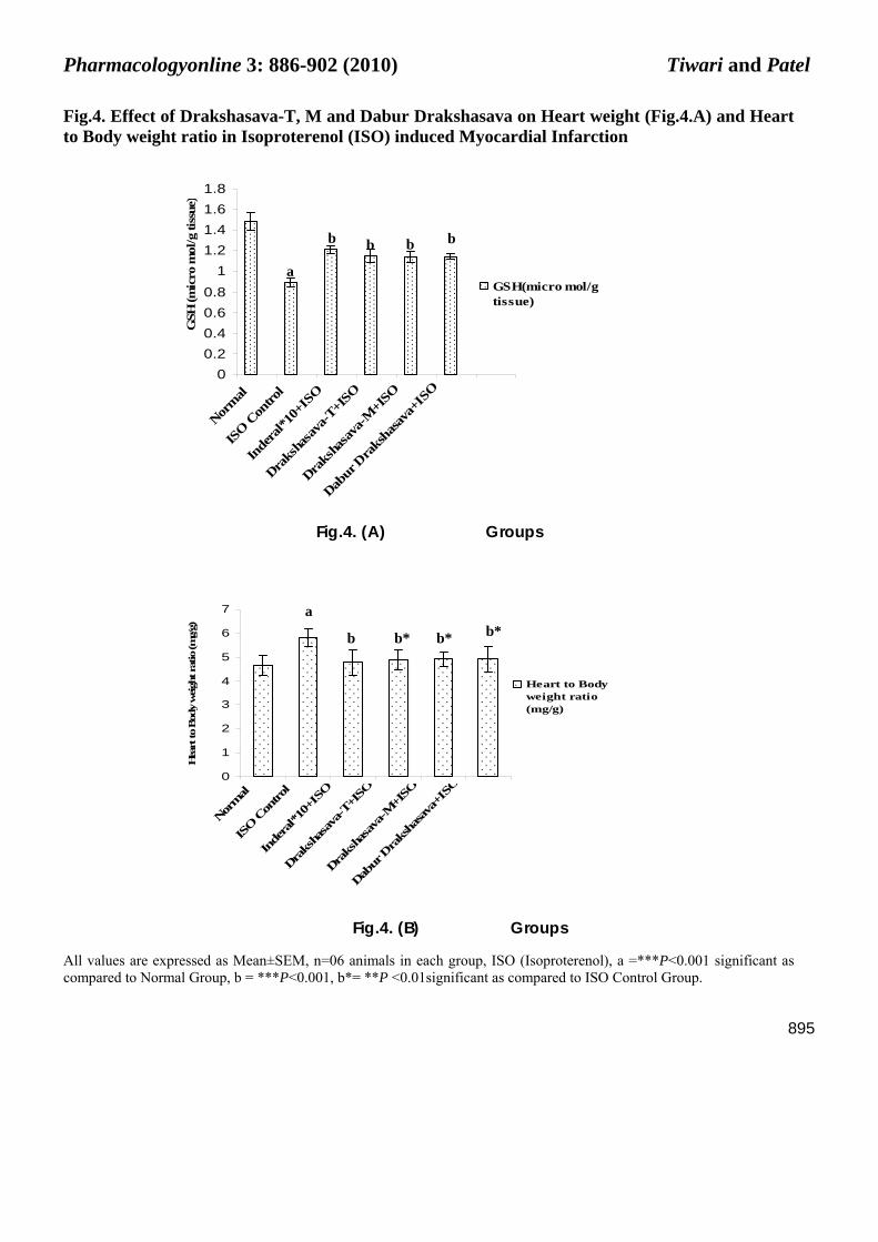
ISO control group showed significant (P<0.001) increase in heart weight and heart to body weight ratio as compared to normal group while Drakshasava-T, Drakshasava-M and Dabur
Pharmacologyonline 3: 886-902 (2010) Tiwari and Patel
891
Drakshasava treated groups significantly (P<0.001, P<0.01) reduced the increased heart weight and heart to body weight ratio as compared to ISO control group(Fig.4).
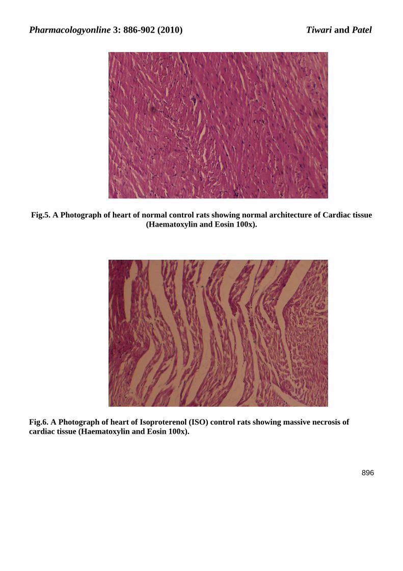
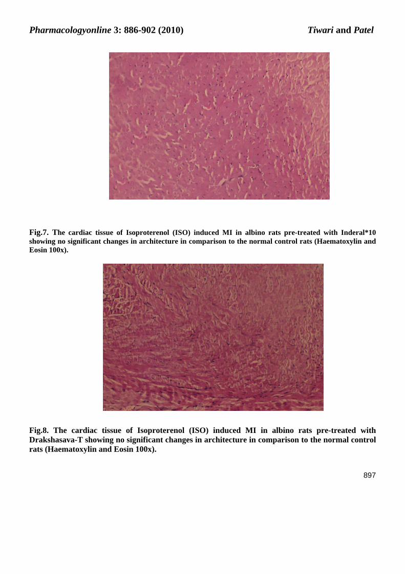
On histopathological examination, in ISO control group (Fig.6), significant myocardial -membrane damage and infiltration of inflammatory cells along with massive necrosis of heart muscle fibres was observed as compared to normal group (Fig.5) which showed normal architecture of heart on histological examination. Pre-treatment with standard cardioprotective agent Inderal*10 at the dose of 10 mg/kg orally and Drakshasava-T, Drakshasava-M and Dabur Drakshasava at the dose of 2 ml/kg orally, significantly prevented myocardial necrosis as indicated by significant reduction in the infiltration of inflammatory cells, vacuolar change as well as oedema in ISO induced MI in albino rats as compared to ISO control group (Fig.7-10).
Fig.1: Effect of Drakshasava-T, M and Dabur Drakshasava on Serum LDH and CK-MB (Fig.1. A) and Serum ALT and AST (Fig.1. B) In Isoproterenol (ISO) induced Myocardial Infarction in albino rats
0
100
200
300
400
500
600
Normal
ISO C
ontro
l
Indera
l*10+
ISO
Draksh
asav
a-T+IS
O
Draksh
asav
a-M+IS
O
Dabur D
raksh
asava
+ISO
U/L LDH(U/L)
CK-MB(U/L)
Fig.1 (A) Groups
a
a
a
b b
b
b
b b
b b
Pharmacologyonline 3: 886-902 (2010) Tiwari and Patel
892
050
100150200250300350
Norm
al
ISO C
ontro
l
Inde
ral*1
0+IS
O
Draks
hasa
va-T
+ISO
Draks
hasa
va-M
+ISO
Dabur
Dra
ksha
sava
+ISO
IU/L ALT(IU/L)
AST(IU/L)
Fig.1 (B) Groups
All values are expressed as Mean±SEM, n=06 animals in each group, ISO (Isoproterenol), a =***P<0.001 significant as compared to Normal Group, b = ***P<0.001 significant as compared to ISO Control Group.
a
a
b
bb
b
b
b
b
b
Pharmacologyonline 3: 886-902 (2010) Tiwari and Patel
893
Fig.2. Effect of Drakshasava-T, M and Dabur Drakshasava on Serum Lipid Profile in Isoproterenol (ISO) induced Myocardial Infarction in Albino rats
050
100150200250300350
Normal
ISO Contro
l
Inderal*10
+ ISO
Drakshasav
a-T+ISO
Drakshasav
a-M+IS
O
Dabur Drak
shasa
va+ISO
(mg/
dl) Cholesterol
HDLLDLTriglycerides
Groups
All values are expressed as Mean±SEM, n=06 animals in each group, ISO (Isoproterenol), a =***P<0.001 significant as compared to Normal Group, b = ***P<0.001 significant as compared to ISO Control Group.
a
b
a
a
a
b b b
b b b
bb
b
b
bb
bb
b
Pharmacologyonline 3: 886-902 (2010) Tiwari and Patel
894
Fig.3. Effect of Drakshasava-T, M and Dabur Drakshasava on Heart MDA (Fig.3.A) and GSH (Fig.3.B) in Isoproterenol (ISO) induced Myocardial Infarction in Albino rats
0
50
100
150
200
250
300
Norm
al
ISO C
ontro
l
Inde
ral*1
0+IS
O
Draks
hasa
va-T
+ISO
Draks
hasa
va-M
+ISO
Dabur
Dra
ksha
sava
+ISO
MD
A(n
mol
/g ti
ssue
)
MDA(nmol/g tissue)
Fig.3. (A) Groups
00.20.40.60.8
11.21.41.61.8
Normal
ISO C
ontro
l
Inde
ral*1
0+IS
O
Draks
hasa
va-T
+ISO
Draks
hasa
va-M
+ISO
Dabur
Dra
ksha
sava+
ISO
GSH
(mic
ro m
ol/g
tiss
ue)
GSH(micro mol/gtissue)
Fig.3. (B) Groups
All values are expressed as Mean±SEM, n=06 animals in each group, ISO (Isoproterenol), a =***P<0.001 significant as compared to Normal Group, b = ***P<0.001 significant as compared to ISO Control Group.
a
b
b b b
b
a
b b b
Pharmacologyonline 3: 886-902 (2010) Tiwari and Patel
895
Fig.4. Effect of Drakshasava-T, M and Dabur Drakshasava on Heart weight (Fig.4.A) and Heart to Body weight ratio in Isoproterenol (ISO) induced Myocardial Infarction
00.20.40.60.8
11.21.41.61.8
Norm
al
ISO C
ontro
l
Inde
ral*1
0+IS
O
Draks
hasa
va-T
+ISO
Draks
hasa
va-M
+ISO
Dabur
Dra
ksha
sava
+ISO
GSH
(mic
ro m
ol/g
tiss
ue)
GSH(micro mol/gtissue)
Fig.4. (A) Groups
0
1
2
3
4
5
6
7
Norm
al
ISO C
ontro
l
Inde
ral*1
0+IS
O
Draks
hasa
va-T
+ISO
Draks
hasa
va-M
+ISO
Dabur
Dra
ksha
sava
+ISO
Hea
rt to
Bod
y w
eigh
t rat
io (m
g/g)
Heart to Bodyweight ratio(mg/g)
Fig.4. (B) Groups
All values are expressed as Mean±SEM, n=06 animals in each group, ISO (Isoproterenol), a =***P<0.001 significant as compared to Normal Group, b = ***P<0.001, b*= **P <0.01significant as compared to ISO Control Group.
b
b b
b a
b*
b* b* b
a
Pharmacologyonline 3: 886-902 (2010) Tiwari and Patel
896
Fig.5. A Photograph of heart of normal control rats showing normal architecture of Cardiac tissue (Haematoxylin and Eosin 100x).
Fig.6. A Photograph of heart of Isoproterenol (ISO) control rats showing massive necrosis of cardiac tissue (Haematoxylin and Eosin 100x).
Pharmacologyonline 3: 886-902 (2010) Tiwari and Patel
897
Fig.7. The cardiac tissue of Isoproterenol (ISO) induced MI in albino rats pre-treated with Inderal*10 showing no significant changes in architecture in comparison to the normal control rats (Haematoxylin and Eosin 100x).
Fig.8. The cardiac tissue of Isoproterenol (ISO) induced MI in albino rats pre-treated with Drakshasava-T showing no significant changes in architecture in comparison to the normal control rats (Haematoxylin and Eosin 100x).
Pharmacologyonline 3: 886-902 (2010) Tiwari and Patel
898
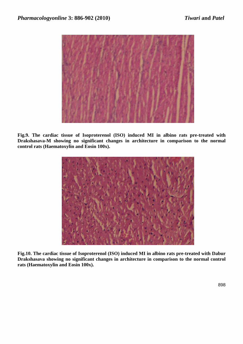
Fig.9. The cardiac tissue of Isoproterenol (ISO) induced MI in albino rats pre-treated with Drakshasava-M showing no significant changes in architecture in comparison to the normal control rats (Haematoxylin and Eosin 100x).
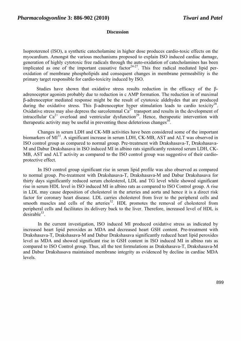
Fig.10. The cardiac tissue of Isoproterenol (ISO) induced MI in albino rats pre-treated with Dabur Drakshasava showing no significant changes in architecture in comparison to the normal control rats (Haematoxylin and Eosin 100x).
Pharmacologyonline 3: 886-902 (2010) Tiwari and Patel
899
Discussion
Isoproterenol (ISO), a synthetic catecholamine in higher dose produces cardio-toxic effects on the myocardium. Amongst the various mechanisms proposed to explain ISO induced cardiac damage, generation of highly cytotoxic free radicals through the auto-oxidation of catecholamines has been implicated as one of the important causative factor24-27. This free radical mediated lipid per-oxidation of membrane phospholipids and consequent changes in membrane permeability is the primary target responsible for cardio-toxicity induced by ISO.
Studies have shown that oxidative stress results reduction in the efficacy of the β-adrenoceptor agonists probably due to reduction in c AMP formation. The reduction in of maximal β-adrenoceptor mediated response might be the result of cytotoxic aldehydes that are produced during the oxidative stress. This β-adrenoceptor hyper stimulation leads to cardio toxicity28. Oxidative stress may also depress the sarcolemmal Ca2+ transport and results in the development of intracellular Ca2+ overload and ventricular dysfunction29. Hence, therapeutic intervention with therapeutic activity may be useful in preventing these deleterious changes30.
Changes in serum LDH and CK-MB activities have been considered some of the important biomarkers of MI31. A significant increase in serum LDH, CK-MB, AST and ALT was observed in ISO control group as compared to normal group. Pre-treatment with Drakshasava-T, Drakshasava-M and Dabur Drakshasava in ISO induced MI in albino rats significantly restored serum LDH, CK-MB, AST and ALT activity as compared to the ISO control group was suggestive of their cardio-protective effect.
In ISO control group significant rise in serum lipid profile was also observed as compared to normal group. Pre-treatment with Drakshasava-T, Drakshasava-M and Dabur Drakshasava for thirty days significantly reduced serum cholesterol, LDL and TG level while showed significant rise in serum HDL level in ISO induced MI in albino rats as compared to ISO Control group. A rise in LDL may cause deposition of cholesterol in the arteries and aorta and hence it is a direct risk factor for coronary heart disease. LDL carries cholesterol from liver to the peripheral cells and smooth muscles and cells of the arteries32. HDL promotes the removal of cholesterol from peripheral cells and facilitates its delivery back to the liver. Therefore, increased level of HDL is desirable33.
In the current investigation, ISO induced MI produced oxidative stress as indicated by increased heart lipid peroxides as MDA and decreased heart GSH content. Pre-treatment with Drakshasava-T, Drakshasava-M and Dabur Drakshasava significantly reduced heart lipid peroxides level as MDA and showed significant rise in GSH content in ISO induced MI in albino rats as compared to ISO Control group. Thus, all the test formulations as Drakshasava-T, Drakshasava-M and Dabur Drakshasava maintained membrane integrity as evidenced by decline in cardiac MDA levels.
Pharmacologyonline 3: 886-902 (2010) Tiwari and Patel
900
In the ISO control group, a significant increase in heart weight and heart weight to body weight ratio was observed which was reversed with Drakshasava-T, Drakshasava-M and Dabur Drakshasava treatment in ISO induced MI in albino rats. It suggests the cardio-protective property of all these test formulations. Furthermore, histopathological examination confirmed the cardio-protective effects of Drakshasava-T, Drakshasava-M and Dabur Drakshasava. Thus, all the test formulations as Drakshasava-T, Drakshasava-M and Dabur Drakshasava were found nearly equally effective to that of standard cardio-protective agent Inderal*10 to attenuate the effect of ISO induced MI.
In summary, the present study strongly suggests that multiple mechanisms may be responsible for the cardio-protective effect of Drakshasava-T, Drakshasava-M and Dabur Drakshasava. All these test formulations as Drakshasava-T, Drakshasava-M and Dabur Drakshasava produced myocardial adaptive changes (augmentation of endogenous antioxidants as GSH) on chronic administration. In addition, they restored the integrity of the myocardium, subsequent to ISO induced oxidative stress. Histopathological assessment further confirmed the cardio-protective effect of all these test formulations. Drakshasava mainly contains dried fruits of Vitis vinifera which are the rich source of phenolic compounds and possess good antioxidant activity. The obtained result suggests that presence of self generated alcohol could be beneficial in the faster absorption of poly-phenolic compounds present in Drakshasava which are responsible for showing scavenging of ISO induced free radicals.
The present study provides scientific basis for the cardio-protective potential of Drakshasava validating its usage in Ayurveda. Considering its safety, efficacy and traditional acceptability, clinical trials should be conducted to support its therapeutic use in ischemic heart diseases.
References
1. Bolli R. Myocardial ischemic metabolic disorder leading to cell death. Rev Post Cardiol 1994; 13: 649-653.
2. Kumar Vinay, Abbas AK, Fausto Nelson. Robbins and Cotran Pathologic Basis of Disease.7th ed. Philadelphia: Saunders Publication; 2004.p. 571-572.
3. Dhar ML, Dhar MM, Dhawan BN, Mehrotra BN and Ray C. Screening of Indian plants for biological activity. J Exp Biol 1968; 6: 232-247.
4. Hertog MGL, Feskens EJM, Hollam PCH, Katan MB, Kromhout D. Dietary antioxidant flavonoids and risk of coronary heart diseases. Lancet 1993; 342: 1007-1020.
5. The Ayurvedic Formulary of India Part-I. Delhi (India): Controller of Publications; 2000. p. 35.
6. Baydar NG, Ozkan G, Sagdic O. Total phenolic contents and antibacterial activities of grape (Vitis vinifera L.) extracts. Food Control 2004; 15:335-339.
Pharmacologyonline 3: 886-902 (2010) Tiwari and Patel
901
7. Frankel EN, Kanner J, German JB, Parks E, Kinsella JE. Inhibition of oxidation of human low-density lipoprotein by phenolic substances in red wine. Lancet 1993; 341(20): 454-457.
8. Mayer AS, Yi OS, Person DA, Waterhouse DL, Frankel EN. Inhibition of human low-density lipoprotein oxidation in relation to composition of phenolic antioxidants in grapes (Vitis vinifera). J Agri Food Chem 1997; 45:1638-1643.
9. Teissedre PL, Frankel EN, Waterhouse AL, Peleg H, German GB. Inhibition of in vitro human LDL oxidation by phenolic antioxidants from grapes and wines. J Sci Food Agri 1996; 70: 55-61.
10. Waterhouse AL. Wine antioxidants may reduce heart disease and cancer. Presentation of American Chemical Society, Washington; 1994
11. Zheng M, Han QD, Xiao RP. Distinct adrenergic receptor subtype signaling in the heart and their path physiological relevance. Acta Physiol Sin 2004; 56: 1-15.
12. Mishra S. Bhaisazya Kalpana Vigyan. Varanasi (India): Chaukambha Bharati Prakashan; 2005. p. 253-254.
13. Alan M, Radhamani S, Ali U, Purushottam KK. Microbiological Screening of Dhataki Flowers. Journal of Research in Ayurveda and Siddha 1984; 2(4):371-375.
14. Rona G, Chapel CI, Balazs T, Gaudry R. An infarct like myocardial lesion and other toxic manifestations produced by isoproterenol in the rat. Arch Pathol 1959; 76: 443-455.
15. Tripathi KD. Essentials of Medical Pharmacology. 6thed. New Delhi (India): Jaypee Brothers Medical Publishers Limited; 2008. p. 137-138,537.
16. Varley H. Practical Clinical Biochemistry. 4thed. NY: William Heinemann; 1967.p. 161-162.
17. Lamprecht W, Stan F, Weisser H, Heinz F. Determination of creatine phosphate and adenosine triphosphate with creatine kinase. In: Methods of Enzymatic analysis. Ed. HU Vergmeyer. NY: Academic Press; 1974. p. 1776-1778.
18. Mohun AF, Cook IGY. Simple methods for measuring serum levels of Glutamic–oxaloacetic and Glutamic-Pyruvic transaminases in routine laboratories. J Clin Pathol 1957; 10 (4): 394-399.
19. Allain CC, Pool LS, Chan CS, Richmond W. Enzymatic determination of serum cholesterol. Clin Chem 1974; 20: 447-475.
20. Friedewald WT, Levy RI, Fredrickson DS. Estimation of the Concentration of Low-density lipoprotein cholesterol in plasma, without use of the preparative ultracentrifuge. Clin Chem 1972; 18: 499-502.
21. Muller PH, Schmulling RM, Liebich HM, Eggstein M. A fully enzymatic triglyceride determination. J Clin Chem 1977; 15: 457-464.
Pharmacologyonline 3: 886-902 (2010) Tiwari and Patel
902
22. Ohkawa H, Ohisi N, Yagi K. Assay for lipid peroxides in animal tissue by thiobarbituric acid reaction. Anal Biochem 1979; 95: 351-358.
23. Ellman GL. Tissue Sulphydril groups. Arch Biochem Biophys 1959; 82: 72-77.
24. Handforth CP. Isoproterenol induced myocardial infarction in animals. Arch Pathol 1962; 73: 161-165.
25. Csapo Z, Dusek J, Rano G. Early alterations of the cardiac muscle cells in isoproterenol induced necrosis. Arch Pathol 1972; 93: 356-365.
26. Singhal PK, Beamish RE, Dhalla NS. Potential oxidative pathways of catecholamines in the formation of lipid peroxides and heart disease. Adv Exp Med Biol 1983; 161: 391-401.
27. Nirmala C, Puvanakrishnan R. Isoproterenol induced myocardial infarction in rats; functional and biochemical alterations. Med Sci Res 1994; 22: 575-577.
28. Haenen GR, Veerman M, Bast A. Reduction of beta- adrenoceptor functions by oxidative stress in heart. Free Radic Biol Med 1990; 9: 279-288.
29. Tappia PS, Heta T, Dhalla NS. Role of oxidative stress in catecholamine induced changes in cardiac sarcolemmal Ca2+ transport. Arch Biochem Biophys 2001; 377: 85-92.
30. Noronha AA, Steen EM, Woolf N. The correlation between catecholamine and lipid per oxidation induced myocardial infarction in heart cells. Basic Res Cadiol 1984; 80: 133-136.
31. Jennings RB, Murry CE, Steenburger CJR, Reimer KA. Acute myocardial ischemia: development of cell injury in sustained ischemia. Circulation 1990; 82: 3-12.
32. Pederson TR. Low density lipoprotein cholesterol lowering is and will be the key to the future of lipid management. Am J cardiol 2001; 87 (5A): 8B-12B
33. Bolden WE, Pearson TA. Raising low levels of High density lipoprotein cholesterol is an important target of therapy. Am J cardiol 2000; 85 (5): 645-650.