phase ii randomized, double-masked, vehicle-controlled trial of ... ii randomized, double... ·...
TRANSCRIPT

Phase II Randomized, Double-Masked,Vehicle-Controlled Trial of RecombinantHuman Nerve Growth Factor forNeurotrophic Keratitis
Stefano Bonini, MD,1 Alessandro Lambiase, MD, PhD,2 Paolo Rama, MD,3 Francesco Sinigaglia, MD,4
Marcello Allegretti, PhD,4 Wendy Chao, PhD,4 Flavio Mantelli, MD, PhD,4 for the REPARO Study Group*
Purpose: To evaluate the safety and efficacy of topical recombinant human nerve growth factor (rhNGF) fortreating moderate-to-severe neurotrophic keratitis (NK), a rare degenerative corneal disease resulting fromimpaired corneal innervation.
Design: Phase II multicenter, randomized, double-masked, vehicle-controlled trial.Participants: Patients with stage 2 (moderate) or stage 3 (severe) NK in 1 eye.Methods: The REPARO phase II study assessed safety and efficacy in 156 patients randomized 1:1:1 to
rhNGF 10 mg/ml, 20 mg/ml, or vehicle. Treatment was administered 6 drops per day for 8 weeks. Patients thenentered a 48- or 56-week follow-up period. Safety was assessed in all patients who received study treatment,whereas efficacy was by intention to treat.
Main Outcome Measures: Corneal healing (defined as <0.5-mm maximum diameter of fluoresceinstaining in the lesion area) was assessed by masked central readers at week 4 (primary efficacy end point) andweek 8 (key secondary end point) of controlled treatment. Corneal healing was reassessed post hoc bymasked central readers using a more conservative measure (0-mm staining in the lesion area and no otherpersistent staining).
Results: At week 4 (primary end point), 19.6% of vehicle-treated patients achieved corneal healing (<0.5-mmlesion staining) versus 54.9% receiving rhNGF 10 mg/ml (þ35.3%; 97.06% confidence interval [CI], 15.88e54.71;P < 0.001) and 58.0% receiving rhNGF 20 mg/ml (þ38.4%; 97.06% CI, 18.96e57.83; P < 0.001). At week 8 (keysecondary end point), 43.1% of vehicle-treated patients achieved less than 0.5-mm lesion staining versus 74.5%receiving rhNGF 10 mg/ml (þ31.4%; 97.06% CI, 11.25e51.49; P ¼ 0.001) and 74.0% receiving rhNGF 20 mg/ml(þ30.9%; 97.06% CI, 10.60e51.13; P ¼ 0.002). Post hoc analysis of corneal healing by the more conservativemeasure (0-mm lesion staining and no other persistent staining) maintained statistically significant differencesbetween rhNGF and vehicle at weeks 4 and 8. More than 96% of patients who healed after controlled rhNGFtreatment remained recurrence free during follow-up. Treatment with rhNGF was well tolerated; adverse effectswere mostly local, mild, and transient.
Conclusions: Topical rhNGF is safe and more effective than vehicle in promoting healing of moderate-to-severe NK. Ophthalmology 2018;125:1332-1343 ª 2018 by the American Academy of Ophthalmology. This isan open access article under the CC BY-NC-ND license (http://creativecommons.org/licenses/by-nc-nd/4.0/).
Supplemental material available at www.aaojournal.org.
With approximately 7000 nerve endings per square milli-meter, the cornea is the most densely innervated tissue inhumans.1 Corneal nerves (deriving from the trigeminalganglion) help maintain transparency in this avasculartissue and participate in ocular surface homeostasis byproducing neurotrophins and facilitating sensory-dependent corneal and tearing reflexes.1,2 Trigeminalnerve damage may cause neurotrophic keratitis (NK) withpartial or total loss of corneal sensation, leading to visualimpairment and potentially permanent blindness. Neuro-trophic keratitis, also known as neurotrophic keratopathy, is
1332 ª 2018 by the American Academy of OphthalmologyThis is an open access article under the CC BY-NC-ND license(http://creativecommons.org/licenses/by-nc-nd/4.0/). Published by Else
a rare disease (estimated prevalence, 1.6e4.2 cases per10 000 persons)3,4 with various underlying causes (mostcommonly herpetic infections and ocular or neurologicsurgeries) that impair corneal innervation.5,6 Neurotrophickeratitis diagnosis, prognosis, and treatment (reviewedelsewhere)3,6 are based on disease severity, which is clas-sified broadly into 3 stages.7 Briefly, stage 1 (mild) NKexhibits ocular surface irregularity and reduced vision,stage 2 (moderate) NK exhibits a nonhealing persistentepithelial defect (PED), and stage 3 (severe) NK exhibitscorneal ulceration involving subepithelial (stromal) tissue,
vier Inc.
https://doi.org/10.1016/j.ophtha.2018.02.022ISSN 0161-6420/18

Bonini et al � rhNGF for Neurotrophic Keratitis
which may progress to corneal perforation. All diseasestages cause some vision loss; however, if untreated,moderate NK progresses to severe disease with associatedrisks of profound vision loss resulting from scarring andcorneal perforation. Conventional therapy for stage 1 aimsto prevent epithelial breakdown, generally byadministering preservative-free artificial tears and dis-continuing toxic topical medications. Stage 2 or 3 therapiesaim to facilitate corneal healing and prevent corneal thin-ning (which may lead to perforation); these include sur-geries and procedures (e.g., tarsorrhaphy, botulinum-induced ptosis, conjunctival flap, amniotic membranetransplantation) to restore ocular surface integrity, butpotentially sacrificing vision and cosmesis.
Strong evidence supports the treatment of NK withneurotrophic factors.8 Nerve growth factor (NGF) hasdemonstrated important roles in maintaining cornealhomeostasis in vitro, ex vivo, and in animal models.9,10
Nerve growth factor is highly conserved among verte-brates,11 and small uncontrolled, open-label studies withmurine NGF (mNGF) produced promising results for thetreatment of corneal neurotrophic ulcers.12,13 Confirmationof results obtained with mNGF have been highly antici-pated14; however, nearly 2 decades passed with no approvedtreatments for NK and no NGF-based treatments availablefor any indication. For NK therapies in general, clinicaldevelopment has been hindered by the paucity of adequatelysized and rigorously designed studies; indeed, only 1 ran-domized controlled trial of NK patients exists in the pub-lished literature to date, and the investigative treatment(topical fibronectin ophthalmic solution) was not superior toplacebo for healing PEDs.15 Thus, the natural history of NKis not completely understood, and approved treatments arenot available for use as comparators for further studies.For NGF in particular, translational development has beenmired by its complex tertiary structure, which complicatesthe manufacturing of recombinant human NGF (rhNGF)suitable for clinical use. To this end, we developed anEscherichia coliederived rhNGF formulation for topicalophthalmic use and demonstrated it to be safe and welltolerated in phase I randomized, double-masked, vehicle-controlled studies in healthy volunteers16 and in NKpatients.17 Herein, we report phase II study results oftopical rhNGF treatment for moderate-to-severe NK.
Methods
Clinical Trial Design
The REPARO (Latin for “repair”) trial was a phase I/II double-masked, randomized, multicenter, vehicle-controlled, parallel-group study that was designed to evaluate the safety and efficacy ofrhNGF eye drops (10 or 20 mg/ml, 6 drops/day for 8 weeks) inpatients with stage 2 or 3 NK. Phase I assessed safety in 18 patientsto support proceeding to phase II and was conducted, analyzed, andreported separately.17 Phase II randomized 156 patients 1:1:1 torhNGF 10 mg/ml, rhNGF 20 mg/ml, or vehicle for an 8-weekcontrolled treatment period. Follow-up duration (48 or 56 weeks)was determined by baseline group assignment and corneal healingstatus during controlled treatment. For vehicle-treated patients,baseline randomization included the possibility of secondary
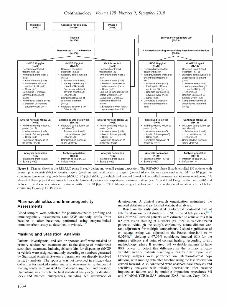
rhNGF treatment (10 or 20 mg/ml) in the event of treatment failureduring the 8-week controlled treatment period, predefined as failureto achieve corneal healing, recurrence of NK after healing, ordeterioration (lesion size increase of �1 mm, best-corrected dis-tance visual acuity [BCDVA] decrease of >5 Early TreatmentDiabetic Retinopathy Study [ETDRS] letters, progression tocorneal melting or perforation, or onset of infection). This patientsubset received 8 weeks of uncontrolled treatment beforecontinuing follow-up (total follow-up, 56 weeks). The phase IIstudy design is diagrammed in Figure 1. The REPARO studygroup is listed in Appendix 1 (available at www.aaojournal.org),and the trial was registered at ClinicalTrials.gov (identifier,NCT01756456).
Patients
Patients (�18 years of age) with NK were diagnosed with stage 2(PED) or stage 3 (corneal ulcer) disease using published criteria.7
The main inclusion criteria were evidence of decreased cornealsensitivity within the corneal lesion and 1 or more cornealquadrants outside the lesion; BCDVA score of 75 ETDRS lettersor fewer (�0.2 logarithm of the minimum angle of resolution,�20/32 Snellen, or �0.625 decimal fraction) in the affected eye;and no objective clinical evidence of improvement of the PED orcorneal ulcer within 2 weeks before study enrollment. The mainexclusion criteria were stage 2 or 3 NK affecting both eyes,active ocular infection or inflammation unrelated to NK, or otherocular disease or severe vision loss in the affected eye. Forcomplete inclusion and exclusion criteria, see Appendix 2(available at www.aaojournal.org).
Efficacy Assessments
The primary efficacy variable was corneal healing, defined as lessthan 0.5-mm fluorescein staining (the lower limit of reliable slit-lamp assessment) in the lesion area, assessed in clinical picturesby masked central readers as a yes-or-no binary variable at week 4(primary end point) and week 8 (prespecified secondary end point).Other secondary variables included visual acuity (BCDVAmeasured in ETDRS letters), corneal sensitivity measured using theCochet-Bonnet aesthesiometer (CBA), and duration of cornealhealing through follow-up.
Exploratory efficacy variables included reflex tearing (Schirmertest wetting distance after 5 minutes), time to onset of healing(>20% reduction in maximum diameter of the corneal lesion frombaseline), and time to corneal healing (<0.5-mm lesion staining)during the controlled or uncontrolled treatment periods. Post hocefficacy variables included change in lesion size and the primaryend point of corneal healing reassessed more conservatively bymasked central readers as 0-mm lesion staining and no otherpersistent staining outside of the lesion.
Safety Assessments
The primary safety variable was incidence of adverse events (AEs).Ocular tolerability was recorded by patients on a visual analogscale (VAS) from 0 to 100 mm (0 ¼ no symptoms; 100 ¼ worstpossible discomfort) for each of 7 different symptoms: foreignbody sensation, burning or stinging, itching, ocular pain, stickyfeeling, blurred vision, and photophobia. An overall VAS scorewas calculated as the mean of individual symptom scores. Othersafety parameters included visual acuity (BCDVA measured inETDRS letters), intraocular pressure, dilated fundus ophthalmos-copy, vital signs, hematologic results, and clinical chemistryresults.
1333
Assessed for eligibility(N=186)
rhNGF 10 g/ml (N=52)
• Received controlled treatment (n=52)
• Withdrew before week 8 (n=6)o Adverse event (n=3)o Inadequate efficacy /
control of NK (n=2)o Other (n=1)
• Completed 8 weeks of controlled treatment (n=46)
• Withdrew at week 8 (n=1)o Decision unrelated to
adverse event (n=1)
Randomized 1:1:1 at baseline(N=156)
rhNGF 20 g/ml(N=52)
• Received controlled treatment (n=52)
• Withdrew before week 8 (n=12)o Adverse event (n=9)o Inadequate efficacy /
control of NK (n=1)o Decision unrelated to
adverse event (n=1)o Other (n=1)
• Completed 8 weeks of controlled treatment (n=40)
• Withdrew at week 8 (n=1)o Other (n=1)
rhNGF 10 g/ml (N=10)
• Received uncontrolled treatment (n=10)
• Withdrew before week 8 of uncontrolled treatment (n=1)o Adverse event (n=0)o Inadequate efficacy /
control of NK (n=1)o Decision unrelated to
adverse event (n=0)o Other (n=0)
• Completed 8 weeks of uncontrolled treatment (n=9)
Entered 48-week follow-up (N=45)
• Withdrew during follow-up period (n=12)o Adverse event (n=6)o Lost to follow-up (n=4)o Other (n=2)
• Completed 48 weeks of follow-up (n=33)
Entered 48-week follow-up (N=39)
• Withdrew during follow-up period (n=6)o Adverse event (n=0)o Lost to follow-up (n=2)o Other (n=4)
• Completed 48 weeks of follow-up (n=33)
Entered 48-week follow-up (N=25)
• Withdrew during follow-up period (n=3)o Adverse event (n=1)o Lost to follow-up (n=1)o Other (n=1)
• Completed 48 weeks of follow-up (n=22)
Continued follow-up (N=9)
• Withdrew during follow-up period (n=2)o Adverse event (n=0)o Lost to follow-up (n=0)o Other (n=2)
• Completed 56 weeks of follow-up (n=7)
Analysis population(N=52)
• Intention to treat (n=52)• Safety (n=52)
Analysis population(N=52)
• Intention to treat (n=52)• Safety (n=52)
Analysis population(N=52)
• Intention to treat (n=52)• Safety (n=52)
Analysis population(N=10)
• Intention to treat (n=10)• Safety (n=10)
rhNGF 20 g/ml (N=13)
• Received uncontrolled treatment (n=13)
• Withdrew before week 8 of uncontrolled treatment (n=0)o Adverse event (n=0)o Inadequate efficacy /
control of NK (n=0)o Other (n=0)
• Decision unrelated to adverse event (n=0)
• Completed 8 weeks of uncontrolled treatment (n=13)
Continued follow-up (N=13)
• Withdrew during follow-up period (n=4)o Adverse event (n=0)o Lost to follow-up (n=1)o Other (n=3)
• Completed 56 weeks of follow-up (n=9)
Analysis population(N=13)
• Intention to treat (n=13)• Safety (n=13)
Entered 56-week follow-up*(N=23)
Allocated according to secondary baseline randomization (N=23)
Ineligible(N=12)
Phase I(N=18)
Phase II(N=156)
Vehicle control(N=52)
• Received controlled treatment (n=52)
• Withdrew before week 8 (n=4)o Adverse event (n=1)o Decision unrelated to
adverse event (n=1)o Other (n=2)
• Entered 56-week follow-up before week 8 (n=8)*
• Completed 8 weeks of controlled treatment (n=40)o Entered 56-week follow-
up at week 8 (n=15)*
Figure 1. Diagram showing the REPARO phase II study design and overall patient disposition. The REPARO phase II study enrolled 156 patients withneurotrophic keratitis (NK) of severity stage 2 (persistent epithelial defect) or stage 3 (corneal ulcer). Patients were randomized 1:1:1 to 10 mg/ml re-combinant human nerve growth factor (rhNGF), 20 mg/ml rhNGF, or vehicle and received 8 weeks of controlled treatment and 48 weeks of follow-up. *A56-week follow-up period was intended for vehicle-treated patients who experienced treatment failure (see Clinical Trial Design section for details) andincluded 8 weeks of uncontrolled treatment with 10 or 20 mg/ml rhNGF (dosage assigned at baseline in a secondary randomization scheme) beforecontinuing follow-up for 48 weeks.
Ophthalmology Volume 125, Number 9, September 2018
Pharmacokinetics and ImmunogenicityAssessments
Blood samples were collected for pharmacokinetics profiling andimmunogenicity assessments (anti-NGF antibody shifts frombaseline to after baseline), performed using enzyme-linkedimmunosorbent assay as described previously.16
Masking and Statistical Analysis
Patients, investigators, and site or sponsor staff were masked toprimary randomized treatment and to the dosage of randomizedsecondary treatment. Indistinguishable kits for dispensing rhNGFor vehicle were assigned randomly according to numbers generatedby Statistical Analysis System programmers not directly involvedin study analysis. The sponsor was not involved in efficacy datacollection for masked central analysis. Assessments by the centralreading center were masked to treatment assignment and duration.Unmasking was restricted to final statistical analysis (after databaselock) and medical emergencies, including NK recurrence or
1334
deterioration. A clinical research organization maintained themasked database and performed statistical analyses.
Based on the only published randomized controlled trial ofNK15 and uncontrolled studies of mNGF-treated NK patients,12,13
60% of rhNGF-treated patients were estimated to achieve less than0.5-mm lesion staining at 4 weeks (vs. 30% in vehicle-treatedpatients). Although the study’s exploratory nature did not war-rant adjustment for multiple comparisons, 2-sided significance ofchi-square testing was adjusted to the Pocock threshold (a ¼0.0294),18 yielding a 97.06% confidence interval (CI) for theprimary efficacy end point of corneal healing. According to thismethodology, phase II required 141 evaluable patients to have80% power to detect this difference in the primary efficacyvariable, and 156 patients assuming a 10% to 20% dropout rate.Efficacy analyses were performed on intention-to-treat pop-ulations, with missing data after baseline using the last observationcarried forward. Also conducted were observed-case analyses andsensitivity analyses, with missing observations after baselineimputed as failures and by multiple imputation procedures MIand MIANALYZE in SAS software (SAS Institute, Cary, NC).

Table 1. Patient Demographics and Baseline Characteristics
Characteristics
Recombinant Human Nerve Growth Factor
Vehicle (N [ 52)10 mg/ml (N ¼ 52) 20 mg/ml (N ¼ 52)
Age (yrs)Mean (SD) 59.0 (17.17) 62.5 (14.01) 60.4 (16.78)Median (minimumemaximum) 61.5 (20e87) 63.5 (18e95) 60.5 (23e91)
Female gender, no. (%) 30 (57.7) 30 (57.7) 35 (67.3)Ethnicity, no. (%)Hispanic, Latino, or Spanish 6 (11.5) 9 (17.3) 5 (9.6)N/A 4 (7.7) 1 (1.9) 6 (11.5)
Race, no. (%)Asian 1 (1.9) 0 1 (1.9)Black 0 0 1 (1.9)White 46 (88.5) 51 (98.1) 45 (86.5)N/A 5 (9.6) 1 (1.9) 5 (9.6)
Primary NK diagnosis, no. (%)Stage 2 21 (40.4) 27 (51.9) 28 (53.8)Stage 3 31 (59.6) 25 (48.1) 24 (46.2)
Underlying cause, no. (%)Diabetes mellitus 3 (5.8) 4 (7.7) 4 (7.7)Dry eye disease 6 (11.5) 6 (11.5) 5 (9.6)Herpetic eye disease* 15 (28.8) 11 (21.2) 18 (34.6)Neurosurgical procedure
Acoustic neuroma 2 (3.8) 1 (1.9) 3 (5.8)Auditive neurosurgery 0 1 (1.9) 0Cerebellar metastasis 0 1 (1.9) 0Cerebral epidermoid cyst aspiration 0 1 (1.9) 0Craniotomy for glioma 1 (1.9) 0 0Facial nerve reconstruction 1 (1.9) 0 0Meningioma excision 0 1 (1.9) 1 (1.9)Schwannoma 1 (1.9) 1 (1.9) 3 (5.8)Unspecified 1 (1.9) 2 (3.8) 0
Nonviral infectionAmoebic keratitis 0 2 (3.8) 0Unspecified 1 (1.9) 0 1 (1.9)
Ocular surface injury/inflammationChemical burn 4 (7.7) 2 (3.8) 3 (5.8)Unspecified 1 (1.9) 3 (5.8) 2 (3.8)
Ocular surgery or procedureCataract surgery/scleral buckle/vitrectomy 1 (1.9) 1 (1.9) 1 (1.9)Corneal transplantation 0 0 1 (1.9)Keratoplasty 2 (3.8) 0 0Maxillofacial surgery (eyelid suture) 1 (1.9) 0 0Strontium brachytherapy, mitomycin drops 0 0 1 (1.9)Unspecified 5 (9.6) 4 (7.7) 4 (7.7)
OtherAtopic dermatitis 1 (1.9) 0 0Corneal hypoesthesia 0 1 (1.9) 0Facial palsy resulting from measles 1 (1.9) 0 0Goldenhar syndrome 0 0 1 (1.9)Graves-Basedow disease 0 1 (1.9) 0Lagophthalmos 0 0 1 (1.9)Miller-Fisher syndrome 1 (1.9) 0 0Multifactorial (HSV, keratoplasty, burn, diabetes) 0 1 (1.9) 0Neurovascular encephalopathy 0 1 (1.9) 0Paraneoplastic neuropathy (lung cancer) 0 1 (1.9) 0Pemphigoid 0 1 (1.9) 1 (1.9)Polyneuropathy, traumatic erosion 0 1 (1.9) 0
Stroke 1 (1.9) 2 (3.8) 0Systemic medication 1 (1.9) 0 0Topical medication (glaucoma medication) 0 1 (1.9) 1 (1.9)Unknown origin 1 (1.9) 1 (1.9) 0
(Continued)
Bonini et al � rhNGF for Neurotrophic Keratitis
1335

Table 1. (Continued.)
Characteristics
Recombinant Human Nerve Growth Factor
Vehicle (N [ 52)10 mg/ml (N ¼ 52) 20 mg/ml (N ¼ 52)
Venous sinus thrombosis 1 (1.9) 0 0Viral conjunctivitis (unspecified) 0 0 1 (1.9)
HSV ¼ herpes simplex virus; N/A ¼ not available (ethnicity and race were not collected in all countries); NK ¼ neurotrophic keratitis; SD ¼ standarddeviation.*Includes herpes simplex, herpes zoster, and recurrent herpetic keratitis.
Ophthalmology Volume 125, Number 9, September 2018
For binary secondary and exploratory efficacy end points,2-sided significance was set at a ¼ 0.05. Change in BCDVA scorefrom baseline to week 8 was analyzed by an analysis of covariancemodel using treatment group and baseline BCDVA score. Mixed-effects repeated-measures models using treatment, visit, andbaseline measurements were used to assess changes in lesion size(maximum dimension) and reflex tearing (Schirmer test wettingdistance) from baseline to weeks 4 and 8. The time to onset ofhealing (>20% reduction in maximum diameter of the corneallesion from baseline) and corneal healing (<0.5-mm maximumdiameter of fluorescein staining) were analyzed using Kaplan-Meier methods and the log-rank test (for the controlled treatmentperiod) and descriptive statistics (for the uncontrolled treatmentperiod). Data collected during follow-up also were analyzed usingdescriptive statistics.
Study Oversight
Approval was obtained for the study protocol, amendments, andstudy-related documents (including informed consent) from theinstitutional review board of Sapienza University of Rome and anindependent ethics committee from each country with 1 or moreparticipating sites (Appendix 1, available at www.aaojournal.org).The study complied with the Declaration of Helsinki, relevant partsof the Code of Federal Regulations Title 21, and good clinicalpractice and good laboratory practice guidelines. Writteninformed consent was obtained before study-related procedures.Compliance was assessed at each visit and verified by studymonitors during onsite visits.
Results
Patients and Treatment
The REPARO investigators (Appendix 1, available atwww.aaojournal.org) represented 39 sites in 9 European countries(Belgium, France, Germany, Hungary, Italy, Poland, Portugal,Spain, and the United Kingdom); 32 sites in 6 countries enrolled 1or more patients. Figure 1 provides an overview of patientdisposition (including reasons for withdrawal). Of 186 patientsscreened from January 2013 through May 2015, 174 wereenrolled: 18 in phase I17 and 156 in phase II. Patient demographicsand baseline characteristics were well balanced in the REPAROphase II study, with no clinically notable differences betweentreatment groups (Table 1). Consistent with publishedliterature,5,6,13,19 common underlying causes included herpetic eyedisease (44 patients) and ocular or neurologic surgery (21 patientseach). Prior treatments for NK (most commonly artificial tears, gels,or ointments and topical antibiotics) are listed in Appendix 3(available at www.aaojournal.org).
1336
Efficacy Outcomes
Table 2 summarizes efficacy analyses at weeks 4 and 8 (lastobservation carried forward). Corneal healing (<0.5-mm lesionstaining) was achieved at week 4 (primary end point) in 19.6% ofvehicle-treated patients versus 54.9% receiving rhNGF 10 mg/ml(þ35.3%; 97.06% CI, 15.88%e54.71%; P < 0.001) and 58.0%receiving rhNGF 20 mg/ml (þ38.4%; 97.06% CI, 18.96%e57.83%; P< 0.001). Corneal healing at week 8 (key secondary endpoint) was achieved in 43.1% of vehicle-treated patients versus74.5% receiving rhNGF 10 mg/ml (þ31.4%; 97.06% CI, 11.25%e51.49%; P ¼ 0.001) and 74.0% receiving rhNGF 20 mg/ml(þ30.9%; 97.06% CI, 10.60%e51.13%; P ¼ 0.002). Table 3summarizes the post hoc reanalysis of corneal healing using themore conservative definition (0-mm lesion staining and no otherpersistent staining). This confirmed statistically significant differ-ences between rhNGF and vehicle, with consistently higher per-centages healed in the rhNGF 20-mg/ml group at both week 4 andweek 8. Observed-case, worst-case (missing observations afterbaseline imputed as failures), and multiple imputation analysesproduced similar results (not shown). Differences between rhNGFgroups were not statistically significant.
Figure 2A shows representative images of corneal fluoresceinstaining at baseline through week 8. Lesion size changes frombaseline (determined by the reading center) were analyzed posthoc for clinically significant differences between treatments (Fig2B). At week 4, least squares mean lesion size change frombaseline was 49.8% with rhNGF 20 mg/ml, 39.5% with rhNGF10 mg/ml, and 8.9% with vehicle. At week 8, lesion size changewas 76.0% with rhNGF 20 mg/ml, 58.4% with rhNGF 10 mg/ml,and 26.2% with vehicle. Overall, rhNGF-treated patients exhibi-ted greater (but statistically nonsignificant) lesion size reductionsfrom baseline versus vehicle-treated patients, trending towardsignificance in rhNGF 20 mg/ml versus vehicle at week 8 (P ¼0.102; 95% CI, e109.61% to 9.98%).
Visual acuity outcomes were assessed as changes from baselineto week 8. As shown in Figure 3, compared with vehicle-treatedpatients, least squares mean change in BCDVA score (ETDRSletters) from baseline to week 8 was significantly different in pa-tients receiving rhNGF 10 mg/ml (P ¼ 0.022), but not in thosereceiving rhNGF 20 mg/ml (P ¼ 0.213). However, the differencebetween rhNGF doses was not significant (P ¼ 0.305). Best-corrected distance visual acuity assessed as a gain of 15 ETDRSletters (yes or no) from baseline to week 8 produced similar results(Table 4). Compared with vehicle, 15-letter gains were achieved bymore patients receiving rhNGF 10 mg/ml (þ27.5%; 95% CI,8.33%e46.67%; P ¼ 0.008) and rhNGF 20 mg/ml (þ19%; 95%CI, 0.91%e38.83%; P ¼ 0.068), with no statistically significantdifference between rhNGF doses (P ¼ 0.421).
Corneal sensitivity during the controlled treatment period wasmeasured directly in the corneal lesion and outside quadrants usingthe CBA as secondary efficacy variable, and indirectly by Schirmer

Table 2. Primary Efficacy Analysis of Corneal Healing (<0.5-mm Lesion Staining)
Results
Recombinant Human Nerve Growth Factor
Vehicle (N [ 52)*10 mg/ml (N ¼ 52)* 20 mg/ml (N ¼ 52)*
Healed at week 4, no. (%) 28/51 (54.9) 29/50 (58.0) 10/51 (19.6)Difference (rhNGF e vehicle), % 35.3 38.4
97.06% CI 15.88e54.71 18.96e57.83P value <0.001 <0.001
Difference (rhNGF 20 mg/ml e rhNGF 10 mg/ml), % 3.197.06% CI e18.38 to 24.58P value 0.754
Healed at week 8, no. (%) 38/51 (74.5) 37/50 (74.0) 22/51 (43.1)Difference (rhNGF e vehicle), % 31.4 30.9
97.06% CI 11.25e51.49 10.60e51.13P value 0.001 0.002
Difference (rhNGF 20 mg/ml e rhNGF 10 mg/ml), % e0.597.06% CI e19.46 to 18.44P value 0.953
CI ¼ confidence interval; rhNGF ¼ recombinant human nerve growth factor.*Number of patients randomized to each treatment.
Bonini et al � rhNGF for Neurotrophic Keratitis
testing of reflex tearing as an exploratory variable. Compared withvehicle, more patients receiving rhNGF 10 or 20 mg/ml exhibitedimprovement in corneal sensitivity (measured by CBA) from base-line to weeks 4 and 8, but the differences between treatment groupswere not significant (Table S5, available online atwww.aaojournal.org). Figure S4 (available at www.aaojournal.org)shows results of Schirmer tests of reflex tearing. Least squaresmean change from baseline was greater in the rhNGF-treatedgroups compared with those receiving vehicle, with differencesreaching statistical significance between rhNGF 10 mg/ml andvehicle groups at week 4 (P ¼ 0.047) and week 8 (P ¼ 0.010).Comparisons between patients receiving rhNGF 20 mg/ml andvehicle were not significant at week 4 (P ¼ 0.234) or week 8 (P ¼0.201). However, comparisons between rhNGF doses also were notsignificant at either week 4 (P ¼ 0.442) or week 8 (P ¼ 0.191).
Figure 5 illustrates exploratory Kaplan-Meier analyses oftime-to-event variables for the controlled treatment period. The
Table 3. Post Hoc Efficacy Analysis of Corneal Healing (
Results
Recom
10 mg/ml (N
Healed at week 4, no. (%) 25/51Difference (rhNGF e vehicle), % 35.3
97.06% CI 16.78eP value <0.0
Difference (rhNGF 20 mg/ml e rhNGF 10 mg/ml), % 9.097.06% CI e12.55 tP value 0.3
Healed at week 8, no. (%) 32/51 (Difference (rhNGF e vehicle), % 29.4
97.06% CI 8.82e5P value 0.0
Difference (rhNGF 20 mg/ml e rhNGF 10 mg/ml), % 9.397.06% CI e10.96 tP value 0.3
CI ¼ confidence interval; rhNGF ¼ recombinant human nerve growth factor.*Number of patients randomized to each treatment.
median time to onset of healing (20% reduction in maximumlesion diameter from baseline), which was 14 days in patientsreceiving vehicle (95% CI, 14e28 days), compared with 8 daysin patients receiving rhNGF 10 mg/ml (95% CI, 7e14 days; P ¼0.002) and 14 days in patients receiving rhNGF 20 mg/ml (95%CI, 7e14 days; P ¼ 0.015). For time to corneal healing (<0.5-mm lesion staining), median time was 56 days (95% CI, 42daysenot estimable) in patients receiving vehicle, comparedwith 29 days in patients receiving rhNGF 10 mg/ml (95% CI,20e55 days; P ¼ 0.002) and 28 days in patients receivingrhNGF 20 mg/ml (95% CI, 19e55 days; P ¼ 0.002).
Follow-up data (not powered for efficacy analyses) are pre-sented using descriptive statistics. Of patients receiving vehicleduring 8-week controlled treatment, 23 experienced treatmentfailure (failure to achieve corneal healing, recurrence of NK afterhealing, or deterioration) and entered the 56-week follow-upperiod, which included 8 weeks of uncontrolled rhNGF treatment
0-mm Lesion Staining, No Other Persistent Staining)
binant Human Nerve Growth Factor
Vehicle (N [ 52)*¼ 52)* 20 mg/ml (N ¼ 52)*
(49) 29/50 (58) 7/51 (13.7)44.3
53.80 25.80e62.7501 <0.001
o 30.516662.7) 36/50 (72.0) 17/51 (33.3)
38.70.01 18.72e58.6203 <0.001
o 29.4721
1337

Figure 2. Images showing assessment of corneal lesion size on clinical pictures. A, Representative images showing the progression of a typical oval, par-acentral, neurotrophic corneal lesion from baseline through week 8 in a patient treated with 20 mg/ml recombinant human nerve growth factor (rhNGF).Top row, Photographs of the cornea illuminated with diffuse white light. Bottom row, Corneal lesion healed at week 8 as assessed by the central readingcenter on fluorescein staining (green) photographs obtained under cobalt-blue light illumination. B, Bar graph showing post hoc analysis of least squaresmean percentage change from baseline in maximum dimension of persistent epithelial defect or corneal ulcer after the 8-week controlled treatment period.Error bars represent standard error. Magnitude change in lesion size was greater in patients in the rhNGF treatment groups compared with the vehicle group(not reaching statistical significance), with a trend toward significance in 20 mg/ml rhNGF versus vehicle treatment at week 8 (P ¼ 0.102; 95% confidenceinterval, e109.61 to 9.98).
Ophthalmology Volume 125, Number 9, September 2018
(Fig 1). Per a secondary baseline randomization scheme, 10patients received 10 mg/ml rhNGF and 13 received 20 mg/mlrhNGF. At the end of uncontrolled treatment, corneal healing(<0.5-mm lesion staining, assessed by the investigator) was ach-ieved in 3 of 10 patients (30%) receiving 10 mg/ml rhNGF and in 8of 13 patients (61.5%) receiving 20 mg/ml rhNGF. Figure S6(available at www.aaojournal.org) shows Kaplan-Meier plots oftime-to-event variables for the 8-week uncontrolled treatmentportion of the 56-week follow-up period. Onset of healing wasassessed as 20% reduction in maximum lesion diameter from thelast measurement of the controlled treatment period. Median timeto onset of healing was 14.5 days (range, 7e55 days) in the 10-mg/ml rhNGF group and 7 days (range, 7e42 days) in the 20-mg/mlrhNGF group. Median time to corneal healing (<0.5-mm lesion
1338
staining) in the 10-mg/ml rhNGF group was 15 days (range, 14e27days) and 21 days (range, 7e42 days) in the 20-mg/ml rhNGFgroup.
Of patients who achieved corneal healing (<0.5-mm lesionstaining) and completed follow-up, very few experienced recur-rence of the PED or corneal ulcer. Of those who healed aftercontrolled treatment and completed 48-week follow-up, recurrencewas experienced by 1 of 20 patients in the vehicle group (4.8%), 1of 27 patients in the rhNGF 10-mg/ml group (3.6%), and 1 of 28patients in the rhNGF 20-mg/ml group (3.4%). Of patients whohealed after uncontrolled treatment and completed 56 weeks offollow-up, recurrence was experienced by 0 of 4 patients in therhNGF 10-mg/ml group and 2 of 6 patients (33%) in the rhNGF 20-mg/ml group.

0
2
4
6
8
10
12
14
16
18
20
Vehicle
rhNGF 10 µl/ml
rhNGF 20 µl/ml
LSm
ean
Cha
nge
from
Bas
elin
e
Figure 3. Bar graph showing secondary efficacy analysis of visual acuityscore during controlled treatment. Least squares mean (LSmean) changefrom baseline in best-corrected distance visual acuity measured in EarlyTreatment Diabetic Retinopathy Study letters was analyzed using ananalysis of covariance model (treatment þ baseline score). Compared withvehicle-treated patients, LSmean change from baseline to week 8 wasgreater in the rhNGF-treated groups, with the difference reaching statisticalsignificance between patients receiving vehicle and those receiving 10 mg/mlrecombinant human nerve growth factor (rhNGF; P ¼ 0.022), but not20 mg/ml rhNGF (P ¼ 0.213). However, the comparison between rhNGFdoses also was not significant (P ¼ 0.305).
Bonini et al � rhNGF for Neurotrophic Keratitis
Safety Outcomes
Table 6 summarizes treatment-related AEs during controlledtreatment, which occurred in 25 patients: 6 (11.5%) receivingrhNGF 10 mg/ml, 9 (17.3%) receiving rhNGF 20 mg/ml, and 10(19.2%) receiving vehicle. Two patients receiving rhNGF 10 mg/ml, 9 receiving rhNGF 20 mg/ml, and 4 receiving vehicle experi-enced AEs leading to discontinuation of study treatment. Addi-tional phase II safety results (treatment-related AEs duringuncontrolled treatment and follow-up periods) are presented inAppendix 4 (available at www.aaojournal.org). Overall, 17 patients(10.9%) experienced serious AEs during controlled treatment: 3
Table 4. Secondary Efficacy Analysis of Patients Achieving 1
Results
Recom
10 mg/ml (N
15-letter gain in BCDVA at week 4, no. (%) 18/49 (Difference (rhNGF e vehicle), % 15.8
95% CI e2.36 toP value 0.0
Difference (rhNGF 20 mg/ml e rhNGF 10 mg/ml), % e2.695% CI e22.41 tP value 0.7
15-letter gain in BCDVA at week 8, no. (%) 24/48 (Difference (rhNGF e vehicle), % 27.5
95% CI 8.33e4P value 0.0
Difference (rhNGF 20 mg/ml e rhNGF 10 mg/ml), % e8.595% CI e29.21 tP value 0.4
BCDVA ¼ best-corrected distance visual acuity; CI ¼ confidence interval; rhNPatients without a yes-or-no response available at week 4 and week 8 are not c*Number of patients randomized to each treatment.
receiving rhNGF 10 mg/ml, 9 receiving rhNGF 20 mg/ml, and 5receiving vehicle. No serious AEs were considered related tostudy treatment.
Changes from baseline VAS scores were analyzed by repeated-measures analysis of covariance (controlled treatment period) ordescriptive statistics (follow-up period). Decreases in VAS scoreswere observed in all groups, indicating improvement in oculartolerability, but differences between groups were not statisticallysignificant for the controlled treatment period or otherwise note-worthy during follow-up.
Patients whose NK worsened during the study were dis-continued (and respective treatments unmasked) per protocol. Ofvehicle-treated patients, 12 experienced deterioration (2 patients atweek 4, 4 patients at week 6, and 6 patients at week 8), versus 4receiving rhNGF 10 mg/ml (1 patient at week 4, 1 patient at week 6,and 2 patients at week 8) and 4 receiving rhNGF 20 mg/ml (1patient at week 4, no patients at week 6, and 3 patients at week 8).
Eight deaths were reported during the study: 2 during controlledtreatment (1 receiving rhNGF 10 mg/ml and 1 receiving rhNGF 20mg/ml) and 6 during follow-up (4 patients in the rhNGF 10-mg/mlgroup and 1 each in the 20-mg/ml and vehicle groups). All eventsleading to death (Appendix 4, available at www.aaojournal.org)were considered unrelated to study treatment.
Pharmacokinetics and Immunogenicity
As shown in Figure S7 (available at www.aaojournal.org), only 5patients (3 receiving rhNGF 10 mg/ml and 2 receiving rhNGF 20mg/ml) demonstrated serum NGF concentrations more than thelower limit of quantification of 32.000 pg/ml at any time pointtested. Consistent with phase I studies of rhNGF,16,17 these re-sults likely represent individual fluctuations of endogenous NGFindependent of study treatment. No anti-NGF antibodies weredetected at any time point during controlled or uncontrolledtreatment periods or follow-up.
Discussion
This study demonstrated that topical rhNGF safely andeffectively improves corneal epithelial integrity in moderate
5-Letter Gains in Best-Corrected Distance Visual Acuity
binant Human Nerve Growth Factor
Vehicle (N [ 52)*¼ 52)* 20 mg/ml (N ¼ 52)*
36.7) 14/41 (34.1) 9/43 (20.9)13.2
33.97 e5.72 to 32.1597 0.175
o 17.239850.0) 17/41 (41.5) 9/40 (22.5)
19.06.67 e0.91 to 38.8308 0.068
o 12.1421
GF ¼ recombinant human nerve growth factor.onsidered in this table. The significance level is 0.05.
1339

Figure 5. Exploratory analyses of Kaplan-Meier time-to-event variables during controlled treatment. A, Median time to onset of healing (>20% reductionin maximum diameter of the corneal lesion from baseline) was 14 days in patients receiving vehicle (95% confidence interval [CI], 14e28 days), versus 8days in patients receiving rhNGF 10 mg/ml (95% CI, 7e14 days; P ¼ 0.002) and 14 days in patients receiving rhNGF 20 mg/ml (95% CI, 7e14 days; P ¼0.015). B, Median time to corneal healing (<0.5-mm lesion staining) was 56 days (95% CI, 42 daysenot estimable) in patients receiving vehicle, versus 29days in patients receiving rhNGF 10 mg/ml (95% CI, 20e55 days; P ¼ 0.002) and 28 days in patients receiving rhNGF 20 mg/ml (95% CI, 19e55 days; P ¼0.002). rhNGF ¼ recombinant human nerve growth factor.
Ophthalmology Volume 125, Number 9, September 2018
to severe NK, confirming results achieved using mNGF.12,13
Although previous reports demonstrated clinical effective-ness of mNGF 200 mg/ml,12,13 preclinical pharmacologictests demonstrated higher potency of E. coliederivedrhNGF versus mNGF: notably, higher affinity for humanTrkA (high-affinity NGF receptor) and approximately 10-fold potency in inducing proliferation of human TF1 cellsexpressing TrkA (Dompé Farmaceutici SpA, unpublisheddata, 2012). Thus, rhNGF 20 mg/ml was selected as theequivalent therapeutic dose, and 10 mg/ml (lowest concen-tration compatible with analytical and manufacturing re-quirements) for dose-response purposes. Both rhNGF dosesdemonstrated robust efficacy results of corneal healing after
1340
4 to 8 weeks of treatment. Healing was maintained throughfollow-up for more than 96% of rhNGF-treated patients.
The use of intense topical lubricants and close follow-upin vehicle-treated patients showed the natural course of NKusing this conservative treatment approach. A subset ofpatients receiving constant lubrication with vehicle for up to8 weeks demonstrated epithelial regrowth and closure of anNK lesion; however, lubrication alone may have a higherrisk of disease progression and persistence of a smallcorneal lesion (<0.5 mm), which may pose a risk of com-plications (e.g., superinfection and a relapse to more severeNK). Because healthy corneas may demonstrate some de-gree of corneal staining,20 we compared 2 different

Table 6. Summary of Treatment-Related Adverse Events* by System Organ Class and Preferred Term (Controlled Treatment Period)
Body System (MedDRA Preferred Term)
Recombinant Human Nerve Growth Factor
Vehicle (N [ 52)y10 mg/ml (N ¼ 52)y 20 mg/ml (N ¼ 52)y
No. of EventsReported
No. of Patients(%)
No. of EventsReported
No. of Patients(%)
No. of EventsReported
No. of Patients(%)
Any adverse event 10 6 (11.5) 15 9 (17.3) 20 10 (19.2)Eye disorders 7 5 (9.6) 10 7 (13.5) 16 9 (17.3)Eye pain 0 0 4 4 (7.7) 3 2 (3.8)Blepharitis 1 1 (1.9) 1 1 (1.9) 1 1 (1.9)Corneal neovascularization 0 0 1 1 (1.9) 1 1 (1.9)Eye irritation 1 1 (1.9) 0 0 1 1 (1.9)Eye pruritus 0 0 1 1 (1.9) 1 1 (1.9)Vision blurred 0 0 0 0 2 2 (3.8)Abnormal sensation in eye 0 0 0 0 1 1 (1.9)Asthenopia 0 0 0 0 1 1 (1.9)Conjunctival hyperemia 0 0 0 0 1 1 (1.9)Corneal deposits 0 0 1 1 (1.9) 0 0Corneal epithelium defect 0 0 0 0 1 1 (1.9)Dry eye 0 0 0 0 1 1 (1.9)Eye discharge 1 1 (1.9) 0 0 0 0Eyelid edema 0 0 0 0 1 1 (1.9)Eyelid pain 2 1 (1.9) 0 0 0 0Lacrimation increased 1 1 (1.9) 0 0 0 0Macular fibrosis 0 0 1 1 (1.9) 0 0Ocular hyperemia 0 0 0 0 1 1 (1.9)Photophobia 1 1 (1.9) 0 0 0 0Visual acuity reduced 0 0 1 1 (1.9) 0 0
General disorders and administration site conditions 1 1 (1.9) 0 0 3 3 (5.8)Disease progressionz 1 1 (1.9) 0 0 2 2 (3.8)Instillation site pain 0 0 0 0 1 1 (1.9)
Nervous system disorders 2 2 (3.8) 1 1 (1.9) 1 1 (1.9)Headache 1 1 (1.9) 1 1 (1.9) 1 1 (1.9)Neuralgia 1 1 (1.9) 0 0 0 0
Blood and lymphatic system disorders 0 0 1 1 (1.9) 0 0Neutropenia 0 0 1 1 (1.9) 0 0
Cardiac disorders 0 0 1 1 (1.9) 0 0Arrhythmia 0 0 1 1 (1.9) 0 0
Infections and infestations 0 0 1 1 (1.9) 0 0Corneal abscess 0 0 1 1 (1.9) 0 0
Investigations 0 0 1 1 (1.9) 0 0Blood pressure increased 0 0 1 1 (1.9) 0 0
MedDRA ¼ Medical Dictionary for Regulatory Activities.Percentages are calculated using the population number in each treatment group as the denominator.*Treatment-related adverse events are those events recorded by the investigator as having a possible, probable, or highly probable relationship to studytreatment.yNumber of patients who received each treatment in the specified study period.zDisease progression was defined as increase in lesion size of 1 mm or more, decrease in best-corrected distance visual acuity by more than 5 Early TreatmentDiabetic Retinopathy Study letters, progression in lesion depth to corneal melting or perforation, or onset of infection.
Bonini et al � rhNGF for Neurotrophic Keratitis
definitions of corneal healing. Our results suggest that themore conservative measure of corneal healing (0-mmlesion staining and no other persistent staining) is morereliable than the conventional measure (<0.5-mm lesionstaining) for evaluating corneal healing. Although bothmeasures produced consistent results, the more conservativeassessment showed more consistent differences betweenrhNGF and vehicle, allowing more definitive discriminationof treatment effect.
Clinical efficacy of topical rhNGF for treating NK alsowas supported by improvement on other clinically relevantend points, including corneal lesion size, time to cornealhealing (or onset of healing), BCDVA, corneal sensitivity
measured by CBA, and reflex tearing (which also may reflectcorneal sensitivity not detectable by CBA). Although we didnot observe statistically significant differences between bothrhNGF doses and vehicle in these variables at every timepoint, the sample size was based on the dichotomous (yes-or-no) primary end point and was not powered to detect smallbut clinically significant differences in secondary, explor-atory, or post hoc variables. To this point, the rhNGF 10-mg/ml group (but not the rhNGF 20-mg/ml group) exhibitedstatistically significant differences compared with the vehiclegroup in some secondary end points (such as visual acuityand reflex tearing); however, for the same end points, dif-ferences between rhNGF doses did not reach statistical
1341

Ophthalmology Volume 125, Number 9, September 2018
significance. Thus, it is difficult to draw conclusions on doseresponsiveness. Nonetheless, patients receiving rhNGFgenerally had better trends of improvement for most efficacyend points versus patients receiving vehicle.
Visual acuity was assessed as a secondary efficacy endpoint, although it does not necessarily reflect NK severity orhealing status. For example, in stage 2 NK, absence of theepithelium may have little or no impact on vision, whereasre-epithelialization in the central or paracentral cornea cancause optical aberrations (and hence reduced vision).Figure 2A illustrates this latter point; it would not besurprising that this patient still had reduced vision after 8weeks of controlled rhNGF 20-mg/ml treatment, despiteachieving corneal healing with 0-mm lesion staining andno other persistent staining.
No safety concerns arose; most AEs were ocular, mild,and transient and did not require discontinuing or correctivetreatments. The predominant treatment-related AE was eyepain; others included abnormal sensation in the eye, excesslacrimation, photophobia, eyelid pain, and eye or eyelidirritation, which may reflect therapeutic actions of rhNGFand normal healing. Indeed, restoring corneal innervationand sensitivity (which, in turn, will promote corneal healing)can be associated with increased ocular surface symptoms.No immunogenicity to NGF was detected in this study;furthermore, consistent with phase I results,16,17 most pa-tients showed undetectable serum NGF, no systemic AEs, orboth. Taken together, these pharmacokinetic and immuno-genicity results suggest unlikely systemic absorption oraccumulation of topical ophthalmic rhNGF.
Neurotrophic keratitis is a challenging disease with ahigh unmet need for treatments that improve cornealsensitivity (which is crucial for restoring corneal epithelialintegrity) and promote healing without surgery or compro-mising vision. In this study, topical rhNGF demonstratedfavorable benefit-to-risk ratios for patients with moderate-to-severe NK, confirming that rhNGF is a feasible approachto treating NK. The neuroprotective effects of rhNGF alsomay be extended to other ophthalmic indications withneurodegenerative components, including glaucoma,21
macular degeneration,22 and retinitis pigmentosa.23
Acknowledgment
In memory of Eugenio Aringhieri (1959e2018), Chief ExecutiveOfficer, Dompé Farmaceutici SpA.
References
1. Müller LJ, Marfurt CF, Kruse F, Tervo TM. Corneal nerves:structure, contents and function. Exp Eye Res. 2003;76(5):521e542.
2. Shaheen BS, Bakir M, Jain S. Corneal nerves in health anddisease. Surv Ophthalmol. 2014;59(3):263e285.
3. Sacchetti M, Lambiase A. Diagnosis and management ofneurotrophic keratitis. Clin Ophthalmol. 2014;8:571e579.
4. Mastropasqua L, Massaro-Giordano G, Nubile M,Sacchetti M. Understanding the pathogenesis of neurotrophic
1342
keratitis: the role of corneal nerves. J Cell Physiol.2017;232:717e724.
5. Chang BH, Groos EB. Neurotrophic keratitis. In:Krachmer JH, Mannis MJ, Holland EJ, eds. Cornea. St. Louis:Mosby/Elsevier; 2011:1101e1108.
6. Mantelli F, Nardella C, Tiberi E, et al. Congenital cornealanesthesia and neurotrophic keratitis: diagnosis and manage-ment. Biomed Res Int. 2015;2015:805876.
7. Mackie IA. Neuroparalytic keratitis. In: Fraunfelder FT,Roy FH, Grove J, eds. Current Ocular Therapy. 4 ed. Phila-delphia: W. B. Saunders; 1995:452e454.
8. Hefti F. Neurotrophic factor therapy for nervous systemdegenerative diseases. J Neurobiol. 1994;25(11):1418e1435.
9. Lambiase A, Sacchetti M, Bonini S. Nerve growth factortherapy for corneal disease. Curr Opin Ophthalmol.2012;23(4):296e302.
10. Lambiase A, Mantelli F, Sacchetti M, et al. Clinical applica-tions of NGF in ocular diseases. Arch Ital Biol. 2011;149(2):283e292.
11. Hallböök F, Ibanez CF, Persson H. Evolutionary studies ofthe nerve growth factor family reveal a novel memberabundantly expressed in Xenopus ovary. Neuron. 1991;6(5):845e858.
12. Lambiase A, Rama P, Bonini S, et al. Topical treatment withnerve growth factor for corneal neurotrophic ulcers. N Engl JMed. 1998;338(17):1174e1180.
13. Bonini S, Lambiase A, Rama P, et al. Topical treatment withnerve growth factor for neurotrophic keratitis. Ophthalmology.2000;107(7):1347e1351. discussion 1351e1352.
14. Smith RE, Sadun AA. Clearing the cornea with nerve growthfactor. N Engl J Med. 1998;338(17):1222e1223.
15. Gordon JF, Johnson P, Musch DC. Topical fibronectinophthalmic solution in the treatment of persistent defects of thecorneal epithelium. Chiron Vision Fibronectin Study Group.Am J Ophthalmol. 1995;119(3):281e287.
16. Ferrari MP, Mantelli F, Sacchetti M, et al. Safety and phar-macokinetics of escalating doses of human recombinant nervegrowth factor eye drops in a double-masked, randomizedclinical trial. BioDrugs. 2014;28(3):275e283.
17. Bonini S, Lambiase A, Rama P, et al. Phase I trial of recom-binant human nerve growth factor for neurotrophic keratitis.Ophthalmology. 2018;125:1468e1471.
18. Pocock SJ. Group sequential methods in the design andanalysis of clinical trials. Biometrika. 1977;64(2):191e199.
19. Hsu HY, Modi D. Etiologies, quantitative hypoesthesia, andclinical outcomes of neurotrophic keratopathy. Eye ContactLens. 2015;41(5):314e317.
20. Dundas M, Walker A, Woods RL. Clinical grading of cornealstaining of non-contact lens wearers. Ophthalmic Physiol Opt.2001;21(1):30e35.
21. Lambiase A, Aloe L, Centofanti M, et al. Experimental andclinical evidence of neuroprotection by nerve growth factoreye drops: implications for glaucoma. Proc Natl Acad Sci U SA. 2009;106(32):13469e13474.
22. Lambiase A, Coassin M, Tirassa P, et al. Nerve growth factoreye drops improve visual acuity and electrofunctional activityin age-related macular degeneration: a case report. Ann IstSuper Sanita. 2009;45(4):439e442.
23. Lenzi L, Coassin M, Lambiase A, et al. Effect of exogenousadministration of nerve growth factor in the retina of rats withinherited retinitis pigmentosa. Vision Res. 2005;45(12):1491e1500.

Bonini et al � rhNGF for Neurotrophic Keratitis
Footnotes and Financial Disclosures
Originally received: June 22, 2017.Final revision: February 9, 2018.Accepted: February 13, 2018.Available online: April 10, 2018. Manuscript no. 2017-1439.1 Ophthalmology Department, Campus Bio-Medico University, Rome,Italy.2 Sense Organs Department, Sapienza University, Rome, Italy.3 Cornea Unit, San Raffaele Scientific Institute, Milan, Italy.4 Dompé Farmaceutici SpA, Milan, Italy.
Presented in part at: Association for Research in Vision and OphthalmologyAnnual Meeting, May 2017, Baltimore, Maryland; and European Society ofOphthalmology 2017 Congress, June 2017, Barcelona, Spain.
*A complete listing of the members of the REPARO Study Group isavailable in Appendix 1 (available at www.aaojournal.org).
Investigators: Alessandro Lambiase, MD e Sense Organs Department,Sapienza University, Rome, Italy; Paolo Rama, MD e San Raffaele Scien-tific Institute, Milan, Italy; Elisabeth Messmer, MD e Klinikum der Uni-versität München, Germany; Pasquale Aragona, MD e Azienda OspedalieraUniversity of Messina, Italy; Gerd Geerling, MD e Department ofOphthalmology, Heinrich-Heine-University, Düsseldorf, Germany; LeonardoMastropasqua, MD e Gabriele D’Annunzio University, Chieti, Italy; RitaMencucci, MD e Careggi University Hospital, Florence, Italy; John Dart,MD e National Institute of Health Research Biomedical Research Centre,Moorfields Eye Hospital, NHS Foundation Trust, UCL Institute ofOphthalmology, London, United Kingdom; Andrea Leonardi, MD e
Department of Neuroscience, Ophthalmology Unit, University of Padua,Italy; Jesus Montero, MD e Cartuja Vision, Sevilla, Spain; MaurizioRolando, MD e Ophthalmology Department, University of Genoa, Italy;Thomas Reinhard, MD e Universitäts-Augenklinik Freiburg, Germany;Claus Cursiefen, MD e University of Cologne, Cologne, Germany; JaimeEtxebarria, MD e Hospital de Cruces, Vizcaya, Spain; Eric Gabison, MD e
Fondation Ophtalmologique Adolphe de Rothschild, & Université ParisDiderot, Paris, France; Jacek P. Szaflik, MD, PhD e Department ofOphthalmology, Medical University of Warsaw, SPKSO Ophthalmic Uni-versity Hospital, Warsaw, Poland; Nacim Bouheraoua, MD, PhD - Quinze-Vingts National Ophthalmology Hospital, UPMC-Sorbonne Universities,INSERM UMR S 968, Institut de la Vision, CNRS, UMR 7210, Paris,France; Maria De La Paz, MD e Barraquer Eye Center, Barcelona, Spain;Maite Sainz de la Maza, MD e Hospital Clinic de Barcelona, Spain; EdwardWylegala, MD e Medical University of Silesia -District Railway HospitalKatowice Poland; Francisco Figueiredo, MD, PhD e Department ofOphthalmology, Royal Victoria Infirmary and Newcastle University, New-castle Upon Tyne, United Kingdom; Paolo Fogagnolo, MD e ClinicaOculistica, ASST Santi Paolo e Carlo, Università degli Studi di Milano,Milano, Italy; Parwez Hossain, MD e Southampton General Hospital,University of Southampton, United Kingdom; Katrin Lorenz, MD e
Department of Ophthalmology, University Medical Center, JohannesGutenberg-University Mainz, Germany; Pierre-Yves Robert, MD e CHYDupuytren, Limoges, France; José Benitez del Castillo, MD e HospitalClinico San Carlos, Madrid, Spain; Catherine Creuzot-Garcher, MD e
Hopital François Mitterrand, CHU Dijon, France; Friedrich Kruse, MD e
Universitätsklinikum Erlangen, Germany; François Malecaze, MD e CHUToulouse-Purpan, Toulouse, France; Jesús Merayo-Lloves, MD e InstitutoUniversitario Fernández-Vega. University of Oviedo, Spain; Saaeha Rauz,
MD e University of Birmingham, United Kingdom; Jorge Alio, MD eVissum Corporación Oftalmológica de Alicante, Spain; Fiona Carley, MD e
Manchester Royal Eye Hospital, Manchester, United Kingdom; RamaeshKanna, MD e Hospital of Glasgow, United Kingdom; Carina Koppen,MD e Universitair Ziekenhuis Antwerpen, Edegem, Belgium; Janos Nem-eth, MD e Semmelweis University, Budapest, Hungary; Joaquim NetoMurta, MD e University Hospital Coimbra, EPE, Coimbra, Portugal; LuisTorrao, MD e Centro Hospitalar de São João, Porto, Portugal.
Financial Disclosure(s):The author(s) have made the following disclosure(s): S.B.: Consultant/advisor and Licensed intellectual property � Dompé Farmaceutici SpA(Milan, Italy)
A.L.: Consultant/advisor and Licensed intellectual property � DompéFarmaceutici SpA (Milan, Italy)
P.R.: Scientific advisory board � Dompé Farmaceutici SpA (Milan, Italy)
F.S.: Consultant and Employee � Dompé Farmaceutici SpA (Milan, Italy)
M.A., W.C., and F.M.: Employees � Dompé Farmaceutici SpA (Milan,Italy)
Supported by Dompé Farmaceutici SpA, Milan, Italy. The sponsor partic-ipated in the design and conduct of the study; data collection for pharma-cokinetics and immunogenicity assessments; management, analysis, andinterpretation of the data; and preparation and review of the manuscript. Thesponsor was not involved in efficacy data collection for masked centralanalysis.
HUMAN SUBJECTS: Human subjects were included in this study. Theinstitutional review board of Sapienza University of Rome and an inde-pendent ethics committee from each country with 1 or more participatingsites approved the study, and informed consent to participate in the studywas obtained from all patients. The study was performed in accordance withthe tenets of the Declaration of Helsinki, Code of Federal Regulations, andGood Clinical Practice guidelines.
No animal subjects were used in this study.
Author Contributions:
Conception and design: Bonini, Lambiase, Rama, Sinigaglia, Allegretti,Mantelli
Analysis and interpretation: Bonini, Lambiase, Rama, Sinigaglia, Allegretti,Chao, Mantelli
Data collection: Bonini, Lambiase, Rama, Allegretti, Mantelli
Obtained funding: None
Overall responsibility: Bonini, Lambiase, Rama, Sinigaglia, Allegretti,Chao, Mantelli
Abbreviations and Acronyms:AE ¼ adverse event; BCDVA ¼ best-corrected distance visual acuity;CBA ¼ Cochet-Bonnet aesthesiometer; CI ¼ confidence interval;ETDRS ¼ Early Treatment Diabetic Retinopathy Study; mNGF ¼ murinenerve growth factor; NGF ¼ nerve growth factor; NK ¼ neurotrophickeratitis; PED ¼ persistent epithelial defect; rhNGF ¼ recombinant humannerve growth factor; SE ¼ standard error; VAS ¼ visual analog scale.
Correspondence:Alessandro Lambiase, MD, PhD, Sense Organs Department, SapienzaUniversity, Viale del Policlinico 155, Rome, Italy, 00100. E-mail:[email protected].
1343