phase microscope studies o livinf g blood-cells of the tunicates
TRANSCRIPT
Phase Microscope Studies of Living Blood-cells of theTunicates under Normal and Experimental Conditions, -with
a Description of a New Type of Motile Cell Appendage
By WARREN ANDREW, PH.D., M.D.(From the Department of Anatomy, School of Medicine, Indiana University, Iioo W. Michigan
Street, Indianapolis 7, Indiana)
With one plate (fig. 4)
SUMMARY
The living blood-cells of tunicates have been studied by means of phase contrastmicroscopy. The species used were Ascidia atra Lesueur, Ecteinascidia turbinata, andClavelina picta, all of which are common in the Bermuda region. Blood-cells in bloodtaken directly from the heart, or from the vessels, or elsewhere, particularly in thetunic of Ecteinascidia turbinata and Clavelina picta, were observed for periods rangingup to 24 h.
A new type of appendage is described for those cells which we have called vacuolatecells ('Q cells' of Fulton, 'signet-ring' and 'colorless morula cell' of George). Individualcells show from 1 to 5 appendages, which are beaded and actively undulate. Fragmentsfrom the appendages continue to undulate. Such fragments bear some resemblance toblood-platelets of higher vertebrates in their strongly adhesive properties.
In view of the morphological features, motility characteristics, and apparent func-tions, the terms lymphocyte and macrophage are considered appropriate for the finelygranular and coarsely granular amoebocytes. No active swimming motions of theorange cells, nor of any other corpuscles were seen; but all of the types of cells, colouredand colourless, with the exception of the vacuolate cells, exhibit amoeboid progression.
No transformation of vacuolate into green cells was brought about in our experi-ments with varying strengths of acid, contrary to the earlier findings of Fulton. Manytypes of blood-cells in the tunicates have 'fixed cell' counterparts in the tissues. It issuggested that the large 'bladder cells' of the tunic of A. atra Lesueur may be suchcounterparts of the vacuolate cells of the blood.
INTRODUCTION
THE peculiar interest which attaches to the tunicates as primitive membersof the group Chordata and the unusual biochemical and physiological
features of their circulatory system (Henze, 1911, 1912, 1913; Hecht, 1918)have stimulated several studies of the blood of these organisms.
The presence of compounds of the element vanadium in the blood-corpuscles and the periodic reversal, at intervals of a few minutes, of thedirection of circulation of the blood, are among the distinctive features.Two facts are of more direct importance in relation to the cellular compositionof the blood: (1) the existence of pigment-carrying blood-cells, and (2) theapparent lack of distinction in these animals of blood-cells from cells of tissuefluid and wandering cells throughout the tissues, indicating the probabilityof a very free interchange between the circulatory channels and the tissues.rQiiarterly Journal of Microscopical Science, Vol. 102, part 1, pp. 89-105, 1961.]
90 Andrew—Blood-cells of Tunicates
The two authors who have contributed the most comprehensive articleson the blood-cells of tunicates have set up completely different classificationsof the cell types, and their conclusions differ in regard to important charac-teristics of some of the types which obviously correspond in the two classifica-tions. Such differences of opinion relate to the question of motility of the cell,presence or absence of a nucleus, functional significance, and genetic relation-ship to other types of blood-cells. The unresolved problems relating to theblood-cells seemed to us to lend themselves well to studies with the phasemicroscope, whereby details of structure and behaviour, invisible or veryvaguely seen previously, might be expected to be brought out clearly.
Fulton (1920) concentrated his studies upon one species, the large blackascidian, Ascidia air a. He described 10 types of blood-cells or 'corpuscles'.Of these, three are pigmented: the green, orange, and blue cells. He dividesthe 7 types of unpigmented cells into two large groups, the motile or amoeboidcells and the non-motile or quiescent cells. In the motile group he designatesthree subdivisions A-i, A-2, and A-3 (A for Amoeboid). Under the quiescentgroup he designates four subdivisions Q-i, Q-2, Q-3, and Q-4 (Q for Quies-cent). We shall discuss later some of the details of his description of thesegroups in comparison with our own observations.
Fulton gives approximate figures for the numbers of each type of cell, butemphasizes the difficulties involved in such counts. He found the green cellsto constitute 60%; the Q cells (quiescent, colourless) 21%; the colourlessamoebocytes, 16%; the blue cells, 2 to 3%; and the orange cells, 1 to 2%.
George (1926, 1930, 1939) studied the blood of A. atra also, but in additioncarried out comparative studies within the group of tunicates, including workon Perophora viridis, Ecteinascidia turbinata, Clavelina oblongata, and Sym-plegtna viride, all of which are colonial species.
For A. atra George (1939) lists 8 typesof blood-cells. The pigmented celltypes agree in general with those of Fulton. (The latter mentioned an in-frequently seen 'brown cell', but did not list it in his summary. George failedto see such brown cells at any time.)
The unpigmented cells of George are: (1) the finely granular amoebocytes,which are said to resemble mammalian lymphocytes; (2) the coarsely granularamoebocytes, which have a peripheral layer of clear hyaloplasm and possessmarked phagocytic properties; (3) the vesicular signet-ring type, a cell whichhas a single large vacuole and a thin peripheral layer of cytoplasm, thickenedin the region where the nucleus is present; (4) the colourless morula cells,composed of 3 vacuolated lobes and a peripheral cap-like region with thenucleus and much of the cytoplasm; and (5) the amoeboid compartment cells,which show themselves to have a coarsely reticulated cytoplasm, at least afterstaining with neutral red, but which appear hyaline when alive and unstained.
George (1930) discusses the classification of Fulton and lists the corre-spondence of the categories in that classification to those in his own. For theunpigmented cells, George feels that the A-i cells correspond to his ownamoeboid compartment cells; the A-2 cells to his coarsely granular amoeboid

Andrew—Blood-cells of Tunicates 91
cells. George does not seem to have a class corresponding to the A-3 cells,which Fulton describes as 'nearly spherical, highly granular and continuallyin rapid "swimming" motion'. The Q-i and Q-2 cells of Fulton correspond toGeorge's signet-ring type and the Q-3 and Q-4 cells (bilobed and trilobedrespectively) correspond to George's 'colorless morula' type.
With these facts in mind, it will be less difficult to understand a comparisonof our own observations with those of these authors and to consider the relativevalidity of the observations on these cell types which they made without theadvantages of phase contrast microscopy.
MATERIALS AND METHODS
We have studied the blood-cells of three species of ascidians common inthe waters around Bermuda: A. atra Lesueur, a large black solitary form, onwhich we have concentrated our attention; E. turbinata, a colonial type withzooids showing a bright orange pigment in the mantle and the gonads; andC. picta, a colonial form with zooids which are transparent except in theperipharyngeal zone and the endostyle, which are a fine purple. For therecognition and naming of these forms we have consulted Van Name (1902)and Berrill (1932).
Adult animals were used for the most part in this study. Twenty-fourspecimens of A. atra, ranging from 5 to 9 cm in length, were used, and alarge number of zooids of E. turbinata and C. picta, each zooid from a differentcolony, were studied.
In the case of E. turbinata, where we found tadpoles readily available fromseveral colonies, we made a survey of the blood-cells of the tadpole and ofa few recently metamorphosed animals, for comparison with those of themature forms.
The blood was obtained in either of two ways: (1) by cutting a flap in thetunic to expose the body of the animal, inserting an o-8-mm oxalated capillarytube directly into the beating heart, and withdrawing a drop of blood; or (2)piercing the tunic and mantle with the detachable blade of a scalpel, neverpreviously used, and picking up with a pipette a small portion of the cloud ofblood which immediately poured forth into the sea-water. In the first methodthere was, of course, no dilution; in the second the dilution was with thesea-water into which the blood issued. Actually, the appearance and be-haviour of the cells of the blood, as obtained by the two methods, are verysimilar. The blood removed by either method was placed directly on a No.1, 22X5o-mm cover-glass and a 22-mm square cover-glass was put over it.The cover-glasses with the blood were transferred to the mechanical stage ofthe microscope. The instrument used was the Zeiss inverted plankton micro-scope with phase-contrast condenser III Z/6. Bright field (positive) phase con-trast was used.
Magnifications of X400 and X 1000 were found to be most useful. In everycase the preparations were examined over a period of many hours and in a

92 Andrew—Blood-cells of Tunicates
number of instances in which the dilution method was used, activity of thecells continued for as much as 24 h.
We were interested in the possibility of observing the various types ofblood-cells in some other tissue environment and made various attempts to doso with freshly removed tissues, including branchiae, body-wall, gonads, andtunic. It is in the tunic that by far the most successful observations of thistype can be made. The ground-substance of the tunic of C. picta is almostcompletely transparent and the cells in it can be seen with the greatest clarityunder phase. The tunic of E. turbinata has a considerable degree of trans-parency although the orange pigment somewhat obscures the picture. Thetunic of A. atra, with its thick black appearance in the gross aspect, at firstseemed somewhat discouraging; but thin hand-cut sections revealed a greatconcentration of pigment peripherally and a transparent quality of all of thedeeper layers, with the cells readily observable.
In addition, pieces of all of the tissues studied in the living animal werefixed in 10% formalin and carried through the paraffin method. Sections werecut at 6 /J., and stained with haematoxylin and eosin or Malory's trichromeconnective-tissue stain.
OBSERVATIONS
The blood of A. atra shows a constant preponderance of green cells,moderate numbers of amoebocytes and 'vacuolate' cells, and scattered bluecells and orange cells. We have not made an attempt to determine the exactnumbers of the types in this study, but we would feel in general agreementwith Fulton (1920), whose figures we have given above, although we mustdiffer with him on the characteristics and probable relationship of the types.
With the phase microscope the amoeboid cells, both the so-called finelygranular and coarsely granular types, show a tendency to adhere to the glassand to begin progressive motion almost as soon as the preparation is made.The changes in shape are rapid (fig. 1). A conspicuous feature is the presenceon many of the cells of long, needle-like pseudopodia. These usually appearto be free of the glass and occasionally present a 'to and fro' motion whichseems active rather than passive. A second conspicuous feature is a suddenproduction of clear, bleb-like pseudopodia from any portion of the surface ofthe cell, indicating progression in the direction toward which they protrude.The suddenness of formation of these blebs is a constant feature. The endo-plasm then flows slowly into them.
There is a question in our mind as to whether some of the amoeboid cellsmay actually have filamentous projections as they float in the blood. At least,we have been unable to begin our observation rapidly enough after extravasa-tion to see such cells only in a rounded condition. In this regard, the rathersluggish rate of flow and the periodic reversal of its direction might be ofsignificance in not maintaining a rounded form.
The change of shape of the amoebocytes and their progressive motionpersists for from one to several hours in our preparations and they show no

Andrew—Blood-cells of Tunicates 93
tendency to disintegrate. In three instances we have seen division of amoe-bocytes. A constriction first appears and is drawn out into a long bridge. Dur-ing this process, as seems general in dividing cells, the pseudopodia are drawnin and the two cells present a rather smooth outline until division is com-pleted. As far as we could tell, these three divisions, seen in separate prepara-
909 A.M.
FIG. 1. A blue cell from the blood of A. atra, showing the rapid changes in form duringprogressive motion in a cover-glass preparation. Positive phase contrast.
tions from different specimens of the black tunicate, were normal and eachgave rise to two amoebocytes of approximately equal size.
The large amoebocytes vary somewhat in appearance. In many of them thecytoplasm appears as an almost homogeneous grey mass with only a greaterclarity marking the blunt pseudopodia at their formation. In some, on theother hand, the ectoplasmic portion is sharply distinguished from the endo-plasm, appearing as wide, clear sheets surrounding it. In such cases theendoplasm contains granules and particularly lipid droplets. That these twoappearances are only two aspects of the same type of cell seems evident fromthe presence of intermediate forms, from the behaviour of the cells, and fromthe fact that the cells of the two appearances vary inversely in number indifferent specimens, the cells of the homogeneous type being more numerouswhen the granular is less numerous, and conversely.
The small amoebocytes, on the contrary, vary little in appearance from cellto cell or from animal to animal. They resemble the large lymphocyte ofvertebrates in showing a clear nucleus, usually with a distinct nucleolus, and afairly large amount of cytoplasm. The cytoplasm appears a homogeneous

94 Andrew—Blood-cells of Tunicates
grey. Amoeboid activity occurs but seems far less marked than in the largeamoebocytes.
Phase-contrast microscopy clearly reveals a third type of amoebocytepresent in the blood. This is a cell with a strikingly reticulated cytoplasm(fig. 2, A). Dark bands form a very regular type of network, with a light grey,
WOju
FIG. 2. Colourless compartment cells from E. turbinata. A, cell from the blood; B, cells fromconnective tissue of the tail of a newly hatched larva. Positive phase contrast.
often almost entirely clear, cytoplasm in its interstices. The nucleus of suchcells is usually seen without difficulty. It is rather small, contains powderychromatin, and generally does not show a nucleolus. This type of amoeboidcell moves slowly and its ectoplasmic border is narrow and inconspicuous.
The coloured cells of A. air a, green, blue, and orange, have been groupedtogether by earlier authors apparently simply because of the possession ofcolour. Actually, there seems to be a greater resemblance in morphologicaland behavioural features between some of the coloured types and the colourlesstypes than between the different coloured forms. In this regard, the blue cellof the blood seems to us to be very similar to the large amoebocyte. In fact,apart from the presence of the blue pigment, its resemblance in form andmotion to the homogeneous phase of the large amoebocyte is striking (seefig i). The blue pigment itself is marked by a considerable variation inappearance, being present in some cells as minute granules of rather uniformsize, iri others as coarse spheroidal 'clumps' of material varying considerablyin size. We shall describe later the appearance of these cells in the tunic andtheir probable relationship with those of the blood.
Before describing the other two types of coloured cells, we shall give ourown findings on the group of cells which Fulton describes as Quiescent (Q-i,
Andrew—Blood-cells of Tunicates 95
Q-2, Q-3, and Q-4) and which George apparently lists under at least twogroupings, namely, the vesicular signet-ring cells and the colourless morulacells.
These cells are marked particularly by the presence of from one to threespheroidal vacuoles, which we find to have a light yellowish tinge, althoughthe earlier authors appear to us justified in not including such cells as colouredcells on the basis of this faint colour. The vacuole apparently contains ahighly refractive material which contrasts strongly with the light grey cyto-plasm. The cytoplasm is always scanty but in some cases, particularly in thecells with one vacuole, it forms a well-defined cap, in which the nucleus isperceived by phase contrast with some difficulty.
The cells with one vacuole, which we shall call the unilocular vacuolatedcells, are far more numerous, in a ratio of more than 10 to one, than the othertwo types of vacuolated cells combined (those with two vacuoles or bilocularvacuolate cells and those with three vacuoles or trilocular vacuolate cells).
A striking feature of the unilocular vacuolate cells is the presence in thevacuole of a solid body, a small spheroidal concretion which is constantlymoving slowly from place to place within the vacuole, thus changing itsposition with relation to the cytoplasmic cap and the nucleus (fig. 3, A, c). Inaddition, there may be one or a few very minute granules which showBrownian motion. The consistency of the material in the vacuole would thusappear to be that of a non-viscous fluid.
Are the unilocular vacuolate cells actually quiescent? We have not seenany progressive movement by these cells, either in the blood or tunic. How-ever, we have on a number of occasions seen lobe-like pseudopods formedfrom the crescent of cytoplasm, indicating at least an ability to change shapeto a slight extent. The smooth spheroidal contour of the vacuole, however,has never been seen to change, a point to remember in relation to the contentsof the green cells.
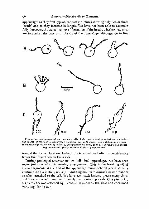
The completely new feature of the unilocular vacuolate cells which phase-contrast microscopy has revealed to us is the property which they have ofputting out one or several appendages. There are long, thin, beaded processeswhich undergo a curious active undulation that may last for many hours.We first observed this phenomenon in a group of such cells in one of ourearly cover-glass preparations which had remained under the microscope forabout 85 min.
We have since seen these appendages on vacuolate cells in preparationsimmediately after the blood had been removed. In fact, such preparations,when there is still a slight flow of fluid, seem to offer the finest view of them,and every individual cell may be seen to have from one to four such appen-dages extending out from the cell body. The length of the appendages varies,from about the diameter of the cell (7 to 9^) up to 4 or 5 times this magnitude,and in exceptional cases even much longer. We have seen one appendage over90 [x. in length.
Observation of individual cells has permitted us to see these beaded
96 Andrew—Blood-cells of Tunicates
appendages as they first appear, as short structures showing only two or three'beads' and as they increase in length. We have not been able to ascertainfully, however, the exact manner of formation of the beads, whether new onesare formed at the base or at the tip of the appendage, although we incline
9:41
FIG. 3. Various aspects of the vacuolate cells of A. atra. A and B, variations in numberand length of the motile processes. The central cell in B shows fragmentation of a process,the detached piece remaining active, c, changes in form of the body of a vacuolate cell occurr-
ing over a short period of time. Positive phase contrast.
toward the former location. Indeed, the terminal bead often is considerablylarger than the others in the series.
During prolonged observations on individual appendages, we have seenmany instances of an interesting phenomenon. This is the breaking off ofseveral segments at the end of the appendage. Such isolated pieces actuallycontinue the distinctive, actively undulating motion in almost the same manneras when attached to the cell. We have seen such isolated pieces many timesand have observed them continuously over various periods. One piece of 5segments became attached by its 'basal' segment to the glass and continued'writhing' for 65 min.

100/U
FIG. 4W. ANDREW

Andrew—Blood-cells of Tunicates 97
The beaded appendages appear to be formed from the cytoplasm of theunilocular vacuolate cells without any participation of the material of thevacuole, which remains undiminished in size. By positive phase-contrastmicroscopy the 'beads' appear black, the very short intervening portions ofthe appendage grey. We have never seen any of the yellow, refractiveappearance of the vacuole in any of the appendages.
It is easily understandable how earlier workers, using direct microscopy,would not have seen the beaded appendages. We have obtained fields in whicha number of such appendages could be seen with the greatest clarity bypositive phase contrast, changed back then to the ordinary optical system,and found them at first to be entirely invisible. Knowing exactly where anappendage is, it can with difficulty be identified without phase contrast, butsurely would be passed over readily.
The other two kinds of vacuolate cells, the bilocular and trilocular ones,resemble the unilocular in the appearance of the vacuolar material. We haveonly occasionally found in one of the locules a granule or concretion like thatwhich is almost constantly present in the unilocular vacuolate cell, and wehave not seen formation of appendages from them, although, since thesecells are far less common, such formation may have been missed by us. Inany event, however, if the appendages do occur on these other vacuolate cells,they would appear to be less common than on the unilocular ones.
The two remaining types of blood-cells are the green cells and the orangecells. The green cells, the most abundant of all of the types, are of about thesame size as the unilocular vacuolate cells. Their most prominent feature,with phase contrast or without, is the presence in each cell of a number ofsomewhat wedge-shaped green bodies (fig. 4, B). These bodies are oftenirregularly placed but may present a sort of blastula-like arrangement, withthe narrower portion of each wedge directed toward a clear central area.
We have not seen any concretions in the green cells nor any granulesexhibiting Brownian motion. In fact, the green material of these cells appearsto be a viscid substance, in contrast to the material in the yacuoles of thevacuolate cells.
Fulton described a transformation of his Q-2 cells into green cells, as aresult of treatment of the cells with weak acid. We studied the effects of suchtreatment, using phase contrast and three different concentrations of aceticacid. A drop of acetic acid, 0-025, °'S> or o-i N, was added to a drop of ascidianblood and the results studied. Preparations were made both with and without
FIG. 4. (plate). Tissue of A. atra, fixed in formalin, stained with Harris's haematoxylinand eosin.
A, interior of a branchial vessel. Two large macrophages are seen near the centre of thefield and a blue cell in the lower left portion.
B, interior of a branchial vessel. Two green cells are present, with a smaller macrophageabove them. The green cell nearest to the centre of the field shows an eccentrically locatednucleus.
c, tunic, showing a small blood-vessel, the lacunae of several of the large vacuolate cells,and the fibrillar appearance of the intercellular material.
2421.1 H

98 Andrew—Blood-cells of Tunicates
a cover-glass. The only clear-cut change which we found in the vacuolatecells was with o-i N acetic acid. Here, there appeared to be a solution of thevacuolar contents, and there was rapid swelling and a pushing of the concre-tion to the periphery of the cell. We were chiefly interested in the effect inthe vacuolate cells and we followed 100 of these cells for periods ranging from5 min. to a half-an-hour, after use of each of the three strengths of acid. Wedid also observe the other cell types during such treatment.
Table 1 shows the changes in different types of cells at the various strengthsof the acid.
TABLE I
Effect on blood cells of A. atra of treatment with acetic acid of different strengths
Normalityof acid
0-025
0-05
o - i
Amoebocytes
Cessationofmotion
Tendencyto with-drawPseudo-pods
Roundingup
Cellular Alterations
Green
No visiblechange
No visiblechange
Accentua-tion ofoutlinesof greenbodieswithincell
Blue
Cessationofmotion
Tendencyto with-drawpseudo-pods
Roundingup
Orange
No visiblechange
No visiblechange
Accentua-tion ofoutlinesof orangebodieswithincell
VacuolateNo visible
change
Moderateswelling
Marked swell-ing. Solu-tion ofvacuolatecontents.Cessationofbrownianmotion.Concretionto peri-phery
The orange cells resemble the green cells in size and form. The contentsof the cell, however, may be in the form of 'clumps' of orange pigment or inthat of more separate granules and fusiform bodies. The clumps are morereminiscent of the bodies within the green cell but are not as definitely wedge-shaped and the 'blastula-like arrangement' has not been seen. The problemof motility of these cells will be discussed later.
A comparative study of the blood of E. turbinata and of C. picta shows greatsimilarities to that of A. atra but also some important differences. Both ofthese species show the three types of amoebocytes, the three kinds of vacuolatecells, and the green cells. They both lack blue cells. E. turbinata also has con-spicuous orange cells in the blood, lacking in C. picta. E. turbinata also hasanother type of cell, an amoeboid cell with its cytoplasm filled with refractive,colourless spheroids which roll over one another as the animal moves. C. pictahas a brown cell in the blood, not seen in either of the other species. This
Andrew—Blood-cells of Tunicates 99
cell has one to three vacuoles containing a brown fluid with granules present-ing Brownian motion.
The green cells of these two colonial species resemble morphologicallythose of A. atra but the green substance itself is far paler and sometimesappears almost clear. The vacuolate cells of these two species also differ some-what from those of the black tunicate. The crescent of the unilocular vacuolatecells is less prominent and the concretion in the vacuole is a less constantfeature.
'Blood-cells' in other tissues of tunicates
We have confined our study of the blood-cells, or perhaps it would bebetter to say 'the blood-cell types', to tissues in which these may be dis-tinguished in the living state. A concentration of orange cells on the branchialpapillae of A. atra was seen in all the specimens which we examined. Suchcells were very seldom identified, except within blood-channels, in the tunicof this same animal, while in E. turbinata the tunic shows numerous largeorange cells which constitute the 'pigment cells' lending colour to the zooids.The orange 'cells' here are several times the size of those in the blood, havelarge numbers of fusiform orange granules, and send processes out widelyinto the surrounding tissue. While we were able to identify cell types in othertissues, we have been able to observe cell behaviour only in the tunic.
Cell motion seems to be at a minimum in the thick tunic of A. atra but tobe very active in the tunic of both E. turbinata and C. picta. The preparationsof the tunic of A. atra were made by cutting thin sections through its thicknessand placing these on a cover-glass in a drop of sea-water. With the invertedmicroscope it was not necessary to place a cover-glass over the preparation.
Studied in this way with low power, the main mass of the tunic, all excepta thin layer at the periphery, shows an almost transparent mass with the bluecells and greyish, fibroblast-like cells, scattered through it. Examined underhigher powers, the transparent mass resolves itself into a mass of very largespheroidal cells which appear rather closely packed together. The substanceof these cells is of an extreme clarity. In many of them a narrow crescent oflight grey appearance can be seen. In nearly every one of them a concretioncan be seen, strongly reminiscent of those in the unilocular vacuolate cells ofthe blood. The only difference is a greater tendency to some elongation orirregularity of the concretion in the cells of the tunic.
Closer study shows that a few of the tunic cells actually are bilocular ortrilocular in character, adding further to their resemblance to the vacuolatecells of the blood.
The blue cells of the tunic resemble those of the blood in general appearanceand in range of variation in the aspect of their granules. They seem, however,to be 'fixed cells'. We have been unable to see any motion whatever in them.Indeed, no motion by any cells outside the blood-channels has been seen inthe tunic of this ascidian.
In the colonial species the thin, transparent tunic, just removed and placed

ioo Andrew—Blood-cells of Tunicates
in sea-water on a cover-glass, is a scene of great activity. All the types ofblood-cells which are seen to move in preparations of the blood are seen herealso and all are in motion. The green cells in the tunic are more active thanthose in preparations of the blood and indeed are fully as active as the largeamoebocytes. Their activity helps to demonstrate the viscid character of thegreen substance, which flows with the cytoplasm, slowly separates intodroplets by constrictions of the cell-body, and often shows slow re-fusion ofsuch droplets, as of those of an oil of a thick consistency.
In the tunic of the two colonial species we have seen none of the large,clear, spheroidal cells so conspicuous in that of A. atra. The matrix is almostentirely clear, but a few delicate fibres are seen scattered through it in irregularfashion.
Blood of tadpoles of Ecteinascidia turbinata
In the tadpoles of E. turbinata the blood-vascular system and circulatingblood are seen in the living animal. Free blood was readily obtained byseparation of the tail from the trunk under the binocular dissecting micro-scope. Study of cover-glass preparations reveals the presence of small amoebo-cytes (lymphocytes), amoeboid compartment (reticulated) cells, green cells,and orange cells. These types of cells show the same kinds of motion as inblood from the adult specimens. Large amoebocytes and vacuolar cells appearto be absent.
A point of some interest was the finding in the clear tail-fins of the tadpolesof cells which have cytoplasm almost identical in appearance with the amoe-boid compartment cells of the blood but which are fixed cells, as determinedby prolonged observation. These cells also show a definite orientation inrelation to the delicate fibres near to them, such that processes of the cellsrun parallel and in close relation to such fibres.
DISCUSSION
The variation in the appearance of the large amoebocytes is noteworthy.We have described these cells as having in many cases a rather homogeneouscytoplasm, in others showing granules, lipid droplets, and even phagocytizedcells. We have also pointed out that the cells with more inclusions generallyshow broad sheets of clear ectoplasm. While the differences probably are of afunctional nature, it seems important to stress their occurrence. We are in-clined to agree with George's change from the expression used in his 1930paper, 'coarsely granular amoebocytes', to that used in his 1939 paper,'macrophages*; for these cells surely are reminiscent, in structure and apparentfunction, of the macrophages of vertebrates, which they resemble also in thevariety of aspect which they present.
Such large amoebocytes, with their differences in functional aspect, evidentlyare represented in Fulton's classification by the A-i and A-2 cells, which heindicates as being of the same general size (10 to 15 JX in diameter in the
Andrew—Blood-cells of Tunicates 101
living state) and as differing chiefly in the granularity of the cytoplasm of theA-2 cells.
A feature of the amoeboid motion of the large amoebocytes which hasseemed prominent to us is the frequency of occurrence of a sharply pointedpseudopod oriented in the direction of travel. This is so definite in these cellsthat while progressing they may almost be said to show an 'anterior' and a'posterior' extremity. Not infrequently the pointed end will develop a flexurea few JU, back from its tip, and as it turns the remainder of the body will follow.Thus the direction of the point will indicate the direction in which the cell isprogressing or toward which it is turning. A cell may go for considerabledistances without changing its shape greatly. On the other hand, it may restrather quietly in one area and repeatedly put out blunt, bleb-like pseudopods,undergoing an almost constant transformation of shape and appearance.
The small amoebocyte, or finely granular amoebocyte of George's earlierwork, later called by him the lymphocyte (George, 1939) seems to correspondin Fulton's classification to the A-3 cell, which is 8 to 9 fi, in diameter in theliving state.
These cells resemble the lymphocytes of vertebrates not only in appearancebut in manner of progression. During amoeboid motion they elongate buthave a blunter 'anterior' end. The nucleus, as with the mammalian lymphocyte,is 'dragged' in the posterior end.
Our own third type of amoebocyte, the coarsely reticulated type, seems tohave no parallel in Fulton's classification. We cannot doubt, however, that itdoes correspond to George's 'amoeboid compartment cell' (George, 1930,figs. 9, 10, 53, 63). This is in spite of the fact that to George these cells were'hyaline when alive and unstained'. He used weak neutral red in sea-water,which brought out the reticulated cytoplasm and angular vacuoles after abouthalf-an-hour. By phase contrast these features, ordinarily invisible, are seenvery readily. Such cells possess both blunt and pointed pseudopods, formedfrom an outer layer of non-reticulated cytoplasm (ectoplasm).
Perhaps our most interesting finding in relation to amoeboid compartmentcells is the discovery of their counterpart as 'fixed tissue' cells in at least onelocation, namely, in the tail-fin of tadpoles of E. turbinata, where we havedescribed them (see above) as in intimate relationship with the fibres.
The relation between cells of the blood and fixed cells in the tissues becomesof much interest in regard to the blue cells and orange cells. In A. atra it isthe blue cells that give the bluish-black colour to the tunic. Fulton (1920, p.409) says: 'As far as can be ascertained there is no essential difference mor-phologically between the blue cells in the blood and the pigment cells in thetest.' He does state also, however, that the pseudopodia of the test cells arenever seen to contract. This we have confirmed in prolonged observations onthin pieces of test. To our mind such a difference is rather an important one,in that we seem to be dealing, in the test, with a fixed type of cell, which,therefore, is not the same as the blood-cell but rather its fixed counterpart.
The group of cell types which we have described as a series, namely the

102 Andrew—Blood-cells of Tunicates
unilocular, bilocular, and trilocular vacuolate cells were treated in two quitedifferent ways by Fulton and by George. Fulton, as we have seen, did considerthem as a series, which he designated as the Quiescent Cells (Q-i, Q-2, Q-3,and Q-4). He recognized the large vacuole of the unilocular type (his Q-i andQ-2) and the frequent presence of a small concretion within the vacuole.This concretion he called the 'excretory granule' and claimed to have seen itin process of elimination from the cell on several occasions. He pictures the.'granule' as a somewhat rectangular structure, while we have found it almostalways to be spheroidal. We have watched some of these cells for several hoursbut have not been able to observe 'excretion' of the granule. Certainly thegranule moves about rather freely in the liquid contents of the vacuole.
The bilocular and trilocular vacuolate elements were recognized by Fulton,and also later by George, to be individual cells, but George does not relatethem to the unilocular form. Rather, he describes the unilocular vacuolatecells as 'signet-ring cells', has no special category for bilocular cells, anddescribes as 'colorless morula' cells the cells which we have spoken of as 'tri-locular vacuolate'. While we do not feel that Fulton's system of naming thesecell types by letters and numbers is a good one, we also feel that the terms'signet-ring' and 'colorless morula' are not adequate descriptions of thesecell types. The latter term, indeed, is misleading, since, although withoutthat intent, it certainly would seem to indicate a group of several cells.
The striking feature of the unilocular cells, the active beaded appendagesseen by phase contrast, were not seen by the earlier workers. What theirsignificance may be, it is difficult even to speculate. It seems not unlikely tous, however, that they might have some 'usefulness' or function even in thecirculating blood. The cells themselves possibly might require a stabilizationmechanism, particularly as they do show a definite polarity. It is even tempt-ing to speculate here on a possibly equilibratory function of the concretion.In any event, we do not think it likely that these appendages represent simplyan 'abnorrnal' phenomenon in extravasated blood.
The apparent 'vitality' of these appendages is rather surprising, for we havefound an occasional one in motion over 24 h after extravasation. We haveseen such appendages undergo a separation of the terminal segments, a smallchain of cytoplasmic beads thus being freed into the water. Such a chaincontinues its active undulating motion. In one case we continued to observea chain of 5 'beads' for 65 min after its separation from the long appendageof a cell. In this case, and in a number of others, the proximal 'bead' of theisolated chain was seen to be attached to the glass, while the rest of its lengthcontinued to undulate.
One of the characteristics of the beaded processes suggests a possiblefunction for them. This is the property of adhesiveness of the individual beads,which causes them to stick to the substrate. Such adhesiveness reminds us ofthe same characteristic as is found in the blood-platelets of higher vertebrates.The 'beads' resemble the platelets also in being fragments of cytoplasm de-rived from cells but continuing a certain independent existence after separation

Andrew—Blood-cells of Tunicates 103
from the parent cell. Whether the role of these cytoplasmic fragments intunicates may be related to clotting of blood, as in vertebrates, is a questionwhich will require experimental studies for its elucidation.
Our study of the histological structure of the fresh tunic has indicated arather striking resemblance between the vacuolate cells of the blood and thevery large, so-called 'bladder-cells' of the tunic. The chordoid type of cellis found in the tunic of many ascidians (Seeliger & Hartmeyer, 1911), butof the three studied here only A. atra possesses such cells. These 'bladder-cells' (Blasenzellen of German authors) are actually connective tissue, or, wemight even say, skeletal elements, for they form the bulk of the tunic. Theyare much like the chorda cells of various invertebrate forms and the noto-chordal cells of the vertebrate embryo. By far the greater part of the cell is ahuge, fluid-filled vacuole. A narrow crescent of cytoplasm can be made outon many of them but is lacking or invisible on others. Of the greatest interestis the almost constant presence in each cell of a small concretion in the vacuole,as in the vacuolate cells of the blood. The concretion in the tunic cells is moreoften placed peripherally in the vacuole and more frequently somewhatelongated than in the blood-cell. An occasional cell of the tunic shows two orthree 'lobes', as though an incomplete fusion of vacuoles had occurred.
Rapkine and Damboviceanu (1925), working with A. mentula and usingindicators and the micro-injection technique, found the pH of the vacuolatecells and green cells to be much lower than that of the other blood cells (2-8as compared to 6-o). The pH of the large vesicular elements of the tunic(bladder-cells) was very nearly 2-0.
The subject of the cell population of the ascidian mantle is a complex one.Saint Hilaire (1931), in a description of 31 species of ascidians, has listed nofewer than 14 types of cells of the tunic. In any one species, however, thenumber of types may not be great, and in A. atra we have identified, outsidethe blood-vessels, only the large 'bladder-cells', blue cells, finely granularamoebocytes (lymphocytes), and coarsely granular amoebocytes (macro-phages).
Huus (1937) states that little is known about the extent to which the cellspenetrating into the tunic are already differentiated ones or are young cellforms which differentiate into the special cell types only after settling down inthe tunic.
The nature of the cells in the ascidian mantle is of particular interestbecause of the presence of 'animal cellulose' (tunicin) in it. Our knowledgeof this substance has been confirmed and amplified by several authors (SaintHilaire, 1931; Grassman, Zechmeister, Toth, & Stadler, 1933). The celluloseis present in the interstitial matrix of the tunic and seems to permeate the fibres.
Henze thought that there might well be a relation between the vanadiumchromogens of the blood-cells and the formation of cellulose—that the vana-dium chromogen represents a strong reducing system and might be analogousin this way to the mechanism of the chloroplasts, making use of carbondioxide. Winterstein (1909) indicated that carbon dioxide is used or bound

104 Andrew—Blood-cells of Tunicates
by the blood, since there is such a small content in the plasma. Florkin (1934)also found less carbon dioxide in the blood than in sea-water.
There is some reason, then, for linking the unusual chemical constitution ofthe blood with the presence of cellulose in the tunic. A migration of cells fromthe blood into the substance of the tunic might account for the transport ofmaterials picked up in the blood.
The remaining two types of cells common to the blood of all three speciesare the green cells and the orange cells. Fulton (1920) speaks of the green cellsas ' . . . in a perfectly quiescent state'; while George (1930), speaking of thesame species, A. atra, calls them 'noticeably amoeboid'.
We have observed motion of the green cells many times. The viscous natureof the green material, in fact, is well seen in the way in which it flows into thepseudopodia. It always maintains a sharply defined interface with the cyto-plasm. The green substance blackens readily with osmium tetroxide anddisappears if a little 1 % acetic acid is added to the preparation. It seems to bea fatty, probably nutritive substance.
Fulton (1920) described the orange cells as showing a 'rapid swimmingmotion', while George (1930) described them as amoeboid. We have observedthe formation of 'pseudopods' or extensions from the cell-body, blunt pro-cesses which may project several /x.
In regard to a 'swimming motion', we have watched orange cells many timesand have found them often to show a slow but free movement through thefluid while surrounding cells of other types were either entirely quiescent orundergoing amoeboid motion on the glass. On a few of these cells we havemade out a long, thin structure which resembles a solitary cilium or flagellumbut whether it really is a distinctive 'swimming organ' remains problematical.We have never seen orange cells undergoing progressive amoeboid motion.
The possible relationship between the vacuolate cells and the green cellsis a subject on which Fulton (1920) and George (1930) differed completely.According to Fulton, a weak solution of an acid added to a 'fresh smear' ofascidian blood caused some of the vacuolate cells (his Q-2 cells and someof the larger Q-i cells) to take on a green tinge and to develop lines of divisionor furrows. In this way, green cells were supposed to develop from thevacuolate cells. Fulton believes that this is the normal course of events—thatgreen cells arise from the vacuolate cells.
Of the supposed transformation of vacuolate into green cells, George (1930,p. 390) said that it 'does not seem tenable in the light of our present knowledgeof the structure of ascidian blood cells'.
Our own results, derived from experiments with three different concen-trations of acetic acid (table 1), have given no evidence of transformation ofvacuolate cells but have shown a rather ready tendency of the contents of thevacuoles to dissolve in a medium more acid than that of the blood. Thedisappearance of the contents of the vacuole leaves a swollen cell with an'empty' appearance and with its concretion, now motionless, in a peripheralposition.

Andrew—Blood-cells of Tunicates 105
This work was carried out at the Bermuda Biological Station during thesummer of 1959. The author is sincerely grateful to Dr. William Sutcliffe,Director of the Station, and his staff for many courtesies. Appreciation isdue to Mr. James F. Glore, Medical Artist at Indiana University, for aid withthe illustrations. The research was supported by U.S. Public Health ServiceGrant, H-4330.
REFERENCES
BERRILL, N. J., 1932. 'Ascidians of the Bermudas.' Biol. Bull., 62, 77.CUENOT, L., 1891. 'fhudes sur le sang et les glandes lymphatiques dans la serie animale
(2e Partie: Invertebres).' Arch. Zool. exp. gen., 9, 13.FLORKIN, M., 1934. 'Sur un caractere souvent mal interpret du milieu interieur des Ascidies.'
C.R. Soc. Biol., Paris, 117, 1226.FULTON, J. F., 1920. 'The blood of Ascidia atra Lesueur; with special reference to pigmenta-
tion and phagocytosis.' Acta Zoologica, Arg. I, 3, 381.GEORGE, W. C, 1926. 'The histology of the blood of Perophora viridis (ascidian).' J. Morph.
Physiol., 41, 311., 1930. 'The histology of the blood of some Bermuda ascidians.' Ibid., 49, 385., 1939. 'A comparative study of the blood of the tunicates.' Quart. J. Micr. Sci., 81, 391.
GRASSMANN, W., ZECHMEISTER, L., T6TH, L. & STADLER, R., 1933. 'Uber den enzy-matischen Abbau der Cellulose und ihrer Spaltprodukte. II. Mitteilung uber enzy-matische Spaltung von Polysacchariden.' Liebig's Annalen der Chemie, 503, 167.
HECHT, S., 1918. 'The physiology of Ascidia atra Lesueur. III. The blood system.' Amer. J.Physiol., 45, 157.
HENZE, M., 1911. 'Untersuchungen uber das Blut der Ascidien. I. Die Vanadiumverbindungder Blutkorperchen.' Z. physiol. Chem., 72, 494.1912. 'Untersuchungen uber das Blut der Ascidien.' II. Ibid., 79, 215.1913. 'Untersuchungen uber das Blut der Ascidien. III. Mitteilung. Ibid., 86, 340.
Huus, J., 1937. Ascidiaceae-Tethyodeae, in Handbuch der Zoologie, edited by Kiikenthal andKrumbach, vol. 5, 2 Halfte. Das Blut, p. 614; Testa oder Zellulosemantel, p. 556. Berlin(Gruyter).
RAPKINE, L., & DAMBOVICEANU, A., 1925. 'Sur le pH interieur de certains elements du sanget de la tunique chez Ascidia mentula.' C.R. Soc. Biol. Paris, 93, 1427.
SAINT-HILAIRE, K., 1931. 'Morphogenetische Untersuchungen des Ascidienmantels.'Zool. Jahrb. 54, 435.
SEELIOER, O., & HARTMEYER, R., 191 I . Tunicata (Manteltiere), I. Abteilung. Die Appen-dicularien und Ascidien, in H. G. Bronn's Klassen und Ordnungen des Tierreichs, vol. 3,Supplement, Der dussere Celhdosemantel, p. 210; Das Blut, p. 553.
VAN NAME, W. G., 1902. 'The ascidians of the Bermuda Islands.' Trans. Conn. Acad. Artsand Sci. 11, 325.
WINTERSTEIN, H., 1909. 'Zur Kenntnis der Blutgase wirbelloser Seetiere.' Biochem. Z,,19. 384-