phenalenone-type phytoalexins mediate resistance of … · phenalenone-type phytoalexins mediate...
TRANSCRIPT
Phenalenone-type phytoalexins mediate resistance ofbanana plants (Musa spp.) to the burrowing nematodeRadopholus similisDirk Hölschera,b,1,2, Suganthagunthalam Dhakshinamoorthyc,1, Theodore Alexandrovd,e,f,g, Michael Beckerh,Tom Bretschneideri, Andreas Buerkertb, Anna C. Creceliusj, Dirk De Waelec,k, Annemie Elsenl, David G. Heckelm,Heike Heklaun, Christian Hertwecki, Marco Kaio, Katrin Knopj, Christoph Krafftp, Ravi K. Maddulao, Christian Matthäusp,Jürgen Poppp,q, Bernd Schneidera, Ulrich S. Schubertj,r, Richard A. Sikoras, Aleš Svatošo, and Rony L. Swennenc,t,u
aNuclear Magnetic Resonance Research Group, oMass Spectrometry Research Group, mDepartment of Entomology, Max Planck Institute for Chemical Ecology,07745 Jena, Germany; bDepartment of Organic Plant Production and Agroecosystems Research in the Tropics and Subtropics, University of Kassel, 37213Witzenhausen, Germany; cLaboratory of Tropical Crop Improvement, Division of Crop Biotechnics, Faculty of Bioscience Engineering, Department ofBiosystems, University of Leuven, 3001 Leuven, Belgium; dCenter for Industrial Mathematics, Department of Mathematics, University of Bremen, 28359Bremen, Germany; eSteinbeis Innovation Center for Scientific Computing in Life Sciences, 28359 Bremen, Germany; fScientific Computing in Life Sciences(SCiLS) GmbH, 28359 Bremen, Germany; gSkaggs School of Pharmacy and Pharmaceutical Science, University of California, San Diego, La Jolla, CA 92093;hDepartment of Applications Matrix-Assisted Laser Desorption/Ionization - Time of Flight (MALDI-TOF), Bruker Daltonik GmbH, 28359 Bremen, Germany;iDepartment of Biomolecular Chemistry, Leibniz Institute for Natural Product Research and Infection Biology, 07745 Jena, Germany; jLaboratory of Organicand Macromolecular Chemistry, Jena Center for Soft Matter, qInstitute for Physical Chemistry and Abbe Center of Photonics, Department of Chemistry andEarth Sciences, Friedrich Schiller University of Jena, 07743 Jena, Germany; kUnit of Environmental Sciences and Management, Department of Nematology,North-West University, Potchefstroom 2520, South Africa; lDepartment of Research and Development, Soil Service of Belgium, 3001 Leuven Heverlee,Belgium; nDepartment of Geobotany, Martin Luther University of Halle-Wittenberg, 06108 Halle, Germany; pWorkgroup Spectroscopy/Imaging, Institute ofPhotonic Technology, 07745 Jena, Germany; rDutch Polymer Institute, 5612 AB, Eindhoven, The Netherlands; sInstitute of Crop Science and ResourceConservation-Phytomedicine, Rheinische Friedrich-Wilhelms-Universität Bonn, 53115 Bonn, Germany; tBioversity International, 3001 Leuven, Belgium; anduInternational Institute of Tropical Agriculture, Croydon CR9 3EE, United Kingdom
Edited by Jerrold Meinwald, Cornell University, Ithaca, NY, and approved November 14, 2013 (received for review August 8, 2013)
The global yield of bananas—one of the most important foodcrops—is severely hampered by parasites, such as nematodes,which cause yield losses up to 75%. Plant–nematode interactionsof two banana cultivars differing in susceptibility to Radopholussimilis were investigated by combining the conventional and spa-tially resolved analytical techniques 1H NMR spectroscopy, matrix-free UV-laser desorption/ionization mass spectrometric imaging, andRaman microspectroscopy. This innovative combination of analyticaltechniques was applied to isolate, identify, and locate the banana-specific type of phytoalexins, phenylphenalenones, in the R. similis-caused lesions of the plants. The striking antinematode activity ofthe phenylphenalenone anigorufone, its ingestion by the nema-tode, and its subsequent localization in lipid droplets within thenematode is reported. The importance of varying local concentra-tions of these specialized metabolites in infected plant tissues, theirinvolvement in the plant’s defense system, and derived strategiesfor improving banana resistance are highlighted.
plant protection | induced plant defense | matrix-free LDI-MSI
Bananas and plantains (Musa spp.) are among the world’smost important food and cash crops, with a global pro-
duction of about 138 million tons in 2010. These crops are part ofa well-balanced human diet and are a major food staple for morethan 400 million people in the tropics (1, 2). About 82% of theworld’s banana production is consumed locally, particularly inIndia, China, and many African countries (Table S1) (1, 2). Exportof bananas to the northern hemisphere represents an importantsource of employment in countries such as Costa Rica, Ecuador,Colombia, and the Philippines (Table S2) (1, 2). Banana yields areseverely hampered by fungi, insects, and plant-parasitic nematodes.The burrowing nematode, Radopholus similis (Cobb, 1893) Thorne,1949, is the key nematode pathogen, causing yield losses up to75% (3). R. similis is found in all major banana-producingregions of the world; its best-known hosts are bananas, blackpepper, Citrus spp. (4), and coffee (5). R. similis causes extensiveroot lesions that can lead to toppling of banana plants (6).Plant-parasitic nematodes have been effectively managed
through the use of nematicides. However, their high toxicity hasadverse effects on humans and their toxic residues are known to
accumulate through nontarget organisms in the food chain (7).After the withdrawal of many effective nematicides, such as methylbromide, from the market (8), organophosphate and carbamatenematicides are still intensively applied to banana and thereforecontinue to threaten the health of agricultural workers and theenvironment (9). Although several biological control approaches,including the application of both single and multiple controlorganisms—such as Fusarium oxysporum, Paecilomyces lilacinus,Trichoderma atroviride isolates, and Bacillus firmus—have provedpromising under greenhouse conditions, the control they conferto banana plants most probably does not protect plants for morethan one cycle in the field, and most of these organisms have yetto be tested under field conditions (10).
Significance
The ongoing decline of banana yields caused by pathogens andthe use of toxic chemicals to manage them has attracted con-siderable attention because of the importance of bananas asa major staple food for more than 400 million people. We dem-onstrate that secondary metabolites (phenylphenalenones) ofMusa are the reason for differences in cultivar resistance, anddetected the phenylphenalenone anigorufone in greater con-centrations in lesions in roots of a nematode-resistant cultivarthan in those of a susceptible one. An in vitro bioassay identifiedanigorufone as the most active nematostatic and nematocidalcompound. We discovered that large lipid–anigorufone complexdroplets are formed in the bodies of Radopholus similis exposedto anigorufone, resulting in the nematode being killed.
Author contributions: D.H., S.D., A.B., D.G.H., B.S., R.A.S., and R.L.S. designed research; D.H.,S.D., M.B., T.B., A.C.C., H.H., M.K., K.K., R.K.M., and C.M. performed research; D.H., S.D., T.A.,T.B., A.C.C., D.D.W., A.E., M.K., K.K., C.K., C.M., J.P., U.S.S., and A.S. analyzed data; and D.H.,S.D., T.A., T.B., A.C.C., D.D.W., A.E., C.H., M.K., K.K., C.K., C.M., J.P., B.S., U.S.S., R.A.S., A.S.,and R.L.S. wrote the paper.
The authors declare no conflict of interest.
This article is a PNAS Direct Submission.1D.H. and S.D. contributed equally to this work.2To whom correspondence should be addressed. E-mail: [email protected].
This article contains supporting information online at www.pnas.org/lookup/suppl/doi:10.1073/pnas.1314168110/-/DCSupplemental.
www.pnas.org/cgi/doi/10.1073/pnas.1314168110 PNAS | January 7, 2014 | vol. 111 | no. 1 | 105–110
AGRICU
LTURA
LSC
IENCE
S
The in-depth investigation of the plant–nematode interactionsat the cellular and molecular level could lead to the developmentof more rational and efficient control strategies (11). The pro-duction of toxic, herbivore-deterrent or -repellent secondary meta-bolites, which is typical for many plant defense systems, is particu-larly interesting in this context. Musa cultivars resistant to R. similishave been identified, especially the cultivar Yangambi km5 (Ykm5)(12). Histochemical and ultrastructural investigations of lesionscaused by R. similis in Ykm5 revealed the accumulation of phenoliccompounds in response to infection (13). Unfortunately, many ofthese studies were based solely on histochemical staining methodsand did not identify the chemical structures of nematicidal sec-ondary metabolites (7, 14, 15). Initial phytochemical analyses of R.similis-infected roots of the Musa cultivar Pisang sipulu identifiedthe phenylphenalenone anigorufone (1) as a phytoalexin producedin response to nematode damage and confirmed earlier suggestionsof the significant role of phytoalexins in the plant defense system(16). Phenylphenalenones are a group of special phenylpropanoid-derived natural products (17), which are known as Musaceae phy-toalexins (18). The activity of phenylalanine ammonia lyase (EC4.3.15), the entry-point enzyme of the phenylpropanoid pathway, iscorrelated to the biosynthesis of specific phenylpropanoids involvedin defense and was substantially induced in nematode infected rootsof Ykm5 (19). Phenylphenalenone-related compounds show bi-ological activity against bacteria, fungi, algae, and diatoms (18, 20–22). The formation of these compounds has been elicited in bananaleaves by Mycosphaerella fijiensis (Black Sigatoka leaf streak dis-ease), in the fruit peels by Colletotrichum musae (anthracnose dis-ease), and in roots and rhizomes by F. oxysporum f. sp. cubense(Panama disease) and R. similis (16, 18, 21, 23).
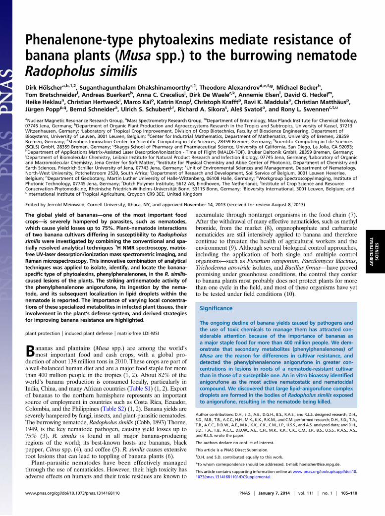
ResultsRoot Damage and Root Lesions. In the greenhouse, potted GrandeNaine (GN) and Ykm5 plants were inoculated with R. similis.Roots of 20-wk-old plants were monitored for nematode damage12 wk after infection. Root damage was more severe in GN thanin Ykm5. Visual observation of infected GN roots revealed large,continuous, and tunnel-like lesions (Fig. 1 A and B). Moreover,root damage extended to the root bases near the GN corms (Fig.1C). Newly formed root lesions were light red in color. Olderlesions were dark red-brownish. In Ykm5, no corm infection wasobserved and both newly formed and older root lesions weresmall, discontinuous, dark red-brown in color, and nonexpanding(Fig. 1 D and E). Uninfected roots of Ykm5 and GN, used ascontrols, showed no lesions and appeared healthy upon visualinspection.
Isolation and Identification of Metabolites. R. similis lesions fromGN and Ykm5 roots were manually dissected from infected rootsunder the microscope, and metabolites were extracted from le-sion material using ethanol. The intensely red-colored lesionswere easily visually distinguishable from healthy, beige-coloredroot tissues. Equal amounts of roots from uninfected plants wereused as controls. Liquid-liquid separation from all of the abovefour samples resulted in CHCl3, ethyl acetate, 1-butanol, andaqueous subfractions. Preliminary 1H NMR analysis of sub-fractions of the R. similis-infected GN and Ykm5 root materialrevealed signals in the aromatic range of the spectrum of theCHCl3 extract, whereas the other subfractions did not show ar-omatic signals of notable intensity. The CHCl3 subfractions werefurther analyzed for the occurrence of phenylphenalenones. Ina first step, the CHCl3 subfractions of all of the samples werepurified by semipreparative HPLC. The individually collectedfractions were subjected to 1H NMR spectroscopy for identifi-cation and the structures of all compounds were identified astypical metabolites and major phytoalexins of Musa species (Fig.S1) (18, 21, 23). The phytochemical profiles and the number ofsecondary metabolites in GN and Ykm5 (Fig. S1) were slightly
different. Anigorufone (1), hydroxyanigorufone (2), irenolone(5), 4-phenyl-1H,3H-benzo[de]isochromene-1,3-dione (7), and4-(4-hydroxyphenyl)-1H,3H-benzo[de]isochromene-1,3-dione (8)were detectable in the extracts of lesions from both cultivars.(2S,3R)-2,3-Dihydro-2,3-dihydroxy-9-phenylphenalen-1-one (3),isoanigorufone (4), and anigorootin (9) were exclusively found inYkm5, and methylirenolone (6) only in GN. Twice as muchanigorufone (1), the major specialized metabolite of lesions inboth cultivars, was found in Ykm5 compared with GN. Addi-tionally, all minor compounds were present in smaller amountsper kilogram of root material in GN compared with Ykm5. Theamounts of the isolated compounds are shown in Table S3.
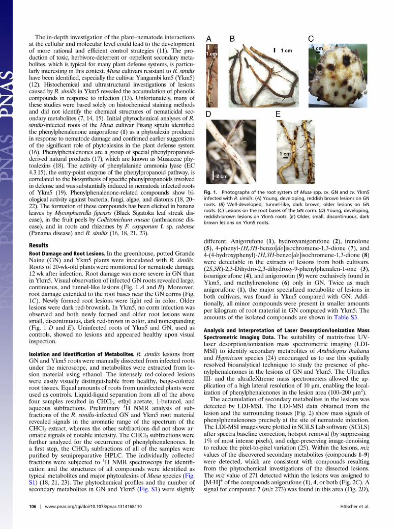
Analysis and Interpretation of Laser Desorption/Ionization MassSpectrometric Imaging Data. The suitability of matrix-free UV-laser desorption/ionization mass spectrometric imaging (LDI-MSI) to identify secondary metabolites of Arabidopsis thalianaand Hypericum species (24) encouraged us to use this spatiallyresolved bioanalytical technique to study the presence of phe-nylphenalenones in the lesions of GN and Ykm5. The UltraflexIII- and the ultrafleXtreme mass spectrometers allowed the ap-plication of a high lateral resolution of 10 μm, enabling the local-ization of phenylphenalenones in the lesion area (100–200 μm2).The accumulation of secondary metabolites in the lesions was
detected by LDI-MSI. The LDI-MSI data obtained from thelesion and the surrounding tissues (Fig. 2) show mass signals ofphenylphenalenones precisely at the site of nematode infection.The LDI-MSI images were plotted in SCiLS Lab software (SCiLS)after spectra baseline correction, hotspot removal (by suppressing1% of most intense pixels), and edge-preserving image-denoisingto reduce the pixel-to-pixel variation (25). Within the lesions, m/zvalues of the discovered secondary metabolites (compounds 1–9)were detected, which are consistent with compounds resultingfrom the phytochemical investigations of the dissected lesions.The m/z value of 271 detected within the lesions was assigned to[M-H]+ of the compounds anigorufone (1), 4, or both (Fig. 2C). Asignal for compound 7 (m/z 273) was found in this area (Fig. 2D),
Fig. 1. Photographs of the root system of Musa spp. cv. GN and cv. Ykm5infected with R. similis. (A) Young, developing, reddish brown lesions on GNroots. (B) Well-developed, tunnel-like, dark brown, older lesions on GNroots. (C) Lesions on the root bases of the GN corm. (D) Young, developing,reddish-brown lesions on Ykm5 roots. (E) Older, small, discontinuous, darkbrown lesions on Ykm5 roots.
106 | www.pnas.org/cgi/doi/10.1073/pnas.1314168110 Hölscher et al.
as well as signals of compound 2 and its 4-phenyl analog 5 (m/z287) (Fig. 2E). The signalm/z 289 was attributable to the secondarymetabolites 3, 8, or both (Fig. 2F). A signal for the O-methylphenylphenalenone (6) of m/z 301 was also detectable (Fig. 2G).Furthermore, using the ultrafleXtreme, we were able to detecta signal for the dimer 9 at m/z 573.The areas of the lesions were strikingly different from the
healthy, uninfected surrounding regions with regard to the pre-sence of secondary metabolites, both in the isolation experimentsand in the LDI-MSI investigations. Nevertheless, an elevated ionsignal background in single LDI measurements caused by highlaser intensities was observed within the uninfected regions ofthe tissue. As the color mapping indicates only the signal in-tensity at specific raster points and not the occurrence of a cer-tain compound, color signals appear where only background isdetected (Fig. S2).
Effect of Phenylphenalenones in R. similis Motility Bioassay. To testfor antinematode properties of the phenylphenalenone-type phy-toalexins, an in vitro bioassay on the motility of R. similis wasperformed with 13 selected phenylphenalenones and relatedcompounds (Fig. S1 and Table S4). The percentage of nematodemotility inhibition (nematostatic effect) was monitored after 24,48, and 72 h of exposure to the compounds.At both tested concentrations (50 and 100 ppm), anigorufone
(1), and compounds 2 and 13 showed a nematostatic effect as of24 h of incubation. This effect was either constant (compound13) or increased over time up to 72 h (1 and 2). Anigorufone (1)showed the strongest activity, with 89% of the nematodes be-coming immotile after 72 h of incubation. Similar nematostaticeffects were observed for compounds 2, 4, 8, and 4-hydroxy-2-methoxy-9-phenylphenalenone (11) at a concentration of 100
ppm. Exposure to 50 ppm resulted in higher percentages ofimmotile nematodes of 48 h of exposure, in comparison with the1% ethanol control. The compounds 5, 9, methoxyanigorufone(10), musanolone C (12), and perinaphthenone (15) revealedinconsistent nematostatic effects. Methylirenolone (6) and dihy-droxyanigorootin (14) did not show any substantial inhibition ofnematode motility (not included in Table S4). The solvent (1%ethanol) exhibited a negligible effect on nematode motility com-parable to sterile distilled water.
Dosage Effect of Anigorufone (1) on R. similis Motility Bioassay.Among the phenalenones tested in the R. similis motility bio-assay, anigorufone (1) was the most active compound at a con-centration of 100 ppm (Table S4). Therefore, anigorufone (1)was selected for a more detailed investigation of its nematostaticpotential. The data obtained at 10, 20, 40, 50, and 100 ppmclearly showed a concentration-dependent effect of anigorufone(1) on nematode motility (Fig. S3). At an incubation time of 24 h,the percentage of immotile versus the total number of nematodesincreased from nonsignificant 26%, at a concentration of 10 ppm,to significant 50.2% at 40 ppm and 74.3% at 100 ppm. Hence,significantly (P < 0.05) higher levels of nematostatic effects wereobserved from 40 ppm upwards compared with the control (1%ethanol). Increasing the concentration to 150 ppm resulted inpartial precipitation of anigorufone (1). Compared with the val-ues obtained after 24 h, the motility of nematodes further de-creased at incubation times of 48 and 72 h (Fig. S3). Theconcentration of anigorufone (1), which inhibited the motility of50% of the nematodes (IC50) in the bioassay, was 59 μg/mL for anincubation time of 24 h. For an exposure time of 48 h, IC50 was38 μg/mL, and only 23 μg/mL for 72 h of exposure.
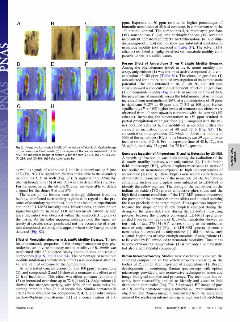
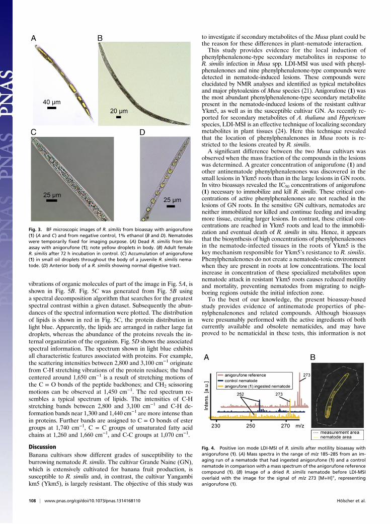
Nematode Ingestion of Anigorufone (1) and Its Detection by LDI-MSI.A surprising observation was made during the evaluation of theR. similis motility bioassay with anigorufone (1). Under brightfield microscopy (BF), yellow droplets were seen in parts ofthe bodies of nematodes exposed to high concentrations ofanigorufone (1) (Fig. 3). These droplets were readily visible becauseof the natural transparency of the nematode cuticle. Nematodescontaining such yellow droplets were subjected to LDI-MSI toidentify the yellow pigment. The drying of the nematodes on theindium tin oxide (ITO)-coated conductive glass slides and thehigh-field vacuum conditions of the LDI-MSI process preservedthe position of the nematodes on the slides and allowed pointingthe laser precisely at the target region. This aspect was importantbecause the shape of the yellow droplets changed during thedrying on the glass slides and during the LDI-MSI measuringprocess, because the droplets converged. LDI-MSI spectra re-corded from yellow regions of R. similis nematodes showed anion peak of m/z 273 [M+H]+, corresponding to the molecularmass of anigorufone (1) (Fig. 4). LDI-MSI spectra of controlnematodes not exposed to anigorufone (1) did not show sucha signal. Ingestions of large enough amounts of anigorufone (1)to be visible by BF always led to nematode mortality. Thus, it hasbecome obvious that anigorufone (1) is not only a nematostaticbut also a nematocidal compound.
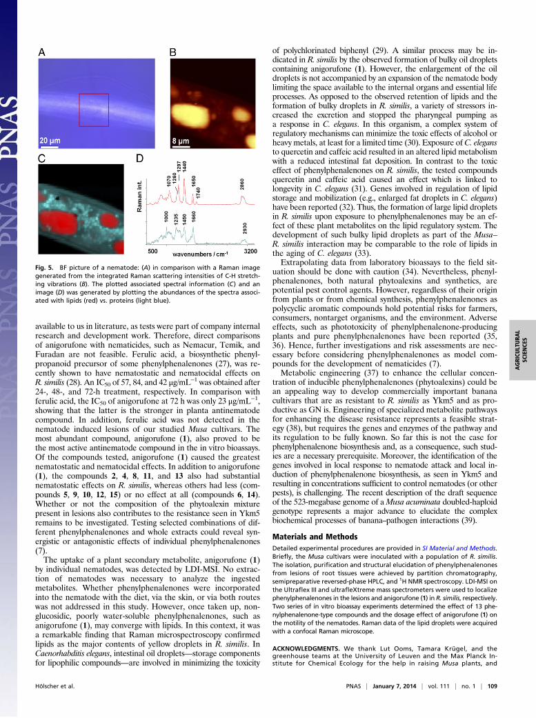
Raman Microspectroscopy. Studies were conducted to analyze thechemical composition of the yellow droplets appearing in thebodies of R. similis after ingestion of anigorufone (1). Recentdevelopments in combining Raman spectroscopy with opticalmicroscopy provided a new noninvasive technique to assess andimage biological samples and processes. The technique has re-cently been successfully applied to identify and visualize lipiddroplets in nematodes (26). Fig. 5A shows a BF image of partof a R. similis nematode using a 60×/NA = 1 water-immersionobjective. The Raman image, reconstructed from the integratedareas of the scattering intensities originating from C-H stretching
Fig. 2. Negative ion mode LDI-MSI of the lesions of Ykm5. (A) Optical imageof the lesions on Ykm5 roots. (B) The region of the lesions subjected to LDI-MSI. The molecular image of section B for the m/z (C) 271, (D) 273, (E) 287,(F) 289, and (G) 301. (H) False color scale bar.
Hölscher et al. PNAS | January 7, 2014 | vol. 111 | no. 1 | 107
AGRICU
LTURA
LSC
IENCE
S
vibrations of organic molecules of part of the image in Fig. 5A, isshown in Fig. 5B. Fig. 5C was generated from Fig. 5B usinga spectral decomposition algorithm that searches for the greatestspectral contrast within a given dataset. Subsequently the abun-dances of the spectral information were plotted. The distributionof lipids is shown in red in Fig. 5C, the protein distribution inlight blue. Apparently, the lipids are arranged in rather large fatdroplets, whereas the abundance of the proteins reveals the in-ternal organization of the organism. Fig. 5D shows the associatedspectral information. The spectrum shown in light blue exhibitsall characteristic features associated with proteins. For example,the scattering intensities between 2,800 and 3,100 cm−1 originatefrom C-H stretching vibrations of the protein residues; the bandcentered around 1,650 cm−1 is a result of stretching motions ofthe C = O bonds of the peptide backbones; and CH2 scissoringmotions can be observed at 1,450 cm−1. The red spectrum re-sembles a typical spectrum of lipids. The intensities of C-Hstretching bands between 2,800 and 3,100 cm−1 and C-H de-formation bands near 1,300 and 1,440 cm−1 are more intense thanin proteins. Further bands are assigned to C = O bonds of estergroups at 1,740 cm−1, C = C groups of unsaturated fatty acidchains at 1,260 and 1,660 cm−1, and C-C groups at 1,070 cm−1.
DiscussionBanana cultivars show different grades of susceptibility to theburrowing nematode R. similis. The cultivar Grande Naine (GN),which is extensively cultivated for banana fruit production, issusceptible to R. similis and, in contrast, the cultivar Yangambikm5 (Ykm5), is largely resistant. The objective of this study was
to investigate if secondary metabolites of theMusa plant could bethe reason for these differences in plant–nematode interaction.This study provides evidence for the local induction of
phenylphenalenone-type secondary metabolites in response toR. similis infection in Musa spp. LDI-MSI was used with phenyl-phenalenones and nine phenylphenalenone-type compounds weredetected in nematode-induced lesions. These compounds wereelucidated by NMR analyses and identified as typical metabolitesand major phytoalexins of Musa species (21). Anigorufone (1) wasthe most abundant phenylphenalenone-type secondary metabolitepresent in the nematode-induced lesions of the resistant cultivarYkm5, as well as in the susceptible cultivar GN. As recently re-ported for secondary metabolites of A. thaliana and Hypericumspecies, LDI-MSI is an effective technique of localizing secondarymetabolites in plant tissues (24). Here this technique revealedthat the location of phenylphenalenones in Musa roots is re-stricted to the lesions created by R. similis.A significant difference between the two Musa cultivars was
observed when the mass fraction of the compounds in the lesionswas determined. A greater concentration of anigorufone (1) andother antinematode phenylphenalenones was discovered in thesmall lesions in Ykm5 roots than in the large lesions in GN roots.In vitro bioassays revealed the IC50 concentrations of anigorufone(1) necessary to immobilize and kill R. similis. These critical con-centrations of active phenylphenalenones are not reached in thelesions of GN roots. In the sensitive GN cultivars, nematodes areneither immobilized nor killed and continue feeding and invadingmore tissue, creating larger lesions. In contrast, these critical con-centrations are reached in Ykm5 roots and lead to the immobili-zation and eventual death of R. similis in situ. Hence, it appearsthat the biosynthesis of high concentrations of phenylphenalenonesin the nematode-infected tissues in the roots of Ykm5 is thekey mechanism responsible for Ykm5’s resistance to R. similis.Phenylphenalenones do not create a nematode-toxic environmentwhen they are present in roots at low concentrations. The localincrease in concentration of these specialized metabolites uponnematode attack in resistant Ykm5 roots causes reduced motilityand mortality, preventing nematodes from migrating to neigh-boring regions outside the initial infection zone.To the best of our knowledge, the present bioassay-based
study provides evidence of antinematode properties of phe-nylphenalenones and related compounds. Although bioassayswere presumably performed with the active ingredients of bothcurrently available and obsolete nematicides, and may haveproved to be nematicidal in these tests, this information is not
Fig. 3. BF microscopic images of R. similis from bioassay with anigorufone(1) (A and C) and from negative control, 1% ethanol (B and D). Nematodeswere temporarily fixed for imaging purpose. (A) Dead R. similis from bio-assay with anigorufone (1); note yellow droplets in body. (B) Adult femaleR. similis after 72 h incubation in control. (C) Accumulation of anigorufone(1) in small oil droplets throughout the body of a juvenile R. similis nema-tode. (D) Anterior body of a R. similis showing normal digestive tract.
Fig. 4. Positive ion mode LDI-MSI of R. similis after motility bioassay withanigorufone (1). (A) Mass spectra in the range of m/z 185–285 from an im-aging run of a nematode that had ingested anigorufone (1) and a controlnematode in comparison with a mass spectrum of the anigorufone referencecompound (1). (B) Image of a dried R. similis nematode before LDI-MSIoverlaid with the image for the signal of m/z 273 [M+H]+, representinganigorufone (1).
108 | www.pnas.org/cgi/doi/10.1073/pnas.1314168110 Hölscher et al.
available to us in literature, as tests were part of company internalresearch and development work. Therefore, direct comparisonsof anigorufone with nematicides, such as Nemacur, Temik, andFuradan are not feasible. Ferulic acid, a biosynthetic phenyl-propanoid precursor of some phenylphenalenones (27), was re-cently shown to have nematostatic and nematocidal effects onR. similis (28). An IC50 of 57, 84, and 42 μg/mL−1 was obtained after24-, 48-, and 72-h treatment, respectively. In comparison withferulic acid, the IC50 of anigorufone at 72 h was only 23 μg/mL−1,showing that the latter is the stronger in planta antinematodecompound. In addition, ferulic acid was not detected in thenematode induced lesions of our studied Musa cultivars. Themost abundant compound, anigorufone (1), also proved to bethe most active antinematode compound in the in vitro bioassays.Of the compounds tested, anigorufone (1) caused the greatestnematostatic and nematocidal effects. In addition to anigorufone(1), the compounds 2, 4, 8, 11, and 13 also had substantialnematostatic effects on R. similis, whereas others had less (com-pounds 5, 9, 10, 12, 15) or no effect at all (compounds 6, 14).Whether or not the composition of the phytoalexin mixturepresent in lesions also contributes to the resistance seen in Ykm5remains to be investigated. Testing selected combinations of dif-ferent phenylphenalenones and whole extracts could reveal syn-ergistic or antagonistic effects of individual phenylphenalenones(7).The uptake of a plant secondary metabolite, anigorufone (1)
by individual nematodes, was detected by LDI-MSI. No extrac-tion of nematodes was necessary to analyze the ingestedmetabolites. Whether phenylphenalenones were incorporatedinto the nematode with the diet, via the skin, or via both routeswas not addressed in this study. However, once taken up, non-glucosidic, poorly water-soluble phenylphenalenones, such asanigorufone (1), may converge with lipids. In this context, it wasa remarkable finding that Raman microspectroscopy confirmedlipids as the major contents of yellow droplets in R. similis. InCaenorhabditis elegans, intestinal oil droplets—storage componentsfor lipophilic compounds—are involved in minimizing the toxicity
of polychlorinated biphenyl (29). A similar process may be in-dicated in R. similis by the observed formation of bulky oil dropletscontaining anigorufone (1). However, the enlargement of the oildroplets is not accompanied by an expansion of the nematode bodylimiting the space available to the internal organs and essential lifeprocesses. As opposed to the observed retention of lipids and theformation of bulky droplets in R. similis, a variety of stressors in-creased the excretion and stopped the pharyngeal pumping asa response in C. elegans. In this organism, a complex system ofregulatory mechanisms can minimize the toxic effects of alcohol orheavy metals, at least for a limited time (30). Exposure of C. elegansto quercetin and caffeic acid resulted in an altered lipid metabolismwith a reduced intestinal fat deposition. In contrast to the toxiceffect of phenylphenalenones on R. similis, the tested compoundsquercetin and caffeic acid caused an effect which is linked tolongevity in C. elegans (31). Genes involved in regulation of lipidstorage and mobilization (e.g., enlarged fat droplets in C. elegans)have been reported (32). Thus, the formation of large lipid dropletsin R. similis upon exposure to phenylphenalenones may be an ef-fect of these plant metabolites on the lipid regulatory system. Thedevelopment of such bulky lipid droplets as part of the Musa–R. similis interaction may be comparable to the role of lipids inthe aging of C. elegans (33).Extrapolating data from laboratory bioassays to the field sit-
uation should be done with caution (34). Nevertheless, phenyl-phenalenones, both natural phytoalexins and synthetics, arepotential pest control agents. However, regardless of their originfrom plants or from chemical synthesis, phenylphenalenones aspolycyclic aromatic compounds hold potential risks for farmers,consumers, nontarget organisms, and the environment. Adverseeffects, such as phototoxicity of phenylphenalenone-producingplants and pure phenylphenalenones have been reported (35,36). Hence, further investigations and risk assessments are nec-essary before considering phenylphenalenones as model com-pounds for the development of nematicides (7).Metabolic engineering (37) to enhance the cellular concen-
tration of inducible phenylphenalenones (phytoalexins) could bean appealing way to develop commercially important bananacultivars that are as resistant to R. similis as Ykm5 and as pro-ductive as GN is. Engineering of specialized metabolite pathwaysfor enhancing the disease resistance represents a feasible strat-egy (38), but requires the genes and enzymes of the pathway andits regulation to be fully known. So far this is not the case forphenylphenalenone biosynthesis and, as a consequence, such stud-ies are a necessary prerequisite. Moreover, the identification of thegenes involved in local response to nematode attack and local in-duction of phenylphenalenone biosynthesis, as seen in Ykm5 andresulting in concentrations sufficient to control nematodes (or otherpests), is challenging. The recent description of the draft sequenceof the 523-megabase genome of aMusa acuminata doubled-haploidgenotype represents a major advance to elucidate the complexbiochemical processes of banana–pathogen interactions (39).
Materials and MethodsDetailed experimental procedures are provided in SI Material and Methods.Briefly, the Musa cultivars were inoculated with a population of R. similis.The isolation, purification and structural elucidation of phenylphenalenonesfrom lesions of root tissues were achieved by partition chromatography,semipreparative reversed-phase HPLC, and 1H NMR spectroscopy. LDI-MSI onthe Ultraflex III and ultrafleXtreme mass spectrometers were used to localizephenylphenalenones in the lesions and anigorufone (1) in R. similis, respectively.Two series of in vitro bioassay experiments determined the effect of 13 phe-nylphenalenone-type compounds and the dosage effect of anigorufone (1) onthe motility of the nematodes. Raman data of the lipid droplets were acquiredwith a confocal Raman microscope.
ACKNOWLEDGMENTS. We thank Lut Ooms, Tamara Krügel, and thegreenhouse teams at the University of Leuven and the Max Planck In-stitute for Chemical Ecology for the help in raising Musa plants, and
Fig. 5. BF picture of a nematode: (A) in comparison with a Raman imagegenerated from the integrated Raman scattering intensities of C-H stretch-ing vibrations (B). The plotted associated spectral information (C) and animage (D) was generated by plotting the abundances of the spectra associ-ated with lipids (red) vs. proteins (light blue).
Hölscher et al. PNAS | January 7, 2014 | vol. 111 | no. 1 | 109
AGRICU
LTURA
LSC
IENCE
S

Alexandra zum Felde for editorial help in the preparation of the manu-script. This study was supported in part by German Research Foundation[Deutsche Forschungsgemeinschaft (DFG)] Grants HO 4380/1 and SCHN 450/10 (to D.H. and B.S.); an Interfaculty Council for Development Cooperation,University of Leuven PhD scholarship (to S.D.); a grant by coopérationeuropéenne dans le domaine de la recherche scientifique et technique
action 872 for a short-term scientific mission to the Max Planck Institutefor Chemical Ecology (S.D.); a grant from the European Union 7th Frame-work Programme (Grant 305259) (to T.A.); the Dutch Polymer Institute(technology area high-throughput experimentation) (U.S.S.); ThüringerMinisterium für Bildung, Wissenschaft, und Kultur Grant B515-07008 (toU.S.S.); and the DFG (U.S.S.).
1. FAOSTAT (2011) Production (Crops) quantities of banana and plantains for 2011. Foodand Agriculture Organization of United Nations. Available at faostat3.fao-org/fao-stat-gateway/go/to/download/Q/QC/E. Accessed October 15, 2013.
2. FAOSTAT (2011) Trade (Crops and livestock products) quantities of banana andplantains for 2011. Food and Agriculture Organization of United Nations. Available atfaostat3.fao-org/faostat-gateway/go/to/download/T/TP/E. Accessed October 15, 2013.
3. Sarah JL (1999) Diseases of Banana, Abacá and Ensete, ed Jones DR (CABI Publishing,Wallingford, United Kingdom), pp 295–303.
4. Moens M, Perry RN (2009) Migratory plant endoparasitic nematodes: A group rich incontrasts and divergence. Annu Rev Phytopathol 47:313–332.
5. Campos VP, Villain L (2005) Plant Parasitic Nematodes in Subtropical and TropicalAgriculture, eds Luc M, Sikora RA, Bridge J (CABI Publishing, Wallingford, UnitedKingdom), 2nd Ed, pp 529–579.
6. Leach R (1958) Blackhead toppling disease of bananas. Nature 181(4603):204–205.7. Chitwood DJ (2002) Phytochemical based strategies for nematode control. Annu Rev
Phytopathol 40:221–249.8. Quénéhervé P, Valette C, Topart P, Tezenas du Montcel H, Salmon F (2009) Nematode
resistance in bananas: Screening results on some wild and cultivated accessions ofMusa spp. Euphytica 165(1):123–126.
9. Wesseling C, et al. (2010) Symptoms of psychological distress and suicidal ideationamong banana workers with a history of poisoning by organophosphate or N-methylcarbamate pesticides. Occup Environ Med 67(11):778–784.
10. zum Felde A, et al. (2009) The nematode burrowing of banana: Strategies for con-trolling the uncontrollable. Acta Hortic 828:101–108.
11. Grunewald W, Cannoot B, Friml J, Gheysen G (2009) Parasitic nematodes modulatePIN-mediated auxin transport to facilitate infection. PLoS Pathog 5(1):e1000266.
12. Dochez C, Whyte M, Tenkouano A, Ortiz R, De Waele D (2005) Response of eastAfrican highland bananas and hybrids to Radopholus similis. Nematology 7(5):655–666.
13. Valette C, Andary C, Geiger JP, Sarah JL, Nicole M (1998) Histochemical and cyto-chemical investigations of phenols in roots of banana infected by the burrowingnematode Radopholus similis. Phytopathology 88(11):1141–1148.
14. Beckman CH (2000) Phenolic-storing cells: Keys to programmed cell death and peri-derm formation in wilt disease resistance and in general defence responses in plants?Physiol Mol Plant Pathol 57(3):101–110.
15. Wuyts N, et al. (2007) Potential physical and chemical barriers to infection by theburrowing nematode Radopholus similis in roots of susceptible and resistant banana(Musa spp.). Plant Pathol 56(5):878–890.
16. Collingborn FMB, Gowen SR, Mueller-Harvey I (2000) Investigations into the bio-chemical basis for nematode resistance in roots of three musa cultivars in response toRadopholus similis infection. J Agric Food Chem 48(11):5297–5301.
17. Cooke RG, Edwards JM (1981) Naturally occurring phenalenones and related com-pounds. Fortschr Chem Org Naturst 40:153–190.
18. Luis JG, Fletcher WQ, Echeverri F, Grillo TA (1994) Phenalenone-type phytoalexinsfrom Musa acuminata—Synthesis of 4-phenyl-phenalenones. Tetrahedron 50(37):10963–10970.
19. Wuyts N, De Waele D, Swennen R (2006) Activity of phenylalanine ammonia-lyase,peroxidase and polyphenol oxidase in roots of banana (Musa acuminata AAA, cvsGrande Naine and Yangambi km5) before and after infection with Radopholus sim-ilis. Nematology 8(2):201–209.
20. Winters K, Batterton JC, Van Baalen C (1977) Phenalen-1-one: Occurrence in a fuel oiland toxicology to microalgae. Environ Sci Technol 11(3):270–272.
21. Otálvaro F, et al. (2007) Phenalenone-type compounds from Musa acuminata var.“Yangambi km 5” (AAA) and their activity againstMycosphaerella fijiensis. J Nat Prod70(5):887–890.
22. Jitsaeng K, Schneider B (2010) Metabolic profiling of Musa acuminata with Spor-obolomyces salmonicolor. Phytochem Lett 3(2):84–87.
23. Hirai N, Ishida H, Koshimizu K (1994) A phenalenone-type phytoalexin from Musaacuminata. Phytochemistry 37(2):383–385.
24. Hölscher D, et al. (2009) Matrix-free UV-laser desorption/ionization (LDI) mass spec-trometric imaging at the single-cell level: Distribution of secondary metabolites ofArabidopsis thaliana and Hypericum species. Plant J 60(5):907–918.
25. Alexandrov T, et al. (2010) Spatial segmentation of imaging mass spectrometry datawith edge-preserving image denoising and clustering. J Proteome Res 9(12):6535–6546.
26. Klapper M, et al. (2011) Fluorescence-based fixative and vital staining of lipid dropletsin Caenorhabditis elegans reveal fat stores using microscopy and flow cytometryapproaches. J Lipid Res 52(6):1281–1293.
27. Schmitt B, Hölscher D, Schneider B (2000) Variability of phenylpropanoid precursors inthe biosynthesis of phenylphenalenones in Anigozanthos preissii. Phytochemistry53(3):331–337.
28. Wuyts N, de Waele D, Swennen R (2006) Effects of plant phenylpropanoid pathwayproducts and selected terpenoids and alkaloids on the behavior of the plant-parasiticnematodes Radopholus similis, Pratylenchus penetrans and Meloidogyne incognita.Nematology 8(1):89–101.
29. Menzel R, et al. (2007) Cytochrome P450s and short-chain dehydrogenases mediatethe toxicogenomic response of PCB52 in the nematode Caenorhabditis elegans. J MolBiol 370(1):1–13.
30. Jones D, Candido EPM (1999) Feeding is inhibited by sublethal concentrations oftoxicants and by heat stress in the nematode Caenorhabditis elegans: Relationship tothe cellular stress resonance. J Exp Zoolog A Comp Exp Biol 284(2):147–157.
31. Pietsch K, et al. (2011) Hormetins, antioxidants and prooxidants: Defining quercetin-,caffeic acid- and rosmarinic acid-mediated life extension in C. elegans. Biogerontology12(4):329–347.
32. Ashrafi K, et al. (2003) Genome-wide RNAi analysis of Caenorhabditis elegans fatregulatory genes. Nature 421(6920):268–272.
33. Hou NS, Taubert S (2012) Function and regulation of lipid biology in Caenorhabditiselegans aging. Front Physiol 3:143.
34. Spence KO, Lewis EE, Perry RN (2008) Host-finding and invasion by entomopathogenicand plant-parasitic nematodes: Evaluating the ability of laboratory bioassays to predictfield results. J Nematol 40(2):93–98.
35. Darwin C (1866) On the Origin of Species by Means of Natural Selection (John Murray,London), 4th British Ed, p 12. Available at darwin-online.org.uk/Variorum/1866/1866-12-dns.html. Accessed November 25, 2013.
36. Flors C, et al. (2006) Phototoxic phytoalexins. Processes that compete with the pho-tosensitized production of singlet oxygen by 9-phenylphenalenones. PhotochemPhotobiol 82(1):95–103.
37. Misawa N (2011) Pathway engineering for functional isoprenoids. Curr Opin Bio-technol 22(5):627–633.
38. Collinge DB, Jørgensen HJL, Lund OS, Lyngkjaer MF (2010) Engineering pathogenresistance in crop plants: Current trends and future prospects. Annu Rev Phytopathol48:269–291.
39. D’Hont A, et al. (2012) The banana (Musa acuminata) genome and the evolution ofmonocotyledonous plants. Nature 488(7410):213–217.
110 | www.pnas.org/cgi/doi/10.1073/pnas.1314168110 Hölscher et al.