phenotyping of transgenic pigs to determine the
TRANSCRIPT

Phenotyping of transgenic pigs to determine the suitability of xenografts in the treatment of
aortic diseases
Strauss E (1,2), Wroblewska J, Wawrzynska L, Tomczak J, Pukacki F, Kruszyna L, Zdun M,
Frackowiak H, Oszkinis G
(1) Poznan University of Medical Sciences &
(2) Institute of Human Genetics, Polish Academy of Sciences

Disclosure of Interest
Speaker name: Ewa Strauss
• I do not have any potential conflict of interest
Financial support:The National Centre for Research and Development grant number INNOMED/I/17 /NCBR/2014 from the Innovative Economy Operational Programme founds, in the framework of the European Regional Development Fund.

BACKGROUND
•The synthetic materials used in the surgical treatmentof aortic diseases can cause graft infection, whichrequires the replacement aortic prostesis with biological material, such as auto- or allografts.
•Due to the shortage of multiorgan donors, many patients with the prosthesis infection cannot be treated successfully
•The domestic pig may be the perfect donor of easily accessible blood vessels for transplantation

AIM
• Under the MEDPIG project a series of „humanized” pigs were generated to omit species incompatibility
• We focused on the ex vivo studies of the aorta & skin grafts from these transgenic pigs, to determine the suitability of transgenic xenografts in the treatment of aortic diseases.
• In a pilot in vivo study functionality of the aortic grafts were tested through cross transplantation(betwee transgenic pigs).

POLITRANSGENIC: GAL, FUT, HLA, ZNF
Human genes that regulate the complement system are inserted by the method of DNA microinjection. The introduction of the human genes may mask the porcine epitopes.
MATERIAL
VESSELS & SKIN FROMTRANSGENIC ANIMALS
DOMESTIC PIGRACE: POLAND LANDRACE
VESSELS & SKIN FROMCONTROL ANIMALS
SAMPLES OF VESSELS & SKIN
HUMAN
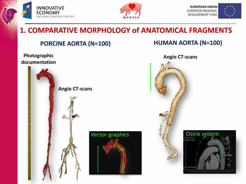
PORCINE AORTA (N=100) HUMAN AORTA (N=100)
1. COMPARATIVE MORPHOLOGY of ANATOMICAL FRAGMENTS
Vector graphics Osirix system
Photographicdocumentation
Angio CT-scans
Angio CT-scans

RESULTS
Other possibilities of xenografts application• Combining fragments of 2 blood vessels• Vascular patches• Non-anatomical prosteis (by-pass)• Acellular matrices
PIG HUMAN
A B D O M I N A LT H O R A C I C
PIG HUMAN
PIG HUMANPIG HUMAN
PIG HUMAN PIG HUMAN
DIA
ME
TE
R
69% P<.0001 57% P<.0001
WA
LL
TH
ICK
NE
SS
82%, P=.009 97%, P=.76 NS
P/H=122 % P<.001 144% P<.0001
LE
NG
HT

There were no histological markers indicating the presence of
atherosclerotic plaques or inflammation
Thoracic aortaDifference in tunica media histology:
• shortened elastic fibers• an increased number of collagen fibers• numerous clusters of smooth muscle cells• unusual presence of blood vessels
Abdominal aortaDifference in tunica media histology:
• increased number of smooth muscle cells, indicating rather the muscle type of arteries
2.1. RESULTS: LIGHT MICROSCOPY (H+E)
2. ULTRASTRUCTURE OF THE AORTA

Morphology of the endothelial cell layer of the porcine aorta
A. Aorta of transgenic pig (this study)Normal porcine endothelia show a continuity and a slight, physiologicalnuclear bulging and alignment in the direction of flow.
Examples of endothelial injury caused by iodinatedcontrast media (data from the literature)*B and C. massive cell bulging, widened irregular intercellular spaces
*- Am J Physiol Renal Physiol 303: F1592–F1598, 2012.
BA C
2.2. RESULTS: SCANNING ELECTRON MICROSCOPY

0
10
20
30
0 20 40
Flexibility Relaxation The curve of the aortic wall tension Texture analyzer
For
ce (N)
Elongation
Nanoidenter Agilent G200
3.2. NANO-SCALE
7 tests performed at different locations
3. BIOMECHANICAL STUDIES
3.1. MACRO-SCALE

Effect of the cryopreservation of pig aortic tissue (period 30-180 days)
H+E staining of fresh and cryopreserved aortas grafts

3.2. RESULTS
The nano-scale mechanical properties of the porcine aorta
(intima & internal elastic membrane)
FRESH
CONTROL TRANSGENIC
ABDOMINALTHORACIC
CONTROL TRANSGENICMO
DU
LUS
OF
ELA
STIC
ITY
[k
Pa]
CRYOPRESERVED
CONTROL TRANSGENIC
ABDOMINALTHORACIC
CONTROL TRANSGENICCONT_T TRANS_T CONT_A TRANS_A
50
100
150
200
RubberSoft tissues
DiamondEngineering materials
CONT_T TRANS_T CONT_A TRANS_A
50
100
150
200
250
Glass
0.01–0.1 GPa 1050 - 1210 GPa
The Young’s modulus [E]

4. PRELIMINARY IN VIVO FUNCTIONAL STUDY (4 TG animals)
Cross-transplantation procedure Two months after surgery
Before transplantation After transplantation
Genetic analysis of imbreding
24 markers
Angio-CT scans

THORACIC AORTA
Aortic EndothelialCell in vitro culture
Molecular and functional validation (20 TG + 20 Control)
Brigt field microscopic image of in vitro primary porcine aortic endothelial cells
acLDL uptake
Fluorescent microscopy image and cytometric analysis of CD31 expression and acLDL uptake
CD31 expression
5. IN VITRO STUDIES OF CYTOTOXICITY

Level of cleavege products of the C3, C4 and C5 components of the complement system in porcineendothelial cells
Relative level of complement-dependant cytotoxicity, measured by LDH release in porcine endothelial cells.
RESULTS: CYTOMETRIC ANALYSIS

TRANSGENIC
vs CONTROL
CONTROL SERUM-
TREATED
vs
CONTROL UNTREATED
TRANSGENIC
SERUM- TREATED
vs
CONTROL
SERUM- TREATED
AFFYMETRIX PORCINE MICROARRAYSPRIMARY PORCINE AORTIC ENDOTHELIAL CELLS
RESULTS: WHOLE-GENOME EXPRESSION ANALYSIS

ACELLULAR BIOMATERIALS – ACELLULAR SKIN GRAFTSTreatment of ischemic wounds
H+E staining of acelluler dermis
SEM image of acellular dermis
• Skin derived from transgenic pigs, submittedto chemical and enzymatical acellularization
• Free from Porcine Endogenous Retroviruses• Radiation sterilization• Temporary dressing
Currently used in this project in the treatment of burn wounds (accidents in the mining industry) and for donor fields.

SUMMARY
• Morphological, histological, mechanical and functionalfeatures of the porcine aorta, indicate the potentialusefulness of aortic transgenic xenografts for the treatment of aortic diseases
• Tissue of poly-transgenic pigs are less immunogenic to human, as compared to those from non-transgenicanimals
• Acellularization and cryopreservation may extend the applicability of tissue of transgenic pigs in medicine
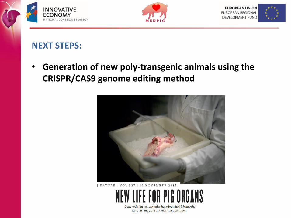
NEXT STEPS:
• Generation of new poly-transgenic animals using the CRISPR/CAS9 genome editing method

SCIENTIFIC COOPERATION
Uniwersytet Przyrodniczy w Poznaniu
INSTITUTE OF HUMAN GENETICS PASProf. Ryszard Słomski
UNIVERSITY OF LIFE SCIENCES IN POZNAN
• Department of anatomy of animalsProf. Hieronim FrąckowiakVet Maciej Zdun
• Department of Biotechnology & MicrobiologyProf. Tomasz JankowskiPhD Wojciech BiałasStudents (1)
TECHNOLOGY PARKWielkopolska Center for Advanced TechnologiesPhD Marek NowickiStudents (1)
NATIONAL RESEARCH INSTITUTE OF ANIMAL PRODUCTION IN BALICE/KRAKOWPhD Vet, Jaroslaw WieczorekProf. Jerzy SmorągPhD Anna Radko
CENTRE FOR EXPERIMENTAL AND INNOVATIVE MEDICINE IN KRAKOWPhD Vet, Agnieszka Pietsch-FulbiszewskaPhD Michał Nowakowski
POZNAN UNIVERSITY OF MEDICAL SCIENCES
• Department of General and Vascular SurgeryProf. MD, Grzegorz OszkinisProf. MD, Fryderyk PukackiPhD MD, Łukasz KruszynaPhD MD, Maciej ZielińskiMD, Hubert StępakMD, Paweł Zawadzki
• Laboratory of Translational MedicinePhD Ewa Strauss PhD med. Joanna WróblewskaMsc Jolanta TomczakMsc Lidia Wawrzyńska
• Department of Histology and EmbriologyProf. Maciej ZabelPhD Agnieszka MalińskaPhD Marcin Ruciński
• Wielkopolska Cancer CentreProf. MD, Andrzej Mackiewicz

Thank you!