phosphatidylserine on viable sperm and phagocytic
TRANSCRIPT

ARTICLE
Phosphatidylserine on viable sperm and phagocyticmachinery in oocytes regulate mammalianfertilizationClaudia M. Rival1,2,3, Wenhao Xu2, Laura S. Shankman1,2, Sho Morioka1,2, Sanja Arandjelovic 1,2,
Chang Sup Lee1,2,4, Karen M. Wheeler3, Ryan P. Smith3, Lisa B. Haney1,2, Brant E. Isakson5, Scott Purcell6,
Jeffrey J. Lysiak1,3* & Kodi S. Ravichandran 1,2,7*
Fertilization is essential for species survival. Although Izumo1 and Juno are critical for initial
interaction between gametes, additional molecules necessary for sperm:egg fusion on both
the sperm and the oocyte remain to be defined. Here, we show that phosphatidylserine
(PtdSer) is exposed on the head region of viable and motile sperm, with PtdSer exposure
progressively increasing during sperm transit through the epididymis. Functionally, masking
phosphatidylserine on sperm via three different approaches inhibits fertilization. On the
oocyte, phosphatidylserine recognition receptors BAI1, CD36, Tim-4, and Mer-TK contribute
to fertilization. Further, oocytes lacking the cytoplasmic ELMO1, or functional disruption of
RAC1 (both of which signal downstream of BAI1/BAI3), also affect sperm entry into oocytes.
Intriguingly, mammalian sperm could fuse with skeletal myoblasts, requiring PtdSer on sperm
and BAI1/3, ELMO2, RAC1 in myoblasts. Collectively, these data identify phosphatidylserine
on viable sperm and PtdSer recognition receptors on oocytes as key players in sperm:egg
fusion.
https://doi.org/10.1038/s41467-019-12406-z OPEN
1 The Center for Cell Clearance, School of Medicine, University of Virginia, 1340 Jefferson Park Avenue, Pinn Hall, Charlottesville, VA 22903, USA.2Department of Microbiology, Immunology, and Cancer Biology, School of Medicine, University of Virginia, 1340 Jefferson Park Avenue, Pinn Hall,Charlottesville, VA 22903, USA. 3 Department of Urology, School of Medicine, University of Virginia, 1340 Jefferson Park Avenue, Pinn Hall, Charlottesville,VA 22903, USA. 4 College of Pharmacy and Research Institute of Pharmaceutical Sciences, Gyeongsang National University, 501 Jinju-daero, Jinju,Gyeongnam 52828, Republic of Korea. 5 Department of Molecular Physiology and Biological Physics, School of Medicine, University of Virginia, 1340Jefferson Park Avenue, Pinn Hall, Charlottesville, VA 22903, USA. 6 Reproductive Medicine and Surgery Center of Virginia, 595 Martha Jefferson Dr.,Charlottesville, VA 22911, USA. 7 Department of Biomedical Molecular Biology, Ghent University, and the UGent-VIB Center for Inflammation Research,Technologiepark 71, 9052 Ghent, Belgium. *email: [email protected]; [email protected]
NATURE COMMUNICATIONS | (2019) 10:4456 | https://doi.org/10.1038/s41467-019-12406-z | www.nature.com/naturecommunications 1
1234
5678
90():,;
Sexual reproduction requires a productive fusion between thehaploid male and female gametes1,2. Prior to the fusion ofthe gametes, a critical step is the proper recognition between
specific ligand(s) on the sperm and appropriate binding partner(s) on the egg. Recent studies both at the functional and structurallevels have unequivocally established a critical role for the spermsurface protein Izumo1 and the corresponding GPI-anchoredreceptor Juno on the oocyte, with blocking or loss of eitherprotein affecting fertilization3,4. Signaling downstream of Juno inoocytes is yet to be defined, as Juno is a GPI-anchored protein.Further, 3D structure studies5,6 suggest that the Izumo1:Junointeraction is unlikely to lead to fusion and, when Izumo1 wasexogenously expressed on Cos-7 cells7, oocyte binding to thesecells occurred but did not proceed to fusion. The tetraspaninfamily member CD9 on the oocyte has also been linked tomammalian fertilization8. CD9 has no known ligand and it isthought to modify the membrane curvature9,10. Thus, it has beensuggested that other players on both sperm and the oocyte likelycontribute toward gamete fusion (after the Izumo1:Juno inter-action)1,2.
Here, using complimentary approaches on the oocyte andsperm, we show that Phosphatidylserine (PtdSer), exposed onviable sperm, is recognized by specific receptors located on themicrovilli of the oocyte to promote sperm:egg fusion. The sig-naling pathway ELMO1/RAC1, downstream of the PtdSerreceptors BAI1/3, also participates in this event. This pathway isalso conserved in the fusion of sperm with myoblasts. Takentogether, our results shed light into the molecular mechanism ofsperm:egg fusion.
ResultsPtdSer exposed on viable sperm is required for fertilization. Aspart of our studies on apoptotic germ cell clearance in the testes11,we noticed Annexin V staining on freshly isolated sperm from thecauda epididymis (Fig. 1a, b), suggestive of PtdSer exposure(Fig. 1c, d). Although PtdSer has been noted on sperm pre-viously12–14, it was considered to mark dead or non-viable spermbecause PtdSer is a key eat-me signal on cells undergoing apop-tosis, which facilitates recognition and uptake by phagocytes15.Interestingly, PtdSer can also be transiently exposed on viablecells in certain conditions (such as activated B and T cells,myoblasts, or macrophages16–19), and therefore, we furtherinvestigated the relevance of PtdSer exposure on the sperm.
Annexin V staining was prominently seen both on the spermhead and the midpiece, but was absent in the tail (Fig. 1c, d).During spermatogenesis, after exiting the testis, sperm transitsthrough different segments of the epididymis: the caput, thecorpus and the cauda (Fig. 1a). Classical experiments have shownthat only the caudal sperm is capable of fertilization20. Therefore,we assessed PtdSer exposure on sperm as it transits through theepididymis. We noticed a progressive increase in PtdSer exposureon sperm isolated from different segments of the epididymis, withthe cauda epididymis, which contains the fertilization-competentsperm, displaying the highest percentage of PtdSer-positive sperm(Fig. 1e). This also indicates that PtdSer externalization is notmerely an effect of sperm isolation. When we addressed whetherPtdSer exposure on sperm changes with capacitation, a processknown to occur in the female reproductive tract21, we found afurther increase in the percentage of PtdSer-positive sperm aftercapacitation in vitro (Supplementary Fig. 1a). The acrosomereaction is another process that occurs on sperm in the femaletract. When we induced the acrosome reaction in vitro with theionophore A23187, PtdSer continued to be detectable on sperm.Further, as Izumo1 is exposed on caudal sperm after theacrosome reaction and is a central player in fertilization4, we
asked whether PtdSer colocalizes with Izumo1 on sperm. Izumo1(detected via antibody), as well as PtdSer (via Annexin V), werecolocalized in the equatorial region of the sperm head (known tobe involved in sperm:oocyte fusion2) (Supplementary Fig. 2). AsAnnexin V can bind both PtdSer and phosphatidylethanolamine(PtdEtn)22, we tested PtdSer exposure on the sperm using asecond reagent. We have previously established that the solubleextracellular fragment of the PtdSer recognition receptor BAI1(BAI1-TSR fused to the glutathione-S-transferase; GST) bindsPtdSer but not PtdEtn23; BAI1-TSR (but not the control GSTprotein) preferentially bound to the sperm head withoutsignificant midpiece binding (Fig. 1f, g). Consistent with thisobservation, when we used Duramycin, which binds PtdEtn butnot PtdSer22, the binding was noted prominently in the midpiecewith much less binding to the head (Supplementary Fig. 1b).These data suggested specific exposure of PtdSer on the heads offreshly isolated sperm from the cauda epididymis.
To address whether the sperm with PtdSer exposed on theirsurface are viable, we performed time-lapse microscopy, revealingthat the PtdSer+ sperm were motile (Fig. 1h, and Supplementalmovie 1). Further, when we analyzed the expression on the caudalsperm of cleaved caspase 3 (CC3), a marker for apoptosis, theAnnexin V+ sperm were negative for CC3 (SupplementaryFig. 1c). Only after 24 h incubation ex vivo, some of the spermshowed CC3 staining (Supplementary Fig. 1d). These datasuggested that PtdSer is exposed on the surface of viable andmotile sperm.
We next asked whether the PtdSer exposure on sperm isfunctionally important for fertilization. To test this, we performedin vitro fertilization using capacitated caudal sperm and oocytesfrom super-ovulated C57BL/6 female mice, and quantified theemergence of two-cell embryos (see schematic in Fig. 1i). Wedeliberately chose three different reagents to target PtdSer, as eachhave unique features that are complementary, and can inform usbetter about the role of PtdSer during fertilization: (1) Annexin V(which has the highest affinity); (2) BAI1-TSR peptide derivedfrom the extracellular region of the PtdSer receptor BAI1 fused tothe GST (lower affinity than Annexin V but greater specificity forPtdSer); and (3) the soluble head group of the lipid phosphati-dylserine, which has the lowest affinity of the three blockingagents but can act as a competitive inhibitor for PtdSerrecognition receptors. Pleasingly, all three reagents supportedthe hypothesis that PtdSer on sperm contributes to fertilization.Annexin V caused > 85% reduction in fertilization in three out offour independent experiments (Fig. 1j, k). Masking PtdSer onsperm with BAI1-TSR also significantly reduced fertilization, andconsistent with the BAI1-TSR being of lower affinity thanannexin V, the inhibition of fertilization with BAI1-TSR was lesspronounced (Fig. 1l). Addition of the soluble Phospho-L-Serinehead group also resulted in significant reduction in fertilization inevery experiment (Fig. 1m, n). The partial inhibition (30–40%)was expected as the monomeric soluble head groups of PtdSerhave to compete against multi-valent PtdSer recognition on thesperm surface. The advantage of using the Phospho-L-serineblocking is that we could directly compare its effect againstthe stereoisomer Phospho-D-Serine as a control (carrying thesame charge), and this did not inhibit fertilization. Of note, weconfirmed that progressive motility of the sperm was not affectedafter masking PtdSer with Annexin V or BAI1-TSR (Supplemen-tary Fig. 1e, f). Collectively, these three approaches suggest thatrecognition of PtdSer on sperm (in addition to the well-describedIzumo1) can contribute to in vitro fertilization.
PtdSer receptors on oocytes contribute to fertilization. To testwhether the surface of oocytes contains potential PtdSer-binding
ARTICLE NATURE COMMUNICATIONS | https://doi.org/10.1038/s41467-019-12406-z
2 NATURE COMMUNICATIONS | (2019) 10:4456 | https://doi.org/10.1038/s41467-019-12406-z | www.nature.com/naturecommunications

sites, we took a simplified approach using the binding of thefluorescently labeled 2 µm carboxylate modified beads (2CMB),which are known to bind Annexin V and compete with PtdSer-exposing apoptotic cells23. Zona pellucida (ZP)-free oocytes wereisolated from wild-type C57BL/6 female mice and were incubatedwith red-fluorescent 2CMB for 2 h. At the end of the incubation
period, the oocytes were stained with CD9 antibody to identifythe microvillar region that is known to interact with sperm, fixed,and analyzed by microscopy (Fig. 2a). Oocytes readily boundmultiple beads, and this interaction was restricted to the micro-villus region of the oocytes (Fig. 2b). Importantly, this bindingwas significantly decreased when the beads were pretreated with
Swim 15 min
Annexin Vstaining
Evaluation bymicroscopy
b
In vitro fertilizationControlor GST
Annexin Vor BAI1-TSR
24 h
Evaluation oftwo-cell embryos
Testis
Caput
Cauda
Annexin V/Hoechst
*
**
* *
*
**
**
* *
*
*
*
*
*
*
*
*
*
*
GST only/Hoechst
% A
nnex
in V
+ s
perm
0
20
40
60
80
Caput Corpus Cauda
***
*
Motility of Annexin V+ sperm
t: 00:04 t: 00:09 t: 00:14
Sperm trajectory
*BAI1-TSR (PtdSer)/Hoechst
Sperm head: Sperm midpiece:
e
(No of eggs)
Control Annexin V
O -Phospho-D-Serine
O -Phospho-L-Serine
h
****
* ** **
*
**
*
* **
(No of eggs)Control(290)
Annexin V(264)
**100
80
60
40
20
0
100
80
60
40
20
0
1.2
1
0.8
0.6
0.4
0.2
0
% F
ertil
ized
egg
s
% F
ertil
ized
egg
s
Fer
tiliz
atio
n in
dex
GST(152)
BAI1-TSR(161)
D-Serine(76)
L-Serine(80)
k l
* ***
**
f g
i j
m n
c da
Cor
pus
Epi
didy
mis
NATURE COMMUNICATIONS | https://doi.org/10.1038/s41467-019-12406-z ARTICLE
NATURE COMMUNICATIONS | (2019) 10:4456 | https://doi.org/10.1038/s41467-019-12406-z | www.nature.com/naturecommunications 3

the PtdSer-masking agent BAI1-TSR (Fig. 2b, c). Although PtdSeris an eat-me signal on apoptotic cells, extensive confocal sec-tioning of the bead-bound oocytes did not reveal obvious inter-nalization of the beads under these conditions. These dataindicated the existence of potential PtdSer-binding molecules inthe microvillar region on the surface of oocytes. As maskingPtdSer on sperm significantly reduced the fertilization rate, wehypothesized that the PtdSer recognition receptors on the oocytesmay contribute to the steps toward sperm:egg fusion.
Several PtdSer recognition receptors with redundant functionshave been identified on phagocytes to engage the PtdSer exposedon the apoptotic targets23–26. Therefore, we hypothesized thatone or more such PtdSer recognition receptor(s) on the oocytesmay engage the sperm during fertilization. In a previousbioinformatics analysis of oocyte genes, members of the BAIfamily as well as CD36 were reported to be expressed in bothmouse and human oocytes27. When we assessed the mRNAexpression of BAI family members and CD36, we found readilydetectable expression of BAI1, BAI3, and CD36 in mouse oocytes(Fig. 2d). BAI members belong to the type II adhesion family ofGPCRs (hence, also referred to as ADGBR family) with longextracellular region containing domains capable of directlybinding PtdSer23,25,28–32; CD36 is a member of the scavengerreceptor family, and has also been linked to the binding ofPtdSer24,33–35. CD36 is also reported to function cooperativelywith BAI1 on endothelial cells36. Immunofluorescence micro-scopy using antibodies, which recognize both BAI1 and BAI3(referred to from here onwards as BAI1/3) or CD36, gave aprominent signal in the sperm-binding microvillar region(Fig. 2e); this staining pattern was also similar to the stainingpreviously noted with concanavalin A37 (Fig. 2e), Juno andCD97,9 (Supplementary Fig. 3a). When we assessed the expres-sion of other known direct PtdSer-binding receptors, we founddetectable expression of the message for Timd4 but not Stab2(Fig. 2d). Among the TAM family of receptors that can alsorecognize PtdSer (indirectly, via the bridging molecules Gas6 orprotein S26,38), Mertk, but not Tyro3 and Axl were noted onoocytes39. Immunohistochemistry of whole mouse ovariesrevealed that BAI1 expression is detectable in oocytes from theearliest stages of folliculogenesis, with positive staining fromprimordial follicles through tertiary follicles (Fig. 2f). Similarly,we could readily detect staining for BAI1/3 and CD36 on humanoocytes (discarded/unused oocytes acquired from clinical in vitrofertilization procedures) (Supplementary Fig. 3b). Furthermore,expression of BAI1/3 on oocytes was detectable via immunohis-tochemistry on tissue sections of human ovary (SupplementaryFig. 3c).
In the efferocytosis field, the PtdSer recognition by PtdSerreceptors is known to include redundant mechanisms, as thecharged head group of the lipid PtdSer can be recognized in apolyvalent fashion by multiple receptors to provide sufficientavidity and specificity38,40,41. Therefore, we decided to test all ofthe five potential PtdSer receptors detected in oocytes—CD36,BAI1, BAI3, Tim-4, and Mer-TK—via approaches that targetthem either singly or in combination. Interestingly, CD36 hasbeen shown to cooperatively function with BAI1 in endothelialcells36. Therefore, to test the potential multi-pronged interactioninvolving both CD36 and BAI1/3, we tested the effect ofantibodies targeting CD36 or BAI1/3 (via antibody thatrecognizes both BAI1/BAI3), either alone or combination (seeschematic in Fig. 2g). Although antibodies to either BAI1/3 orCD36 alone did not inhibit fertilization (Supplementary Fig. 3d),a combination of antibodies targeting both BAI1/3 and CD36caused a reproducible and statistically significant inhibition offertilization in vitro (Fig. 2h, i). As a positive control, antibody toJuno could strongly inhibit fertilization (Supplementary Fig. 3f).Next, we used a more direct assay for the sperm entry intooocytes. During fertilization, the nucleus of the sperm decon-denses after entry into the oocyte cytoplasm42–44. This earlystep of fertilization can be scored using oocytes loaded with theDNA binding dye 4′,6-diamidino-2-phenylindole (DAPI), andthe appearance of DAPI-stained decondensed sperm DNA(Fig. 2j)42–44. After establishing and validating this assay, wetested the efficacy of the BAI1/3- and CD36-blocking antibodieson the sperm entry into the oocyte. We found a 67% decreasein the percentage of oocytes with decondensed sperm DNA(Fig. 2k, l), suggesting that blocking three of the five PtdSerreceptors (BAI1/BAI3, and CD36) expressed on oocytes can alsoimpair fertilization in vitro, complementary to the masking ofPtdSer on the sperm.
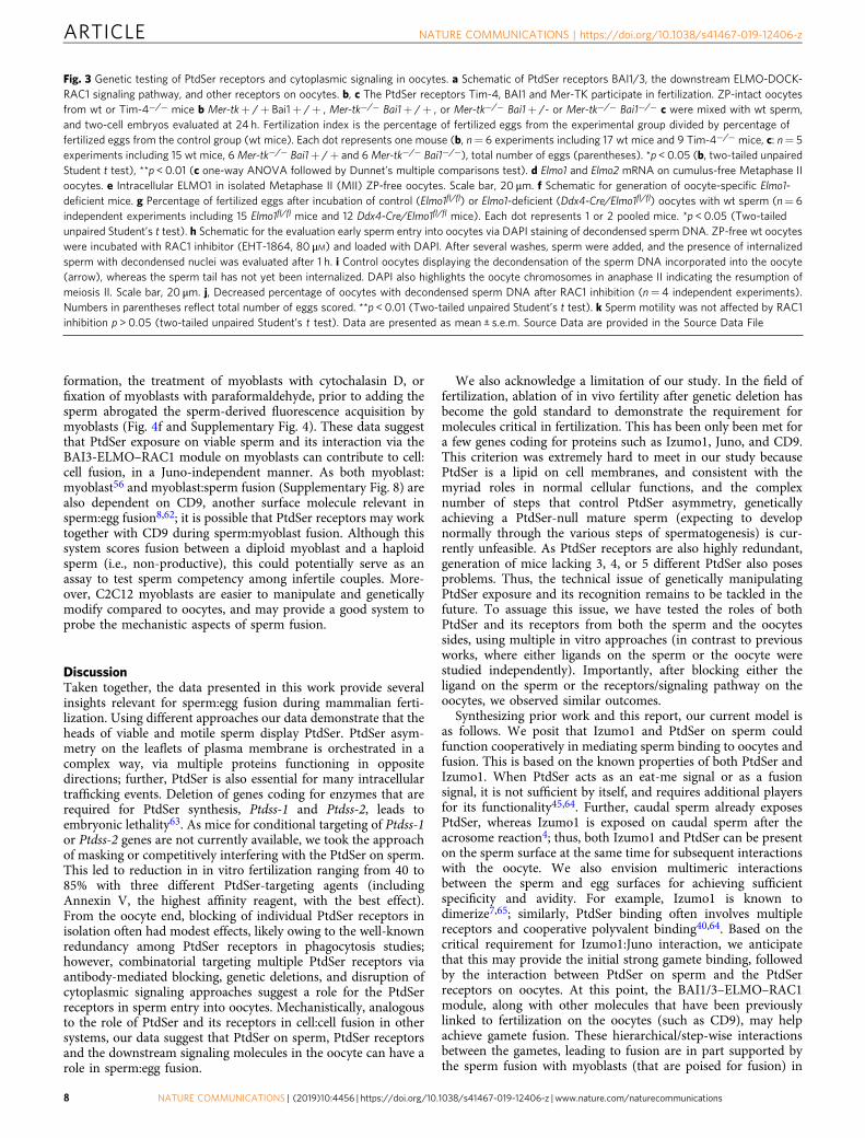
Next, we wanted to genetically test the contribution of oocytePtdSer receptors to fertilization. Because of the extensivefunctional redundancy among PtdSer recognition receptors it iswidely reported that single knockout of PtdSer receptors oftenshow partial defects in apoptotic cell clearance, and defects arebetter revealed by deletion of more than one receptor45–48. Wetested three of the PtdSer receptors expressed on oocytes usingsingle or double knockout mice: Tim-4, BAI1, and Mer-TK. Tim-4 directly binds PtdSer while Mer-TK binds PtdSer indirectlythrough the bridging molecules Gas6 or Protein S [note: oocytesalso express Gas639]. Mice deficient in Tim-4 alone showed amodest but statistically significant reduction in the percentage offertilized eggs (Fig. 3b). We then tested the role ofMertk and Bai1genetically. We performed in vitro fertilization assays with
Fig. 1 Phosphatidylserine on live sperm is important for in vitro fertilization. a Depiction of the mouse testis and epididymis. b Sperm from different regionsof the epididymis were allowed to swim/disperse in TYH+ BSA medium, stained with Annexin V and Hoechst, and evaluated by microscopy. c, d AnnexinV staining of sperm. Asterisks denote sperm heads, and arrows midpiece. Scale bar, 20 μm. e Percentage of Annexin V + sperm from the caput (n= 9mice), corpus (n= 8 mice), and cauda (n= 15 mice) epididymis, with each dot representing one mouse (six independent experiments). *p < 0.05, ***p <0.001 (one-way non parametric ANOVA was followed by Kruskal–Wallis test for multiple comparisons). f, g, Sperm from cauda epididymis were incubatedwith 50 μg/ml GST only f or GST-BAI1-TSR g, washed, fixed and visualized by GST immunofluorescence. Scale bar, 20 μm. h Snap shots of a moviedepicting motility of live Annexin V + (green) sperm (t: time in min). The trajectory of a single sperm is traced by a white dotted line. Scale bar, 30 μm.i Schematic of the in vitro fertilization assay: cumulus oocyte complexes isolated from wt super-ovulated females were incubated with caudal spermpreviously capacitated, in the presence or absence of 10 μg/ml Annexin V, 50 μg/ml GST, or 50 μg/ml GST-BAI1-TSR. The percentage of fertilized eggs(two-cell embryos) was evaluated after 24 h. j Multiple two-cell embryos fertilization with control sperm (left panel, arrows), whereas fewer fertilized eggswere observed with Annexin V (right panel, arrows). Scale bar, 100 μm. k, l Annexin V masking of PtdSer on sperm (k, n= 4 experiments) or GST-BAI1-TSR (l, n= 4 experiments) reduces fertilization. The total number of eggs analyzed is shown in parentheses. Each line represents one experiment and thematching experiments are shown (shape and color). Error bars are s.e.m. *p < 0.05, **p < 0.01 (Two-tailed unpaired Student’s t test). m, n Greaterunfertilized oocytes (asterisks) seen after competition with O-Phospho-L-Serine compared with O-Phospho-D-Serine (top). Scale bar, 100 μm. Each linerepresents one experiment and matching experiments are shown with the same shape and color (n= 4 experiments). Error bars are s.e.m. **p < 0.01 (Two-tailed unpaired Student’s t test). Source Data are provided in the Source Data File
ARTICLE NATURE COMMUNICATIONS | https://doi.org/10.1038/s41467-019-12406-z
4 NATURE COMMUNICATIONS | (2019) 10:4456 | https://doi.org/10.1038/s41467-019-12406-z | www.nature.com/naturecommunications

oocytes isolated either from Mertk−/− or Bai1−/− mice, or micedouble deficient for both Mertk and Bai1. Although oocytesisolated from single deficient mice (either Mertk−/− or Bai1−/−)could be fertilized similar to wild-type eggs (Fig. 3c andSupplementary Fig. 3e), the double deficient Mertk−/−Bai1−/−
oocytes show a significant reduction in fertilization (Fig. 3c).Collectively, these data suggest that even with the considerableredundancy among the PtdSer receptor family, a statisticallysignificant effect can be observed in in vitro fertilization withoocytes deficient in specific PtdSer recognition receptors.
Zona pellucida-free oocytes
2 μm carboxylatemodified beads
(2CMB,PtdSer mimic)
2 hCD9 +
Hoechststaining
& fix± GST
orBAI1-TSR
Evaluation of2CMB
binding tooocytes
g h
Untreatedor
isotype Ig
CD36 Ab+
BAI1/3 Ab
wt oocytes
In vitro fertilization
24 h
(No of oocytes)
a b Beads CD9 Hoechst
Untreated BAI1-TSR
c
Microvilli (ConA)Hoechst
Mouse metaphase II oocytes Mouse ovaries
BAI1/3 Hoechst
CD36 Hoechst BAI1/3 CD36 Hoechst
BAI1/3 Connexin43 Hoechst
BAI1/3 Hematoxilind
e f
j k
Control CD36 + BAI1/3 Abs
1° follicles
2° follicles
3° follicles
wt oocytes
(No of eggs)
mIgA+ sIgG(307)
CD36 +BAI1/3 Abs
(310)
Fer
tiliz
atio
n in
dex
mIgA+ sIgG
(72)
CD36 +BAI1/3 Abs
(84)(No of eggs)
% O
ocyt
es w
ithde
cond
ense
d sp
erm
DN
A
i
l DAPI-loaded fertilized oocyte
Sperm DNA
Evaluationof two-cellembryos
Untreatedor
isotype Ig
CD36 Ab+
BAI1/3 Ab
DAPI-loadedwt oocytes
In vitro fertilization
3 h
Evaluationof DAPI+ sperm
DNAdecondensation
in oocytes
Sperm DNA
% o
ocyt
es w
ith>
5 be
ads/
oocy
te
Untreated(76)
GST(75)
BAI1-TSR(72)
mR
NA
leve
ls(r
elat
ive
to G
AP
DH
)
0.0
0.2
0.4
0.6
0.8
1.0
1.2
0
20
40
60
80
**
**
0
20
40
60
80
100
***
0.000
0.005
0.010
0.015
0.020
nd nd
Bai1Bai2Bai3
Timd4
Stab2
cd36
NATURE COMMUNICATIONS | https://doi.org/10.1038/s41467-019-12406-z ARTICLE
NATURE COMMUNICATIONS | (2019) 10:4456 | https://doi.org/10.1038/s41467-019-12406-z | www.nature.com/naturecommunications 5

The BAI1/3-ELMO1-Rac1 signaling axis affects fertilization.With respect to signaling downstream of PtdSer recognitionreceptors, CD36 has a rather short cytoplasmic tail without anobvious direct signaling, and CD36 can cooperatively signal withBAI136. Among the PtdSer receptors, signaling downstream ofthe BAI family members is one of the best characterized23,25,49.Both BAI1 and BAI3 have long cytoplasmic tails that associatewith the adapter proteins ELMO1 and/or ELMO2 (depending onthe cell type), with subsequent signaling (in complex Dock familyproteins) and activation of the small GTPase RAC123,49–52. GTP-bound active RAC1 promotes actin cytoskeletal remodelingduring adhesion, phagocytosis, and cell:cell fusion events(Fig. 3a). In oocytes, we detected both Elmo1 and Elmo2 mRNA,with Elmo1 expression higher than Elmo2 (Fig. 3d). At theprotein level, ELMO1 expression was readily detected byimmunofluorescence in isolated oocytes (Fig. 3e). To assessthe importance of ELMO1 in fertilization, we crossed mice car-rying floxed Elmo1 alleles (Elmo1fl/fl) with Ddx4-Cre mice53,which express the Cre recombinase specifically in oocytes fromthe earliest stages (Fig. 3f). We super-ovulated the Ddx4-Cre/Elmo1fl/fl female mice, isolated the oocytes, and performedin vitro fertilization assays using caudal sperm from wild-typemice. We noted a significant reduction in in vitro fertilizationwith oocytes from Ddx4-Cre/Elmo1fl/fl mice, compared withcontrol mice (Fig. 3g). The partial reduction is consistent withthe continued expression of ELMO2, which can substitute forELMO111. Incidentally, female nematodes lacking the ELMOhomolog ced-12 have been shown to have lower fecundity, withfewer progeny produced51.
ELMO proteins (together with Dock family members) functionas upstream activators of the small GTPase RAC1, whichregulates actin cytoskeletal rearrangements51 (Fig. 3a). Asgenetically testing the requirement for RAC1 is not feasibleowing to the role of RAC1 during oocyte development and othersteps after the sperm entry54, we took a pharmacologicalapproach and used the sperm DNA decondensation assay tomore directly score the sperm entry into oocytes. We treatedoocytes with the RAC1 inhibitor EHT-1864 (see methods) andalso added EHT-1864 during co-incubation of sperm and oocytes(note that both sperm and oocytes were harvested from wild-typemice) (Fig. 3h, i). EHT-1864 caused a significant reduction in thenumber of oocytes with decondensed sperm DNA (Fig. 3j).Importantly, this effect did not appear to be owing to the RAC1inhibitor affecting sperm, as sperm incubated with EHT-1864alone under the assay conditions showed no reduction in themotility (Fig. 3k). These data suggest that oocyte BAI1/3 andCD36, as well as the ELMO–RAC1 signaling module downstreamof BAI1/3 contribute to the functional steps of fertilization.
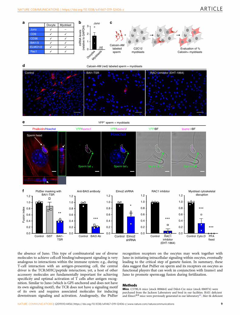
PtdSer-dependent fusion of sperm with skeletal myoblasts. Ourresults up to this point suggest that PtdSer on the sperm and itsreceptors BAI1/3, CD36, Tim-4, and Mer-TK on the oocyte canpromote fusion via the ELMO–RAC1 signaling pathway. Ourlaboratory and others19,25,49 have previously demonstrated thatPtdSer exposure on skeletal myoblasts is important for the fusionbetween myoblasts to form myotubes, and this occurs in a BAI1/3–ELMO–RAC1-dependent manner19,25,49,55. Intriguingly, whenwe examined the expression of genes linked to the sperm:eggfusion in myoblasts, we found that oocytes and myoblasts bothexpressed the membrane proteins CD9, CD36, BAI1, and BAI3,as well as cytoplasmic ELMO2, and RAC1 (Fig. 4a)8,25,49,56–59,whereas Juno expression was not detected in myoblasts (Fig. 4b).Therefore, we asked whether caudal sperm could fuse with ske-letal myoblasts, as it has been shown with other somatic cells60,61,and whether this Juno-independent fusion was mediated by theBAI1-ELMO1-RAC1 module expressed by myoblasts.
We labeled caudal sperm with a red-fluorescent cytoplasmicdye (Calcein-AM)60, incubated them with C2C12 mousemyoblasts, and looked for myoblasts that acquire the sperm-derived Calcein-AM staining (Fig. 4c). Of note, there was nomyoblast:myoblast fusion when they were in growth medium in anon-confluent state. However, some of these myoblasts areknown to be poised for fusion25. Remarkably, we could readilydetect transfer of sperm-derived Calcein-AM into few of themyoblasts in a quantifiable manner (Fig. 4d). To further addressthe fusion between sperm and skeletal myoblasts, we took threeadditional approaches. First, labeling sperm with another dye(DiI) produced similar results as scored by fusion with myoblasts(Supplementary Fig. 4). Second, we isolated sperm from miceexpressing transgenic yellow fluorescent protein (YFP) and, afterincubating them with myoblasts, we detected for YFP/GFP (greenfluorescent protein) and the sperm-specific protein Izumo1within the fusing myoblasts by immunostaining. We couldreadily observe the signal for YFP, and the sperm specific proteinIzumo1, in addition to the DNA from the sperm head within themyoblasts (Fig. 4e). As a third approach, we used electronmicroscopy to detect the presence of sperm within myoblasts.The midpieces (containing multiple mitochondria), and the tailsfrom multiple sperm could be detected inside the cytoplasm of amyoblast (Supplementary Fig. 5c). Of note, we do not detect anyobvious membrane surrounding the sperm structures, suggestingthat the sperm is not contained within a phagocytic vesicle.Interestingly, aminophospholipid asymmetry on myoblasts differsfrom fibroblasts16, and this may, in part, explain sperm fusionwith myoblasts but not fibroblasts (not shown). As C2C12myoblasts are an immortalized cell line, we also tested whethersperm could fuse with cultured mouse primary myoblasts, which
Fig. 2 BAI1/3 and CD36 are expressed on oocytes and contribute to fertilization. a Schematic for the assay to determine PtdSer recognition moieties onmouse oocytes. b Oocytes-bound multiple untreated carboxylate beads within the microvillar region (left), but not the BAI1-TSR-treated beads (right).Scale bar, 20 μm. c Summary of three experiments using carboxylate binding oocytes. ***p < 0.001 (Two-tailed unpaired Student’s t-test). d, PtdSerexpression (qPCR) on cumulus-free Metaphase II oocytes from wild-type female mice (n= 4 experiments). e Concanavalin A (ConA) staining (markingmicrovilli) overlap with staining for BAI1/3 and CD36 on ZP-free oocytes. Scale bar, 20 μm. f Immunofluorescence (top) and immunohistochemistry(bottom) staining of mouse ovaries with BAI1/3 antibodies. Connexin-43 (green) marks follicular cells, ovarian follicles (dotted lines) are indicated.Arrows: oocytes in secondary or tertiary follicles; arrowheads: oocytes in primary follicles. Scale bar, 50 μm. g, Schematic of fertilization assays assessingthe role of BAI1/3 and CD36. h, i Multiple two-cell embryos are observed in the control group (left, arrows) but reduced after CD36 and BAI1/3 antibodytreatment (right). Scale bar, 100 μm. The compilation of data from 7 independent experiments is shown in i. Each dot represents one experiment, withmatching experiments shown (shape and color). sIgG, sheep IgG, mIgA, mouse IgA. **p < 0.05 (two-tailed unpaired Student’s t test). j Schematic of in vitrofertilization assays to test BAI1/3 and CD36 in sperm entry. Untreated or antibody-treated oocytes were loaded with DAPI, and mixed with sperm. Thepercentage of oocytes with decondensed sperm DNA was evaluated after 3 h. k, l A representative image of a fertilized oocyte with one decondensedsperm DNA k. Scale bar: 20 μm. Quantitation of oocytes with decondensed sperm DNA after blocking with CD36 and BAI1/3 antibodies l. Each dotrepresents one experiment (n= 3 independent experiments) and the matching experiments are shown (shape and color). **p < 0.01 (two-tailed unpairedStudent t test). Data are presented as mean ± s.e.m. Source Data are provided in the Source Data File
ARTICLE NATURE COMMUNICATIONS | https://doi.org/10.1038/s41467-019-12406-z
6 NATURE COMMUNICATIONS | (2019) 10:4456 | https://doi.org/10.1038/s41467-019-12406-z | www.nature.com/naturecommunications

also express BAI1/3 and CD36 (Supplementary Fig. 6a), and thiswas indeed the case (Supplementary Fig. 6b, c). Further, when weincubated sperm with primary bone marrow- derived macro-phages, the sperm-derived Calcein-AM was not dispersed withinthe cytoplasm of macrophages as was the case with myoblasts, butrather the sperm appeared to be phagocytosed by the macro-phages (Supplementary Fig. 7).
We next asked whether this sperm:myoblast fusion event alsodepends on PtdSer exposure on the sperm and the BAI1-ELMO–RAC1 module on the myoblast. First, blocking PtdSer onthe sperm (via BAI1-TSR) significantly decreased the fusion of
sperm to myoblasts (Fig. 4d, f). Second, antibody-mediatedblocking of BAI proteins [BAI3 is expressed at a much higherlevel than BAI1 in C2C12 myoblasts49) potently blocked spermfusion to myoblasts (Fig. 4f). Third, C2C12 myoblasts withknockdown of Elmo2 [the predominant ELMO isoform expressedin C2C12 myoblasts49] showed a significantly reduced Calcein-AM acquisition from the labeled sperm (Fig. 4f). Fourth, theRAC1 inhibitor EHT-1864 also potently blocked the sperm:myoblast fusion (Fig. 4d, f and Supplementary Fig. 4). Consistentwith the BAI1/3–ELMO–RAC1 module being involved incytoskeletal rearrangements in cell:cell fusion during myotube
Mer-tk +/+ –/– –/–
Bai1 +/+ +/+ –/–
(No. of eggs) (780) (342) (190)
0.0
0.3
0.6
0.9
1.2
mR
NA
leve
ls(r
elat
ive
to G
AP
DH
)
Elmo1 Elmo2
hj
g
Pro
gres
sive
spe
rmm
otili
ty (
%)
k
Mouse MII oocytes
ELMO1/Hoechst(intracellular staining)
DAPI-loaded oocytes (control)
Sperm tail
Spermhead
Anaphase II
Spermhead
Anaphase II
i
(n ) (19) (16)
Control
RAC1 inhibitor(EHT-1864)
PMSGhCG +
+
wt sperm
1 h
wt sperm
Evaluation ofDAPI+ sperm
DNAdecondensation
in oocytes
wt femalemice
(No. of eggs)
Fer
tiliz
atio
n in
dex
(No. of eggs) (328) (317)
Elmo1: fl/fl fl/flCre: – +
d
e
f
Fer
tiliz
atio
n in
dex
Tim-4
Actin cytoskeleton rearrangement
BAI1/3
ELMO1/2
Dock180
TSR
Rac1-GDP Rac1-GTP
?
CD36 CD9
Juno
Mer-tk
Integrinsαβ
a b
Tim4–/–(382)
wt(676)(No. of eggs)
*
% O
ocyt
es w
ithde
cond
ense
d sp
erm
DN
A
0.0
0.5
1.0
1.5 *
Control(75)
EHT-1864(49)
Control EHT-1864
Fer
tiliz
atio
n in
dex
c **
0
20
40
60
80
100
0
20
40
60
**
In vitro fertilization
In vitro fertilization
Evaluation of two-cell embryos
Elmo1fl/fl Cre–female mice
Elmo1fl/flfemale mice
Elmo1fl/flDdx4-Cre+male mice
Elmo1fl/fl Cre+female mice
Elmo1 deficientoocytes
Elmo1 sufficientoocytes
0.00
0.01
0.02
0.03
0.04
0.0
0.20.6
0.8
1.0
1.2
DAPI-loaded ZP-free
wt oocytes
++
NATURE COMMUNICATIONS | https://doi.org/10.1038/s41467-019-12406-z ARTICLE
NATURE COMMUNICATIONS | (2019) 10:4456 | https://doi.org/10.1038/s41467-019-12406-z | www.nature.com/naturecommunications 7
formation, the treatment of myoblasts with cytochalasin D, orfixation of myoblasts with paraformaldehyde, prior to adding thesperm abrogated the sperm-derived fluorescence acquisition bymyoblasts (Fig. 4f and Supplementary Fig. 4). These data suggestthat PtdSer exposure on viable sperm and its interaction via theBAI3-ELMO–RAC1 module on myoblasts can contribute to cell:cell fusion, in a Juno-independent manner. As both myoblast:myoblast56 and myoblast:sperm fusion (Supplementary Fig. 8) arealso dependent on CD9, another surface molecule relevant insperm:egg fusion8,62; it is possible that PtdSer receptors may worktogether with CD9 during sperm:myoblast fusion. Although thissystem scores fusion between a diploid myoblast and a haploidsperm (i.e., non-productive), this could potentially serve as anassay to test sperm competency among infertile couples. More-over, C2C12 myoblasts are easier to manipulate and geneticallymodify compared to oocytes, and may provide a good system toprobe the mechanistic aspects of sperm fusion.
DiscussionTaken together, the data presented in this work provide severalinsights relevant for sperm:egg fusion during mammalian ferti-lization. Using different approaches our data demonstrate that theheads of viable and motile sperm display PtdSer. PtdSer asym-metry on the leaflets of plasma membrane is orchestrated in acomplex way, via multiple proteins functioning in oppositedirections; further, PtdSer is also essential for many intracellulartrafficking events. Deletion of genes coding for enzymes that arerequired for PtdSer synthesis, Ptdss-1 and Ptdss-2, leads toembryonic lethality63. As mice for conditional targeting of Ptdss-1or Ptdss-2 genes are not currently available, we took the approachof masking or competitively interfering with the PtdSer on sperm.This led to reduction in in vitro fertilization ranging from 40 to85% with three different PtdSer-targeting agents (includingAnnexin V, the highest affinity reagent, with the best effect).From the oocyte end, blocking of individual PtdSer receptors inisolation often had modest effects, likely owing to the well-knownredundancy among PtdSer receptors in phagocytosis studies;however, combinatorial targeting multiple PtdSer receptors viaantibody-mediated blocking, genetic deletions, and disruption ofcytoplasmic signaling approaches suggest a role for the PtdSerreceptors in sperm entry into oocytes. Mechanistically, analogousto the role of PtdSer and its receptors in cell:cell fusion in othersystems, our data suggest that PtdSer on sperm, PtdSer receptorsand the downstream signaling molecules in the oocyte can have arole in sperm:egg fusion.
We also acknowledge a limitation of our study. In the field offertilization, ablation of in vivo fertility after genetic deletion hasbecome the gold standard to demonstrate the requirement formolecules critical in fertilization. This has been only been met fora few genes coding for proteins such as Izumo1, Juno, and CD9.This criterion was extremely hard to meet in our study becausePtdSer is a lipid on cell membranes, and consistent with themyriad roles in normal cellular functions, and the complexnumber of steps that control PtdSer asymmetry, geneticallyachieving a PtdSer-null mature sperm (expecting to developnormally through the various steps of spermatogenesis) is cur-rently unfeasible. As PtdSer receptors are also highly redundant,generation of mice lacking 3, 4, or 5 different PtdSer also posesproblems. Thus, the technical issue of genetically manipulatingPtdSer exposure and its recognition remains to be tackled in thefuture. To assuage this issue, we have tested the roles of bothPtdSer and its receptors from both the sperm and the oocytessides, using multiple in vitro approaches (in contrast to previousworks, where either ligands on the sperm or the oocyte werestudied independently). Importantly, after blocking either theligand on the sperm or the receptors/signaling pathway on theoocytes, we observed similar outcomes.
Synthesizing prior work and this report, our current model isas follows. We posit that Izumo1 and PtdSer on sperm couldfunction cooperatively in mediating sperm binding to oocytes andfusion. This is based on the known properties of both PtdSer andIzumo1. When PtdSer acts as an eat-me signal or as a fusionsignal, it is not sufficient by itself, and requires additional playersfor its functionality45,64. Further, caudal sperm already exposesPtdSer, whereas Izumo1 is exposed on caudal sperm after theacrosome reaction4; thus, both Izumo1 and PtdSer can be presenton the sperm surface at the same time for subsequent interactionswith the oocyte. We also envision multimeric interactionsbetween the sperm and egg surfaces for achieving sufficientspecificity and avidity. For example, Izumo1 is known todimerize7,65; similarly, PtdSer binding often involves multiplereceptors and cooperative polyvalent binding40,64. Based on thecritical requirement for Izumo1:Juno interaction, we anticipatethat this may provide the initial strong gamete binding, followedby the interaction between PtdSer on sperm and the PtdSerreceptors on oocytes. At this point, the BAI1/3–ELMO–RAC1module, along with other molecules that have been previouslylinked to fertilization on the oocytes (such as CD9), may helpachieve gamete fusion. These hierarchical/step-wise interactionsbetween the gametes, leading to fusion are in part supported bythe sperm fusion with myoblasts (that are poised for fusion) in
Fig. 3 Genetic testing of PtdSer receptors and cytoplasmic signaling in oocytes. a Schematic of PtdSer receptors BAI1/3, the downstream ELMO-DOCK-RAC1 signaling pathway, and other receptors on oocytes. b, c The PtdSer receptors Tim-4, BAI1 and Mer-TK participate in fertilization. ZP-intact oocytesfrom wt or Tim-4−/− mice b Mer-tk+ /+ Bai1+ /+ , Mer-tk−/− Bai1+ /+ , or Mer-tk−/− Bai1+ /- or Mer-tk−/− Bai1−/− c were mixed with wt sperm,and two-cell embryos evaluated at 24 h. Fertilization index is the percentage of fertilized eggs from the experimental group divided by percentage offertilized eggs from the control group (wt mice). Each dot represents one mouse (b, n= 6 experiments including 17 wt mice and 9 Tim-4−/− mice, c: n= 5experiments including 15 wt mice, 6Mer-tk−/− Bai1+ /+ and 6Mer-tk−/− Bai1−/−), total number of eggs (parentheses). *p < 0.05 (b, two-tailed unpairedStudent t test), **p < 0.01 (c one-way ANOVA followed by Dunnet’s multiple comparisons test). d Elmo1 and Elmo2 mRNA on cumulus-free Metaphase IIoocytes. e Intracellular ELMO1 in isolated Metaphase II (MII) ZP-free oocytes. Scale bar, 20 μm. f Schematic for generation of oocyte-specific Elmo1-deficient mice. g Percentage of fertilized eggs after incubation of control (Elmo1fl/fl) or Elmo1-deficient (Ddx4-Cre/Elmo1fl/fl) oocytes with wt sperm (n= 6independent experiments including 15 Elmo1fl/fl mice and 12 Ddx4-Cre/Elmo1fl/fl mice). Each dot represents 1 or 2 pooled mice. *p < 0.05 (Two-tailedunpaired Student’s t test). h Schematic for the evaluation early sperm entry into oocytes via DAPI staining of decondensed sperm DNA. ZP-free wt oocyteswere incubated with RAC1 inhibitor (EHT-1864, 80 μM) and loaded with DAPI. After several washes, sperm were added, and the presence of internalizedsperm with decondensed nuclei was evaluated after 1 h. i Control oocytes displaying the decondensation of the sperm DNA incorporated into the oocyte(arrow), whereas the sperm tail has not yet been internalized. DAPI also highlights the oocyte chromosomes in anaphase II indicating the resumption ofmeiosis II. Scale bar, 20 μm. j, Decreased percentage of oocytes with decondensed sperm DNA after RAC1 inhibition (n= 4 independent experiments).Numbers in parentheses reflect total number of eggs scored. **p < 0.01 (Two-tailed unpaired Student’s t test). k Sperm motility was not affected by RAC1inhibition p > 0.05 (two-tailed unpaired Student’s t test). Data are presented as mean ± s.e.m. Source Data are provided in the Source Data File
ARTICLE NATURE COMMUNICATIONS | https://doi.org/10.1038/s41467-019-12406-z
8 NATURE COMMUNICATIONS | (2019) 10:4456 | https://doi.org/10.1038/s41467-019-12406-z | www.nature.com/naturecommunications
the absence of Juno. This type of combinatorial use of diversemolecules to achieve cell:cell binding/subsequent signaling is veryanalogous to interactions within the immune system: e.g., duringT-cell interaction with an antigen-presenting cell, the centraldriver is the TCR:MHC/peptide interaction; yet, a host of otheraccessory molecules are fundamentally important for achievingspecificity and optimal activation of T cells after antigen recog-nition. Similar to Juno (which is GPI-anchored and does not haveits own signaling motif), the TCR does not have a signaling motifof its own and requires associated molecules for inducingdownstream signaling and activation. Analogously, the PtdSer
recognition receptors on the oocytes may work together withJuno in initiating intracellular signaling within oocytes, eventuallyleading to the critical step of gamete fusion. In summary, thesedata suggest that PtdSer on sperm and its receptors on oocytes asfunctional players that can work in conjunction with Izumo1 andJuno to promote sperm:egg fusion during fertilization.
MethodsMice. C57BL/6 mice (stock 000664) and Ddx4-Cre mice (stock 006974) werepurchased from the Jackson Laboratory and bred in our facilities. BAI1 deficientand Elmo1fl/fl mice were previously generated in our laboratory11. Mer-tk-deficient
Oocyte Myoblast
Juno –
CD9
CD36
BAI1/3
ELMO1/2
Rac1
mR
NA
leve
ls(r
elat
ive
to G
AP
DH
)
Calcein-AMlabeledsperm
C2C12myoblasts
Evaluation of %Calcein+ myoblasts
Control BAI1-TSR RAC1 inhibitor (EHT-1864)
a b c
Calcein-AM (red) labeled sperm + myoblastsd
e
Phalloidin/Hoechst YFP/Izumo1 YFP/Izumo1/
HoechstYFP/BF Izumo1/BF
Sperm head
Sperm tail Sperm tailSperm tailSperm tail
YFP+ sperm + myoblasts
Fus
ion
inde
x
f
Control GST BAI1-TSR
PtdSer masking withBAI1-TSR
Control BAI3 Ab
Anti-BAI3 antibody
Control Elmo2shRNA
Elmo2 shRNA
Control RAC1inhibitor
(EHT-1864)
RAC1 inhibitor
Control Cyto.D PFAfixed
Myoblast cytoskeletaldisruption
0.0
0.2
0.4
0.6
0.8
1.0
1.2
0.0
0.2
0.4
0.6
0.8
1.0
1.2
0.0
0.2
0.4
0.6
0.8
1.0
1.2
0.0
0.2
0.4
0.6
0.8
1.0
1.2
0.0
0.2
0.4
0.6
0.8
1.0
1.2
*****
*
***
***
***
0
1
2
3
nd
Juno
Oocyte
s
Myo
blasts
+
NATURE COMMUNICATIONS | https://doi.org/10.1038/s41467-019-12406-z ARTICLE
NATURE COMMUNICATIONS | (2019) 10:4456 | https://doi.org/10.1038/s41467-019-12406-z | www.nature.com/naturecommunications 9

mice (stock 011122) were purchased from the Jackson Laboratory and crossed withBAI1 deficient mice in our facilities. Tim-4-deficient mice were kindly provided byDr. Vijay Kuchroo (Brigham and Women’s Hospital, MA). Yellow fluorescentprotein (YFP) expressing mice (stock 006148) were crossed to E2A-Cre mice (stock003724), both from the Jackson Laboratory. All animal procedures were approvedby and performed according to the guidelines of the Institutional Animal Care andUse Committee (IACUC) at the University of Virginia.
Sperm staining. To stain sperm with Annexin V, the caput, corpus, and caudaepididymis of adult (> 10 weeks old) male mice were dissected and the sperm wereallowed to disperse for 15 min in capacitating medium [TYH + bovine serumalbumin (BSA) (119 mM NaCl, 4.7 mM KCl, 1.71 mM CaCl2, 1.2 mM KH2PO4,25.1 mM NaHCO3, 5.56 mM glucose, 0.51 mM Na pyruvate, 1% phenol red, sup-plemented with 4 mg/ml BSA, penicillin, and streptomycin)66]. For capacitationstudies, caudal sperm were allowed to swim in capacitating medium (describedabove) or non-capacitating medium (lacking CaCl2, NaHCO3 (replaced with 4-(2-hydroxyethyl)-1-piperazineethanesulfonic acid) and 4 mg/ml BSA) and incubatedin the respective medium for additional 90 min. Sperm were counted and 1 × 10⁶
cells were washed in 1×binding buffer and stained with Alexa-Fluor 568 conjugatedAnnexin V (Life Technologies) for 15 min. After washing with binding buffer andstaining with Hoechst, sperm were placed on slides and 6–8 photographs weretaken per sample. The percentage of Annexin V + sperm (stained on head,midpiece, or midpiece and head) was calculated.
To visualize the motility of Annexin V+ sperm, caudal sperm were obtained asdescribed above, capacitated for 90 min in TYH + BSA at 37 °C, 5% CO2, andstained with Annexin V conjugated with Alexa-Fluor 488 (Life Technologies) for15 min at room temperature. Sperm aliquots were placed on 20 μm chamber slides(Leja) and analyzed in a Ziess LSM880 confocal microscope (Microscope FacilityCore, University of Virginia). To detect PtdSer, sperm was also incubated withBAI1-TSR-GST or GST only (50 μg/ml), washed and fixed with 2%paraformaldehyde and placed on slides. GST was detected with a specific antibody(GST Ab, Santa Cruz, sc-138B) followed by a biotinylated secondary antibody andNA-Texas Red.
To test the staining sperm with Duramycin, sperm from the caudaepididymides were obtained as described above, incubated with Duramycin-LC-biotin conjugate (Molecular Targeting Technologies), washed and stained withNA-Texas Red (Southern Biotech). After an additional wash, sperm were stainedwith Hoechst and mounted in coverslips.
To stain sperm with the dyes Calcein-AM or DiI, caudal sperm were allowed toswim in TYH + 0.75 mM Methyl-beta-cyclodextrin (MBCD)67 for 15 min, countedand stained with 4 μM Calcein red orange-AM (Invitrogen) for 30 min in PBS atroom temperature or with 5 μg/ml DiI (Invitrogen) for 30 min at 37 °C in HBSS, aspreviously described60. Cells were washed, counted and resuspended in Dulbecco'smodified Eagle's medium (DMEM) medium supplemented with 20% heatinactivated FBS.
In vitro fertilization (IVF) assays. IVF was performed as previously described68.In brief, 4–5 weeks old female mice were super-ovulated by intraperitoneal injec-tions of 5 IU of pregnant mare serum gonadotropin (Prospec) and 5 IU of humanchorionic gonadotropin (hCG, Sigma) 48 h later. Metaphase II oocytes wererecovered 13 h after hCG, and were: (1) directly used as cumulus oocyte complexes,(2) freed from cumulus cells by a brief incubation with 3 mg/ml hyaluronidase(Sigma) in 0.01% poly vinyl alcohol/FHM medium (EMD Millipore). Theseoocytes were labeled as ZP-intact oocytes; or (3) freed from cumulus cells and ZPusing Tyrode’s solution (Sigma) (ZP-free oocytes). Sperm were collected from thecauda epididymides of adult (> 10 week old) wild-type male mice and capacitatedfor 90 min in TYH + BSA medium (described above, sperm staining section).
All the incubations were performed in medium drops under paraffin oil (Sigma) at37 °C, 5% CO2 atmosphere.
To evaluate the role of PtdSer exposed on sperm, cumulus oocyte complexes(n= 30–60) were inseminated with 2–4 × 10⁴ sperm capacitated for 60–90 min inTYH + BSA (in most of the experiments) or in TYH + MBCD67 (in theexperiment to test the effect of Annexin V in fertilization, Fig. 1k). During the last30 min of capacitation, sperm were incubated with 10 μg/ml Annexin V(Ebiosciences), 50 μg/ml GST or 50 μg/ml GST-BAI1-TSR. After co-culture ofoocytes and sperm in 100 μl drops of high calcium human tubal fluidsupplemented with 1 mM reduced glutathione medium for 4 h67, eggs were washedseveral times and transferred to KSOM medium (EMD Millipore). The percentageof two-cell embryos (fertilized eggs) was determined at 24 h post insemination.Sperm motility upon incubation with control medium, Annexin V, GST, or GST-BAI1-TSR was graded as progressive motility, non-progressive motility, andimmotility, according to the criteria of the 5 Edition of the World HealthOrganization (WHO) Laboratory Manual for the Examination and Processing ofHuman Semen. The non-progressive motility and immotility numbers weregrouped together. To test the soluble head group of PtdSer, O-Phospho-L-Serine(Sigma) and its control O-Phospho-D-Serine (Sigma) in IVF assays, ~ 20 ZP-freeoocytes isolated from wt mice were incubated 1 × 10³ capacitated sperm in 20-μldroplets for 1 h in the presence of 1 mM O-Phospho-L-Serine or o-Phospho-D-Serine. After washes and an overnight incubation the percentage of two-cellembryos was evaluated.
To genetically test the role of PtdSer recognition receptors in the fertilizationassays, BAI1−/−, ELMO1−/−, Tim-4−/−, Mer-tk−/−, BAI1−/−, and wild-typefemale mice were super-ovulated as described above and cumulus oocytescomplexes were inseminated with 2 × 10⁵ capacitated sperm in 100 μl drops ofTYH + BSA for 4 h. After several washes, oocytes were transferred to KSOMmedium and the percentage of two-cell embryos was evaluated at 24 h postinsemination.
To evaluate the role of BAI1/3 and CD36 using blocking antibodies (Abs), weincubated cumulus-free ZP-intact oocytes with BAI1/3 (50 μg/ml, R&D Systems,AF4969), CD36 (10 μg/ml, clone CFR D-2712, Hycult, HM1074), Juno (10 μg/ml,clone TH6, Biolegend, 125102), CD9 (50 μg/ml, clone KMC8, BD Biosciences,553758) antibodies or isotype controls: mouse IgA, (10 μg/ml, SouthernBiotechnologies, 0106-14) or sheep IgG, (50 μg/ml, R&D systems, 5-001-A) inTYH + BSA + 5% FBS (to avoid ZP hardening) for 1 h at 37 °C, 5% CO2. Afterseveral washes in TYH + BSA (to eliminate the FBS) 25–30 oocytes wereinseminated with 4 × 10⁴ capacitated sperm in 100 μl TYH + BSA in the presenceor absence of specific antibodies or isotype controls for 3 h. Oocytes were washedin TYH + BSA and transferred to KSOM medium for an overnight incubation at37 °C, 5% CO2. The fertilization index was calculated as the percentage of fertilizedeggs (two-cell embryos) in the experimental group divided the percentage offertilized eggs observed in the control group.
To score fertilization via the sperm DNA decondensation assay, we incubatedcumulus-free ZP-intact oocytes with CD36, BAI1/3, or the isotype controls asdescribed above, and then loaded the oocytes with 10 μg/ml DAPI (BioRad) for 20min at 37 °C, 5% CO2, and washed several times in TYH + BSA3. Oocytes wereinseminated for 3 h, washed and fixed with 0.25% glutaraldehyde and 0.1%paraformaldehyde for 20 min at room temperature. The percentage of oocytes withDAPI+ decondensed sperm nuclei (typically 1–2/oocyte) was evaluated bymicroscopy.
To evaluate the role of RAC1, ZP-free oocytes were prepared as describedabove, loaded with 10 μg/ml DAPI (BioRad) for 20 min at 37 °C, 5% CO2, andwashed several times in TYH + BSA3. Twenty oocytes, either untreated orpreviously incubated with RAC1 inhibitor (80 μM, EHT-1864, Sigma) for 1–2 hwere inseminated with 1000 capacitated sperm for 1 h in 20 μl drops of TYH +BSA medium at 37 °C, 5% CO2. RAC1 inhibitor was also present the fertilizationsteps. After three washes, oocytes were fixed in 0.25% glutaraldehyde and 0.1%
Fig. 4 Sperm:myoblast fusion via PtdSer and the BAI3-ELMO2-RAC1 signaling axis. a Oocytes and myoblasts express similar molecules. Juno expression isreadily detected on oocytes but not myoblasts. Bars represent mean ± s.e.m. c, Schematic of the sperm:myoblast fusion assay. Caudal epididymal spermwere labeled with Calcein-AM (red) and co-cultured with murine C2C12 myoblasts. After 4 h, myoblasts were washed, stained with Hoechst dye (to stainnuclei) and the percentage of Calcein-AM+ myoblasts was evaluated by microscopy. d Representative images depicting myoblasts that fuse with sperm tobecome Calcein-AM+ (3–10% per field) under control conditions (left panel), whereas this is greatly reduced when the sperm was pretreated with BAI1-TSR to mask PtdSer (middle panel) or after treatment of the myoblasts with the RAC1 inhibitor (right panel). Scale bar, 50 μm. e Detection of sperm insidethe myoblasts. YFP+ sperm were co-incubated with C2C12 myoblasts for 4 h. Myoblasts were washed, fixed, and stained by immunofluorescence withantibodies to YFP/GFP (green) and Izumo1 (pink). Phalloidin (red) and Hoechst (blue) were used to stain the actin cytoskeleton and DNA, respectively. Onthe left panel, the dense sperm nucleus contained within the phalloidin+ cytoplasm is shown on the cross-sectional plane. White arrows: sperm nucleus;green arrow: sperm tail; dotted line: outline of the sperm head. Scale bar: 5 μm. f Reduced percentage of Calcein-AM+ myoblasts after masking of PtdSeron sperm using BAI1-TSR, antibody to BAI3, shRNA-mediated knockdown of Elmo2 in myoblasts, treatment of myoblasts with the RAC1 inhibitor EHT-1864or after pretreatment of myoblasts with cytoskeletal disruption via cytochalasin D (Cyto. D) or paraformaldehyde (PFA) fixation. (n= 3 independentexperiments for each condition except for RAC1 inhibitor, n= 4 independent experiments). Bar charts show mean ± s.e.m. *p < 0.05, **p < 0.001, ***p <0.0001 (one-way ANOVA followed by Dunnet’s multiple comparisons test and two-tailed unpaired Student’s t test). Each dot represents one experiment.Source Data are provided in the Source Data File
ARTICLE NATURE COMMUNICATIONS | https://doi.org/10.1038/s41467-019-12406-z
10 NATURE COMMUNICATIONS | (2019) 10:4456 | https://doi.org/10.1038/s41467-019-12406-z | www.nature.com/naturecommunications
paraformaldehyde for 20 min at room temperature, washed and mounted. Thepercentage of oocytes with DAPI+ decondensed sperm nuclei (typically 1–2/oocyte) was evaluated by microscopy.
Staining of mouse/human eggs with antibodies. ZP-free oocytes were obtainedfrom wild-type female mice after removal of cumulus cells and the ZP via treat-ment with hyaluronidase and Tyrode’s solution, respectively. Live oocytes wereincubated with antibodies to BAI1/3 (R&D Systems), CD36 (Hycult), CD9 (BDPharmingen, 553758), Juno (Biolegend, 125102), or both CD36 + BAI1/3 Abs inTYH + BSA drops under paraffin oil at 37°C, 5% CO2 for 1 h. Antibodies con-centrations were the same as described above (IVF section). For the staining ofmicrovilli, oocytes were incubated with 50 μg/ml fluorescein isothiocyanate-Concanavalin A37 (Vector, FL-1001) for 5 min. Cells were fixed in 4% paraf-ormaldehyde (PFA) + 1% BSA for 30 min at room temperature, washed andincubated with biotinylated secondary or Alexa-Fluor 488 secondary antibodies for1 h at room temperature. After washing, cells were stained with NA-Texas Red andHoechst in 1% BSA. For ELMO1 staining, oocytes were fixed in 4% PFA + 1% BSAand permeabilized with 0.5% triton X-100 + 5% BSA and the stained with anELMO1 antibody (Abcam, ab2239) overnight at 4 °C. ZP-intact human oocyteswere stained with BAI1/3 or CD36 antibodies and fixed as described with mouseoocytes. Informed consent was obtained from female IVF patients undergoingtreatment at the Reproductive Medicine and Surgery Center of Virginia to usediscarded, de-identified unfertilized human oocytes following approval by theSentara Martha Jefferson Hospital Institutional Review Board (IRB #06-017).
The presence of functional PtdSer receptors on mouse ZP-free wild-typeoocytes was evaluated by incubation with 2 μm carboxylate modified red (580/605)beads (1:400, Invitrogen, FluoSpheres) for 2 h in TYH + BSA at 37 °C, 5% CO2.Beads were pretreated with medium only, 50 μg/ml GST or 50 μg/ml GST-BAI1-TSR for 30 min. Oocytes were washed six times in TYH + BSA, stained with CD9antibody for 30 min at 37 °C, 5% CO2 in TYH + BSA, fixed in 4% PFA + 1% BSAand stained with Hoechst. The percentage of oocytes with bound beads wasdetermined by microscopy. We scored oocytes with > 5 bound beads, as accuratelycounting individual beads, particularly in oocytes of the control groups that boundmultiple beads was unfeasible.
Immunostaining of ovarian tissue. Mouse ovaries obtained from 4–6 week-oldwild-type female mice were fixed in Bouin’s solution for 6 h and embedded inparaffin (Histology Core, University of Virginia). Sections were deparaffinized andhydrated, and stained with BAI1/3, Connexin-43 (Abcam, ab11370), or ELMO1antibodies as described above for isolated oocytes. Human ovary was stained withBAI1/3 Ab (R&D Systems) by the Biorepository and Tissue Research Facility at theUniversity of Virginia.
Sperm:myoblast fusion experiments. C2C12 murine skeletal muscle myoblasts(ATCC) were maintained at sub-confluent densities in DMEM medium supple-mented with 20% heat inactivated FBS at 37 °C, 8.5% CO2. Cells were trypsinized,counted and plated in four-well LabTek II Permanox chamber slides (25,000 cells/well). After 24 h, myoblasts were treated with appropriate buffer alone, or 5 μg/mlCytochalasin D (Sigma), 80 μM RAC1 inhibitor (EHT-1864), or incubated with Abs(BAI3 [R&D Systems, MAB39651] or CD9 [BD Biosciences]) for 30 min at 37 °C,8.5% CO2 prior to the sperm co-incubation. Alternatively, myoblasts were fixedwith 4% PFA for 20 min and washed several times. Calcein-AM + sperm(250,000 sperm/well) were added and incubated for 4 h at 37 °C, 8.5% CO2. Theblocking agents were also present during the co-incubation except for CytochalasinD, which was used only during the preincubation to avoid affecting the sperm.Sperm were pretreated with 50 μg/ml GST or 50 μg/ml GST-BAI1-TSR for 30 minand then added to the myoblasts. C2C12 myoblasts with shRNA-mediated Elmo2knockdown have been described previously25. After the 4 h co-incubation, myo-blasts were washed, stained with Hoechst and mounted. Cells were analyzed thesame day and 6–8 fields/well were photographed using the Axio Imager 2 withApotome (Zeiss). Image J cell counter plugin software was used to quantitate nucleinumber. The fusion index was calculated as the percentage of Calcein-AM+
myoblasts in the experimental group normalized to the control untreated cells.Primary myoblasts were isolated from 3–6 weeks old mice as previously
described69,70 and plated in Matrigel-coated dishes. Detection of BAI1/3, CD36 andCD9 was performed by immunofluorescence as described above. For the detectionof YFP sperm within myoblasts, YFP + sperm was co-cultured with C2C12myoblasts for 4 h as described above, and fixed in 3.7% formaldehyde for 5 min atroom temperature. Cells were permeabilized with 0.5% Triton X-100 for 10min andincubated overnight at 4 °C with antibodies to GFP/YFP (Abcam, ab6673) andIzumo1 (ProSci, 8233) diluted in 0.1% Tween-20 and 0.5% BSA. Secondaryantibodies conjugated with Alexa-Fluor 647 or biotin were used. Streptavidin–-Texas Red, Phalloidin (Invitrogen) and Hoechst were also used. Myoblasts wereanalyzed with a LSM880 confocal microscope. The YFP/GFP antibody staining wasnecessary as the endogenous fluorescence of YFP was too weak to detect afterfixation. For electron microscopy, unlabeled sperm were co-cultured with C2C12myoblasts for 4 h. Cells were washed to remove the residual unfused sperm,trypsinized, and washed again before fixation with 2.5% glutaraldehyde and 2%paraformaldehyde, and processing for analysis via electron microscopy.
Quantitative RT-PCR. Total RNA was extracted from cumulus-free ZP-intactoocytes from wild-type female mice using Trizol (Ambion) and the cDNA wassynthesized using QuantiTect Reverse Transcription Kit (Qiagen) according tomanufacturer’s instructions. qPCR for mouse Bai1, Bai2, Bai3, Tim-4, Stab2,Elmo1, Elmo2, cd36, cd52, Juno, or housekeeping gene Gapdh was performed usingTaqman probes (Applied Biosystems) using StepOnePlus Real Time PCR System(ABI). Cd52 was used to determine oocyte contamination with cumulus cells.
Statistical analysis. Statistical significance was determined using GraphPad Prism5 or 6 using unpaired Student’s two-tailed t test, one-sample t test, Mann–Whitneytest, or one-way analysis of variance as according to test requirements. No inclu-sion/exclusion criteria were pre-established. A p value of < 0.05 (*), < 0.01 (**), or <0.001 (***) were considered significant.
Data availabilityAll the relevant data supporting the key findings of this study are available within thearticle and its Supplementary Information files or from the corresponding authors uponreasonable request. The source data underlying Figs. 1e, k, l, n, 2c, d, i, l, 3b, c, d, g, j, k,4b, f, and Supplementary Figs. 1a, 1e, 1f, 3f, 3d, 3e, 4d, 8b are provided as a Source Datafile. A reporting summary for this Article is available as a SupplementaryInformation file.
Received: 17 April 2019; Accepted: 29 August 2019;
References1. Okabe, M. Sperm-egg interaction and fertilization: past, present, and future.
Biol. Reprod. 99, 134–146 (2018).2. Bianchi, E. & Wright, G. J. Sperm meets egg: the genetics of mammalian
fertilization. Annu. Rev. Genet. 50, 93–111 (2016).3. Bianchi, E., Doe, B., Goulding, D. & Wright, G. J. Juno is the egg Izumo
receptor and is essential for mammalian fertilization. Nature 508, 483–487(2014).
4. Inoue, N., Ikawa, M., Isotani, A. & Okabe, M. The immunoglobulinsuperfamily protein Izumo is required for sperm to fuse with eggs. Nature 434,234–238 (2005).
5. Ohto, U. et al. Structure of IZUMO1-JUNO reveals sperm-oocyte recognitionduring mammalian fertilization. Nature 534, 566–569 (2016).
6. Aydin, H., Sultana, A., Li, S., Thavalingam, A. & Lee, J. E. Moleculararchitecture of the human sperm IZUMO1 and egg JUNO fertilizationcomplex. Nature 534, 562–565 (2016).
7. Inoue, N. et al. Molecular dissection of IZUMO1, a sperm protein essential forsperm-egg fusion. Development 140, 3221–3229 (2013).
8. Miyado, K. et al. Requirement of CD9 on the egg plasma membrane forfertilization. Science 287, 321–324 (2000).
9. Runge, K. E. et al. Oocyte CD9 is enriched on the microvillar membrane andrequired for normal microvillar shape and distribution. Dev. Biol. 304,317–325 (2007).
10. Jankovicova, J., Simon, M., Antalikova, J., Cupperova, P. & Michalkova, K.Role of tetraspanin CD9 molecule in fertilization of mammals. PhysiologicalRes. 64, 279–293 (2015).
11. Elliott, M. R. et al. Unexpected requirement for ELMO1 in clearance ofapoptotic germ cells in vivo. Nature 467, 333–337 (2010).
12. Martin, G., Sabido, O., Durand, P. & Levy, R. Phosphatidylserineexternalization in human sperm induced by calcium ionophore A23187:relationship with apoptosis, membrane scrambling and the acrosome reaction.Hum. Reprod. 20, 3459–3468 (2005).
13. de Vries, K. J., Wiedmer, T., Sims, P. J. & Gadella, B. M. Caspase-independentexposure of aminophospholipids and tyrosine phosphorylation in bicarbonateresponsive human sperm cells. Biol. Reprod. 68, 2122–2134 (2003).
14. Gadella, B. M. & Harrison, R. A. Capacitation induces cyclic adenosine 3’,5’-monophosphate-dependent, but apoptosis-unrelated, exposure ofaminophospholipids at the apical head plasma membrane of boar sperm cells.Biol. Reprod. 67, 340–350 (2002).
15. Hichri, R. et al. Apoptotic sperm biomarkers and the correlation betweenconventional sperm parameters and clinical characteristics. Andrologia 50,e12813 (2017).
16. Sessions, A. & Horwitz, A. F. Myoblast aminophospholipid asymmetry differsfrom that of fibroblasts. FEBS Lett. 134, 75–78 (1981).
17. Marguet, D., Luciani, M. F., Moynault, A., Williamson, P. & Chimini, G.Engulfment of apoptotic cells involves the redistribution of membranephosphatidylserine on phagocyte and prey. Nat. Cell Biol. 1, 454–456 (1999).
18. Dillon, S. R., Mancini, M., Rosen, A. & Schlissel, M. S. Annexin V binds toviable B cells and colocalizes with a marker of lipid rafts upon B cell receptoractivation. J. Immunol. 164, 1322–1332 (2000).
NATURE COMMUNICATIONS | https://doi.org/10.1038/s41467-019-12406-z ARTICLE
NATURE COMMUNICATIONS | (2019) 10:4456 | https://doi.org/10.1038/s41467-019-12406-z | www.nature.com/naturecommunications 11

19. van den Eijnde, S. M. et al. Transient expression of phosphatidylserine at cell-cell contact areas is required for myotube formation. J. Cell Sci. 114,3631–3642 (2001).
20. Hoppe, P. C. Fertilizing ability of mouse sperm from different epididymalregions and after washing and centrifugation. J. Exp. Zool. 192, 219–222(1975).
21. Ikawa, M., Inoue, N., Benham, A. M. & Okabe, M. Fertilization: a sperm’sjourney to and interaction with the oocyte. J. Clin. Invest 120, 984–994 (2010).
22. Richard, A. S. et al. Virion-associated phosphatidylethanolamine promotesTIM1-mediated infection by Ebola, dengue, and West Nile viruses. Proc. NatlAcad. Sci. USA 112, 14682–14687 (2015).
23. Park, D. et al. BAI1 is an engulfment receptor for apoptotic cells upstream ofthe ELMO/Dock180/Rac module. Nature 450, 430–434 (2007).
24. Fadok, V. A., Warner, M. L., Bratton, D. L. & Henson, P. M. CD36 is requiredfor phagocytosis of apoptotic cells by human macrophages that use either aphosphatidylserine receptor or the vitronectin receptor (alpha v beta 3). J.Immunol. 161, 6250–6257 (1998).
25. Hochreiter-Hufford, A. E. et al. Phosphatidylserine receptor BAI1 andapoptotic cells as new promoters of myoblast fusion. Nature 497, 263–267(2013).
26. Lemke, G. Phosphatidylserine is the signal for TAM receptors and theirligands. Trends Biochem. Sci. 42, 738–748 (2017).
27. Stanton, J. L. & Green, D. P. A set of 840 mouse oocyte genes with well-matched human homologues. Mol. Hum. Reprod. 7, 521–543 (2001).
28. Hamann, J. et al. International union of basic and clinical pharmacology. XCIV.Adhesion G protein-coupled receptors. Pharm. Rev. 67, 338–367 (2015).
29. Nishimori, H. et al. A novel brain-specific p53-target gene, BAI1, containingthrombospondin type 1 repeats inhibits experimental angiogenesis. Oncogene15, 2145–2150 (1997).
30. Zhu, D. et al. BAI1 regulates spatial learning and synaptic plasticity in thehippocampus. J. Clin. Invest 125, 1497–1508 (2015).
31. Bolliger, M. F., Martinelli, D. C. & Sudhof, T. C. The cell-adhesion G protein-coupled receptor BAI3 is a high-affinity receptor for C1q-like proteins. Proc.Natl Acad. Sci. USA 108, 2534–2539 (2011).
32. Sigoillot, S. M. et al. The secreted protein C1QL1 and its receptor BAI3 controlthe synaptic connectivity of excitatory inputs converging on cerebellarPurkinje cells. Cell Rep. 10, 820–832 (2015).
33. Tait, J. F. & Smith, C. Phosphatidylserine receptors: role of CD36 in binding ofanionic phospholipid vesicles to monocytic cells. J. Biol. Chem. 274,3048–3054 (1999).
34. Greenberg, M. E. et al. Oxidized phosphatidylserine-CD36 interactions playan essential role in macrophage-dependent phagocytosis of apoptotic cells. J.Exp. Med 203, 2613–2625 (2006).
35. Silverstein, R. L. & Febbraio, M. CD36, a scavenger receptor involved inimmunity, metabolism, angiogenesis, and behavior. Sci. Signal 2, re3 (2009).
36. Klenotic, P. A. et al. Histidine-rich glycoprotein modulates the anti-angiogeniceffects of vasculostatin. Am. J. Pathol. 176, 2039–2050 (2010).
37. Johnson, M. H., Eager, D., Muggleton-Harris, A. & Grave, H. M. Mosaicism inorganisation concanavalin A receptors on surface membrane of mouse egg.Nature 257, 321–322 (1975).
38. Rothlin, C. V., Carrera-Silva, E. A., Bosurgi, L. & Ghosh, S. TAM receptorsignaling in immune homeostasis. Annu Rev. Immunol. 33, 355–391 (2015).
39. Kim, K., Lee, S. E. & Kee, K. A. Expression of Gas6 receptors, Tyro3, Axl, andMertk, in oocytes and embryos and effects of Mertk RNAi on the oocytematuration. Dev. Reprod. 16, 195–204 (2012).
40. Ravichandran, K. S. & Lorenz, U. Engulfment of apoptotic cells: signals for agood meal. Nat. Rev. Immunol. 7, 964–974 (2007).
41. Gregory, C. D. & Pound, J. D. Cell death in the neighbourhood: directmicroenvironmental effects of apoptosis in normal and neoplastic tissues. J.Pathol. 223, 177–194 (2011).
42. Conover, J. C. & Gwatkin, R. B. Pre-loading of mouse oocytes with DNA-specific fluorochrome (Hoechst 33342) permits rapid detection of sperm-oocyte fusion. J. Reprod. Fertil. 82, 681–690 (1988).
43. Ohnami, N. et al. CD81 and CD9 work independently as extracellularcomponents upon fusion of sperm and oocyte. Biol. Open 1, 640–647 (2012).
44. Ravaux, B., Garroum, N., Perez, E., Willaime, H. & Gourier, C. A specificflagellum beating mode for inducing fusion in mammalian fertilization andkinetics of sperm internalization. Sci. Rep. 6, 31886 (2016).
45. Ravichandran, K. S. Find-me and eat-me signals in apoptotic cell clearance:progress and conundrums. J. Exp. Med. 207, 1807–1817 (2010).
46. Zagorska, A., Traves, P. G., Lew, E. D., Dransfield, I. & Lemke, G.Diversification of TAM receptor tyrosine kinase function. Nat. Immunol. 15,920–928 (2014).
47. Yanagihashi, Y., Segawa, K., Maeda, R., Nabeshima, Y. I. & Nagata, S. Mousemacrophages show different requirements for phosphatidylserine receptorTim4 in efferocytosis. Proc. Natl Acad. Sci. USA 114, 8800–8805 (2017).
48. Penberthy, K. K. et al. Context-dependent compensation amongphosphatidylserine-recognition receptors. Sci. Rep. 7, 14623 (2017).
49. Hamoud, N., Tran, V., Croteau, L. P., Kania, A. & Cote, J. F. G-proteincoupled receptor BAI3 promotes myoblast fusion in vertebrates. Proc. NatlAcad. Sci. USA 111, 3745–3750 (2014).
50. Geisbrecht, E. R. et al. Drosophila ELMO/CED-12 interacts with Myoblast cityto direct myoblast fusion and ommatidial organization. Dev. Biol. 314,137–149 (2008).
51. Gumienny, T. L. et al. CED-12/ELMO, a novel member of the CrkII/Dock180/Racpathway, is required for phagocytosis and cell migration. Cell 107, 27–41 (2001).
52. Brugnera, E. et al. Unconventional Rac-GEF activity is mediated through theDock180-ELMO complex. Nat. Cell Biol. 4, 574–582 (2002).
53. Gallardo, T., Shirley, L., John, G. B. & Castrillon, D. H. Generation of a germcell-specific mouse transgenic Cre line, Vasa-Cre. Genesis 45, 413–417 (2007).
54. Halet, G. & Carroll, J. Rac activity is polarized and regulates meiotic spindlestability and anchoring in mammalian oocytes. Dev. Cell 12, 309–317 (2007).
55. Jeong, J. & Conboy, I. M. Phosphatidylserine directly and positively regulatesfusion of myoblasts into myotubes. Biochem. Biophys. Res. Commun. 414,9–13 (2011).
56. Charrin, S. et al. Normal muscle regeneration requires tight control of musclecell fusion by tetraspanins CD9 and CD81. Nat. Commun. 4, 1674 (2013).
57. Tachibana, I. & Hemler, M. E. Role of transmembrane 4 superfamily (TM4SF)proteins CD9 and CD81 in muscle cell fusion and myotube maintenance. J.Cell Biol. 146, 893–904 (1999).
58. Park, S. Y., Yun, Y. & Kim, I. S. CD36 is required for myoblast fusion duringmyogenic differentiation. Biochem. Biophys. Res. Commun. 427, 705–710(2012).
59. Vasyutina, E., Martarelli, B., Brakebusch, C., Wende, H. & Birchmeier, C. Thesmall G-proteins Rac1 and Cdc42 are essential for myoblast fusion in themouse. Proc. Natl Acad. Sci. USA 106, 8935–8940 (2009).
60. Mattioli, M., Gloria, A., Mauro, A., Gioia, L. & Barboni, B. Fusion as the resultof sperm-somatic cell interaction. Reproduction 138, 679–687 (2009).
61. Bendich, A., Borenfreund, E. & Sternberg, S. S. Penetration of somaticmammalian cells by sperm. Science 183, 857–859 (1974).
62. Kaji, K. et al. The gamete fusion process is defective in eggs of Cd9-deficientmice. Nat. Genet. 24, 279–282 (2000).
63. Arikketh, D., Nelson, R. & Vance, J. E. Defining the importance ofphosphatidylserine synthase-1 (PSS1): unexpected viability of PSS1-deficientmice. J. Biol. Chem. 283, 12888–12897 (2008).
64. Segawa, K. & Nagata, S. An apoptotic ‘Eat Me’ signal: phosphatidylserineexposure. Trends cell Biol. 25, 639–650 (2015).
65. Inoue, N. & Wada, I. Monitoring dimeric status of IZUMO1 during theacrosome reaction in living spermatozoon. Cell Cycle 17, 1279–1285 (2018).
66. Naito, K., Fukuda, Y. & Toyoda, Y. Effects of porcine follicular fluid on malepronucleus formation in porcine oocytes matured in vitro. Gamete Res. 21,289–295 (1988).
67. Guan, M. et al. In vitro fertilization in mice using the MBCD-GSH protocol.Curr. Protoc. Mouse Biol. 4, 67–83 (2014).
68. Nagy, A. Manipulating the mouse embryo: a laboratory manual, Edn. 3rd.(Cold Spring Harbor Laboratory Press, Cold Spring Harbor, N.Y., 2003).
69. Enwere, E. K. et al. TWEAK and cIAP1 regulate myoblast fusion through thenoncanonical NF-kappaB signaling pathway. Sci. Signal. 5, ra75 (2012).
70. Millay, D. P. et al. Myomaker is a membrane activator of myoblast fusion andmuscle formation. Nature 499, 301–305 (2013).
AcknowledgementsWe thank members of the Ravichandran laboratory for discussions and critical readingof manuscript; Virginia Rubianes and Sheri VanHoose (Histology Core, UVa) and PatPramoonjago (Biorepository and Tissue Research Facility, UVa). We also acknowledgethe early discussions on this topic with Dr. John Herr that provided some of the inputs.This work is supported by grants to K.S.R. from the NIGMS (GM064709), the Center forCell Signaling at the University of Virginia, and the Odysseus I award from the FWO(Belgium). Additional support was provided via the T32HL007284 and a fellowship fromthe American Cancer Society (L.S.S.). S.M. is supported by grants from the Mishima-Kaiun Memorial Foundation and The Kanae Foundation for the Promotion of MedicalScience. This project has also received funding from the European Research Council(ERC) under the European Union’s Horizon 2020 research and innovation programme(grant agreement No. 835243).
Author contributionsC.R. designed, performed, and analyzed most of the experiments in this study with inputfrom K.S.R and J.J.L. W.X. performed and assisted with in vitro fertilization assays. L.S.assisted with the microscopy experiments and live imaging of sperm. K.W. performedsperm analysis and assisted with the in vitro fertilization experiments. C.S.L. performedthe gene expression analysis. S.M. produced reagents and assisted in myoblasts experi-ments and transfection of cells. S.A. assisted with the myoblasts experiments. R.S. assistedwith the sperm analysis and discussions. L.H. assisted with maintenance and genotypingof mouse colony. K.S.R, J.J.L., and C.R. wrote the manuscript with input from co-authors.
ARTICLE NATURE COMMUNICATIONS | https://doi.org/10.1038/s41467-019-12406-z
12 NATURE COMMUNICATIONS | (2019) 10:4456 | https://doi.org/10.1038/s41467-019-12406-z | www.nature.com/naturecommunications

Competing interestsThe authors declare no competing interests.
Additional informationSupplementary information is available for this paper at https://doi.org/10.1038/s41467-019-12406-z.
Correspondence and requests for materials should be addressed to J.J.L. or K.S.R.
Reprints and permission information is available at http://www.nature.com/reprints
Peer Review Information Nature Communications thanks the anonymous reviewers fortheir contribution to the peer review of this work.
Publisher’s note Springer Nature remains neutral with regard to jurisdictional claims inpublished maps and institutional affiliations.
Open Access This article is licensed under a Creative CommonsAttribution 4.0 International License, which permits use, sharing,
adaptation, distribution and reproduction in any medium or format, as long as you giveappropriate credit to the original author(s) and the source, provide a link to the CreativeCommons license, and indicate if changes were made. The images or other third partymaterial in this article are included in the article’s Creative Commons license, unlessindicated otherwise in a credit line to the material. If material is not included in thearticle’s Creative Commons license and your intended use is not permitted by statutoryregulation or exceeds the permitted use, you will need to obtain permission directly fromthe copyright holder. To view a copy of this license, visit http://creativecommons.org/licenses/by/4.0/.
© The Author(s) 2019
NATURE COMMUNICATIONS | https://doi.org/10.1038/s41467-019-12406-z ARTICLE
NATURE COMMUNICATIONS | (2019) 10:4456 | https://doi.org/10.1038/s41467-019-12406-z | www.nature.com/naturecommunications 13