phosphorylation site on the gabab receptor
TRANSCRIPT

phosphorylation site on the GABAB receptor:
Inauguraldissertation
zur Erlangung der Würde eines Doktors der Philosophie vorgelegt der
von
Said Abdel Aziz
aus Basel-Stadt
Zürich, 2011
Original document stored on the publication server of the University of Basel edoc.unibas.ch
This work is licenced under the agreement „Attribution Non-Commercial No Derivatives – 2.5 Switzerland“. The complete text may be viewed here: creativecommons.org/licenses/by-nc-nd/2.5/ch/deed.en

Genehmigt von der Philosophisch-Naturwissenscha�lichen Fakultät auf Antrag von Prof. Dr. Bernhard Be�ler
Prof. Dr. Markus A. Rüegg
Basel, 8. Dezember 2009
Prof. Dr. Eberhard Parlow
Dekan

Dedicated to my parents
Table of contents
I. Aim of the thesis ......................................................................................................................1
II. List of abbreviations ................................................................................................................2
III. Introduction .............................................................................................................................4
3.1 γ-aminobutyric acid (GABA) – Main inhibitory neurotransmitter in the vertebrate CNS and its receptors .................................................................................................................4
3.2 GABAB receptors - Molecular structure and physiology ..........................................................5
3.2.1 Molecular Structure of GABAB receptors......................................................................6
3.2.1.1 Heteromerization of GABAB receptor subunits GABAB1 and GABAB2 ..................6 3.2.1.2 Molecular structure of GABAB receptor subunits GABAB1 and GABAB2 ..............7 3.2.1.3 GABAB1 and GABAB2 subunits distribution in the brain ........................................7
3.2.2 GABAB receptors effector mechanisms ........................................................................9 3.2.2.1 Blocking of Ca2+channels ........................................................................................9 3.2.2.2 Opening of K+ channels ...........................................................................................9 3.2.2.3 Down regulation of adenylate cyclase activity .................................................. 10
3.2.3 GABAB receptor, diseases and drugs ......................................................................... 11 3.2.3.1 Addiction ............................................................................................................... 11 3.2.3.2 Epilepsy ................................................................................................................. 11 3.2.3.3 Nociception........................................................................................................... 12 3.2.3.4 Depression and anxiety ....................................................................................... 12 3.2.3.5 Baclofen (LioresalTM) ............................................................................................ 13 3.2.3.6 γ-hydroxybutyric acid (XyremTM) ........................................................................ 13
3.3 Phosphorylation regulates GABAB receptor function .......................................................... 15
3.3.1 Central kinases in the mammalian brain ................................................................... 16
3.3.1.1 CaMKII ................................................................................................................... 16 3.3.1.2 PKA ........................................................................................................................ 17 3.3.1.3 PKC ........................................................................................................................ 18 3.3.1.4 AMPK..................................................................................................................... 19
3.4 GABAB phosphorylation sites: Identification and physiological relevance ......................... 22
3.4.1 PKA site: serine 892 on the GABAB2 subunit ............................................................. 22 3.4.2 AMPK sites: serine 783 on the GABAB2 subunit and serine 917 on the GABAB
subunit .................................................................................................................... 25
IV. Results ................................................................................................................................... 27
4.1 NMDA receptor activation decreases surface GABAB receptors by CaMKII- mediated phosphorylation of GABAB1 at serine 867 (Manuscript) ...................................................... 27
i

4.1.1 Abstract ........................................................................................................................ 27 4.1.2 Introduc�on ................................................................................................................. 28 4.1.3 Experimental Procedures ........................................................................................... 29
4.1.3.1 Neuronal cultures and transfec�on .................................................................... 29 4.1.3.2 Treatment protocols for neuronal culture ......................................................... 29 4.1.3.3 Immunocytochemistry and quan�fica�on ......................................................... 30 4.1.3.4 Co-Immunoprecipita�on ..................................................................................... 31 4.1.3.5 GST-fusion proteins.............................................................................................. 31 4.1.3.6 Pull-Down assay ................................................................................................... 32 4.1.3.7 In vitro kinase assay ............................................................................................. 32 4.1.3.8 Reverse phase-high pressure liquid chromatography (RP-HPLC) ..................... 32 4.1.3.9 Electro spray ioniza�on mass spectrometry (ESI-MS/MS) ................................ 33
4.1.4 Results .......................................................................................................................... 33
4.1.4.1 Glutamate-induced decrease of cell surface GABAB receptors is NMDA receptor dependent ........................................................................................... 33
4.1.4.2 NMDA receptor-induced decrease of cell surface GABAB receptors is mediated through CaMKII .................................................................................................. 34
4.1.4.3 CaMKII binds and phosphorylates GABAB receptors ......................................... 34 4.1.4.4 Iden�fica�on of a CaMKII phosphoryla�on site within the carboxyl -terminal of
GABAB1 ............................................................................................................. 35 4.1.4.5 Endogenous CaMKII phosphorylates serine 867 ............................................... 35
4.1.5 Discussion .................................................................................................................... 36
4.2 Supplemental results ................................................................................................................ 46
4.2.1 Analysis of the GABAB1 subunit carboxyl-terminus for phosphoryla�on targets ... 46 4.2.2 Glutathione S-tranferase fusion system .................................................................... 48 4.2.3 In vitro phosphoryla�on using purified or endogenous kinases ............................. 49 4.2.4 High pressure liquid chromatography (HPLC) and Mass spectrometry (MS) ......... 51 4.2.5 The phosphospecific pS867 an�body ........................................................................ 54
4.2.5.1 Genera�on of the pS867 an�body ..................................................................... 54 4.2.5.2 Characteriza�on of the pS867 an�body ............................................................. 54 4.2.5.3 In vivo phosphoryla�on of the GABA B1 isoform ................................................ 55 4.2.5.4 NMDA treatment of cor�cal cultures increases S867 phosphoryla�on .......... 57 4.2.5.5 Preliminary immunohistochemical results with the pS867 an�body .............. 58
4.2.6 The phosphoryla�on of S867 is physiological relevant ............................................ 59
V. Final discussion ..................................................................................................................... 62
VI. References ............................................................................................................................ 67
VII. Appendix ................................................................................................................................... I
A. Curriculum Vitae .........................................................................................II B. Acknowledgements .................................................................................. III
ii

I. Aim of the thesis
The GABAB receptor is the metabotropic receptor for γ-aminobutyric acid (GABA), the main inhibitory
neurotransmitter in the mammalian central nervous system (CNS). The functional receptor is a
heteromer consisting of a GABAB1 and GABAB2 subunit. Two isoforms from the GABAB1 subunit exist;
the GABAB1a and the GABAB1b isoform, whose distribution pattern differs throughout the brain. GABAB
receptors manifest their inhibitory action by influencing adenylate cyclase activity, presynaptic
voltage sensitive Ca2+ channels and postsynaptic rectifying K+ channels. The large variety of
neurological and psychiatric disorders e.g. addiction, epilepsy, nociception and depression caused by
GABAB receptors dysfunctions, highlight the importance for GABAB regulatory mechanisms. One such
regulatory mechanism is phosphorylation which is the principal way to regulate GABAB receptor
functioning. In vivo, two phosphorylation sites on GABAB receptors were identified; serine 892 and
serine 783 and both sites play a role in synaptic plasticity. Phosphorylation of serine 892 prolongs
the inhibitory impact of GABAB receptors activity on the CNS and serine 783 is ascribed for its
neuroprotective benefits.
The aim of this thesis was to study the modulatory effect of phosphorylation on GABAB receptor
functioning. The identification of serine 867, a novel physiological relevant phosphorylation site on
GABAB receptors, contrasts with the previous serine sites. Indeed, the serine 867 is positioned on the
GABAB1 subunit, constrasting with the GABAB2 localisation of the serines 892 and 783. Interestingly,
serine 867 phosphorylation is mediated by CaMKII, an abundant and relevant kinase in the CNS.
Consequently, it is proposed that serine 867 phosphorylation could regulate surface availability of
GABAB receptors under neuronal activation leading to synaptic plasticity modulation.
My thesis is divided in three sections; introduction, results and discussion part. The introduction will
provide background information about the structure, distribution and physiological role of GABAB
receptors. The influence of phosphorylation on the GABAB receptors will be clarified by the
itemization of the significant kinases in the CNS and an explanation of GABAB receptor
phosphorylation sites serine 892 and serine 783. The results part includes a manuscript of a potential
publication and supplemental results. The final discussion will focus on the consequences of the
serine 867 phosphorylation on CNS function.
7

II. List of abbreviations
1a-/- mice transgenic GABAB1a subunit isoform deficient mice
1b-/- mice transgenic GABAB1b subunit isoform deficient mice
AMPA α-amino-3-hydroxy-5-methyl-4-isoxazole propionic acid
AMPK 5’AMP-dependent protein kinase
APV D-(-)-2-Amino-5-phosphonopentanoic acid
Ca2+-CaM Ca2+and Calmodulin
CaMKII Ca2+and Calmodulin-dependent kinase
CID collision-induced dissociation
Cl- chloride
CNS central nervous system
DAG diacylglycerol
DAPI 4’, 6-Diamidin-2’-phenylindoldihydrochlorid
DIV day in vitro
eGFP enhanced green fluorescent protein
EPSC excitatory postsynaptic current
ER endoplasmic reticulum
ESI-MS electrospray-ionisation mass spectrometry
GABA gamma-amino butyric acid
GABAA gamma-amino butyric acid type A receptor
GABAB gamma-amino butyric acid type B receptor
GABABC gamma-amino butyric acid type C receptor
GDP guanosine diphosphate
GHB gamma-hydroxybutyrate
GIRK G-protein-coupled inwardly-rectifying potassium channels
GPCR G-protein coupled receptor
GST glutathione s-transferase
GTP guanosine-5'-triphosphate
HPLC high pressure liquid chromatography
IPSC fast inhibitory postsynaptic current
Da Dalton
KN-93 N-2-3-4-Chlorophenyl-2-propenyl-methylamino-methyl-
phenyl-N-2-hydroxyethyl-4-methoxy-benzene-sulphonamide
LTP long term potentiation
MCAO middle cerebral artery occlusion
8

mGluR metabotropic glutamate receptor
MS mass spectrometry
NMDA N-methyl-D-aspartate
NMDAR NMDA-receptor
PDE phosphodiesterase
PKA cyclic AMP-dependent protein kinase
PKC protein kinase C
PKI protein kinase inhibitor
PSD postsynaptic density
RP-HPLC reverse-phase high pressure liquid chromatography
SD standard deviation
SDS-PAGE sodium dodecylsulfate polyacrylamide gel electrophoresis
TR-FRET time-resolved fluorescence resonance energy transfer
9

III. Introduction
3.1 γ-aminobutyric acid (GABA) – Main inhibitory neurotransmitter in the vertebrate CNS
and its receptors
Around 60 years ago, three groups first described GABA in the mammalian brain (Awapara, 1950,
Roberts, 1950, Udenfriend, 1950). Four years later, Kuffler (Kuffler, 1954) and Florey (Florey, 1954)
discovered the inhibitory activity of GABA on crayfish. Following that discovery, it took less than 10
years for GABA to become generally accepted as the principal inhibitory transmitter, especially in
vertebrate brain (Krnjevic, 1963, Krnjevic, 1965, Jasper, 1969, Obata, 1969, Krnjevic, 1967, Dreifuss,
1969, Obata, 1967, Galindo, 1969, Curtis, 1970). There are two major classes of GABA receptors: on
one hand, ligand-gated ion channels (ionotropic) GABAA/C receptors and on the other, G-protein-
coupled (metabotropic) GABAB receptors. Ionotropic GABAA/C receptors (Chebib, 1999) directly gate
chloride channels; the inward flow of negatively charged chloride ions quickly inhibits the
postsynaptic cells. Ionotropic GABAA and GABAC receptors (also known as GABAA-ρ receptors) can be
discriminated by their sensitivity to pharmacological agents such as barbiturates, benzodiazepines,
steroids and bicuculline. Indeed, GABAA but not GABAC receptors are sensitive to such agents.
Beginning of the 1980s, Hill and Bowery identified, also based on the distinct pharmacological profile
for GABA and its analogues (especially bicuculline and baclofen), the second class of GABA receptors
(Hill, 1981); the metabotropic GABAB receptors.
GABAB receptors function via multistep pathways involving guanine nucleotide binding
proteins (G-proteins). Effects of GABAB receptor activation are slow, long lasting and thus considered
as modulatory compared to GABAA/C activation. They even include the induction of long-term
changes in synaptic strength. GABAB receptors generate late inhibitory postsynaptic potentials
(IPSPs) which are important for the fine tuning of inhibitory neurotransmission by increasing
membrane K+ conductance. Late IPSPs have a slower onset and a prolonged duration compared with
fast IPSPs deriving from GABAA/C receptors. Furthermore, postsynaptic GABAB receptors modulate
neurotransmitter release by depressing Ca2+ influx via voltage-activated Ca2+ channels, whereas
postsynaptic GABAB receptors mainly couple to inwardly rectifying K+ channels (Bowery, 2002).
GABAB receptors also inhibit adenylate cyclase; possibly modulating transcription factors (Steiger,
2004) and kinases (Diverse-Pierluissi, 1997, Couve, 2002, Ren, 2003).
10
3.2 GABAB receptors - Molecular structure and physiology
In 1997, Kaupmann et al. successfully isolated cDNAs for the two GABAB1 subunit isoforms; GABAB1a
and GABAB1b (Kaupmann, 1997). Thereupon, GABAB2, the other GABAB receptor subunit was
discovered. The GABAB1 (either GABAB1a or GABAB1b subunit isoform) functionally heteromerizes with
the GABAB2 subunits (Bettler, 2004, Calver, 2002, White, 1998). This organization principle was at
that time completely novel for G-protein coupled receptors (GPCRs). GABAB2 subunits escorts the
GABAB1 subunit to the cell-surface and appears to be the G-proteins linking component of GABAB
receptors. GABAB1 subunits are necessary for agonist binding (Margeta-Mitrovic, 2000, Calver, 2001,
Galvez, 2001, Pagano, 2001). Most researchers in the field expected the existence of many GABAB
receptor subtypes due to the various subcellular distributions and different GABAB pharmacological
properties (e.g. different binding properties for the two GABAB receptor antagonists phaclofen and
CGP (Bonanno, 1992, Gemignani, 1994), Cunningham, 1996, Deisz, 1997, Mohler, 1999, Yamada,
1999, Bowery, 2002), similarly to what was shown for the metabotropic glutamate receptors.
Additionally, GABAB receptors structure is homolog to the metabotropic glutamate receptors
(mGluRs) structure as both receptors belong to the same GPCR family (Conn, 1997). Nonetheless,
there is only one common agreement which is the existence of two GABAB receptor subtypes; the
GABAB(1a,2) heterodimers and the GABAB(1b,2) heterodimers (Bettler, 2004). The broad functional and
pharmacological diversity of the GABAB system emerges from different parameters including the
heteromerizing nature of the GABAB receptors, the homolog but not identical molecular structure of
the different subunits and the localization pattern of two GABAB receptors in different brain regions.
The following chapters analyze these parameters. The description of GABAB receptor localization
pattern will be restricted here to the brain, although functional GABAB receptors can also be found in
peripheral organ and tissues (Ong, 1990), e.g. esophageal sphincter (Smid, 2000), uterus, spleen and
in rat heart myocytes (Calver, 2000). That restriction was taken in regard to the determined extent
of the thesis.
11
3.2.1 Molecular Structure of GABAB receptors
3.2.1.1 Heteromerization of GABAB receptor subunits GABAB1 and GABAB2
Most experiments with cloned GABAB1 and GABAB2 subunits expressed in heterologous cells and
sympathetic neurons show that individual subunits are functionally inert (Filippov, 2000), a
characteristic that differs from other dimeric GPCRs, in which the different subunits are functional
when individually expressed (Bouvier, 2001). Active GABAB receptors assemble into heteromers
composed of GABAB1 and GABAB2 subunits (Marshall, 1999, Mohler, 1999, Bettler, 2004). As
mentioned before, GABAB1 subunits are involved in the binding of GABA (Kaupmann, 1998a),
whereas GABAB2 subunits are responsible for escorting GABAB1 subunits to the cell-surface and for
activating G-proteins (Margeta-Mitrovic, 2000, Margeta-Mitrovic, 2001, Calver, 2002, Galvez, 2001,
Robbins, 2001). Mouse genetic studies addressed the question whether cloned subunits guarantee
classical GABAB functions in vivo. Mice missing either the GABAB1 subunits or GABAB2 subunits, from
here on referred to as GABAB1 deficient or GABAB2 deficient, unveil a complete deficit of typical
biochemical, electrophysiological and behavioral GABAB responses (Prosser, 2001, Schuler, 2001,
Quéva, 2003, Gassmann, 2004). Additionally, GABAB subunit knock-out mice reveal the
predominantly heteromeric nature of native GABAB receptors. They show a substantial down-
regulation of GABAB2 and GABAB1 protein in GABAB1 deficient and GABAB2 deficient mice respectively,
supporting that the native interaction between GABAB1 and GABAB2 subunits prevents their
degradation.
On the other hand GABAB2 deficient mice show small G-proteins dependent GABAB responses
whereas GABAB1 deficient mice did not (Gassmann, 2004), probably due to the inhibition of
constitutively active K+-channels, contrasting with the normally observed activation of K+-channels. It
is unclear whether the atypical GABAB1 responses are of physiological relevance or an artifact of the
knockout situation. The debate about the assembly of GPCRs is going towards higher order
oligomerization structures than dimerization. Time-resolved fluorescence resonance energy transfer
(TR-FRET) even foresights in GABAB receptors transfected HEK 293 and COS cells, a structural and
functional multimeric model for GABAB receptor organization: “dimers of dimers” (Maurel, 2008), but
the in vivo proof of a higher order oligomerization is still missing. Nevertheless, the possibility that
the classical dimeric organization of the GABAB will in future extended to a higher degree of
organization by means of different ways e.g. unknown receptor interactors, cannot be excluded at
that time.
12

3.2.1.2 Molecular structure of GABAB receptor subunits GABAB1 and GABAB2
GABAB subunits share a high degree of homology; by comparing the amino acid sequence of rat
GABAB1b (Kaupmann, 1997) and GABAB2 subunit, we find at protein level, a similarity and an identity
of 45% and 35% respectively, aligning by pileup program human GABAB2 (AF099033) and rat GABAB1b
(Y10370) coding regions (Martin, 1999). Many structural features are shared by the two subunits,
namely, seven-transmembrane-domains, a major characteristic for the GPCR family, an extracellular
chain at the amino terminus and at a long carboxyl-terminal tail. GABAB1 and GABAB2 subunits
interact via the carboxyl-terminal resident coiled-coil domains in (Pagano, 2001). Coiled-coil domains
are dimerization motifs found in numerous proteins, such as leucine-zipper transcription factors, KATP
channels (Zerangue, 1999) or N-methyl-D-aspartate (NMDA) receptors (Scott, 2001). Proximal to
coiled-coil domains, the carboxyl-terminal region of the GABAB1 subunit contains an arginine-based
endoplasmic reticulum (ER) retention signal RSRR. This domain prevents the escape of unassembled
GABAB1 subunits from ER then solely restricting surface expression to correctly assembled
heteromeric receptors. Coiled-coil interaction of both carboxyl-terminals shields the retention signal
then allowing the heteromeric receptor to exit from ER to the surface (Couve, 1998, Margeta-
Mitrovic, 2000, Pagano, 2001, Gassmann, 2005). The GABAB2 subunit carboxyl-terminal is around 100
aminoacids longer than the one from the GABAB1 subunit (Marshall, 1999). Carboxyl-terminals of
GABAB receptor subunits harbor several phosphorylation sites important for regulating receptor
signaling or localization (chapter 3.2). GABAB2 amino-terminal is comparable to the one found on
GABAB1b subunit isoform, but shorter (Martin, 1999). Additional reasons for the diversity in the
GABAB system are the two different isoforms of the GABAB1 subunit, GABAB1a and GABAB1b isoforms.
Further GABAB1 cDNA isoforms than GABAB1a and GABAB1b are not found conserved among different
species and the existence of stable protein products different from the GABAB1a, GABAB1b and GABAB2
could not be demonstrated in vivo. The structural difference in both isoforms is based on the
exclusive presence of a pair of sushi repeats at the amino-terminal of the GABAB1a isoform (Blein,
2004). Sushi repeats, also known as complement control protein modules, or short consensus
repeats, are found in other GPCR as well (Grace, 2004), mediating a wide variety of adhesion protein
interactions (Lehtinen, 2004). Differential promoter usage from the GABAB1 gene generates GABAB1a
and GABAB1b isoforms (Bischoff, 1999, Steiger, 2004).
3.2.1.3 GABAB1 and GABAB2 subunits distribution in the brain
The GABAB receptors are distributed throughout the whole brain; their transcripts and binding sites
are expressed in the brain in almost all neuronal cell populations, in glial cells they are not or only
marginal expressed (Benke, 1999, Bischoff, 1999). The pharmacological difference of the GABAB
responses bases on the differing distribution pattern of the GABAB subunits. In various brain regions
13

including cerebellum, ventrobasal thalamus and hippocampus a diversing pre- and postsynaptic
localization of the GABAB receptor has been described (Kulik, 2002, Kulik, 2003); e.g. in hippocampus,
GABAB receptors are found on GABAergic and glutamatergic terminals, mainly extrasynaptical and
rarely at presynaptic membranes. Most of the GABAB receptors are consequently located far from
the GABA release sites presumably requiring pooling of synaptical released GABA to be activated.
The lack of specific GABAB1 subunit antibodies or pharmacological compounds, demanded the
generation of the GABAB1a and GABAB1b deficient mice, referred as 1a-/- and 1b-/- mice respectively
(Vigot, 2006). The 1a-/- and 1b-/- mice have become an essential tool for studying GABAB receptor
localization and function. Quantitative analysis of immunogold labeled GABAB1 subunits in wild-type
(WT) and 1a-/- or 1b-/- mice unveils a localization of the GABAB1a subunit in the CA1 region of the
hippocampus on presynaptic terminal of the glutamatergic synapses (Vigot, 2006, Guetg, 2009),
whereas the GABAB1b subunit was more abundant at postsynaptic density (Guetg, 2009).
Electrophysiological experiments confirm this distribution, showing that the inhibition of excitatory
postsynaptic currents (EPSCs) at glutamatergic synapses was reduced drastically in 1a-/- mice
compared to WT mice (Vigot, 2006), indicating that presynaptic GABAB responses are mainly
GABAB(1a,2) receptor subtype dependent at CA3-to-CA1 synapses. No difference of the inhibitory
postsynaptic currents (IPSCs) inhibition was observed for 1a-/- and 1b-/- compared to WT mice. Both
GABAB1 subunit isoforms seem to contribute equally to GABAB response on presynaptic GABAergic
terminals. The 1b-/- mice show a 50% postsynaptic reduction in K+-currents compared to WT mice, 1b-
/- mice K+-currents were similar in WT mice, meaning that the postsynaptic GABAB response is
predominantly mediated by GABAB1b isoform (Vigot, 2006). In layer 5 neocortical pyramidal neurons,
inhibition of dendritic Ca2+ spikes is believed to be mediated exclusively by GABAB (1b, 2) receptor
subtypes. Presynaptic inhibition of GABA release was mediated on the contrary through GABAB (1a,
2) receptors (Perez-Garci, 2006). NMDA-independent LTP in lateral amygdale reveals that GABAB1a,
but not GABAB1b is mainly localized at cortical afferents (Shaban, 2006). Similar functional segregation
of GABAB (1a, 2) and GABAB (1b, 2) receptor subtypes was also observed in the thalamus (Ulrich,
2007).
14
3.2.2 GABAB receptors effector mechanisms
The metabotropic GABAB receptor acts through G-proteins activation. GABAB receptors couple to
Giα- and G0α-type G-proteins (Asano, 1986, Campbell, 1993, Greif, 2000, Menon-Johansson, 1993,
Morishita, 1990). They are present on as well inhibitory as excitatory terminals where they either
regulate the release of GABA (autoreceptors) or glutamate (heteroreceptors). Both, autoreceptors
and heteroreceptors, act by inhibiting the presynaptic and voltage sensitive Ca2+ channels. At the
postsynaptic side, GABAB receptors inhibitory actions are mediated through the K+ channels. Both ion
channels are modulated through the Gβγ G-protein subunits. GABAB receptors, finally, also inhibit
adenylate cyclase activity, trough Giα/G0α G-protein subunits. This chapter discusses the inhibitory
mechanisms of GABAB receptors in more detail.
3.2.2.1 Blocking of Ca2+channels
Presynaptic GABAB receptors act through voltage-dependent inhibition of high-voltage activated N
type (Cav2.2) and P/Q type (Cav2.1) Ca2+ channels (Amico, 1995, Cardozo, 1995, Menon-Johansson,
1993, Mintz, 1993, Newberry, 1985, Pfrieger, 1994, Poncer, 1997, Scholz, 1991, Takahashi, 1998).
Both Ca2+ channel types are shown to trigger neurotransmitter release at presynaptic terminals (Wu,
1997). L-type Ca2+ channels seem to get reulated by GABAB receptors; either inhibitory (Amico, 1995,
Maguire, 1989, Marchetti, 1991) or facilitatory (Shen, 1999). However, the effect was shown to be
indirect and protein kinase C (PKC) activity dependent. GABAB receptors also inhibit and disinhibit T-
type Ca2+ channels (Crunelli, 1991, Futatsugi, 1998, Matsushima, 1993, Scott, 1986, Scott, 1990). The
Ca2+ channels inhibition can be modulated by the action potential frequency, where strong
depolarization relieves Ca2+ channels from their G-protein mediated inhibition (Herlitze, 1996, Ikeda,
1996, Zamponi, 1997).
3.2.2.2 Opening of K+ channels
Through the activation of inwardly rectifying GIRK or Kir3 K+ channels GABAB receptors induce late
IPSCs (Luscher, 1997, Schuler, 2001). Ba2+, a Kir3 channel blocker, inhibits the GABAB-induced late
IPSCs (Jarolimek, 1994, Pitler, 1994, Thompson, 1994). The physiological effect of the activation of
Kir3 channels is a K+ efflux resulting in hyperpolarization. The baclofen-induced outward currents are
absent in hippocampal neurons from Kir3.2 and GABAB1 deficient mice (Luscher, 1997, Schuler,
2001). The rectifying properties of synaptical evoked late IPSCs differ between studies. On one hand,
stimulus-evoked and spontaneous late IPSCs in dopaminergic neurons are inwardly rectifying and
similar to the baclofen activated ones (Hausser, 1994). On the other hand, baclofen induces also
linear or even outwardly rectifying conductance, suggesting the contribution of channels other than
15
Kir3, e.g. inactivating voltage gated K+ channels (Saint, 1990) and a small Ca2+ conductance contribute
to late IPSCs.
3.2.2.3 Down regulation of adenylate cyclase activity
The GABAB receptor dependent activation of Gi/0α subunits influences the adenylate cyclase activity.
Indeed, in brain slices it is shown that GABAB receptor agonists inhibit basal or forskolin induced
adenylate cyclase activity (Xu, 1986, Knight, 1996). The physiological confirmation of this effect
comes from cerebral cultures taken from microdialysed living rats’ experiments (Hashimoto, 1997). A
microdialysis cannula was implanted into the striatum of the rats, trough which forskolin was
administrated. The application of GABA and GABAB receptor agonist baclofen dampens the increase
of forskolin stimulated cAMP formation whereas GABAB receptor antagonist CGP5426 abolishes the
reduction in cAMP formation. The main relevance of adenylate cyclase regulation by GABAB
receptors is still unclear. A regulatory role on transcription factors Steiger, 2004) and kinases,
especially cAMP-dependent protein kinase A (PKA) or even on cAMP, Ca2+ and calmodulin dependent
vesicle priming is conceivable (Diverse-Pierluissi, 1997, Couve, 2002, Ren, 2003). Interestingly, few
studies claim that Gβγ subunits of the GABAB receptors activated G-protein can also weakly stimulate
adenylate cyclase activity (Bowery, 2002, Calver, 2002).
16

3.2.3 GABAB receptor, diseases and drugs
A number of clinical studies suggest that GABAB might play an important role in alleviating symptoms
of different maladies like addiction, epilepsy, nociception and multiple sclerosis. This chapter informs
on the current knowledge of GABAB receptors contribution in regard to addiction, epilepsy and
nociception and on baclofen and GHB, two therapeutic relevant GABAB receptors agonists.
3.2.3.1 Addiction
The GABAB agonists reduce human and animal craving for different drugs. Preliminary clinical studies
with cocaine abusing patients show a reduced demand for cocaine after baclofen administration
(Brebner, 2002, Ling, 1998). Animal experiments also support baclofen therapeutic effectiveness
against cocaine abuse. Baclofen suppresses intravenous self-administration of cocaine in rodents
(Brebner, 2000, Campbell, 1999, Roberts, 1996, Shoaib, 1998). A reduced self-administration after
baclofen administration was also observed for heroine (Xi, 1999). GABAB agonists are also effective in
clinical studies against alcoholism (Addolorato, 2002), reducing alcohol intake (Colombo, 2000,
Daoust, 1987, Smith, 1999). GHB application shows promising effects against nicotine dependence
(Dewey, 1998, Dewey, 1999). The GABAB receptor acts inhibitory on the dopaminergic cells,
especially in the nucleus accumbens. The nucleus accumbens is part of the mesolimbic dopamine
system and is believed to be involved in the reward and reinforcement circuitry. Drugs of abuse
increase extracellular dopamine levels in the nucleus accumbens. GABAB receptor activating
compounds could block the effect of drugs of abuse, by decrasing the dopamine release in the
mesolimbic system, and are then interesting therapeutic candidates for the control of addiction (Xi,
1999).
3.2.3.2 Epilepsy
The GABAB receptors participate in the generation of absence seizures within the thalamus (Kim,
1997). The exact receptor-mediated mechanism is still unclear. Animal experiments show that GABAB
agonists increase the probability for seizures. The application of GABAB antagonists on the other
hand reduces the risk for seizures. The activation of postsynaptic GABAB receptors on thalamocortical
neurons produces a prolonged neuronal hyperpolarization facilitating the Ca2+ spiking. That, in turn,
is relayed to the cerebral cortex. One evident phenotype of the GABAB1 deficient mice is the
appearance of absence seizures (Prosser, 2001, Schuler, 2001). Interestingly, the absence of seizures
seen in the GABAB1 deficient mice and GAERS rats, a model for absence epilepsy (Marescaux, 1992),
are different. Indeed, the seizures in the GABAB1 deficient mice are rare and longer in duration which
is indicative for “atypical” absence seizures. Conversely, GAERS rats show seizures characterized by
17

frequent and short EEG bursts. It is conceivable that the different composition of GABAB receptor in
nervous cell system is important in the inherited predisposition to epilepsy (Gambardella, 2003).
However, GAERS rats offer no evidence for differences in the GABAB1 subunit populations or post-
receptor mechanisms. The eventual availability of subtype specific GABAB receptor antibodies would
enable detection of GABAB receptor variants alterations.
3.2.3.3 Nociception
Clinical trials with the GABAB agonist baclofen reveal an antinociceptive property of the GABAB
receptor activity on e.g. vagoglossopharyngeal and ophtalmic-postherpetic neuralgias, diabetic
neuropathy, and migraine (Bowery, 1993, Fromm, 1989, Hering-Hanit, 1999, Sindrup, 2002). Further
animal experiments; rodent models of acute pain, such as tail-flick, hot-plate tests, or chronic pain
models in rats, support this antinociceptive effect of baclofen (Balerio, 2002, Przesmycki, 1998, Cui,
1998, Wiesenfeld-Hallin, 1997). The acute pain tests with GABAB receptor deficient mice confirmed
the antinociceptive role (Schuler, 2001). The GABAB1 deficient mice display a pronounced
hyperalgesia in the hot-plate and tail-flick tests, as well as reduced paw-withdrawal thresholds to
mechanical pressure (Bettler, 2004). These two mice models suggest that the absence of functional
GABAB receptors most probably results in an increased central hyperexcitability of spinal nociceptive
pathways. Intrathecal application of baclofen, that exerts its effects in the brain and the spinal cord,
relieves central pain in patients with spinal lesions or after cerebral strokes (Herman, 1992, Taira,
1995).
3.2.3.4 Depression and anxiety
20 years ago Lloyd et al. (Lloyd, 1985) demonstrated an upregulation in GABAB binding sites occurring
in rat frontal cortex after chronic administration of antidepressant drugs and as a result of
electroconvulsive therapy. First these findings were disputed, but nowadays there is no doubt for the
contribution of the GABAB receptor in the etiology of depression (Enna, 2004). The primary question
is surely whether the modulation or the antagonism of GABAB receptors produces antidepressant-
like or anxiolytic effects. The GABAB1 deficient or GABAB2 deficient mice are more anxious but exhibit
also an antidepressant-like behavior (Mombereau, 2005), suggesting that the loss or blockade of
GABAB receptor functions produce antidepressant-like effects. Several studies performed with GABAB
receptor antagonists in a variety of animal models support that statement (Cryan, 2005). However,
until clinical studies are undertaken, it will not be clear whether antidepressant activity will emerge
from an action at the GABAB receptor. Activation of the GABAB receptor in the dorsal periaqueductal
grey of rats impairs one-way escape in the elevated T maze test, which is consistent with an
18

anxiolytic or panicolytic effect. In addition, baclofen has been reported to reduce the incidence of
panic attacks in patients following the systemic administration.
3.2.3.5 Baclofen (LioresalTM)
Several centrally acting neuronal inhibitory drugs, including benzodiazepines, mediate their effects
by the GABAA receptors activation. In contrast, the only compound in current clinical use, baclofen (β
p-chlorophenyl-GABA), mediates its effects directly through the activation of GABAB receptors.
Baclofen, a common antispastic medicament, was first prescribed in 1972 for therapeutic use (Faigle,
1972), even before GABAB receptors discovery. 30 years later baclofen is still the only GABAB
receptor specific agonist, although the receptors structure was described already a decade ago
(Jones, 1998, Kaupmann, 1998a, White, 1998), acclaiming a clinical poor penetration of the blood-
brain barrier. Its beneficial effects on epilepsy, nociception, depression, anxiety and drug addiction
have been discussed already. Muscle-relaxant properties of baclofen are well established clinically,
making it the drug of choice in the treatment of spasticity associated with cerebral palsy, multiple
sclerosis, stiff-man syndrome and tetanus. Still, the large doses that have to be administered in order
to compensate for the poor blood brain barrier diffusion are rarely tolerated by the patients. Indeed,
the adverse effects of large orally intake of baclofen include dizziness, nausea, sedation and
hallucinations. That has been largely overcome by the intrathecal administration of baclofen using an
indwelling pump. Since this form of administration is locally, it reduces the incidence of adverse
effects and the risk of drug tolerance. Finally it was shown that baclofen suppresses cognitive
behavior in animals (McNamara, 1996). Although this property, baclofen is of little consequence to
clinical medicine, there is the chance that GABAB receptor antagonists might provide a novel
opportunity for treating cognitive impairment. Already some animal models for learning and memory
retention have been established (Bowery, 2002) and the first clinical trials of GABAB receptor
antagonist have started recently (Helm, 2005).
3.2.3.6 γ-hydroxybutyric acid (XyremTM)
The γ-hydroxybutyric acid (GHB) acts mainly through the GABAB receptors (Kaupmann, 2003). GHB is
a naturally occurring, is a short-chain fatty acid related to GABA, rapidly producing effects likened to
a combination of alcohol (euphoria, reduced anxiety, drowsiness, loss of motor control and ecstasy
(enhanced sensuality, emotional warmth) (Galloway, 2000). GHB is used recreationally at raves, or to
heighten sexual pleasure. GHB has also been used as a “health product” for its soporific activity. Body
builders make use of GHB as steroid replacement, although no anabolic effects have yet been
convincingly reported in animals (Nicholson, 2001). The clinical evidence shows GHB abuse produces
severe dependence and withdrawal symptoms. Two large clinical studies where GHB was prescribed
19

as part of treatment program for alcoholism and heroine abuse, showed that 10–15% of patients
became dependent and addicted to GHB (Gallimberti, 2000). GHB combination with alcohol leads to
severe coma. It is unfortunately also used by criminals who are taking advantage of the anterograde
amnesia, muscle relaxation and the fast disinhibition caused by GHB intake, to abuse their unaware
victims.
20
3.3 Phosphorylation regulates GABAB receptor function
Considering that GABAB receptors play a crucial role in synaptic inhibition it is important to
understand the mechanisms involved in their modulation and the impacts of such modulations on
the synaptic function. One major mechanism regulating GPCRs function is phosphorylation of their
intracellular domains by protein kinases (Pitcher, 1998). For the most GPCRs, phosphorylation results
in the reduction of the effector coupling and receptor removal from cell-surface (Tsao, 2000). Both
effects lead to the phenomena called desensitization. Desensitization is described as the decrease of
a receptor response over time despite the continuous presence of the agonist. Ferguson wrote in
2001 (Ferguson, 2001) about GPCR desensitization: “The waning of GPCR responsiveness to agonist
with time represents an important physiological feedback mechanism that protects against both
acute and chronic over stimulation of GPCRs”. They are two major types of receptor desensitization:
the homologous and the heterologous desensitization.
The homologous desensitization results from a receptor phosphorylation by G-protein coupled
receptor kinases (GRKs), leading to the binding of cytoplasmic inhibitory proteins known as arrestins
(Craft, 1995). The binding of arrestins prevents receptor-dependent activation of its associated G-
proteins and, therefore, its effectors. Furthermore, binding of arrestins to GRK phosphorylated
receptors is believed to initiate GPCRs endocytosis, or sequestration (Ferguson, 1996), into the
recycling endosomes (von Zastrow, 1992). In the resensitization the dephosphorylation by a
membrane associated phosphatase returns the internalized receptors to cell-surface (Pitcher, 1995).
On the other hand, the heterologous desensitization means phosphorylation of GPCRs by second
messenger–dependent kinases, e.g. PKA, calcium/calmodulin-dependent kinase II (CaMKII), protein
kinase c (PKC) or 5′-AMP activated protein kinase (AMPK). Receptor phosphorylation by these kinases
impairs receptor-dependent stimulation of their G-proteins (Benovic, 1985, Pitcher, 1992, Hosey,
1999).
The following chapter focuses on the main modulatory kinases in the mammalian brain, being the
serine/threonine kinases CaMKII, PKA, PKC and AMPK. It highlights too their central role in neuronal
processes including LTP. In the second part of this chapter the current knowledge of the
phosphorylation sites on GABAB receptors and their actual physiological relevance, especially in
regard to their regulatory effect on desensitization mechanisms, are explained. Only, the
phosphorylation site serine 883 on the GABAB2 subunits, revealed by phosphoproteomic analysis of
cortical human synaptosomes (DeGiorgis, 2005), will not be included in this discussion due to the
absence of its physiological relevance.
21

3.3.1 Central kinases in the mammalian brain
3.3.1.1 CaMKII
The calcium/calmodulin-dependent kinase II (CaMKII) is very abundant in the CNS (Erondu, 1985) and
is implicated in a wide variety of neurobiological processes (Braun, 1995, Lisman, 2002, Hook, 2001).
CaMKII is encoded by four genes in mammals: α, β, γ, and δ; each of the genes encodes a protein
having an amino-terminal kinase domain, followed by a regulatory region with an autoinhibitory
sequence and a calmodulin binding site. The carboxyl-terminal is usually called association domain
and is responsible for the subunit assembly into large (from 8 to 14 subunits) multimers. Structural
differences between CaMKII isozymes are primarily the consequence of differences within their
variable regions, localized between the regulatory region and association domain (Hudmon, 2002).
All four genes also undergo alternative splicing in these variable regions. In CNS α and β CaMKII
isozymes are predominant (Miller, 1985). The γ and δ isozymes are found at low levels in all tissues
but are enriched in non-neuronal tissues (Tobimatsu, 1989). For instance, the δ CaMKII is found in
the heart whereas α, β and δ in skeletal muscles. The CaMKII is activated by the Ca2+-CaM binding to
its regulatory domain, resulting in the relief of the catalytic domain, from its inhibition by
autoregulatory sequences proximal to CaM binding site. That allows CaMKII to phosphorylate itself
on the autophosphorylatory site threonine 286 and to phsophorylate the CaMKII substrates. As
aforementioned, the CaMKII plays a central role in neuronal processes which is outlined by its
abundant localization in the nervous cell system. The CaMKII is found in dendritic spines and listed as
a major constituent of the postsynaptic density, PSD (Kennedy, 1983). The CaMKII is strongly
involved in the regulation of synaptic plasticity. The PSD is a tiny, amorphous structure located
beneath the postsynaptic membrane of the synapses in the CNS, visible under the electron
microscope as tight complexes of post-synaptic junctional proteins.
The glutamatergic excitatory synaptic transmission in the brain is mediated trough NMDA and AMPA
glutamate receptor activation. Activity-dependent changes in AMPA receptor signaling represent a
key mechanism for the brain plasticity and are the basis of learning and memory formation. The LTP
in hippocampal pyramidal cells is the best established model for plasticity. Although the molecular
mechanisms for LTP are not entirely defined; it is well accepted that the Ca2+ influx through NMDA
receptors initiates the changes seen during LTP (Malenka, 1999, Lisman, 2003, Malenka, 2004,
Collingridge, 2004). The intracellular increases in Ca2+ level activate CaMKII that in turn
phosphorylates the AMPA receptors, leading to their recruitment to the spines PSD (Xie, 2007). The
α-CaMKII deleted mice show impaired CA1 LTP and spatial learning (Silva, 1992a, Silva, 1992b). The
same phenotype is observed in mice with a point mutation preventing autophosphorylation at
threonine 286 (Giese, 1998). The CaMKII even modulates the morphology of dendritic spines through
22

binding and bundling of F actin (Okamoto, 2007) or through the phosphorylation and activation of
guanine-nucleotide exchange factor Rac-1 GEF kalirin-7 (Xie, 2007). That makes of CaMKII a kinase of
primer interest in the CNS research.
3.3.1.2 PKA
The adenosine-3’, 5’-cyclic monophosphate (cAMP) was the first second messenger to be identified.
During the past 30 years, a large number of studies have elucidated the fundamental role of cAMP in
cellular responses to hormones and neurotransmitters. The cAMP activates protein kinase A (PKA).
The PKA consists of a complex of two regulatory ©- and two catalytic ©- subunits. R-subunits are
encoded by four genes Riα, Riβ, RIIα and RIIβ, whereas C-subunits are encoded by three; Cα, Cβ and
Cγ. The cAMP binding to PKA R-subunits induces their dissociation from the C-subunits. The PKA
activity is important for the induction of long-term synaptic, physiological and behavioral changes
and can be seen as another key player in the learning and long-term memory consolidation (de
Toledo-Morrell, 1984, Ardenghi, 1997, Barad, 1998, Bach, 1999, Barros, 2000). The PKA has also a
crucial role in the higher CNS functions; regulatory mechanisms are present in order to control the
PKAs activity. For example, the PKA activity downregulation is achieved through endogenous protein
kinase inhibitors (PKIs). The PKIs are shown to disrupt LTP formation in hippocampus and/or to
impair the long-term memory consolidation. Interestingly, with Rp-cAMPS, the inactive enantiomer
of cAMP, the same effects are observed (Barad, 1998, Spencer, 2002, Hyman, 2001). Application of
the Sp-cAMPS, PKA activators, induces the LTP and enhances memory consolidation (Bach, 1999,
Ardenghi, 1997).
The perfusion of a constitutively active isoform of the PKA catalytic subunit into the CA1 pyramidal
neurons is sufficient to induce persistent, long-lasting synaptic facilitation (Duffy, 2003). Consistently,
evidence from the literature proves that PKA activation enhances the long-term memory function
dependent on the hippocampus, the brain region which is known, from studies of amnesic patients
and experimental animals, to be of eminent importance for long-term memory formation. The PKA
activation has been shown to reverse long-term memory deficits and the extended PKA activity, by
systemic administration of phosphodiesterase (PDE) nhibitors, preventing the cAMP breakdown,
improving the hippocampal function in aged rats (de Toledo-Morrell, 1984). The application of
rolipram, a selective PDE4 inhibitor, or PKA activators (e.g. dopamine receptor agonists) in aged mice
with memory deficits improved their long-term memory consolidation and facilitated LTP (Bach,
1999, Barad, 1998). The enhancing effects of PKA on memory consolidation also appear to extend to
interconnected cortical regions, such as entorhinal and posterior parietal cortex (Ardenghi, 1997).
PKA activation enhanced retrieval in these cortical regions, whereas inhibition of PKA blocked
memory retrieval. That was seen in rats, where PKA activators have been infused bilaterally into the
23

CA1 of rats (Barros, 2000, Barros, 2001).Thus, the activation of PKA in several interconnected cortical
and hippocampal circuits enhance consolidation and possibly the retrieval of memory. Hence, it is
not surprising that PKA is considered as an interesting compound for developing effective
therapeutics for cognitive disorders such as age related cognitive decline, post-traumatic stress
disorder and drug abuse (Arnsten, 2005).
3.3.1.3 PKC
The protein kinase C (PKC) influences the neurite outgrowth or several neuronal functions, for
instance; alcohol actions, ischemic preconditioning and pain The PKC family is separated in three
subgroups; the conventional PKC (cPKC), the novel PKC (nPKC) and the atypical PKC (aPKC). The PKC
consists of, again, an R- and a C-domain tethered together by a hinge region. The C-region is highly
conserved among the different isoforms and to a lesser degree, among the C-region of other
serine/threonine kinases. The second messenger requirement for the activation of PKC differs
according to the isoforms as a result of differences between the R-regions of a same class. The cPKC
isoforms PKCα, PKCβ and PKCγ are diacylglycerol (DAG) and Ca2+ responsive, through the archetypal
C2 domains. The nPKC isoforms PKCδ, PKCε and PKCη are DAG sensitive but Ca2+ insensitive, as their
do not retain Ca2+ at their C2 domains. Finally, the aPKC isoforms PKCζ and PKCι/λ have altered C2
domains and are DAG and Ca2+ insensitive. Their regulation is mediated through their amino-terminal
PB1 domains. The PKCε is expressed at higher levels in the brain, compared to the other tissues
(Shirai, 2008), suggesting its meaningful role in the nervous system. In the PKCε null mice, a higher
sensitivity to the behavioral effects of ethanol is observed conjointly to a reduced rate of ethanol
self-administration. Conditional expression of PKCε in basal forebrain, amygdale and cerebellum of
these mice rescues hypersensitivity and restores the ethanol intake. The hypersensitivity and
avoidance of ethanol in PKCε KO mice appears to be mediated by GABAA activation, since the
allosteric GABAA activators such as pentobarbital and diazepam increase the PKCε KO mice
locomotor activity compared to WT mice (Hodge, 1999, (Choi, 2002).
As mentioned, CaMKII is an essential player for LTP formation. The PKCε, conversely, although
necessary is not sufficient to induce the LTP. In the hippocampus two different types of LTP exist; the
LTP in Schaffer collaterals-CA1 pathway, and the one in the Mossy fibers to CA3 pathway. The first
one is Ca2+-dependent and involves postsynaptic NMDA receptor phosphorylation by PKCγ (Saito,
2002) whereas the second takes place in the presynaptic neurons, where PKCε is enriched consistent
with its role in LTP at this synapse (Koide, 1992, Saito, 1993). Indeed, the presence of PKCε at nerve
terminals is involved in phorbol ester-induced enhancement of glutamate exocytosis and in
phorbolester-induced synaptic potentiation (Saitoh, 2001, Dumuis, 1988). The by transcardial
perfusion applied phorbolester translocated PKCε toward the synaptic side of the nerve terminal. It is
24

thought that PKCε activated by phorbol ester might interact with the microfilament F-actin and
change its conformation, thereby increasing transmitter release. Therefore it is expected that PKCε
contributes to Mossy fiber LTP by increasing the presynaptic neurotransmitter release. However the
mechanism by which PKCε is activated presynaptically during LTP is not fully understood. One
potential mechanism involves the contribution of arachidonic acid as a retrograde messenger
produced by the postsynaptic neuron following NMDA receptor activation. Indeed, the diffusion of
arachidonic acid to the Mossy fiber terminal would then allow presynaptic PKCε activation and would
cause a persistent potentiation of evoked responses (Dumuis, 1988, Kasahara, 1995). The PKCε has
been reported to be also involved in nocioceptor sensitization (Premkumar, 2000, Tominaga, 2001).
Indeed, PKCε directly phosphorylates TRPV1, a capsaicin receptor. TRPV1 is involved in the sensation
of thermal and inflammatory pain (Davis, 2000). Through the PKCε phosphorylation the
desensitization or potentiation of TRPV1 is possible (Mandadi, 2006, Dai, 2004), appointing PKCε as
potential therapeutic target for pain regulation.
Additionally, the PKCε and its specific activators and inhibitors seem to be involved in neuronal
preconditioning and were investigated in hippocampal slices (Raval, 2003, Lange-Asschenfeldt, 2004)
and primary cultured neurons (Wang, 2004, Di-Capua, 2003). The phenomenon “preconditioning”
refers to sub-leathal and mild ischemic insults promoting tolerance against more severe subsequent
ischemic insults in organs such as the heart and the brain. According to these studies, NMDA and
adenosine receptor-mediated preconditioning requires the PKCε activity. Altough the mechanism of
PKCε mediated neuronal preconditioning is not fully understood, these findings suggest that PKCε
may have a protective role in apoplexy. Recently, Shimomura et al. demonstrated a decrease of PKCε
levels at the core of focal cerebral ischemia, and interestingly this decrease was prevented by
hypothermia, a well known neuroprotective mechanism (Shimomura T, 2007). How hypotheramia
exactly alters PKCε levels is currently unknown. In the electron microscopy the PKCε localization at
presynaptic terminals is revealed at nerve fibers (Saito, 1993 Tanaka, 1994). There, it was also shown
that nerve growth factor induced neurite outgrowth is improved by PKCε overexpression and
accordingly, downregulation of PKCε expression inhibits that effect (Hundle, 1995). The PKCε induced
neurite outgrowth is blocked by RhoA, a member of the Ras homolog gene family, indicating an
involvement of RhoA in the PKCε induced neurite outgrowth (Ling, 2004).
3.3.1.4 AMPK
The nervous cell system requires a big part of total body energy, and neurons are particularly
vulnerable to energy deficits as their metabolism is rather inflexible and their nutrient storage
capacity is poor. The adenosine monophosphate (AMP)-activated protein kinase AMPK was known
for the last 35 years as a simple homologue of a yeast non-fermenting gene. Only recently AMPK has
25

been recognized as a central player in the maintenance of balanced cellular and body energy levels.
The AMPK is unsurprisingly highly expressed in the CNS (Turnley, 1999, Culmsee, 2001). During the
high metabolic activity or pathological states of anoxia and ischemia the decrease in cellular ATP
levels rapidly activates AMPK coinciding with AMP increase. The AMPK then increases ATP
generation by increasing cellular glucose uptake and the biogenesis of glucose transporter 4 (Winder,
2001).
The AMPK has a heterotrimeric structure consisting of a catalytic α subunit (α1 and α2) and two non-
catalytic regulatory subunits; β subunit (β1, β2. And β3) and γ subunit (γ1, γ2 and γ3) (Hardie, 2007).
All the three subunits are required for the formation of a stable and fully functional AMPK complex.
Each subunit possesses unique structural components facilitating the characterization of their
specific roles in regulating activity and AMPK functioning in mammalian cells. The catalytic subunits
have a highly conserved amino-terminal domain including the activating phosphorylation site
threonine 172 and an autoinhibitory site.
The AMPK β subunit is a scaffold/docking subunit that contains an amino-terminal myristoylation site
responsible for targeting the AMPK to membranes. Additionally the β subunit is an internal glycogen-
binding domain and a carboxyl-terminal α- and γ-subunit binding domain, essential for the formation
of stable AMPK heterotrimers (Woods, 1996, Iseli, 2005, Towler, 2007, Turnley, 1999). The β subunits
contain several regulatory phosphorylation sites implicated in nuclear localization of AMPK as well as
regulation of AMPK catalytic activity. The γ subunits possess variable amino-terminal region,
followed by highly conserved cystathionine-b-synthase sequence motifs, forming the two functional
Bateman domains. The Bateman domain 1 and Bateman domain 2 are the structures responsible for
binding adenine nucleotides, such as AMP or ATP. The decrease of the ATP/AMP ratio, caused by
enhanced metabolic activity, anoxia, or ischemia, activates the AMPK. AMP binds to the Bateman
domains, induces a conformational change in the heterotrimeric AMPK structure, increasing the
AMPK’s activity.
Under physiological conditions the AMPK signaling, especially in hypothalamus, plays an important
role in the mammalian feeding behavior (Kim, 2004). The majority of identified AMPK substrates are
either enzymes or transcription factors controlling carbohydrate and lipid metabolism (Kahn, 2005).
Pathophysiological regulation of AMPK signaling, e.g. ischemia or anoxia, leads to excitotoxcity,
oxidative and metabolic stresses. The application of AMPK activators protects hippocampal neurons
or astrocytes from the excitoxicity (Culmsee, 2001, Blasquez, 2001). Oxygen and glucose deprived
hippocampal slice cultures show an increase of the activated AMPK (McCullough, 2005). Focal stroke
models where mice are subjected to middle cerebral artery occlusion (MCAO), confirm that increase
in vivo (Murphy, 2003). However, there is some discrepancies in literature, as it is reported that
MCAO subjected mice show decrease of the behavioral deficits after stroke (McCullough, 2005), or
26

even provide sustained neuroprotection for days after the stroke (Li, 2007) following the AMPK
inhibitor treatments. The differences in cell-type and conditions might contribute to the divergent
findings. The AMPK remains an interesting target for the development of medication for stroke
injuries or diseases involving the energy deregulation.
27
3.4 GABAB phosphorylation sites: Identification and physiological relevance
3.4.1 PKA site: serine 892 on the GABAB2 subunit
The phosphorylation of GPCRs generally results in a reduction in the receptor activity, an increase in
the desensitization or the removal of the receptor from the cell-surface (Tsao, 2000). In 2002, Couve
et al. described a controversial effect of the GABAB receptor phosphorylation by the PKA in neurons.
They showed that PKA phosphorylation of the GABAB2 subunits at serine 892 (S892) leads to a
reduction of GABAB receptors desensitization. Also, they provided the preliminary evidence of a
potential participation of GABAB receptors in the well documented phenomenon of β-adrenergic-
dependent facilitation of GABAergic transmission in cerebral cortex and cerebral Purkinje cells
(Sessler, 1995, Saitow, 2000).
Using whole-cell patch clamp recording in primary cultures of rat hippocampal neurons, they showed
that application of baclofen induces K+ channel activation. Short repeated applications of baclofen
result in a clear reduction of the amplitude of the GABAB evoked K+ currents. It is also known that the
GABAB mediated K+ response desensitizes and that intracellular perfusion of cAMP significantly
reduces the agonist and time-dependent desensitization. Consistently, the PKA inhibitors largely
prevent this effect of cAMP perfusion. Following the cAMP perfusion, baclofen induces K+ currents
desensitization, indistinguishably from control neurons and this already from the beginning of the
perfusion. That implies a cAMP-dependent regulation of GABAB receptor mediated K+ responses
trough the activation of PKA. The purified PKA phosphorylates glutathione-S-transferase (GST) fusion
proteins containing the complete carboxyl-terminal domain of the GABAB2 subunit, termed GST-CR2.
Moreover, the detergent-solubilized brain extracts also phosphorylates GST-CR2 proteins suggesting
that endogenous brain PKA is active in brain extracts. The increase of cAMP concentration on brain
extracts augments phosphorylation and Walsh peptide application, a PKA inhibitor, diminishes the
phosphorylation signal. The carboxyl-terminal domain of the GABAB2 subunit contains a strong
consensus site for PKA at S892 (Kennelly, 1991). The GST-CR2 fusion proteins with an alanine
mutation at S892, the GST-CR2 S892A, no longer get phsophorylated by endogenous brain PKA. The
PKA phosphorylation of the complete GABAB receptor is observed in COS cells, transfected with
GABAB1 and GABAB2 complementary DNA (cDNA). After metabolic labeling with [32P] orthophosphate,
immunoprecipitation of a phosphorylated GABAB2 subunit from COS cells lysate was found. However,
no significant phosphorylation of GABAB1 subunit was observed. Interestingly, forskolin treated COS
cells show an increase in GABAB2 subunit phosphorylation, indicating a PKA activity. Consistently,
transfection of alanine substituted GABAB2 subunits S892A cDNA in COS cells show only a weak basal
phosphorylation of the immunoprecipitated GABAB2 subunits. The [35S] methionine labeling and
28
immunofluorescence analysis of WT and S892A exclude differences in protein expression, stability or
membrane targeting, other expressing systems like the HEK 293 or the CHO cells confirmed that
result. The comparison of the tryptic maps from the GABAB2 WT and GABAB2 S892A show the loss of
major, by the [32P] orthophosphate labeled SDS-PAGE gel and trypsin digested phosphorylation
peptides in the map of GABAB2 S892A, accrediting PKA phosphorylation of the recombinant whole
GABAB2 subunit at S892. To investigate the possible functional effect of S892 phosphorylation by PKA
on K+ currents, HEK 293 cells stably expressing Kir3.1 and Kir3.2 K+ channel subunits and transiently
expressing GABAB1 and GABAB2 subunits, were analyzed by whole-cell recordings after the
administration of GABA. In agreement with the results from hippocampal neurons, short-pulses of
GABA application result in K+ current desensitization. The same results were obtained with the
GABAB2 WT and the GABAB2 S892A expressing neurons. The application of cAMP reduces the
desensitization for the WT form, but not for the S892A form, confirming that the S892
phosphorylation is responsible for the GABAB receptor amelioration of the desensitization after PKA
activation. There are many possible ways how S892 modulates K+ response desensitization. The
concentration-response curves for GABAB2 and GABAB2 S892A show no differences in the potency of
GABA on GABAB receptors in presence or absence of cAMP. To study possible effects of cAMP on K+
channels, K+ currents were analyzed in HEK 293 cells expressing Kir3.1 and Kir3.2 K+ channels along
with GABAB1 and either the GABAB2 WT or the GABAB2 S892A. The chord conductance for GABA
induced current is increased in the presence of cAMP for hyperpolarized holding potentials of WT
GABAB receptors consistent with the assumed role of PKA phosphorylation on the GABAB receptor
desensitization reduction. However, the S892A GABAB receptors show overlaying current-voltage
relationships in the presence or absence of cAMP, in accordance with the lack of cAMP effect on
desensitization of mutant receptors. From all the current-voltage relationships, it can be observed
that the reversal potential for the GABA-activated K+ currents is unchanged by cAMP, indicating that
properties of inwardly rectifying K+ channels are unaffected by PKA activation.
The K+ response desensitization might also be regulated by a decreased coupling of GABAB receptors
to Gαi/o subunits. The [35S] GTPγS binding assay within the CHO cells stably transfected with GABAB1
and GABAB2 offers a good mean to solve this question However, there were no differences in the
GABA induced EC50 or the maximal [35S] GTPγS binding between membranes derived from cells
exposed to control medium or to a forskolin containing medium. Attenuation of the K+
desensitization response by the PKA caused by different amounts of functional cell-surface GABAB1 or
GABAB2 heterodimers was excluded using the whole-cell enzyme-linked immunesorbent assay of HEK
293 cells transiently transfected with the GABAB1 and the GABAB2 subunits before and after PKA
activation. The controls are untransfected cells or cells expressing MYC-tagged GABAB1 alone that are
sequestered within the ER show no cell-surface binding of the MYC antibody. The signal was
29

observed in MYC-tagged GABAB1 transfected cells after membrane permeabilization. A robust cell-
surface expression of GABAB1 was detected after coexpression of GABAB2 and the GABAB1 amount
expressed on cell-surface. The cell-surface stability of receptor heterodimers was verified in cells
incubated with the MYC antibody. Loss of the cell-surface antibody was analyzed after the incubation
at 37°C. During the short-term treatment baclofen showed no significant effect on receptor
internalization. The PKA activation, despite the significantly reduced detriment of cell-surface
heterodimers containing GABAB2, had only little effect on the GABAB2 S892A containing dimers.
Altogether, these results imply that phosphorylation of the GABAB2 S892 by PKA strengthens GABAB
receptors at the cell-surface, enhancing the effector coupling of GABAB receptors. In immunoblots of
crude brain membranes anti-pS892, recognized at the S892 phosphorylated GABAB2 subunits. That
detection could be blocked by adding of the chemically phosphorylated synthetic peptides (the ones
used for immunization); confirming specificity of the anti-pS892 antibody and showing S892
phosphorylation in the mammalian CNS. The colocalization studies demonstrate that GABAB2 S892
phosphorylated GABAB receptors are enriched in the periphery of the cell body and abundant within
the edges of neuronal projections. This indicates that a significant pool of phosphorylated GABAB2
subunits is located in vicinity of the cell-surface. In cultured hippocampal cells it was studied with
anti-pS892 antibody whether the native GABAB2 S892 phosphorylation is in consequence of the
active PKA in the CNS. The phosphorylation was studied in the presence or the absence of 8-Br-
SpcAMP, a stable (and membrane permeable) activator of the PKA. The 8-Br-SpcAMP leads to a
significant increase in the GABAB2 S892 phosphorylation, but there are no changes in total amount of
the GABAB receptors. To investigate the role of GABAB receptor activation on GABAB2 S892
phosphorylation, cultured cortical neurons were exposed to baclofen or CGP62349, a GABAB receptor
antagonist, blocking the effect of baclofen. Baclofen decreases basal phosphorylation of the S892,
CGP62349 inhibits that reduction. The baclofen treatment had no effect on total amounts of GABAB
subunits. The activation of Gαi through GABAB receptors results in a reduction of cAMP
concentrations, diminishing the PKA activity and the S892 phosphorylation. The physiological
implications of S892 phosphorylation were recessed using a signaling cAMP level increasing pathway,
which was done through β-adrenergic receptor activation. The noradrenaline application to the
cultured cortical cells, cotransfected with the GABAB2 and β2 adrenergic receptors, shows an increase
of the S892 phosphorylation. The phosphorylation of S892 was monitored after the incubation of
forskolin or isoproterenol (a β-adrenergic agonist) plus 8-Br-Rp-cAMP. The forskolin stimulates as
expected S892 phosphorylation in the cultured neurons, the S892 phosphorylation was also
stimulated on the isoproterenol treatment. The 8-Br-Rp-cAMP pretreatment reduced the
enhancement of the S892 phosphorylation by the β-adrenergic stimulation; the control protein
amounts are unchanged. They also indicate that the signaling pathways modulating cAMP
30

concentration and modify the PKA activation have significant affects on GABAB receptors S892
phosphorylation in cortical and hippocampal neurons. In conclusion, GABAB2 S892 phosphorylation
underlies enhanced coupling of GABAB receptors to inwardly rectifying K+ channels after PKA
activation in hippocampal neurons.
3.4.2 AMPK sites: serine 783 on the GABAB2 subunit and serine 917 on the GABAB1
subunit
Most of the identified AMPK substrates are involved in the control of carbohydrate and lipid
metabolism. Kuramoto et al. showed the first neuroprotective target of AMPK activity, namely, the
GABAB receptors (Kuramoto, 2007). A yeast two-hybrid screen of a rat brain library using the GABAB1
cytoplasmic tail identified the AMPK α1 subunit as interacting protein. The coprecipitation analysis of
endogenous proteins in rat brain extracts confirmed the result. The α1 and α 2 AMPK isoforms and
the GABAB1 and the GABAB2 subunits were found to be associated together. The fluorescence
microscopy in neuronal processes of cultured hippocampal neurons reveals that colocalization,
especially in neuronal processes or in the nucleus. The GST-fusion proteins of carboxyl-terminal tails
of both GABAB receptor subunits; GST-CR1 and GST-CR2, were phosphorylated by the endogenous
AMPK in rat brain extracts and by purified AMPK. The two major phosphorylation sites were
identified combining high pressure liquid chromatography and masspectrometry; the serine 917
(S917) on GABAB1 subunits and the serine 783 (S783) on GABAB2 subunits.
In the HEK 293 cells transiently expressing GABAB1, GABAB2 subunits and the K+ channels Kir 3.1 and
3.2, a time-dependent rundown of K+ currents was observed. The rundown was caused by a GABA
internal perfusion via a patch pipette, containing ATP and GTP. The application of AMP reduced the
rundown. The transfection of the GABAB2 subunits serine 783 alanine substitution mutants (S783A)
inhibits even that rundown. The activation of the cultured hippocampal neurons, where metformin
activated the AMPK, still shows in the GABAB1 S917A mutants the rundown after baclofen
administration. It suggests the S783 phosphorylation has a stabilizing effect on the K+ channels
activation by GABAB receptors. The phosphospecific antibody, anti-pS783, recognizes the AMPK
phosphorylated GABAB2 subunits, but not the S738A mutant. Phenformin, an AMPK activating drug,
increases in transfected HEK 293 as well cultured hippocampal cells the phosphorylation of the S783.
The immunofluorescence experiences using anti-pS783 antibody reveal a minority of cellular GABAB2
subunits is phosphorylated at S783. To assess whether the AMPK is necessary for S783
phosphorylation, an inactive AMPK mutant was transfected into cultured hippocampal neurons. The
S783 phosphorylation signal was significantly decreased in the hippocampal regions CA3 and dentate
gyrus (DG), only on the injured side of the brain; the anti-pS783 immunoreactivity was enhanced. The
31

total GABAB2 amount was unaltered in response to ischemia. To address the question whether the
S783 phosphorylation exerts a neuroprotective effect cultured hippocampal neurons underlying the
catabolism inhibition (using deoxyglucose plus azide) and the glycolysis, causing the AMPK to activate
and the GABAB2 S783 to get phosphorylated, were studied. The expression of the WT or the S783A
mutant of GABAB2 subunits in these cells causes significant decreases in survival after the anoxic
insults in cells expressing the mutant. Kuramoto et al. support with these results previous findings of
the neuroprotective effect of AMPK during metabolic insults (Culmsee, 2001). But they did not
address whether the coupling of the GABAB receptors to the Ca2+ channels was also affected. If
phosphorylation works by promoting coupling between the receptor and G-proteins, like it seems to
be the case, AMPK activation would not only accentuate hyperpolarization in response to
postsynaptic action of GABA, it would promote also its presynaptic effects inhibiting glutamate
release.
32

IV. Results
The results chapter is divided in two parts. First, the actual manuscript for a potential publication
concerning the identified phosphorylation site serine 867 (S867), located on the GABAB1 subunits.
Second, supplemental results are added to explain and actualize the previous findings. This includes
the results from the preliminary sequence analysis showing the first phosphorylation experiments
and indicating the phosphorylation of S867. That part also contains the latest results showing the
predominant GABAB1b subunit isoforms S867 phosphorylation in vivo. Additionally, a potential NMDA
receptor dependent mechanism regulating S867 phosphorylation is presented. Finally the
electrophysiological findings proving the regulatory effect of S867 phosphorylation on the GABAB
receptor function. Some chapters of the second part will flow into the next manuscript.
4.1 NMDA receptor activation decreases surface GABAB receptors by CaMKII-mediated
phosphorylation of GABAB1 at serine 867 (Manuscript)
4.1.1 Abstract
GABAB receptors are G-protein-coupled receptors for gamma-aminobutyric acid (GABA), the main
inhibitory neurotransmitter in the brain. Depending on their subcellular localization GABAB receptors
exert distinct regulatory effects on synaptic transmission. While presynaptic GABAB receptors inhibit
the release of neurotransmitters, postsynaptic GABAB receptors inhibit neuronal excitability by gating
Kir3-type K+ channels. In hippocampal pyramidal neurons postsynaptic GABAB receptors are
abundant around excitatory synapses and their cell surface availability was reported to be controlled
by glutamate. However, the mechanism by which glutamate regulates surface GABAB receptor levels
has not been studied. Here, we report that in primary hippocampal neurons activation of N-methyl-
D-aspartic acid (NMDA) receptors decreases GABAB cell surface levels by promoting their
internalization. This internalization is dependent on Ca2+ influx and can be blocked by inhibiting
CaMKII. We show that serine 867 in the carboxyl-terminus of the GABAB1 subunit (GABAB1S867) is
phosphorylated by CaMKII. Preventing phosphorylation of the GABAB1 carboxyl-terminus by mutation
of GABAB1S867 to alanine renders GABAB receptors refractory to NMDA-mediated internalization.
These results indicate that NMDA receptor activation controls the availability of surface GABAB
receptors through CaMKII-mediated phosphorylation of GABAB1S867. The regulation of GABAB
receptor surface levels may provide a mechanism that increases the time-window of postsynaptic
neuronal excitability upon NMDA receptor activation.
33
4.1.2 Introduction
The modulation of synaptic transmission and strength depends on mechanisms regulating the
number of neurotransmitter receptors at the plasma membrane and consequently their availability
to neurotransmitters. Surface trafficking of the ionotropic neurotransmitter receptor in response to
synaptic activity has been extensively studied (Moss, 2001, Inoue, 2003). In contrast, little is known
about the mechanisms that regulate the trafficking of GABAB receptors in response to synaptic
activity. GABAB receptors are the metabotropic receptors for γ-aminobutyric acid (GABA), the main
inhibitory neurotransmitter in the brain. They exist as obligate heteromers composed of GABAB1 and
GABAB2 subunits. Both subunits are required for normal receptor functioning (Kaupmann, 1998a,
Prosser, 2001, Schuler, 2001, Gassmann, 2004). The GABAB1 subunit binds GABA, whereas the
GABAB2 subunit is responsible for escorting GABAB1 to the cell surface and for G-protein coupling
(Bettler, 2004). Depending on their subcellular localization GABAB receptors exert distinct regulatory
effects on synaptic transmission (Couve, 2000, Bowery, 2002, Ulrich, 2007). Presynaptic GABAB
receptors inhibit the release of GABA (autoreceptors) and other neurotransmitters (heteroreceptors)
(Scanziani, 1992, Jarolimek, 1997, Yamada, 1999). Postsynaptic GABAB receptors inhibit neuronal
excitability by activating Kir3-type K+ channels that shunt excitatory currents and induce membrane
hyperpolarization (Luscher, 1997). Two different GABAB1 subunit isoforms exist, GABAB1a and
GABAB1b, giving rise to the receptor subtypes GABAB(1a,2) and GABAB(1b,2). These receptor subtypes are
differentially distributed in neurons (Kaupmann, 1997, Bischoff, 1999). In hippocampal pyramidal
neurons GABAB1a is most abundant presynaptically, whereas GABAB1b is predominant postsynaptically
(Vigot, 2006, Guetg, 2009).
Basal endocytosis of GABAB receptors can be observed in heterologous cells (Grampp, 2007, Wilkins,
2008) as well as in neurons (Fairfax, 2004, Grampp, 2008), where it is confined to the
somatodendritic compartment (Vargas, 2008). Upon basal endocytosis GABAB receptors are
delivered via dynamin- and clathrin-dependent pathways to lysosomes for degradation (Grampp,
2007, Vargas, 2008).
Contradicting results were found regarding the regulation of surface GABAB receptor levels upon
exposure to agonist. While some reported agonist accelerated endocytosis (Ramoino, 2006, Laffray,
2007, Grampp, 2008), others did not see GABAB receptor internalization upon GABA application
(Couve, 2002, Fairfax, 2004, Grampp, 2007, Vargas, 2008). Interestingly, a recent study demonstrated
that exposure of cortical neurons to glutamate, but not GABA, decreases surface GABAB receptors by
promoting their degradation via proteosomes (Vargas, 2008). Due to the prominent localization of
postsynaptic GABAB receptors in spines opposite glutamatergic boutons (Kulik, 2003, Lopez-Bendito,
2004, Kulik, 2006) the modulation of surface GABAB receptor levels by glutamate could be of
34

functional relevance. However, the glutamate receptors involved as well as the signaling mechanisms
downstream of receptor activation have not been identified. Glutamate release from presynaptic
neurons activates ionotropic as well as metabotropic glutamate receptors. Ionotropic glutamate
receptors are divided into AMPA, NMDA and kainate receptors based on pharmacological and
structural criteria (Collingridge, 1989). AMPA receptors primarily function as Na+ channels, with rapid
activation and deactivation kinetics. They serve as mediators of fast excitatory neurotransmission.
NMDA receptors additionally flux Ca2+ ions and have slower kinetics than AMPA receptors. A unique
property of NMDA receptors is their voltage dependent activation, a result of ion channel
block by extracellular Mg2+ ions. Under certain conditions, the Ca2+-influx through NMDA
receptors promotes the activation of Ca2+/Calmodulin-dependent protein kinase II (CaMKII)
(Malenka, 1989a, Malinow, 1989, Lisman, 2002).
In the current study, we focused on unraveling the mechanism by which glutamate regulates surface
GABAB receptor levels in primary hippocampal neurons, using a pharmacological approach. We
report that NMDA receptor activation decreases surface GABAB receptor levels and provide evidence
that this is at least in part due to increased internalization. Moreover, we show that this mechanism
is CaMKII dependent and involves direct phosphorylation of serine 867 in the carboxyl-terminus of
GABAB1. The regulation of GABAB surface levels by NMDA receptor activation may provide a
mechanism to regulate neuronal excitability and synaptic strength.
4.1.3 Experimental Procedures
4.1.3.1 Neuronal cultures and transfection
High-density primary hippocampal neuronal cultures were prepared from 18.5-day old rat embryos
and cultured in Neurobasalmedium supplemented with B27 (Invitrogen) at a density of ~750
cells/mm2 on poly-L-lysine coated coverslips as described previously (Brewer, 1993, Goslin, 1998). At
day in vitro (DIV) 7, neurons were co-transfected with GABAB1b (HA-GB1b-eGFP or HA-GB1bS867A-
eGFP) and GABAB2 (Myc-GB2) expression vectors using Lipofectamin 2000 (Invitrogen). The HA- and
Myc-tags were located at the very N-terminus of the extracellular domain of GABAB1b and GABAB2,
respectively. Gene expression was under control of the neuron specific synapsin-1 promoter (gift
from T. Oertner/K. Svoboda).
4.1.3.2 Treatment protocols for neuronal culture
All treatments were performed in conditioned medium at 37°C / 5% CO2. Control neurons were
incubated for 30 min with 5 μM glycine (Sigma-Aldrich). Glutamate treatment: neurons were
incubated for 30 min with 50 μM glutamate (Sigma-Aldrich) and 5μM glycine. NMDA treatment:
35

neurons were incubated for 3 min with 75 μM N-Methyl-D-aspartic acid (NMDA, Sigma-Aldrich) and 5
μM glycine, and returned to conditioned medium for 27 min (adapted from (Kim, 2007). APV, KN-93
and EGTA treatment were applied prior to glutamate or NMDA treatment. Treatment procedures
were as follows: APV treatment: neurons were preincubated for 2 h in 100 μM D-(-)-2-Amino-5-
phosphonopentanoic acid (APV, Tocris); KN-93 treatment: neurons were preincubated in 10 μM. N-
[2-[[[3-(4-Chlorophenyl)-2-propenyl]methylamino]methyl]phenyl]-N-(2-hydroxyethyl)-4-
methoxybenzenesulphonamide (KN-93, Tocris) for 2 h; EGTA treatment: neurons were preincubated
in 2 mM Ethylene glycol-bis-(2-aminoethyl)-N,N,N', N'-tetraacetic acid (EGTA, Sigma-Aldrich) for 10
min (adapted from (Faure, 2007)).
4.1.3.3 Immunocytochemistry and quantification
Neurons were fixed with 4% paraformaldehyde (PFA, Sigma-Aldrich) in phosphate buffered saline
(PBS) containing 120 mM sucrose for 20 min, permeabilized with 0.25% Triton-X-100 in PBS for 10
min and blocked for 2 h in 10% normal goat serum (NGS, Invitrogen) in PBS. Primary antibodies were
incubated for 2 h in 10% NGS/PBS; secondary antibodies were incubated for 1 h in 10% NGS/PBS.
Stained neuronal cultures were mounted in FluorSave Reagent (Calbiochem) and viewed on a Leica
TCS SPE confocal microscope. Digital pictures were captured with Leica Software (LAS AF) and
identically processed with ImageJ software (Abramoff, 2004). In order to label surface HA-GB1b-eGFP
protein, neurons were stained with mouse anti-HA antibodies (1:500, Covance) prior to
permeabilization. Total HA-GB1b-eGFP protein was labeled with rabbit anti-eGFP antibodies (1:500,
Molecular Probes) following permeabilization. Internalized HA-GB1b-eGFP protein was labeled as
follows: HA-GB1b-eGFP protein at the cell surface of living neurons was labeled prior to application
of the treatment protocol by incubation with rabbit anti-HA antibodies (1:50, Immunology
Consultants Laboratory) for 30 min at 37°C / 5% CO2. Following application of the treatment protocol,
anti-HA antibodies were removed from HA-GB1b-eGFP remaining at the cell surface by an acid wash
(2 min wash in Neurobasalmedium pH 2.0 on ice, followed by intensive washing with
Neurobasalmedium pH 7.4 on ice). Following fixation and permeabilization, anti-HA antibodies
bound to internalized HA-GB1b-eGFP were labeled with fluorophore-conjugated secondary anti-
rabbit antibodies. An appropriate number of neurons were stained with secondary anti-rabbit
antibodies without permeabilization as a control for the acid wash. Secondary antibodies were: Alexa
Fluor 488 goat anti-mouse, Alexa Fluor 568 goat anti-mouse, Alexa Fluor 488 goat anti-rabbit, Alexa
Fluor 568 goat anti-rabbit (1:500; Molecular Probes). All steps were performed at room temperature
unless indicated differently. The fluorescent intensity of labeled HA-GB1b-eGFP proteins was
measured on the soma of transfected neurons and visualized in single optical planes using ImageJ
software. For quantification of surface HA-GB1b-eGFP protein levels, fluorescent intensity of surface
36
HA-GB1b-eGFP was normalized to the total HA-GB1b-eGFP fluorescent intensity. Statistical analysis
was performed with Graph Pad Software. For each statistical analysis independent sample groups
were analyzed.
4.1.3.4 Co-Immunoprecipitation
Brain membranes from BALB/c mice were prepared as follows. Whole brains were polytron-
homogenized in HEPES buffer (4 mM HEPES pH 7.4, 320 mM sucrose, 1 mM EDTA, 1 mM EGTA)
containing Complete Protease Inhibitor Cocktail (Roche) and centrifuged for 10 min at 1000 x g.
Samples were kept at 4°C throughout the procedure. Supernatants were collected and
ultracentrifuged for 30 min at 48000 x g. The resulting pellets (P2) containing the membrane fraction
were solubilized in RIPA buffer (50 mM Tris-HCl pH 7.4, 150 mM NaCl, 1% NP-40, 0.5% deoxycholate)
and were centrifuged for 30 min at 10000 x g to remove insoluble material. Supernatants were
precleared by 3 h incubation with protein G-agarose beads (Roche). Anti-GABAB1 antibody 174.1
(Malitschek, 1998) and anti-GABAB2 antibody (Chemicon) respectively, were added to the precleared
lysates and incubated for 1 h followed by overnight incubation with protein G-agarose beads. Rabbit
and guinea pig serum were used as a control. Immunoprecipitated complexes were washed
extensively with RIPA buffer, separated by SDS-page and probed with an anti-CaMKIIα antibody
(1:1000, Santa Cruz).
4.1.3.5 GST-fusion proteins
To generate GST-GB1 the full-length carboxyl-terminus of GABAB1 (amino acids 857–960) was
amplified by PCR from rat GABAB1 cDNA (Kaupmann, 1997), digested with BamHI and XhoI, and
subcloned in-frame into pGEX-4T-1 fusion vector (GE Healthcare). GST-fusion protein expression
vectors containing alanine substitutions of serine or threonine residues in the GABAB1 carboxyl-
terminus (GST-GB1S867A, GST-GB1T869A, GST-GB1T872A and GST-GB1T869A/T872A) were
generated by overlapping PCR from GST-GB1. To generate GST-GB2 the full-length carboxyl-terminus
of GABAB2 (amino acid 745-940) was amplified by PCR from rat GABAB2 cDNA (Kaupmann, 1998b),
digested with BamHI- and EcoRI and subcloned in-frame into pGEX-4T-1. GST-fusion proteins were
expressed in E. coli BL21 (DE3) competent cells. Expression of the fusion-proteins was induced by
1mM isopropyl 1-thio-β-D-galactopyranoside (IPTG) for 4 h. Cells were lysed by sonication and GST-
fusion proteins were purified by incubation with Glutathione Sepharose 4B (Amersham Biosciences)
overnight at 4°C and washed with PBS.
37
4.1.3.6 Pull-Down assay
Whole-brain lysates from BALB/c mice were prepared by polytron-homogenization in modified RIPA
buffer (50 mM Tris-HCl pH 7.4, 150 mM NaCl, 1 mM EDTA, 1% NP-40, 0.5% Triton X-100) containing
Complete Protease Inhibitor Cocktail. Lysates were cleared by centrifugation at 15700 x g for 30 min
at 4°C and incubated for 4 h with GST-fusion proteins immobilized on sepharose beads. Following
extensive washing in RIPA buffer, isolated proteins were eluted by boiling in Laemmli buffer,
separated by SDS-page and probed with anti-CaMKII antibody (1:5000, BD Biosciences).
4.1.3.7 In vitro kinase assay
GST-fusion proteins immobilized on sepharose beads were phosphorylated with recombinant CaMKII
(New England Biolabs) or the cytosolic fraction (S2) of whole-brain extracts prepared from BALB/c
mice by polytron-homogenization in HEPES buffer (brain membrane preparation above).
Recombinant CaMKII was activated in phosphorylation buffer for 10 min at 30°C, according to the
manufacturer’s instructions. For whole-brain extracts the following phosphorylation buffer was used:
20 mM HEPES pH7.4, 1.7 mM CaCl2, 0.1 M dithiothreitol, 10 mM MgCl2, 1.6 mM cold ATP. For each
phosphorylation reaction, 20 μg GST fusion-protein was incubated with recombinant CaMKII (500
units) or whole-brain extract (50 μg) in the appropriate phosphorylation buffer and in the presence
of 1 μl [γ-32P]-ATP (3000Ci/mmol) for 30 min at 30°C. Free [γ-32P]-ATP was removed by extensive
washing with ice-cold phosphorylation buffer. To inhibit CaMKII activity, whole-brain extracts were
preincubated with 10 μM KN-93 for 20 min at 4°C. Phosphorylated GST-fusion proteins were either
separated by SDS-page and subjected to autoradiography or subsequently used for RP-HPLC and ESI-
MS/MS analysis.
4.1.3.8 Reverse phase-high pressure liquid chromatography (RP-HPLC)
For HPLC analysis, 20 μg of phosphorylated GST-fusion protein was digested with the endoproteinase
Lys C (Wako Chemicals) followed by a second digestion with trypsin (Promega). Digestion was
stopped by adding TFA to 0.1% (v/v) final concentration (TFA, Applied Biosystems). Insoluble material
was removed by centrifugation (12,000 rpm, 5 min) and the supernatant was subjected to RP-HPLC
on Vydac C18 reverse-phase columns (218TP52, 2.1 x 250 mm; Grace Vydac) connected to a Hewlett
Packard 1090 HPLC system. Bound peptides were eluted at 150 l/min with a linear gradient from
0.1% TFA/2% acetonitrile to 0.09% TFA/75% acetonitrile during 60 min. The effluent was monitored
at 214 nm. Fractions were collected at 1 min intervals. The phosphorylated peptides in the fractions
were located by liquid scintillation counting.
38

4.1.3.9 Electro spray ionization mass spectrometry (ESI-MS/MS)
The radioactively labeled peptides were analysed by capillary liquid chromatography tandem MS
(LC/MS/MS) using a set up of a trapping 300SB C-18 column (0.3x50mm) (Agilent Technologies) and a
separating column (0.1x100mm) that had been packed with Magic 300Å C18 reverse-phase material
(5 μm particle size, Michrom Bioresources Inc.). The columns were connected on line to an Orbitrap
FT hybrid instrument (Thermo Finnigan). A linear gradient from 2 to 80% solvent B (0.1% acetic acid
and 80% acetonitrile in water) in solvent A (0.1% acetic acid and 2% acetonitrile in water) in 85 min
was delivered with a Rheos 2200 pump (Flux Instruments) at a flow rate of 100 μl/min. A pre-column
split was used to reduce the flow to approximately 100 nl/min. The eluting peptides were ionized at
1.7 kV. The mass spectrometer was operated in a data-dependent fashion. The precursor scan was
done in the Orbitrap set to 60,000 resolutions, while the fragment ions were mass analysed in the
LTQ instrument. A top five method was run so that the five most intense precursors were selected
for fragmentation. The MS/MS spectra were then searched against the NCBI non-redundant
databank using TurboSequest or Mascot software (Perkins, 1999, Gatlin, 2000).
4.1.4 Results
4.1.4.1 Glutamate-induced decrease of cell surface GABAB receptors is NMDA
receptor dependent
It was recently described that glutamate controls the availability of cell surface GABAB receptors in
cortical neurons by promoting degradation of endocytosed receptors (Vargas, 2008). However,
identification of the glutamate receptors and signaling cascades involved in this regulation has been
lacking. We addressed the mechanism by which glutamate decreases the level of cell surface GABAB
receptors in cultured neurons using a pharmacological approach. Abundant cell surface expression of
GABAB1b was observed following co-transfection of primary hippocampal neurons with GABAB1b and
GABAB2 expression constructs (HA-GB1b-eGFP and Myc-GB2, respectively). Surface expression was
monitored by staining living cells with antibodies directed against the N-terminal HA-tag prior
permeabilization (Figure 1A). Total HA-GB1b-eGFP levels were detected by anti-eGFP
immunostaining following permeabilization of transfected cultures (Figure 1B). To quantify the level
of surface GABAB1b between different treatments, surface HA-GB1b-eGFP staining was normalized to
total HA-GB1b-eGFP. Upon glutamate treatment (50 μM for 30 min in the presence of 5 μM glycine),
surface GABAB1b levels were significantly reduced to 41% of control (Figure 1A and B). Preincubation
of neurons with the NMDA receptor antagonist APV (100 μM for 2 hours) prevented this glutamate-
induced decrease in surface GABAB1b (Figure 1A und B). This indicates that activation of NMDA
receptors is critical for decreasing surface GABAB1b levels. We next tested whether direct activation of
39
NMDA receptors is sufficient to decrease surface GABAB1b levels. Following a short pulse of NMDA
application (75μM NMDA for 3 min in the presence of 5μM glycine) and recovery in conditioned
medium for 27 min, surface GABAB1b levels were significantly reduced to 28% of control levels (Figure
1A and B). We examined whether the decrease of surface GABAB1b levels after glutamate and NMDA
treatment is caused by the removal of receptors from the cell surface. While basal endocytosis of
surface GABAB1b was detectable under control conditions, the pool of endocytosed GABAB1b was
visibly increased upon glutamate and NMDA treatment. Endocytosis was prevented by preincubation
with APV (Figure 1C). In conclusion, these findings indicate that glutamate reduces the availability of
cell surface GABAB receptors by promoting internalization and that this process is dependent on the
activation of NMDA receptors.
4.1.4.2 NMDA receptor-induced decrease of cell surface GABAB receptors is
mediated through CaMKII
We assessed whether Ca2+-influx through NMDA receptors is critical for the regulation of surface
GABAB receptor levels by glutamate. In the presence of the extracellular Ca2+-chelator EGTA,
glutamate failed to significantly reduce GABAB1b surface levels in transfected hippocampal neurons
(normalized surface GABAB1b: glutamate, 34 ± 6.4% of control, n = 10, ***p < 0.001; glutamate +
EGTA, 79 ± 6.0% of control, n = 10, p > 0.05; mean ± SEM, one-way ANOVA followed by Dunnet post-
hoc test). CaMKII can be activated by the Ca2+ elevation following NMDA receptor activation in the
postsynaptic cell. We therefore investigated whether CaMKII activation is required for the NMDA
receptor induced decrease in cell surface GABAB receptors. Upon preincubation of primary
hippocampal neuronal cultures with the CaMKII inhibitor KN-93, glutamate (Figure 2A) and NMDA
(Figure 2B) failed to reduce surface GABAB1b levels. These experiments demonstrate that CaMKII
activation by NMDA receptors is involved in controlling GABAB receptor levels at the cell surface.
4.1.4.3 CaMKII binds and phosphorylates GABAB receptors
We next investigated whether CaMKII can associate with GABAB receptors. We found that rabbit
anti-GABAB1 or guinea pig anti-GABAB2 antibodies co-immunoprecipitated CaMKII from purified
mouse brain membranes. As a control, rabbit and guinea pig sera did not precipitate CaMKII (Figure
3A). Furthermore GST pull-down assays were used to demonstrate the association of CaMKII with
GABAB receptors. As the carboxyl-termini of GABAB1 and GABAB2 are relatively large domains, in
contrast to the intracellular loops, they represent the most likely targets for modification by kinases
(Fairfax, 2004). GST-fusion proteins containing the entire carboxyl-terminus of GABAB1 (GST-GB1) or
GABAB2 (GST-GB2) were generated. CaMKII from whole-brain lysates was predominantly binding to
GST-GB1, and to a lesser extent to GST-GB2 (Figure 3B). In vitro phosphorylation assays were
40
performed to further evaluate the phosphorylation of GABAB receptor carboxyl-termini by CaMKII.
The GST-fusion proteins were incubated with [γ-32P]-ATP and recombinant CaMKII for 30 min at 30°C
and resolved by SDS-PAGE. CaMKII-dependent phosphorylation was detected by autoradiography
and was observed on GST-GB1 but not GST-GB2 or GST alone (Figure 3C). The degradation of the
GST-GB2 fusion protein seen by Coomassie blue staining is consistent with earlier reports from other
laboratories (Couve, 2002). Our findings indicate that CaMKII is associated with heteromeric GABAB
receptors in the brain, and specifically phosphorylates the carboxyl-terminus of GABAB1.
4.1.4.4 Identification of a CaMKII phosphorylation site within the carboxyl-terminal
of GABAB1
According to phosphorylation prediction programs, the carboxyl-terminus of GABAB1 contains a
number of putative CaMKII phosphorylation sites (Pearson, 1985). To identify residues that are
phosphorylated by CaMKII, proteolytic digests of in vitro phosphorylated GST-GB1 were analyzed by
reverse-phase high-performance liquid chromatography (RP-HPLC) combined with electro spray
ionization mass spectrometry (ESI-MS/MS). GST-GB1 was phosphorylated by recombinant CaMKII in
the presence of [γ-32P]-ATP and digested by LysC and trypsin. Resulting peptides were separated by
RP-HPLC analysis (Figure 4A, upper panel) and fractions collected at 1 min intervals. The majority of
the radiolabel eluted in a single peak in fraction 54 (Figure 4A, lower panel), which was subjected to
ESI-MS/MS analysis. An abundant phosphorylated peptide corresponding to residues 863-884 of the
GABAB1 carboxyl-terminus was identified (GEWQpSETQDTMK) and phosphorylation was assigned to
serine 867 (Figure 4B). Mutational analysis of the GST-GB1 fusion protein revealed that serine 867 is
the only phosphorylation site in the carboxyl-terminus of GABAB1 that is phosphorylated by
recombinant CaMKII (Figure 4C). Specifically, no phosphorylation was observed with GST-GB1S867A,
which contains a S867 to alanine. Moreover, substitution of threonine 869 and/or threonine 872
with alanine (GST-GB1T869A, GST-GB1T872A and GST-GB1T869A/T872A) did not prevent
phosphorylation of the GST fusion proteins. By combining different approaches, we have shown that
recombinant CaMKII is specifically phosphorylating GABAB1867.
4.1.4.5 Endogenous CaMKII phosphorylates serine 867
Next we tested whether serine 867 within the carboxyl-terminus of GABAB1 is phosphorylated by
kinases in brain extractions. GST-GB1 was incubated with the cytosolic fraction of whole brain
extracts in the presence of [γ-32P]-ATP and proteolytic peptides were separated by RP-HPLC (Figure
5). Similar to phosphorylation by recombinant CaMKII, the majority of radiolabel eluted in a single
peak following from the reverse-phase column (Figure 5A). ESI-MS/MS analysis demonstrated that
the GABAB1 carboxyl-terminal peptide GEWQSETQDTMK was a major constituent of the radiolabeled
41
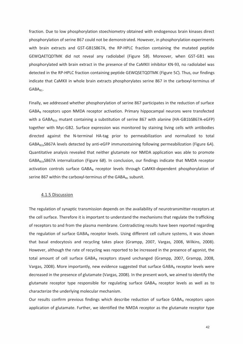
fraction. Due to low phosphorylation stoechiometry obtained with endogenous brain kinases direct
phosphorylation of serine 867 could not be demonstrated. However, in phosphorylation experiments
with brain extracts and GST-GB1S867A, the RP-HPLC fraction containing the mutated peptide
GEWQAETQDTMK did not reveal any radiolabel (Figure 5B). Moreover, when GST-GB1 was
phosphorylated with brain extract in the presence of the CaMKII inhibitor KN-93, no radiolabel was
detected in the RP-HPLC fraction containing peptide GEWQSETQDTMK (Figure 5C). Thus, our findings
indicate that CaMKII in whole brain extracts phosphorylates serine 867 in the carboxyl-terminus of
GABAB1.
Finally, we addressed whether phosphorylation of serine 867 participates in the reduction of surface
GABAB receptors upon NMDA receptor activation. Primary hippocampal neurons were transfected
with a GABAB1b mutant containing a substitution of serine 867 with alanine (HA-GB1bS867A-eGFP)
together with Myc-GB2. Surface expression was monitored by staining living cells with antibodies
directed against the N-terminal HA-tag prior to permeabilization and normalized to total
GABAB1bS867A levels detected by anti-eGFP immunostaining following permeabilization (Figure 6A).
Quantitative analysis revealed that neither glutamate nor NMDA application was able to promote
GABAB1bS867A internalization (Figure 6B). In conclusion, our findings indicate that NMDA receptor
activation controls surface GABAB receptor levels through CaMKII-dependent phosphorylation of
serine 867 within the carboxyl-terminus of the GABAB1 subunit.
4.1.5 Discussion
The regulation of synaptic transmission depends on the availability of neurotransmitter-receptors at
the cell surface. Therefore it is important to understand the mechanisms that regulate the trafficking
of receptors to and from the plasma membrane. Contradicting results have been reported regarding
the regulation of surface GABAB receptor levels. Using different cell culture systems, it was shown
that basal endocytosis and recycling takes place (Grampp, 2007, Vargas, 2008, Wilkins, 2008).
However, although the rate of recycling was reported to be increased in the presence of agonist, the
total amount of cell surface GABAB receptors stayed unchanged (Grampp, 2007, Grampp, 2008,
Vargas, 2008). More importantly, new evidence suggested that surface GABAB receptor levels were
decreased in the presence of glutamate (Vargas, 2008). In the present work, we aimed to identify the
glutamate receptor type responsible for regulating surface GABAB receptor levels as well as to
characterize the underlying molecular mechanism.
Our results confirm previous findings which describe reduction of surface GABAB receptors upon
application of glutamate. Further, we identified the NMDA receptor as the glutamate receptor type
42

responsible for decreasing surface GABAB receptor levels. We demonstrate that GABAB receptors are
internalized upon stimulation with glutamate or NMDA. In a recent report it was shown that
glutamate stimulation may lead to proteolytic degradation of constitutively internalized GABAB
receptors (Vargas, 2008). However, in contrast to the findings presented in our study, the authors did
not detect increased levels of internalized receptors. This discrepancy could be caused by different
experimental conditions.
Preincubation of neurons with an NMDA receptor antagonist prevented the glutamate-induced
reduction of surface GABAB receptor levels, ruling out that other glutamate receptors directly
regulate surface GABAB receptor levels. However, other glutamate receptors, in particular AMPA
receptors, are likely to contribute to the NMDA receptor-mediated effect. NMDA receptor activation
is voltage dependent, requiring removal of the Mg2+ block by depolarization. Under physiological
conditions this is primarily mediated by AMPA receptor activation. It is expected that there is
sufficient basal activity in primary hippocampal neuronal cultures allowing for direct NMDA receptors
activation by NMDA. In conclusion, our results demonstrate that NMDA receptor activation is
essential and sufficient to reduce GABAB surface receptors, however modulatory roles of other
glutamate receptors cannot be ruled out.
NMDA receptors are localized at the postsynaptic density (PSD) of dendritic spines where their
activation allows Ca2+ and Na+ to enter the spine. Glutamate failed to reduce surface GABAB
receptors in the presence of extracellular EGTA, which implicates a crucial role for the Ca2+ influx
following NMDA receptor activation. CaMKII is an established target downstream of NMDA receptor
activation. CaMKII is one of the most abundant serine/threonine kinases in the brain and is enriched
in the PSD. It is activated by binding of Ca2+/Calmodulin (Bennett, 1983, Kennedy, 1983) and
subsequent autophosphorylation (Malenka, 1989b, Bayer, 2001). The GABAB receptor is known to be
localized predominantly perisynaptically (Kulik, 2006, Guetg, 2009) and therefore in the vicinity of
PSD associated proteins. Since CaMKII is coupled to the activation of NMDA receptors in many
physiological processes, it was tested whether CaMKII plays a critical role in NMDA induced surface
reduction of GABAB receptors. Indeed, preincubation with a CaMKII inhibitor failed to decrease
surface GABAB receptors. We show evidence that reduction of surface receptors is mediated trough
phosphorylation of the GABAB receptor by CaMKII. Recent studies show that, upon glutamate
application or LTP induction, increased levels of activated CaMKII are restricted to single spines and
do not incorporate neighboring spines (Zhang, 2008, Lee, 2009) Therefore, glutamate- and NMDA-
induced reduction of surface GABAB receptors most likely occurs postsynaptically on glutamatergic
synapses.
We identified serine 867 within the carboxyl-terminus of GABAB1 (GABAB1S867) as a novel
phosphorylation site that can be phosphorylated by recombinant as well as endogenous CaMKII.
43

Interestingly, the sequence around GABAB1S867 is not conform to the consensus sequence for
CaMKII phosphorylation (Pearson, 1985). GABAB1S867 is located in the juxtamembrane domain which
is recognized as a regulatory region for many transmembrane proteins, in particular receptor
tyrosine kinases. However a similar regulatory role for the juxtamembrane domain in GPCRs has not
been established yet. Although we cannot exclude the existence of additional CaMKII
phosphorylation sites within other domains of GABAB1 or on GABAB2, mutational analysis
demonstrates that GABAB1S867 is essential for glutamate-induced reduction of surface GABAB
receptor levels. Two other phosphorylation sites on the GABAB receptor are described; first, serine
892 on the GABAB2 subunit (GABAB2S892) was shown to be phosphorylated by cyclic AMP-dependent
protein kinase (PKA) (Couve, 2002) and secondly, serine 783 on the GABAB2 subunit (GABAB2S783) by
5’AMP-dependent protein kinase (AMPK) (Scott, 2002, Hardie, 2007, Kuramoto, 2007).
Functionally, both GABAB2S892 and GABAB2S783 phosphorylation lead to stabilization of surface
GABAB receptors and enhance GABAB receptor functions (Couve, 2002, Fairfax, 2004, Kuramoto,
2007). In contrast, our results demonstrate that phosphorylation of GABAB1S867 reduces surface
GABAB receptor levels. Similar to CaMKII, AMPK is activated in response to Ca2+-influx following
neuronal activity. However in contrast to CaMKII activation, which is directly mediated by
calmodulin/Ca2+, AMPK activation requires the activation of calmodulin-dependent kinase kinases, in
particular CaMKKβ. Moreover, AMPK is activated in response to metabolic stresses that deplete
neurons of ATP and increase ADP levels (Hawley, 2005, Witters, 2006, Hardie, 2007). Thus the
availability of GABAB receptors at the plasma membrane is differentially modulated by the carboxyl-
terminal phosphorylation.
While another report showed that stimulation with glutamate stabilizes GABAA receptors at the cell
surface thereby promoting inhibition (Marsden, 2007) our results suggest that NMDA-induced
decrease of GABAB surface receptors strengthens excitatory signals. The bidirectional effect of
glutamate may operate on the fast acting inhibitory GABAA receptor to prevent overexcitation and
neurotoxicity, whereas decrease of slow acting surface GABAB receptor may support memory
formation and contribute to long term potentiation.
44
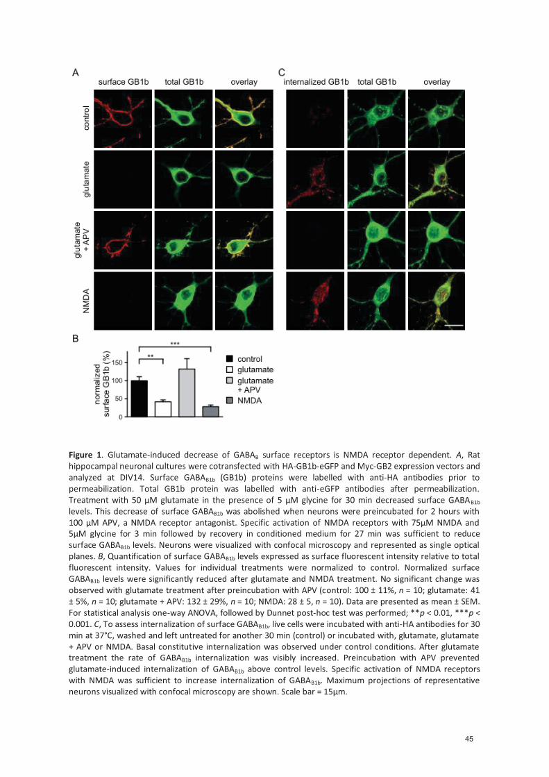
Figure 1. Glutamate-induced decrease of GABAB surface receptors is NMDA receptor dependent. A, Rat hippocampal neuronal cultures were cotransfected with HA-GB1b-eGFP and Myc-GB2 expression vectors and analyzed at DIV14. Surface GABAB1b (GB1b) proteins were labelled with anti-HA antibodies prior to permeabilization. Total GB1b protein was labelled with anti-eGFP antibodies after permeabilization. Treatment with 50 μM glutamate in the presence of 5 μM glycine for 30 min decreased surface GABAB1b levels. This decrease of surface GABAB1b was abolished when neurons were preincubated for 2 hours with 100 μM APV, a NMDA receptor antagonist. Specific activation of NMDA receptors with 75μM NMDA and 5μM glycine for 3 min followed by recovery in conditioned medium for 27 min was sufficient to reduce surface GABAB1b levels. Neurons were visualized with confocal microscopy and represented as single optical planes. B, Quantification of surface GABAB1b levels expressed as surface fluorescent intensity relative to total fluorescent intensity. Values for individual treatments were normalized to control. Normalized surface GABAB1b levels were significantly reduced after glutamate and NMDA treatment. No significant change was observed with glutamate treatment after preincubation with APV (control: 100 ± 11%, n = 10; glutamate: 41 ± 5%, n = 10; glutamate + APV: 132 ± 29%, n = 10; NMDA: 28 ± 5, n = 10). Data are presented as mean ± SEM. For statistical analysis one-way ANOVA, followed by Dunnet post-hoc test was performed; **p < 0.01, ***p < 0.001. C, To assess internalization of surface GABAB1b, live cells were incubated with anti-HA antibodies for 30 min at 37°C, washed and left untreated for another 30 min (control) or incubated with, glutamate, glutamate + APV or NMDA. Basal constitutive internalization was observed under control conditions. After glutamate treatment the rate of GABAB1b internalization was visibly increased. Preincubation with APV prevented glutamate-induced internalization of GABAB1b above control levels. Specific activation of NMDA receptors with NMDA was sufficient to increase internalization of GABAB1b. Maximum projections of representative neurons visualized with confocal microscopy are shown. Scale bar = 15μm.
45
Figure 2. CAMKII is required for the NMDA receptor-induced decrease of surface GABAB receptors. Rat hippocampal neuronal cultures were co-transfected with HA-GB1b-eGFP and Myc-GB2 expression vectors and analyzed at DIV14. Surface GABAB1b (GB1b) labelled with anti-HA antibodies prior to permeabilization were assessed relative to total GABAB1b expression. Values for individual treatments were normalized to control. Preincubation of neurons with the CaMKII inhibitor KN-93 (10 μM) for 2 h prevented the reduction in surface GABAB1b levels following treatment with glutamate (control: 100 ± 12, n = 10; glutamate 24 ± 5, n = 10; glutamate + KN-93: 102 ± 29; n = 6;) or NMDA (control: 100 ± 14, n = 10; NMDA: 29 ± 9, n = 10; NMDA + KN-93: 86 ± 18, n = 6). Data were represented as mean ± SEM. For statistical analysis one-way ANOVA, followed by Tuckey post-hoc test was performed; *p < 0.05, **p < 0.01.
46
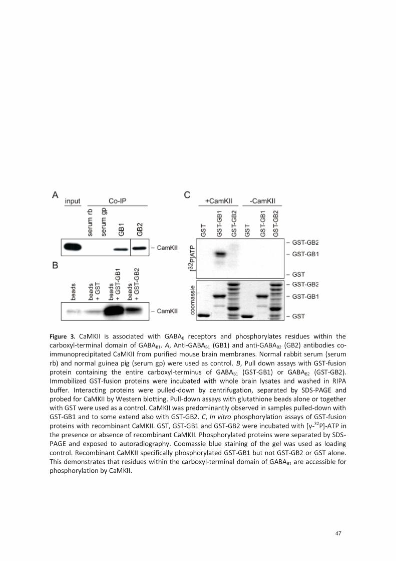
Figure 3. CaMKII is associated with GABAB receptors and phosphorylates residues within the carboxyl-terminal domain of GABAB1. A, Anti-GABAB1 (GB1) and anti-GABAB2 (GB2) antibodies co-immunoprecipitated CaMKII from purified mouse brain membranes. Normal rabbit serum (serum rb) and normal guinea pig (serum gp) were used as control. B, Pull down assays with GST-fusion protein containing the entire carboxyl-terminus of GABAB1 (GST-GB1) or GABAB2 (GST-GB2). Immobilized GST-fusion proteins were incubated with whole brain lysates and washed in RIPA buffer. Interacting proteins were pulled-down by centrifugation, separated by SDS-PAGE and probed for CaMKII by Western blotting. Pull-down assays with glutathione beads alone or together with GST were used as a control. CaMKII was predominantly observed in samples pulled-down with GST-GB1 and to some extend also with GST-GB2. C, In vitro phosphorylation assays of GST-fusion proteins with recombinant CaMKII. GST, GST-GB1 and GST-GB2 were incubated with [γ-32P]-ATP in the presence or absence of recombinant CaMKII. Phosphorylated proteins were separated by SDS-PAGE and exposed to autoradiography. Coomassie blue staining of the gel was used as loading control. Recombinant CaMKII specifically phosphorylated GST-GB1 but not GST-GB2 or GST alone. This demonstrates that residues within the carboxyl-terminal domain of GABAB1 are accessible for phosphorylation by CaMKII.
47
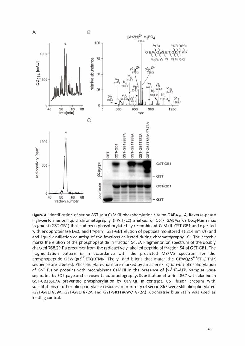
Figure 4. Identification of serine 867 as a CaMKII phosphorylation site on GABAB1. A, Reverse-phase high-performance liquid chromatography (RP-HPLC) analysis of GST- GABAB1 carboxyl-terminus fragment (GST-GB1) that had been phosphorylated by recombinant CaMKII. GST-GB1 and digested with endoproteinase LysC and trypsin. GST-GB1 elution of peptides monitored at 214 nm (A) and and liquid cintillation counting of the fractions collected during chromatography (C). The asterisk marks the elution of the phosphopeptide in fraction 54. B, Fragmentation spectrum of the doubly charged 768.29 Da precursor from the radioactively labelled peptide of fraction 54 of GST-GB1. The fragmentation pattern is in accordance with the predicted MS/MS spectrum for the phosphopeptide GEWQpS867ETQDTMK. The y- and b-ions that match the GEWQpS867ETQDTMK sequence are labelled. Phosphorylated ions are marked by an asterisk. C, In vitro phosphorylation of GST fusion proteins with recombinant CaMKII in the presence of [γ-32P]-ATP. Samples were separated by SDS-page and exposed to autoradiography. Substitution of serine 867 with alanine in GST-GB1S867A prevented phosphorylation by CaMKII. In contrast, GST fusion proteins with substitutions of other phosphorylable residues in proximity of serine 867 were still phosphorylated (GST-GB1T869A, GST-GB1T872A and GST-GB1T869A/T872A). Coomassie blue stain was used as loading control.
48

Figure 5. Endogenous CaMKII phosphorylates GABAB1S867. (A-C) GST-fusion proteins were incubated with the cytosolic fraction of whole mouse brain extracts in the presence of [γ-32P]-ATP and analyzed by RP-HPLC. Elution profiles are shown on the top and scintillation graphs on the bottom. The carboxyl-terminal GABAB1 peptide GEWQSETQDTMK was identified by ESI-MS/MS as a major constituent of a highly phosphorylated fraction (asterisk) of the GST-GB1 effluent (A). The corresponding fraction of the GST-GB1S867A effluent (arrow head) containing peptide GEWQAETQDTMK with a substitution of alanine for serine 867 did not get phosphorylated (B). Following phosphorylation of GB1-GST in the presence of KN-93 no radiolabel was detected in the fraction containing GEWQSETQDTMK (arrow head) (C).
49
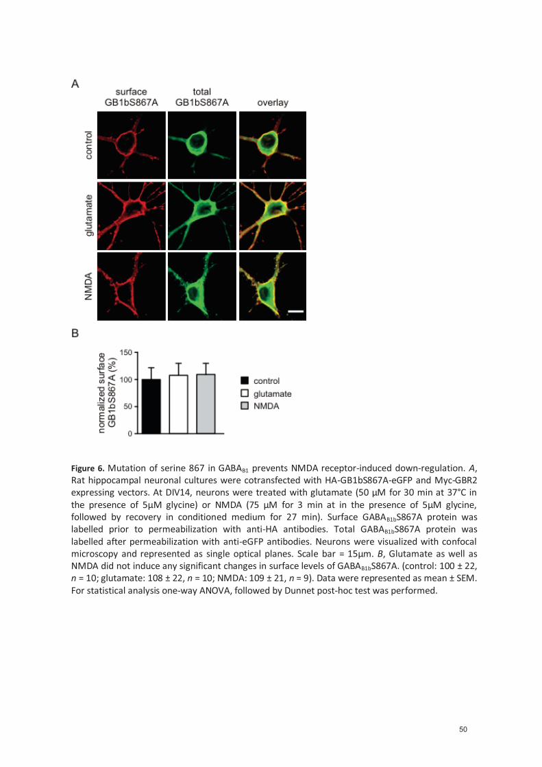
Figure 6. Mutation of serine 867 in GABAB1 prevents NMDA receptor-induced down-regulation. A, Rat hippocampal neuronal cultures were cotransfected with HA-GB1bS867A-eGFP and Myc-GBR2 expressing vectors. At DIV14, neurons were treated with glutamate (50 μM for 30 min at 37°C in the presence of 5μM glycine) or NMDA (75 μM for 3 min at in the presence of 5μM glycine, followed by recovery in conditioned medium for 27 min). Surface GABAB1bS867A protein was labelled prior to permeabilization with anti-HA antibodies. Total GABAB1bS867A protein was labelled after permeabilization with anti-eGFP antibodies. Neurons were visualized with confocal microscopy and represented as single optical planes. Scale bar = 15μm. B, Glutamate as well as NMDA did not induce any significant changes in surface levels of GABAB1bS867A. (control: 100 ± 22, n = 10; glutamate: 108 ± 22, n = 10; NMDA: 109 ± 21, n = 9). Data were represented as mean ± SEM. For statistical analysis one-way ANOVA, followed by Dunnet post-hoc test was performed.
50

Statement of personal contribution to the manuscript:
- Planning and generation of the GST-fusion proteins.
- Design and accomplishement of the pull down assays
- Design and accomplishement of all presented phosphorylation experiments with the purified
and native kinases.
- Design and accomplishement of all HPLC experiments.
- Preparation of the samples for MS analysis.
- Preparation and analysis of the scintillation data.
- Identification of the phosphorylation site S867.
- Writing of the material method part of the phosphorylation eperiments, HPLC, scintillation
and MS analysis.
- Writing of the results part of the phosphorylation eperiments, HPLC, scintillation and MS
analysis.
- Contribution to the finalization of the manuscript.
51
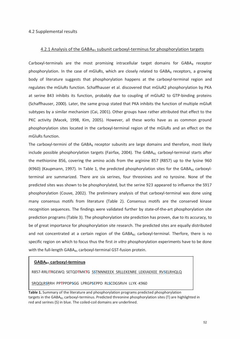
4.2 Supplemental results
4.2.1 Analysis of the GABAB1 subunit carboxyl-terminus for phosphorylation targets
Carboxyl-terminals are the most promising intracellular target domains for GABAB receptor
phosphorylation. In the case of mGluRs, which are closely related to GABAB receptors, a growing
body of literature suggests that phosphorylation happens at the carboxyl-terminal region and
regulates the mGluRs function. Schaffhauser et al. discovered that mGluR2 phosphorylation by PKA
at serine 843 inhibits its function, probably due to coupling of mGluR2 to GTP-binding proteins
(Schaffhauser, 2000). Later, the same group stated that PKA inhibits the function of multiple mGluR
subtypes by a similar mechanism (Cai, 2001). Other groups have rather attributed that effect to the
PKC activity (Macek, 1998, Kim, 2005). However, all these works have as as common ground
phosphorylation sites located in the carboxyl-terminal region of the mGluRs and an effect on the
mGluRs function.
The carboxyl-termini of the GABAB receptor subunits are large domains and therefore, most likely
include possible phosphorylation targets (Fairfax, 2004). The GABAB1 carboxyl-terminal starts after
the methionine 856, covering the amino acids from the arginine 857 (R857) up to the lysine 960
(K960) (Kaupmann, 1997). In Table 1, the predicted phosphorylation sites for the GABAB1 carboxyl-
terminal are summarized. There are six serines, four threonines and no tyrosine. None of the
predicted sites was shown to be phosphorylated, but the serine 923 appeared to influence the S917
phosphorylation (Couve, 2002). The preliminary analysis of that carboxyl-terminal was done using
many consensus motifs from literature (Table 2). Consensus motifs are the conserved kinase
recognition sequences. The findings were validated further by state-of-the-art phosphorylation site
prediction programs (Table 3). The phosphorylation site prediction has proven, due to its accuracy, to
be of great importance for phosphorylation site research. The predicted sites are equally distributed
and not concentrated at a certain region of the GABAB1 carboxyl-terminal. Therfore, there is no
specific region on which to focus thus the first in vitro phosphorylation experiments have to be done
with the full-length GABAB1 carboxyl-terminal GST-fusion protein.
GABAB1 C-terminus
R857-RRLITRGEWQ SETQDTMKTG SSTNNNEEEK SRLLEKENRE LEKIIAEKEE RVSELRHQLQ
SRQQLRSRRH PPTPPDPSGG LPRGPSEPPD RLSCDGSRVH LLYK -K960
Table 1. Summary of the literature and phosphorylation programs predicted phosphorylation targets in the GABAB1 carboxyl-terminus. Predicted threonine phosphorylation sites (T) are highlighted in red and serines (S) in blue. The coiled-coil domains are underlined.
GABAB1 carboxyl-terminus
52
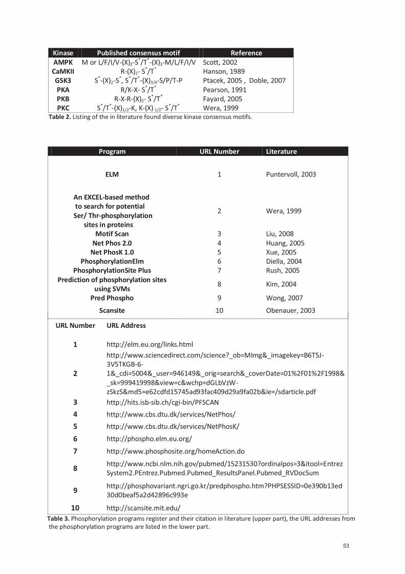
Kinase Published consensus motif Reference AMPK M or L/F/I/V-(X)2-S*/T*-(X)3-M/L/F/I/V Scott, 2002 CaMKII R-(X)2- S*/T* Hanson, 1989 GSK3 S*-(X)2-S*, S*/T*-(X)3/4-S/P/T-P Ptacek, 2005 , Doble, 2007 PKA R/K-X- S*/T* Pearson, 1991 PKB R-X-R-(X)2- S*/T* Fayard, 2005 PKC S*/T*-(X)1/2-K, K-(X) 1/2- S*/T* Wera, 1999
Table 2. Listing of the in literature found diverse kinase consensus motifs.
Program URL Number Literature
ELM 1 Puntervoll, 2003
An EXCEL-based method to search for potential Ser/ Thr-phosphorylation sites in proteins
2 Wera, 1999
Motif Scan 3 Liu, 2008 Net Phos 2.0 4 Huang, 2005
Net PhosK 1.0 5 Xue, 2005 PhosphorylationElm 6 Diella, 2004
PhosphorylationSite Plus 7 Rush, 2005 Prediction of phosphorylation sites
using SVMs 8 Kim, 2004
Pred Phospho 9 Wong, 2007
Scansite 10 Obenauer, 2003
URL Number URL Address
1 http://elm.eu.org/links.html
2
http://www.sciencedirect.com/science?_ob=MImg&_imagekey=B6T5J-3V5TKGB-6-1&_cdi=5004&_user=946149&_orig=search&_coverDate=01%2F01%2F1998&_sk=999419998&view=c&wchp=dGLbVzW-zSkzS&md5=e62cdfd15745ad93fac409d29a9fa02b&ie=/sdarticle.pdf
3 http://hits.isb-sib.ch/cgi-bin/PFSCAN
4 http://www.cbs.dtu.dk/services/NetPhos/
5 http://www.cbs.dtu.dk/services/NetPhosK/
6 http://phospho.elm.eu.org/
7 http://www.phosphosite.org/homeAction.do
8 http://www.ncbi.nlm.nih.gov/pubmed/15231530?ordinalpos=3&itool=EntrezSystem2.PEntrez.Pubmed.Pubmed_ResultsPanel.Pubmed_RVDocSum
9 http://phosphovariant.ngri.go.kr/predphospho.htm?PHPSESSID=0e390b13ed30d0beaf5a2d42896c993e
10 http://scansite.mit.edu/ Table 3. Phosphorylation programs register and their citation in literature (upper part), the URL addresses from the phosphorylation programs are listed in the lower part.
53

4.2.2 Glutathione S-tranferase fusion system
The glutathione s-transferase (GST) fusion system is profiled as the optimal model to study
phosphorylation of the GABAB receptors at the GABAB1 carboxyl-terminal. The GST fusion system
expresses, purifies, and detects recombinant fusion proteins produced in E. coli. It relies on inducible,
high-level gene expression of gene fragments as fusion proteins with a Schistosoma japonicum GST-
tag (Table 4.a). A fusion protein contains the GST-tag localized at the amino terminal and the protein
of interest at the carboxyl-terminal. The GST fusion proteins are purified from bacterial lysates by
means of affinity chromatography using immobilized glutathione (glutathione coated sepharose
beads) while the impurities are removed by washing. Boiling the samples in Laemmli buffer or under
mild, non-denaturing conditions using reduced glutathione elutes the GST fusion proteins from the
immobilized glutathione. The GST fusion system has been used successfully in many applications
including molecular immunology (Toye, 1990), the production of vaccines (Fikrig, 1990, Johnson,
1989) and studies involving protein-protein (Kaelin, 1991) and DNA-protein (Chittenden, 1991)
interactions. In this work, the GST-GB1 protein, including the full-length carboxyl-terminal of GABAB1
(amino acids 857–960, see Table 4.b) was used.
A first important hint, that a phosphorylation site on the GABAB1 carboxyl-terminal is located at a
juxtramembrane site was given from the first in vitro phosphorylation experiments using the GST-
GB1 fusion protein and its truncations. These GST-GB1 fusion proteins were gradually truncated from
their carboxyl-terminal. They were phosphorylated using native and purified kinases. GST-GB1Δ1
(Table 4.c) and GST-GB1Δ2 (Table 4.d), the two truncated forms were generated by PCR from GST-
GB1. The sense primer (5`-GTTCCGCGTGGATCCCGCAGG-3’) is the same for GST-GB1Δ1 and GST-
GB1Δ2. Conversly, the antisense is different for GST-GB1Δ1 (5`-TTTCTCGAGCTACTTCTCTTCCTCGTTGT
T-3’) and GST-GB1Δ2 (5’-TTTCTCGAGCTATGGGGGGTGGCGCCG TGA -3’). The generated PCR
fragments were digested with BamHI- and XhoI and subcloned in-frame into the pGEX-4T-1 plasmid.
The truncated GST-fusion proteins were expressed, induced and lysed as previously described for the
GST-GB1 fusion proteins.
The GST fusion system enabled the discovery of a new phosphorylation site on the GABAB receptors
by in vitro phosphorylation and HPLC-MS analysis. On the other hand, the interaction between the
responsible kinase with the GABAB receptor can be showed in pull-down assay. But one must keep in
mind that the GST fusion system is artificial and a simplifying model. Indeed, this technique does not
take into account phosphorylation influencing factors like folding of the carboxyl-termini or
interaction with binding partners. Nevertheless, the GST fusion system enables the generation of
large amounts (important for further analysis steps like high pressure liquid chromatography) of a
model protein comprising most of the phosphorlylation sites of the native GABAB1carboxyl-terminal.
54
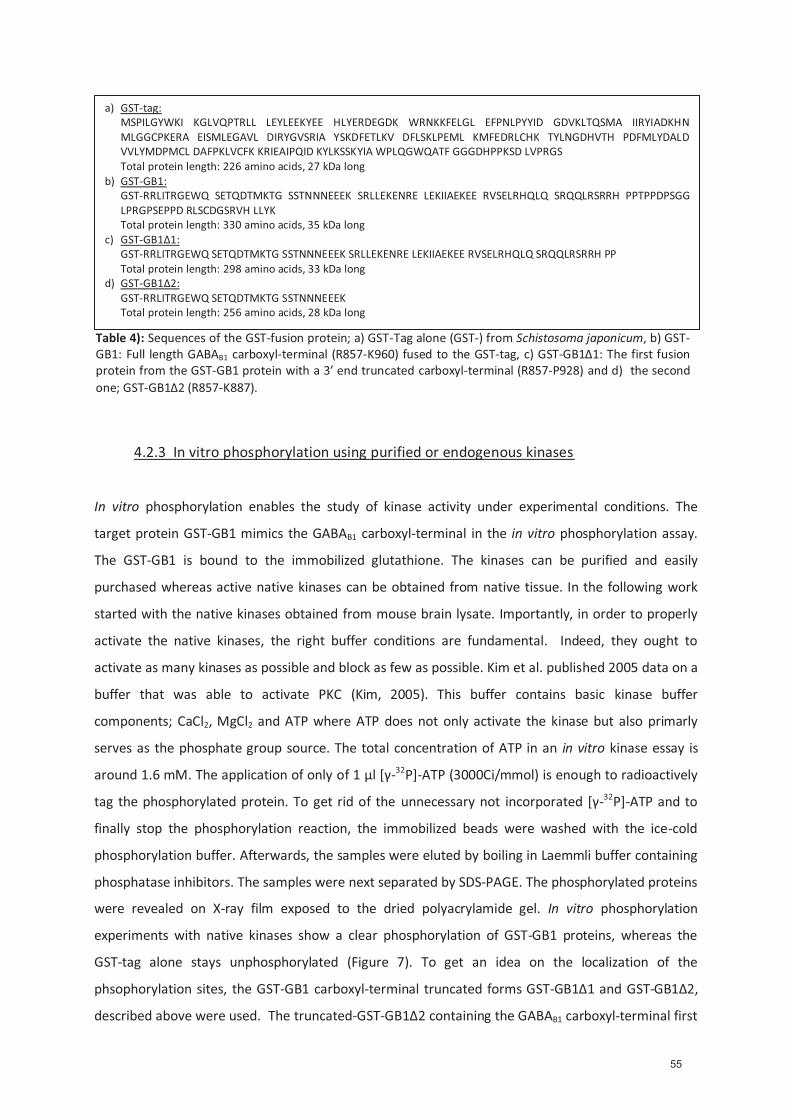
4.2.3 In vitro phosphorylation using purified or endogenous kinases
In vitro phosphorylation enables the study of kinase activity under experimental conditions. The
target protein GST-GB1 mimics the GABAB1 carboxyl-terminal in the in vitro phosphorylation assay.
The GST-GB1 is bound to the immobilized glutathione. The kinases can be purified and easily
purchased whereas active native kinases can be obtained from native tissue. In the following work
started with the native kinases obtained from mouse brain lysate. Importantly, in order to properly
activate the native kinases, the right buffer conditions are fundamental. Indeed, they ought to
activate as many kinases as possible and block as few as possible. Kim et al. published 2005 data on a
buffer that was able to activate PKC (Kim, 2005). This buffer contains basic kinase buffer
components; CaCl2, MgCl2 and ATP where ATP does not only activate the kinase but also primarly
serves as the phosphate group source. The total concentration of ATP in an in vitro kinase essay is
around 1.6 mM. The application of only of 1 μl [γ-32P]-ATP (3000Ci/mmol) is enough to radioactively
tag the phosphorylated protein. To get rid of the unnecessary not incorporated [γ-32P]-ATP and to
finally stop the phosphorylation reaction, the immobilized beads were washed with the ice-cold
phosphorylation buffer. Afterwards, the samples were eluted by boiling in Laemmli buffer containing
phosphatase inhibitors. The samples were next separated by SDS-PAGE. The phosphorylated proteins
were revealed on X-ray film exposed to the dried polyacrylamide gel. In vitro phosphorylation
experiments with native kinases show a clear phosphorylation of GST-GB1 proteins, whereas the
GST-tag alone stays unphosphorylated (Figure 7). To get an idea on the localization of the
phsophorylation sites, the GST-GB1 carboxyl-terminal truncated forms GST-GB1Δ1 and GST-GB1Δ2,
described above were used. The truncated-GST-GB1Δ2 containing the GABAB1 carboxyl-terminal first
a) GST-tag: MSPILGYWKI KGLVQPTRLL LEYLEEKYEE HLYERDEGDK WRNKKFELGL EFPNLPYYID GDVKLTQSMA IIRYIADKHN MLGGCPKERA EISMLEGAVL DIRYGVSRIA YSKDFETLKV DFLSKLPEML KMFEDRLCHK TYLNGDHVTH PDFMLYDALD VVLYMDPMCL DAFPKLVCFK KRIEAIPQID KYLKSSKYIA WPLQGWQATF GGGDHPPKSD LVPRGS Total protein length: 226 amino acids, 27 kDa long
b) GST-GB1: GST-RRLITRGEWQ SETQDTMKTG SSTNNNEEEK SRLLEKENRE LEKIIAEKEE RVSELRHQLQ SRQQLRSRRH PPTPPDPSGG LPRGPSEPPD RLSCDGSRVH LLYK Total protein length: 330 amino acids, 35 kDa long
c) GST-GB1Δ1: GST-RRLITRGEWQ SETQDTMKTG SSTNNNEEEK SRLLEKENRE LEKIIAEKEE RVSELRHQLQ SRQQLRSRRH PP Total protein length: 298 amino acids, 33 kDa long
d) GST-GB1Δ2: GST-RRLITRGEWQ SETQDTMKTG SSTNNNEEEK Total protein length: 256 amino acids, 28 kDa long
Table 4): Sequences of the GST-fusion protein; a) GST-Tag alone (GST-) from Schistosoma japonicum, b) GST-GB1: Full length GABAB1 carboxyl-terminal (R857-K960) fused to the GST-tag, c) GST-GB1Δ1: The first fusion protein from the GST-GB1 protein with a 3’ end truncated carboxyl-terminal (R857-P928) and d) the second one; GST-GB1Δ2 (R857-K887).
55
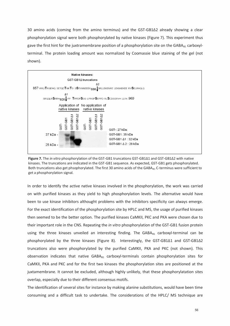
30 amino acids (coming from the amino terminus) and the GST-GB1Δ2 already showing a clear
phosphorylation signal were both phosphorylated by native kinases (Figure 7). This experiment thus
gave the first hint for the juxtramembrane position of a phosphorylation site on the GABAB1 carboxyl-
terminal. The protein loading amount was normalized by Coomassie blue staining of the gel (not
shown).
In order to identify the active native kinases involved in the phosphorylation, the work was carried
on with purified kinases as they yield to high phosphorylation levels. The alternative would have
been to use kinase inhibitors althought problems with the inhibitors specificity can always emerge.
For the exact identification of the phosphorylation site by HPLC and MS, the usage of purified kinases
then seemed to be the better option. The purified kinases CaMKII, PKC and PKA were chosen due to
their important role in the CNS. Repeating the in vitro phosphorylation of the GST-GB1 fusion protein
using the three kinases unveiled an interesting finding. The GABAB1 carboxyl-terminal can be
phosphorylated by the three kinases (Figure 8). Interestingly, the GST-GB1Δ1 and GST-GB1Δ2
truncations also were phosphorylated by the purified CaMKII, PKA and PKC (not shown). This
observation indicates that native GABAB1 carboxyl-terminals contain phosphorylation sites for
CaMKII, PKA and PKC and for the first two kinases the phosphorylation sites are positioned at the
juxtamembrane. It cannot be excluded, although highly unlikely, that these phosphorylatation sites
overlap, especially due to their different consensus motifs.
The identification of several sites for instance by making alanine substitutions, would have been time
consuming and a difficult task to undertake. The considerations of the HPLC/ MS technique are
Figure 7. The in vitro phosphorylation of the GST-GB1 truncations GST-GB1Δ1 and GST-GB1Δ2 with native kinases. The truncations are indicated in the GST-GB1 sequence. As expected, GST-GB1 gets phosphorylated. Both truncations also get phsophorylated. The first 30 amino acids of the GABAB1 C-terminus were sufficient to get a phosphorylation signal.
56

limited and not suitable for the analysis of all the possible phosphorylation sites. The variety of
different possible kinase phosphorylation sites demands the systematic analysis of a single
phosphorylation site. That was the most promising and fastest way to find, under the given
circumstances, a physiological relevant phosphorylation site. The kinase with the most potential was
CaMKII. Indeed, it has a consensus motif appearing in the proteolytic peptide fragments and most
importantly, it is very abundant in the CNS. Additionnaly, indications from the literature (Vargas,
2008) and from different unpublished electrophysiological experiments and localization studies
predict a physiological relevant connection between CaMKII and GABA receptor. All this information
led to the decision of focusing all the efforts on CaMKII actions.
4.2.4 High pressure liquid chromatography (HPLC) and Mass spectrometry (MS)
The mass spectrometry (MS) measures the mass to charge ratio (m/z) of ions. The high pressure
liquid chromatography (HPLC, also sometimes called high performance liquid chromatography) is a
form of column chromatography used to separate, identify and quantify compounds. The HPLC
consists of a column packed with a chromatographic material, generating a specific surface layer
(stationary phase) and a pump that moves the analyte containing mobile phase through the column.
A detector at the end records the elution of the analytes. The retention time varies depending on the
interactions between the stationary phase, the molecules being analyzed and the solvents used.
The combination of liquid chromatography and mass spectrometry allows the detection of analytes
in the picomol range. It was made possible by the availability of the electro spray (ES) technology
(Issaq, 2009). The electro spray technology is a technique that ejects the ions from a solvent
containing the analyte(s) of interest. The electro spray disperses the liquid into a fine aerosol. The ion
formation involves extensive solvent evaporation by mixing water with volatile organic compound
solvents (e.g. methanol, acetonitrile). The first vacuum stage of a MS samples the aerosol through a
Figure 8. The in vitro phosphorylation of GST-GB1 with the purified kinases CaMKII,PKA and PKC. All three kinases phosphosphorylate GST-GB1. This indicates that the GABAB1 carboxyl-terminus could be a target for several kinases.
57

capillary. The solvent evaporates from a charged droplet until it becomes unstable upon reaching its
Rayleigh limit; the droplet size decreases, the charge to surface ratio increases. Ions of equal charge
repel each other and the droplet deforms and emits charged ions. During this process, the droplet
loses a small percentage of its mass (an electron) along with a relatively large percentage of its
charge. The ES technique is so mild that labile analytes remain intact after the electro spray
ionisation (ESI) procedure (Bothner, 1998). For the analysis of phosphorylated peptides, reverse
phase-high pressure liquid chromatography (RP-HPLC) is the chromatography of choice. In RP-HPLC,
the solid phase support is modified by long saturated hydrocarbon chains. The analyte binds to the
stationary phase via hydrophobic interactions. The analyte is dissolved in aqueous trifluoroacetic
acid, the polar solvent. When the concentration of acetonitrile slowly increases during the gradient,
adsorbed peptides elute sequentially from the stationary phase with increasing hydrophobicity. The
detector records eluting peptides at 214 nm, while at 280 nm the aromatic amino acids containing
peptide fragments can be detected.
To increase the stoichiometry of phosphorylation, at least 10μg of the target protein should
be phosphorylated with a final ATP concentration of 20 μM. To facilitate the tracking of the
phosphorylated peptides, the kinase reaction is carried out by spiking the ATP mixture with 1 μl of
radioactive ATP (indicates the specific activity) to reach at least 10% phosphorylation of the protein
of interest. In that experiment, however, we could not reach the desired phosphorylation
stoichiometry using native kinases. Instead, we used purified, highly active CaMKII, to obtain
reasonable phosphorylation. The peptide fragments are generated by a digestion with
endoproteinases lys-c and trypsin (Jeno, 1995). The lys-c cleaves at the carboxyl-terminal end of
lysine whereas the trypsin cleaves peptide bonds carboxyl-terminal of lysine and arginine. The rate of
hydrolysis can be slowed down by acidic residue (glutamic acid or aspartic acid)_on either side of the
cleavage site. A proline on the carboxyl-terminal of the cleavage site can even inhibit digestion by
trypsin without affecting lys-c cleaving activity. The peptide fragments generated are usually
between 5 to 30 amino acids longs. Very short peptides do not bind to the reverse-phase matrix
while very long peptides tend to bind irreversibly to the column. In Table 5 the lys-c and trypsin
digested peptides obtained from GST-GB1 are summarized.
The liquid scintillation counting of the HPLC fragments unveils a strongly phosphorylated peptide.
The MS analysis of this fraction yielded two peptides of 1439.61 Da and 1519.58 Da. The collision-
induced dissociation (CID) spectrum and databank searching showed that the two peptides are
derived from the juxtamembrane position of GST-GB1 and comprise the unphosphorylated
GEWQS867ETQDTMK (Figure 9.a) peptide and the phosphorylated peptide GEWQpS867ETQDTMK with
a phosphorylation at the S867 (Figure 9.b).
58
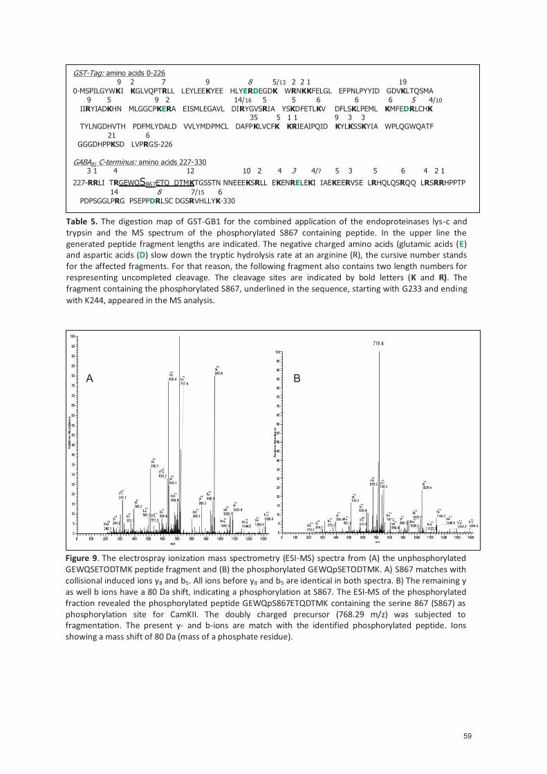
Table 5. The digestion map of GST-GB1 for the combined application of the endoproteinases lys-c and trypsin and the MS spectrum of the phosphorylated S867 containing peptide. In the upper line the generated peptide fragment lengths are indicated. The negative charged amino acids (glutamic acids (E) and aspartic acids (D) slow down the tryptic hydrolysis rate at an arginine (R), the cursive number stands for the affected fragments. For that reason, the following fragment also contains two length numbers for respresenting uncompleted cleavage. The cleavage sites are indicated by bold letters (K and R). The fragment containing the phosphorylated S867, underlined in the sequence, starting with G233 and ending with K244, appeared in the MS analysis.
GST-Tag: amino acids 0-226 9 2 7 9 8 5/13 2 2 1 19 0-MSPILGYWKI KGLVQPTRLL LEYLEEKYEE HLYERDEGDK WRNKKFELGL EFPNLPYYID GDVKLTQSMA 9 5 9 2 14/16 5 5 6 6 6 5 4/10 IIRYIADKHN MLGGCPKERA EISMLEGAVL DIRYGVSRIA YSKDFETLKV DFLSKLPEML KMFEDRLCHK 35 5 1 1 9 3 3 TYLNGDHVTH PDFMLYDALD VVLYMDPMCL DAFPKLVCFK KRIEAIPQID KYLKSSKYIA WPLQGWQATF 21 6 GGGDHPPKSD LVPRGS-226 GABAB1 C-terminus: amino acids 227-330 3 1 4 12 10 2 4 3 4/7 5 3 5 6 4 2 1
227-RRLI TRGEWQS867ETQ DTMKTGSSTN NNEEEKSRLL EKENRELEKI IAEKEERVSE LRHQLQSRQQ LRSRRHPPTP 14 8 7/15 6 PDPSGGLPRG PSEPPDRLSC DGSRVHLLYK-330
Figure 9. The electrospray ionization mass spectrometry (ESI-MS) spectra from (A) the unphosphorylated GEWQSETODTMK peptide fragment and (B) the phosphorylated GEWQpSETODTMK. A) S867 matches with collisional induced ions y8 and b5. All ions before y8 and b5 are identical in both spectra. B) The remaining y as well b ions have a 80 Da shift, indicating a phosphorylation at S867. The ESI-MS of the phosphorylated fraction revealed the phosphorylated peptide GEWQpS867ETQDTMK containing the serine 867 (S867) as phosphorylation site for CamKII. The doubly charged precursor (768.29 m/z) was subjected to fragmentation. The present y- and b-ions are match with the identified phosphorylated peptide. Ions showing a mass shift of 80 Da (mass of a phosphate residue).
A B
59

4.2.5 The phosphospecific pS867 antibody
After showing the phosphorylation of GST-GB1 at S867 by native CaMKII, the next step was to
demonstrate that S867 can be phosphorylated in native GABAB1 subunits. This was done by
generating a phosphospecific antibody pS867. This pS867 antibody specifically recognizes the
phosphorylated S867 site. In the following chapter the generation and characterization of the pS867
antibody will be discussed. The interesting findings of native S867 phosphorylation will be highlighted
and the mechanism increasing its phosphorylation will be explained. Finally the preliminary
immunohistochemical experiments with the pS867 antibody will be outlined.
4.2.5.1 Generation of the pS867 antibody
For the pS867 antibody generation, an S867 phosphorylated peptide ITRGEWQpS867EAQDT (Lamp,
2001) was conjugated to the carrier protein keyhole limpet hemocyanin (KLH) to achieve an
increased immunization. The sequence and phosphorylation of the phosphorylated peptide was
verified by MS analysis. The phosphorylated peptide-KLH conjugate was emulsified with complete
Freund’s adjuvant and injected subcutaneously in New Zealand rabbits four times at biweekly
intervals. Additional injection of the peptide-KLH conjugate in Freund’s incomplete adjuvant boosts
the immunization reaction. One week after the boost, the blood is collected from the central ear
artery and is allowed to clot and retract at 37°C overnight. The serum is decanted and clarified by
centrifugation at 2’500 rpm for 15 minutes. The antibody fraction is then purified by affinity
chromatography on beads. For this, beads were equilibrated with the phosphorylated peptide-KLH
conjugate dissolved in a PBS and NaCl solution. The beads were washed with PBS. The washed beads
were loaded into a plastic column and washed again with PBS until the OD at 280 nm is 0. The elution
of the antibodies was done with a mixture of 200 mM glycine at pH 2.8. The immunoglobulin-
containing fraction is identified by measuring the absorbance at 280 nm. The fractions were dialyzed
against PBS at 4°C overnight. To absorb anti-non-phosphopetide antibody, the generated antibody
solution is applied onto a column and equilibrated with the non-phosphopetide-KLH conjugate. The
non-phosphorylated peptide ITRGEWQSEAQDT. The purified solution is then collected and its
absorbance mesured on more time at 280 nm. The pS867 specificity was finally confirmed by ELISA
where the antibody showed a strong affinity towards the phosphorylated peptide.
4.2.5.2 Characterization of the pS867 antibody
Additionally to the ELISA data, the specificity of the pS867 antibody was examined using the GST-GB1
protein and its S867A substitution, phosphorylated by purified CaMKII. The pS867 antibody
surprisingly does not recognize the unphosphorylated GST-GB1 proteins (Figure 10a, first lane).but
60
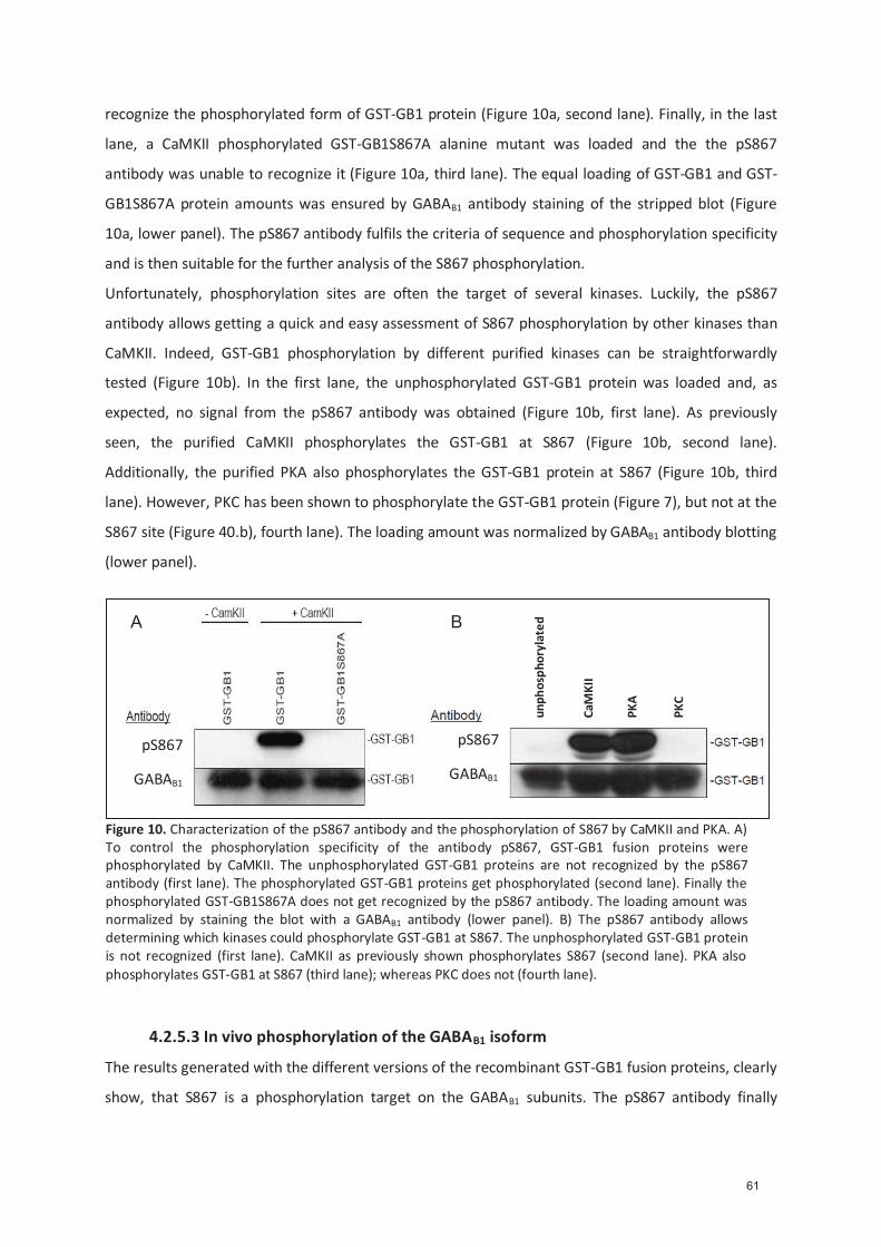
recognize the phosphorylated form of GST-GB1 protein (Figure 10a, second lane). Finally, in the last
lane, a CaMKII phosphorylated GST-GB1S867A alanine mutant was loaded and the the pS867
antibody was unable to recognize it (Figure 10a, third lane). The equal loading of GST-GB1 and GST-
GB1S867A protein amounts was ensured by GABAB1 antibody staining of the stripped blot (Figure
10a, lower panel). The pS867 antibody fulfils the criteria of sequence and phosphorylation specificity
and is then suitable for the further analysis of the S867 phosphorylation.
Unfortunately, phosphorylation sites are often the target of several kinases. Luckily, the pS867
antibody allows getting a quick and easy assessment of S867 phosphorylation by other kinases than
CaMKII. Indeed, GST-GB1 phosphorylation by different purified kinases can be straightforwardly
tested (Figure 10b). In the first lane, the unphosphorylated GST-GB1 protein was loaded and, as
expected, no signal from the pS867 antibody was obtained (Figure 10b, first lane). As previously
seen, the purified CaMKII phosphorylates the GST-GB1 at S867 (Figure 10b, second lane).
Additionally, the purified PKA also phosphorylates the GST-GB1 protein at S867 (Figure 10b, third
lane). However, PKC has been shown to phosphorylate the GST-GB1 protein (Figure 7), but not at the
S867 site (Figure 40.b), fourth lane). The loading amount was normalized by GABAB1 antibody blotting
(lower panel).
4.2.5.3 In vivo phosphorylation of the GABAB1 isoform
The results generated with the different versions of the recombinant GST-GB1 fusion proteins, clearly
show, that S867 is a phosphorylation target on the GABAB1 subunits. The pS867 antibody finally
Figure 10. Characterization of the pS867 antibody and the phosphorylation of S867 by CaMKII and PKA. A) To control the phosphorylation specificity of the antibody pS867, GST-GB1 fusion proteins were phosphorylated by CaMKII. The unphosphorylated GST-GB1 proteins are not recognized by the pS867 antibody (first lane). The phosphorylated GST-GB1 proteins get phosphorylated (second lane). Finally the phosphorylated GST-GB1S867A does not get recognized by the pS867 antibody. The loading amount was normalized by staining the blot with a GABAB1 antibody (lower panel). B) The pS867 antibody allows determining which kinases could phosphorylate GST-GB1 at S867. The unphosphorylated GST-GB1 protein is not recognized (first lane). CaMKII as previously shown phosphorylates S867 (second lane). PKA also phosphorylates GST-GB1 at S867 (third lane); whereas PKC does not (fourth lane).
A B
GABAB1
pS867
GABAB1
pS867
unp
hosp
hory
late
d
CaM
KII
PKA
PKC
61

allows determining if native GABAB1 subunits are phosphorylated at S867. For that, an
immunoprecipitation of the GABAB1 subunit from mouse brain lysate was done.
The mouse brain lysate was prepared from adult wildtype Balb/C (WT). The brains are homogenized
in 0.32 M Sucrose-HEPES buffer (0.32 M sucrose, 4 mM HEPES, 1 mM EDTA, 0.5 M EDTA pH 8, 1 mM
EDTA, 0.5 M EGTA pH 8, complete protease inhibitor cocktail and the phosphatase inhibitor cocktails
1 and 2 (Sigma-Aldrich, St. Louis, MO). The lysates are centrifuged at 2900 g for 10min at 4°C to
remove nucleic debris. Afterwards, the supernatant is ultracentrifuged for 30 min at 4°C at 48000 g.
The pellets are solubilised in 3 ml RIPA buffer (50 mM Tris pH 7.4, 150 mM NaCl, 1 mM EDTA, 1% NP-
40 (IGEPAL), 0.5 % DOC, and complete protease inhibitor cocktail, and the phosphatase inhibitor
cocktails 1 and 2). By sonication and centrifugation at 15’000 g the lysates are finally cleared. Loading
of the prepared WT mouse brain lysate show no phosphorylation signal (Figure 11 input lanes). Only
after immunoprecipitation of the GABAB1 subunits a pS867 signal was obtained (Figure 11, GABAB1
immunoprecipitation lanes). For negative control, an immunoprecipitation was done with adult
GABAB1 knock out Balb/C mice (R1-/-).
For immunoprecipitation, the mouse brain lysate first had to be precleared with agarose G-
protein (Roche) 4°C. Immunoprecipitation is done with protein agarose G, coupled with the mouse
polyclonal GABAB1 antibody (Abcam).
Comparing the immunoprecipitation with the input lanes, a clear accumulation of both GABAB1
subunit isoforms (Figure 11, lower GABAB1 blot) is obvious. This accumulation is specific, as suggested
by the mouse immunoglobulin control (lanes IgM). The pS867 antibody clearly shows S867
phosphorylation at the immunoprecpitated GABAB1 subunits. Moreover, the pS867 antibody unveils
that S867 phosphorylation is predominantly at GABAB1b isoforms.
CaMKII was shown to be concentrated at the postsynaptic terminal (Colbran, 2004). Various
localization studies claim that the GABAB1b isoforms are postsynaptically located on dendritic spines
at the glutamatergic synapses (Vigot, 2006, Ulrich, 2007, Guetg, 2009). It cannot be excluded that
GABAB1a isoforms are also phosphorylated by CaMKII at S867, since GABAB1a isoforms, fewer than
GABAB1b isoforms, are also found on postsynaptic terminal of glutamatergic synapses (Guetg, 2009).
The native S867 phosphorylation was only visible after immunoprecipitation of the GABAB1 subunits.
The S867 phosphorylation, that will be shown later, is part of a modulatory and reversible process. It
is then anticipated that at any time-point, only a small population of native GABAB1 isoforms remains
in its phosphorylated state. By immunoprecipitation the amount of phosphorylated GABAB1 subunits
can be accumulated.
62
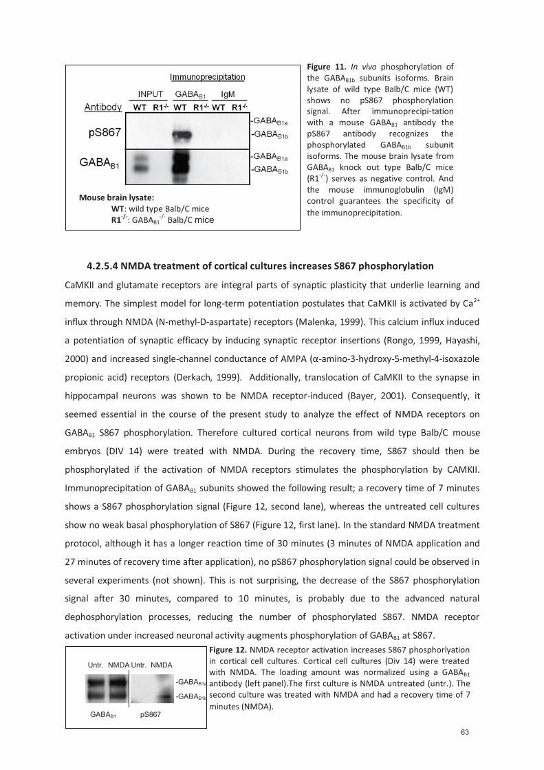
4.2.5.4 NMDA treatment of cortical cultures increases S867 phosphorylation
CaMKII and glutamate receptors are integral parts of synaptic plasticity that underlie learning and
memory. The simplest model for long-term potentiation postulates that CaMKII is activated by Ca2+
influx through NMDA (N-methyl-D-aspartate) receptors (Malenka, 1999). This calcium influx induced
a potentiation of synaptic efficacy by inducing synaptic receptor insertions (Rongo, 1999, Hayashi,
2000) and increased single-channel conductance of AMPA (α-amino-3-hydroxy-5-methyl-4-isoxazole
propionic acid) receptors (Derkach, 1999). Additionally, translocation of CaMKII to the synapse in
hippocampal neurons was shown to be NMDA receptor-induced (Bayer, 2001). Consequently, it
seemed essential in the course of the present study to analyze the effect of NMDA receptors on
GABAB1 S867 phosphorylation. Therefore cultured cortical neurons from wild type Balb/C mouse
embryos (DIV 14) were treated with NMDA. During the recovery time, S867 should then be
phosphorylated if the activation of NMDA receptors stimulates the phosphorylation by CAMKII.
Immunoprecipitation of GABAB1 subunits showed the following result; a recovery time of 7 minutes
shows a S867 phosphorylation signal (Figure 12, second lane), whereas the untreated cell cultures
show no weak basal phosphorylation of S867 (Figure 12, first lane). In the standard NMDA treatment
protocol, although it has a longer reaction time of 30 minutes (3 minutes of NMDA application and
27 minutes of recovery time after application), no pS867 phosphorylation signal could be observed in
several experiments (not shown). This is not surprising, the decrease of the S867 phosphorylation
signal after 30 minutes, compared to 10 minutes, is probably due to the advanced natural
dephosphorylation processes, reducing the number of phosphorylated S867. NMDA receptor
activation under increased neuronal activity augments phosphorylation of GABAB1 at S867.
Figure 11. In vivo phosphorylation of the GABAB1b subunits isoforms. Brain lysate of wild type Balb/C mice (WT) shows no pS867 phosphorylation signal. After immunoprecipi-tation with a mouse GABAB1 antibody the pS867 antibody recognizes the phosphorylated GABAB1b subunit isoforms. The mouse brain lysate from GABAB1 knock out type Balb/C mice (R1-/-) serves as negative control. And the mouse immunoglobulin (IgM) control guarantees the specificity of the immunoprecipitation.
Mouse brain lysate: WT: wild type Balb/C mice
R1-/-: GABAB1-/- Balb/C mice
Figure 12. NMDA receptor activation increases S867 phosphorlyation in cortical cell cultures. Cortical cell cultures (Div 14) were treated with NMDA. The loading amount was normalized using a GABAB1 antibody (left panel).The first culture is NMDA untreated (untr.). The second culture was treated with NMDA and had a recovery time of 7 minutes (NMDA).
-GABAB1a
Untr. NMDA Untr. NMDA
GABAB1 pS867
-GABAB1b
63
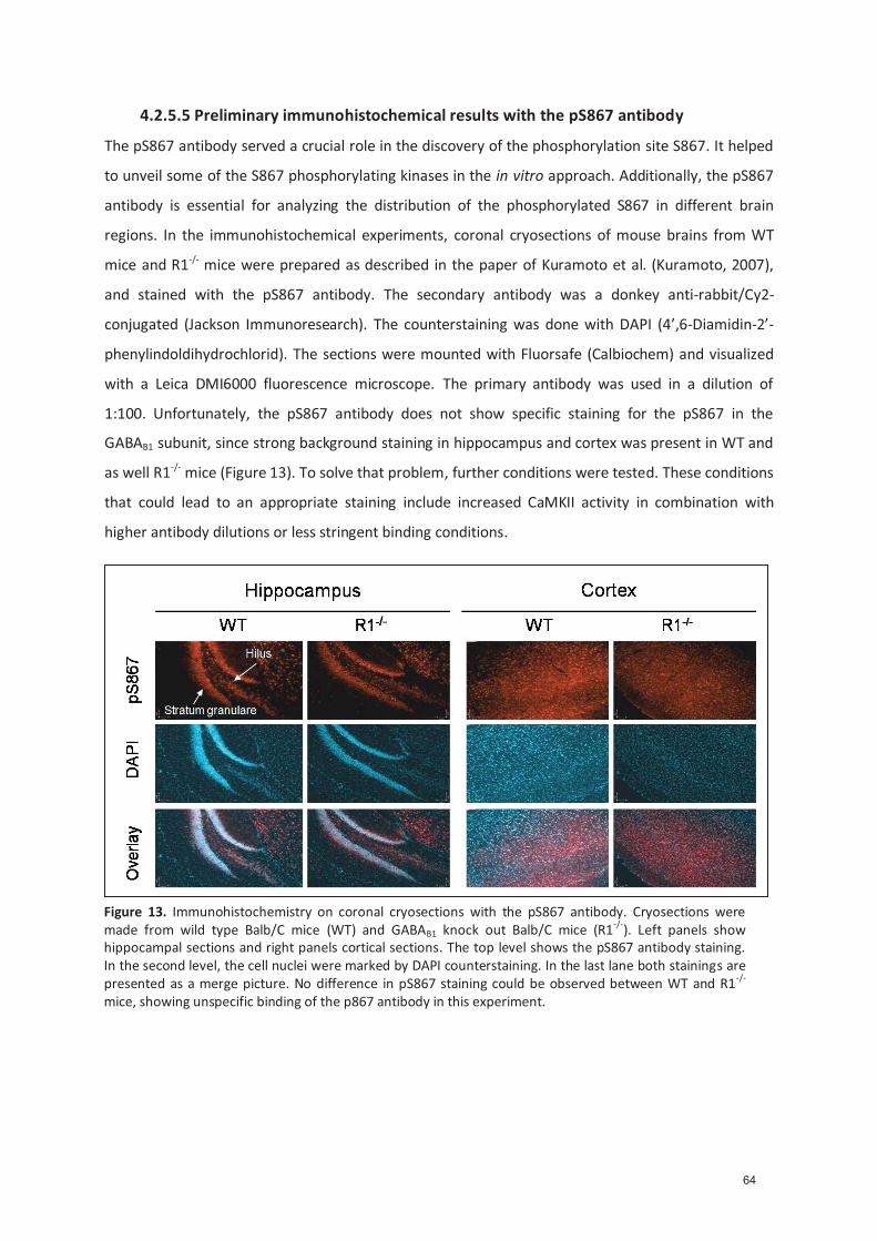
4.2.5.5 Preliminary immunohistochemical results with the pS867 antibody
The pS867 antibody served a crucial role in the discovery of the phosphorylation site S867. It helped
to unveil some of the S867 phosphorylating kinases in the in vitro approach. Additionally, the pS867
antibody is essential for analyzing the distribution of the phosphorylated S867 in different brain
regions. In the immunohistochemical experiments, coronal cryosections of mouse brains from WT
mice and R1-/- mice were prepared as described in the paper of Kuramoto et al. (Kuramoto, 2007),
and stained with the pS867 antibody. The secondary antibody was a donkey anti-rabbit/Cy2-
conjugated (Jackson Immunoresearch). The counterstaining was done with DAPI (4’,6-Diamidin-2’-
phenylindoldihydrochlorid). The sections were mounted with Fluorsafe (Calbiochem) and visualized
with a Leica DMI6000 fluorescence microscope. The primary antibody was used in a dilution of
1:100. Unfortunately, the pS867 antibody does not show specific staining for the pS867 in the
GABAB1 subunit, since strong background staining in hippocampus and cortex was present in WT and
as well R1-/- mice (Figure 13). To solve that problem, further conditions were tested. These conditions
that could lead to an appropriate staining include increased CaMKII activity in combination with
higher antibody dilutions or less stringent binding conditions.
Figure 13. Immunohistochemistry on coronal cryosections with the pS867 antibody. Cryosections were made from wild type Balb/C mice (WT) and GABAB1 knock out Balb/C mice (R1-/-). Left panels show hippocampal sections and right panels cortical sections. The top level shows the pS867 antibody staining. In the second level, the cell nuclei were marked by DAPI counterstaining. In the last lane both stainings are presented as a merge picture. No difference in pS867 staining could be observed between WT and R1-/- mice, showing unspecific binding of the p867 antibody in this experiment.
64
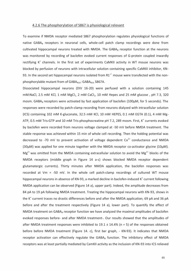
4.2.6 The phosphorylation of S867 is physiological relevant
To examine if NMDA receptor mediated S867 phosphorylation regulates physiological functions of
native GABAB receptors in neuronal cells, whole-cell patch clamp recordings were done from
cultivated hippocampal neurons treated with NMDA. The GABAB receptor function at the neurons
was monitored by recording of baclofen evoked current responses of G-protein coupled inwardly
rectifying K+ channels. In the first set of experiments CaMKII activity in WT mouse neurons was
blocked by perfusion of neurons with intracellular solution containing specific CaMKII inhibitor, KN-
93. In the second set hippocampal neurons isolated from R1-/- mouse were transfected with the non-
phosphorylable mutant from of GABAB1b: GABAB1b S867A.
Dissociated hippocampal neurons (DIV 16-20) were perfused with a solution containing 145
mM:NaCl, 2.5 mM KCl, 1 mM MgCl2, 2 mM CaCl2, 10 mM Hepes and 25 mM glucose , pH 7.3, 320
mosm. GABAB receptors were activated by fast application of baclofen (100μM, for 5 seconds). The
responses were recorded by patch-clamp recording from neurons dialyzed with intracellular solution
(ICS) containing 102 mM K-gluconate, 32.5 mM KCl, 10 mM HEPES, 0.1 mM EGTA (0.1), 4 mM Mg-
ATP, 0.5 mM Tris-GTP and 10 mM Tris-phosphocreatine pH 7.2, 289 mosm. First, K+ currents evoked
by baclofen were recorded from neurons voltage clamped at -50 mV before NMDA treatment. The
stable response was achieved within 15 min of whole cell recording. Then the holding potential was
decreased to -70 mV to prevent activation of voltage dependent Ca2+ conductance and NMDA
(30μM) was applied for one minute together with the NMDA receptor co-activator glycine (10μM).
Mg2+ was omitted from the NMDA containing extracellular solution to avoid the Mg2+ blocks of the
NMDA receptors (middle graph in Figure 14 a-c) shows blocked NMDA receptor dependent
glutamatergic currents). Thirty minutes after NMDA application, the baclofen responses was
recorded at Vm = -50 mV. In the whole cell patch-clamp recordings of cultured WT mouse
hippocampal neurons in absence of KN-93, a marked decline in baclofen-induced K+ current following
NMDA application can be observed (Figure 14 a), upper part). Indeed, the amplitude decreases from
84 pA to 19 pA following NMDA treatment. Treating the hippocampal neurons with KN-93, shows in
the K+ current traces no drastic differences before and after the NMDA application; 69 pA and 36 pA
before and after the treatment respectively (Figure 14 a), lower part). To quantify the effect of
NMDA treatment on GABAB receptor function we have analyzed the maximal amplitudes of baclofen
evoked responses before- and after NMDA treatment-. Our results showed that the amplitudes of
after NMDA treatment responses were inhibited to 19.1 ± 14.4% (n = 5) of the responses obtained
before before NMDA treatment (Figure 14. c), first bar graph, - KN-93). It indicates that NMDA
receptor activation can effectively regulate the GABAB function. The inhibitory effect of NMDA
receptors was at least partially mediated by CamKII activity as the inclusion of KN-93 into ICS relieved
65

NMDA-induced reduction of baclofen responses by nearly 50% (mean K+ amplitude after NMDA
response was to 65.5 ± 20.2%, n = 5) (Figure 14.c), second bar graph).
In the next series of experiments we testedthe hypothesis that mechanism underlying the
NMDA induced inhibition of GABAB responses involves CaMKII-mediated phosphorylation of S867, in
absence of KN-93. Neurons isolated from R1-/- mouse were transfected with WT GABAB1b (GB1b) or
S867A substituted GABAB1b constructs (GB1bS867A) together with GABAB2. The GB1b construct has
before NMDA treatment K+ amplitudes of 51 pA and after NMDA treatment amplitudes of 11 pA
(Figure 14. b), upper part). Exchanging GB1b by the GB1bS867A construct shows that the before
NMDA treatment amplitude was 57 pA before and 32 pA for the after NMDA treatment amplitude
(Figure 14. b) lower part). The WT GABAB1b construct shows 80% (SD 11%) reduction of the K+
response after NMDA application compared to the response before NMDA treatment before the
NMDA application. The K+ current amplitude after NMDA treatment is around 11 pA roughly the
same value as the NMDA treated WT mouse neurons. Although NMDA was applied, the S867A
GABAB1b substitution rescues the K+ response to around 64% (SD 16%) of the baclofen elicited K+
response before NMDA application. The measured K+ amplitude after NMDA is around 32 pA.
The results obtained from the transfected R1-/- mouse neurons show a similar response to NMDA
than the one observed with WT mouse neurons. Indeed, the K+ current amplitude decreases in the
WT mouse neurons and WT GABAB1b construct transfected R1-/- mouse neurons. Conversely, the
decrease in the K+ response was almost prevented in WT mouse neurons in appearance of KN-93 and
S867A GABAB1b construct transfected R1-/- mouse neurons.
Both set of experiments confirmed that NMDA receptor mediated activation of CaMKII
phosphorylates the GABAB1b isoform subunit at S867, leading to a big decrease of GABAB receptor
induced K+ response, probably trough the suggested internalization of the GABAB receptors trough
NMDA receptor activation leads to a drastic increase in intracellular Ca2+ concentration triggering
CaMKII activation. The CaMKII mediated phosphorylation of the GABAB1b subunit isoforms at S867
leads to a strong decrease in postsynaptic inhibitory K+ current induced by baclofen. The measured
decrease indicates a loss of the of the GABAB receptor response. An internalization of surface GABAB
receptors could straightforwardly explain the observed reduction in the evoked K+ currents.
Additionally, a phosphorylation mimetic S867D GABAB1b construct will be generated. The transfection
of that construct should show a reduced K+ response after the NMDA treatment. Further
explanations, on how the S867 phosphorylation could decrease GABAB receptors response, will be
clarified in the following final discussion chapter.
66
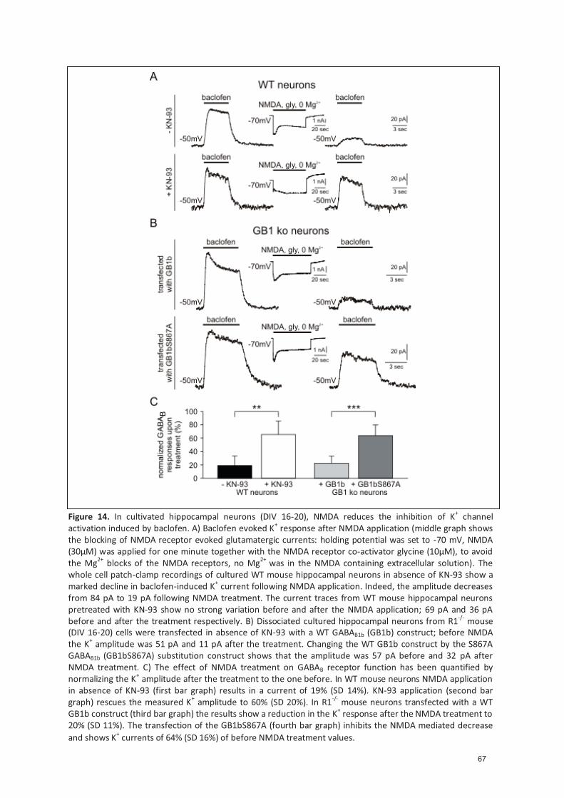
Figure 14. In cultivated hippocampal neurons (DIV 16-20), NMDA reduces the inhibition of K+ channel activation induced by baclofen. A) Baclofen evoked K+ response after NMDA application (middle graph shows the blocking of NMDA receptor evoked glutamatergic currents: holding potential was set to -70 mV, NMDA (30μM) was applied for one minute together with the NMDA receptor co-activator glycine (10μM), to avoid the Mg2+ blocks of the NMDA receptors, no Mg2+ was in the NMDA containing extracellular solution). The whole cell patch-clamp recordings of cultured WT mouse hippocampal neurons in absence of KN-93 show a marked decline in baclofen-induced K+ current following NMDA application. Indeed, the amplitude decreases from 84 pA to 19 pA following NMDA treatment. The current traces from WT mouse hippocampal neurons pretreated with KN-93 show no strong variation before and after the NMDA application; 69 pA and 36 pA before and after the treatment respectively. B) Dissociated cultured hippocampal neurons from R1-/- mouse (DIV 16-20) cells were transfected in absence of KN-93 with a WT GABAB1b (GB1b) construct; before NMDA the K+ amplitude was 51 pA and 11 pA after the treatment. Changing the WT GB1b construct by the S867A GABAB1b (GB1bS867A) substitution construct shows that the amplitude was 57 pA before and 32 pA after NMDA treatment. C) The effect of NMDA treatment on GABAB receptor function has been quantified by normalizing the K+ amplitude after the treatment to the one before. In WT mouse neurons NMDA application in absence of KN-93 (first bar graph) results in a current of 19% (SD 14%). KN-93 application (second bar graph) rescues the measured K+ amplitude to 60% (SD 20%). In R1-/- mouse neurons transfected with a WT GB1b construct (third bar graph) the results show a reduction in the K+ response after the NMDA treatment to 20% (SD 11%). The transfection of the GB1bS867A (fourth bar graph) inhibits the NMDA mediated decrease and shows K+ currents of 64% (SD 16%) of before NMDA treatment values.
67

V. Final discussion
The identification of a CaMKII phosphorylation site on a major neuronal receptor is of a special
significance. All brain functions need, for an optimal functioning, an adapted balance of excitation
and inhibition. The GABAB receptor was the last major brain receptor in the CNS to be cloned
(Kaupmann, 1997). The metabotropic GABAB receptor binds GABA, the main inhibitory
neurotransmitter in the mammalian nervous system. For a long-time, it was generally assumed that
the GABAB receptors system included several pharmacologically distinct receptor subtypes, like the
related mGluR family. However, only two receptor subtypes were identified; GABAB(1a,2) and
GABAB(1b,2) (Kaupmann, 1998a). It is unclear how this small receptor heterogeneity ensures the
differing electrophysiological and pharmacological in vivo responses observed for native GABAB
receptors. As previously discussed, the differing subcellular localization of the two receptor subtypes
is believed to be the reason for the differing behavior of both subtypes (Vigot, 2006, Guetg, 2009).
The inhibitory effects of GABAB receptor activation on the brain are typical for a GPCR, slow and
prolonged. The presynaptic activation of these receptors lead to the modulation of adenylate cyclase
activity or to the blocking of voltage-dependent Ca2+ channels which inhibit neurotransmitters
release. On the other hand, postsynaptic receptors activation leads to a modulation in the gating of
Kir3-type K+ channels.
Surprisingly, GABAB receptor regulating mechanisms remain unidentified to a great extent. However,
it is thought that receptor-associated proteins and direct posttranslational modifications influence
the receptor function. Phosphorylation by protein kinases is known as one of the most important
post-translational modifications (Hunter, 1995). The importance of protein kinases in regulating
cellular activities is already reflected by the large number of protein kinases related genes present in
eukaryotic genomes; protein kinases are one of the largest superfamilies accounting for 1.7% of the
human genome (Manning, 2002). Protein kinases were found to be enriched in the brain, where they
play a critical role in the nervous systems such as memory formation and learning processes. Protein
kinases also influence the GABAB receptors. For instance, PKA phosphorylates the GABAB receptor at
the site S892 causing an elongation of the inhibitory action of GABAB receptors (Couve, 2002). This
observation was first contradictory to the prevailing dogma being that phosphorylation reduces the
GPCRs activity either by increasing receptor desensitization or by removing receptors from the cell
surface. S892 phosphorylation by PKA strengthens the presence of GABAB receptors at cell surface,
thereby enhancing the coupling of GABAB receptors to its effector. S783, another GABAB receptor
phosphorylation site, was later described (Kuramoto, 2007). AMPK phosphorylates S783 thereby
stabilizing the activating effects of the receptors on K+ currents; the K+ current rundown is finally
reduced. AMPK phosphorylation at GABAB receptors is thought to be a novel neuroprotective
68
mechanism, which, under conditions of metabolic stress or ischemia, increases GABAB receptor
function. This consequently reduces excitotoxicity and promotes neuronal survival which diminishes
the harms of a stroke incident. Both S892 and S783 sites are localized on the carboxyl-terminal of the
GABAB2 subunit. For a long time, the GABAB2 subunit seemed to be the most interesting target for
phosphorylation site search on the GABAB receptor; the GABAB2 subunit has the longer carboxyl-
terminal and the GABAB2 subunit is, due to the G-protein interaction, already localized near second
messenger–dependent kinases. Indications for possible phosphorylation sites on the GABAB1 subunit
existed, but were demonstrated only at the in vitro stage, e.g. the S917 was shown on GST-fusion
protein containing the GABAB1 carboxyl-terminal, but not in vivo, and no physiological relevance
could be attributed (Kuramoto, 2007).
In my thesis I identified the first phosphorylation site localized on native GABAB1 subunits, serine 867
(S867). S867 is juxtamembrane positioned on the GABAB1 carboxyl-terminal. It gets phosphorylated
by CaMKII, mostly at the GABAB1b subunit isoform. CaMKII is implicated in a wide variety of neuronal
functions, e.g. memory formation, receptor functioning, structural modification of the cytoskeleton,
axonal transport, and gene expression (Hanson, 1992, Lisman, 1994, Yoshimura, 2000, Yoshimura,
2002). GABAB receptor activity increases the phosphorylation of threonine 286 on α CaMKII (Xu,
2008). Nevertheless, a CaMKII phosphorylation site on the GABAB receptor was never shown. The
present findings finally confirm that CaMKII is regulating, via GABAB1-S867 phosphorylation, GABAB
receptor response.
CaMKII is highly abundant in neurons, especially enriched at synaptic endings (Kelly, 1984) and has a
central role in the regulation of glutamatergic synapses. Its implication in glutamatergic transmission
has emerged largely from the study of LTP, an activity dependent strengthening of synapses that is
thought to underlie some forms of learning and memory. At many excitatory synapses, LTP is
triggered by Ca2+ entry into the postsynaptic cell. Several lines of evidence indicate that CaMKII
detects this Ca2+ elevation and initiates the biochemical cascade that potentiates synaptic
transmission by recruiting AMPA and NMDA receptors to the postsynaptic membrane (Malenka,
1989b, Malinow, 1989, Bayer, 2001, Lisman, 2002). CaMKII might not only induce LTP but might also
be responsible for the persistence of LTP, therefore contributing to memory formation. The strongest
evidence for this idea comes from the observation that CaMKII remains activated for at least one
hour after LTP induction and autophosphorylation of threonine 286 is crucial for its persistent
activation; a mutation that eliminates phosphorylation of this site blocks LTP (Lisman, 2002).
The molecular mechanism underlying the phosphorylation of S867 by CaMKII points in the same
direction as synaptic plasticity. Indeed, regulation of synaptic transmission depends on the
69
availability of surface neurotransmitter receptors and surface GABAB receptor levels were shown to
be decreased in presence of glutamate. The alanine substitution of S867 (S867A) that prevents S867
phosphorylation renders GABAB receptors refractory to NMDA-mediated internalization. KN-93
application or the use of the S867A mutant abolished, in hippocampal dissociated neurons, the
reduction of the GABAB receptor elicited K+ currents after the NMDA treatment. Interestingly, the
receptors ability to evoke inhibitory K+ response was preserved. This result also indicates that S867
phosphorylation leads to an internalization of the GABAB receptors. The glutamate induced reduction
of surface GABAB receptors was postulated to be mediated by a dynamin-dependent clathrin
pathway (Vargas, 2008). To confirm that the NMDA mediated internalization works via the same
pathway, the internalization should be abolished under hypertonic concentrations of sucrose
(Grampp, 2007) or exposure to low K+ concentrations (Bayer, 2001) conditions inhibiting clathrin-
dependent internalizations. Clean and specific inhibition of the dynamin-dependent clathrin pathway
in hippocampal cells was made possible by the use of dynasore. Dynasore has a potent in vitro
inhibitory effect on the GTPase activity of dynamin1, dynamin2 and Drp1but not on other small
GTPases (Macia, 2006). The effects of the dynasore administration can be studied using the
immunofluorescence-based internalization assay. Additionally, cell surface ELISA and biotinylation
assay, with cultured neuronal cells, should be also considered. Optionally, it can be argued that S867
phosphorylation does not contribute to GABAB receptors internalization but only reduce their
recycling to the membrane surface. However, this discrepancy can be solved by transfecting inactive
Rab protein mutant in Rab protein deficient cells (Glodowski, 2007). Rab proteins are guanosine
triphosphateases that mediate the recycling and trafficking of endocytosed vesicles to the cell
surface. The inactive Rab mutant should not reduce the number of surface GABAB receptors if the
phosphorylation is involved in receptor internalization rather than receptor recycling.
The S867 phosphorylation was predominantly found at the native GABAB1b subunit which is, along
with CaMKII, concentrated in the same postsynaptic compartments (Kennedy, 1983). The two GABAB
receptor subtypes GABAB(1a,2) and GABAB1(b,2) produce different responses at axonal and dendritic
effectors when pharmacologically activated by a saturating concentration of baclofen (Perez-Garci,
2006, Shaban, 2006, Vigot, 2006, Ulrich, 2007). The diverse pharmacological reaction of the two
subtypes was explained by the distinct distribution profile of the GABAB1a and GABAB1b subunits to
axonal and dendritic compartments respectively (Vigot, 2006, Guetg, 2009). Their expressions are
also under separate transcriptional control which influence the ratio of receptor subtypes in
individual neurons (Steiger, 2004). S867 phosphorylation represents a selective and specific
modification between the two subunits. Primary, the GABAB1b subunits are controlled by the S867
phosphorylation effect. Transient high NMDA release leads to S867 GABAB1b subunit phosphorylation
consequently reducing the inhibitory GABAB response in dendritic spines. This decrease in inhibition
70

contributes to an increased synaptic transmission efficacy. For this reason, one can anticipate a
contribution of S867 phosphorylation to LTP and long-term depression (LTD) formation. LTD, the
opposing process of LTP, requires also an NMDAR dependent rise in the postsynaptic Ca2+
concentration, which preferentially activates a protein-phosphatase cascade that includes calcineurin
and phosphatase 1 (Carroll, 2001). The phosphatases could potentially dephosphorylate the pool of
S867 phosphorylated GABAB receptors. The internalization of the GABAB would then be reduced
consequently leading to an increased inhibitory transmission in the CNS. One can then expect that
the dephosphorylation of S867 could probably contribute to LTD like phenomenon; the inhibitory
fence of the gabaergic system would be reset and even strengthened.
Further effects on the GABAB receptor system through the S867 phosphorylation by CaMKII are
imaginable. CaMKII phosphorylation was shown to regulate mGluR1 trafficking to different dendritic
compartments (Kielland, 2009) and to influence the availability of Homer3 for its targets in the
postsynaptic densities (Mizutani, 2008). Similarly, GABAB1 subunit phosphorylation could also
potentially leads to a conformational change of the subunit which would increase the binding to
scaffolding proteins such as β-arrestins, as shown for other GPCRs, e.g. anaphylatoxin receptor
(Braun, 2003).
The NMDA receptor mediated Ca2+ influx can activate, in addition to CaMKII, the kinase PKA
(Moyano, 2004). Since PKA was shown in vitro to phosphorylate S867; further attention should be
paid to a possible in vivo phosphorylation of S867 by PKA. This can be done by repeating the NMDA
treatment experiments but in the presence of the cAMP–PKA pathway activator or inhibitor; e.g.
forskolin or the PKA inhibitor peptide14-22 or Walsh peptide (Moreno-Delgado, 2009). PKA also
mediates crucial physiological functions in the brain, especially in processes like LTP (Anwyl, 2009).
GABAB receptors are known to influence PKA activity by decreasing cAMP production, via their
restrictive action on adenylate cyclase. PKA phosphorylates large arrays of proteins involved in mood
disorders; e.g. Rap-1 (a member of the Ras family and is highly expressed in the CNS) (Dwivedi, 2008).
Additionally they are many significant intersections between GABAB receptors and PKA activity.
Indeed, via PKA dependent pathways, presynaptic GABAB receptors decrease the release of GABA
from autoreceptors. This mechanism has an important role in the regulation of cholinergic
transmission in the nucleus basalis of Meynert (Kubota, 2003), where a decrease in acetylcholine
production is a key component in Alzheimer disease etiology (Freund, 2009). PKA and GABAB
receptors are also essential in the sleep-wake behavior of mammals (Datta, 2007). Application of a
PKA activator to Sprague-Dawley rats induces REM sleep, whereas baclofen application has the
opposite effect (Datta, 2007).
71
Concluding, the phosphorylation site S867 can be seen as major part of a feedback mechanism. The
active GABAB receptors decrease the activity of CaMKII and PKA. The physiological S867
phosphorylation by CaMKII and potentially by PKA, antangonizes the decrease in the kinases activity.
S867 phosphorylation reduces the inhibitory activity of GABAB receptors. This feedback mechanism
would ensure a fine tunning of the inhibitory transmission in the CNS that could otherwise not be
guaranteed. S867 is also part of the cross-talk between the glutamatergic and gabaergic nervous
system. The NMDA-dependent phosphorylation of S867 by CaMKII internalizes the whole GABAB
receptor. Consequently, the inhibitory response of the gabaergic system is locally decreased,
whereas the excitatory circuit is enhanced; further recruitment of AMPA and NMDA receptor is then
facilitated, phenomenon also observed during LTP. GABAB receptor subtype specificity of S867
phosphorylation could be seen as an additional explanation for the different GABAB(1a,2) and
GABAB(1b,2) receptors localization. In conclusion, S867 phosphorylation plays an active part in the
orchestration of synaptic plasticity and clearly manifests many possibilities for further scientific
findings on the inhibitory effect of GABAB receptors on the CNS. Especialy the generation of a GABAB1
S867A mice model could be an important landmark in the study of the role of S867 phosphorylation
on the CNS. On such a mice model the regulatory effect of the S867 phosphorylation on the
inhibitory gabaergic is able to get studied under different and the most physiological ways. The
development of new research avenues on that subject enables the development of new innovative
approches for the treatment of neurological and psychiatric diseases.
72
VI. References
Abramoff MD, Magelhaes PJ, Ram SJ (2004) Image Processing with ImageJ. Biophotonics International 11:7
Addolorato G, Caputo F, Capristo E, Domenicali M, Bernardi M, Janiri L, Agabio R, Colombo G, Gessa
GL, Gasbarrini G (2002) Baclofen efficacy in reducing alcohol craving and intake: a preliminary double-blind randomized controlled study. Alcohol Alcohol 37:504-508
Amico C, Marchetti C, Nobile M, Usai C (1995) Pharmacological types of calcium channels and their
modulation by baclofen in cerebellar granules. J Neurosci 15:2839-2848 Anwyl R (2009) Metabotropic glutamate receptor-dependent long-term potentiation.
Neuropharmacology 56:735-740 Ardenghi P, Barros D, Izquierdo LA, Bevilaqua L, Schroder N, Quevedo J, Rodrigues C, Madruga M,
Medina JH, Izquierdo I (1997) Late and prolonged post-training memory modulation in entorhinal and parietal cortex by drugs acting on the cAMP/protein kinase A signalling pathway. Behav Pharmacol 8:745-751
Arnsten AF, Ramos BP, Birnbaum SG, Taylor JR (2005) Protein kinase A as a therapeutic target for
memory disorders: rationale and challenges. Trends Mol Med 11:121-128 Asano T, Ogasawara N (1986) Uncoupling of gamma-aminobutyric acid B receptors from GTP-binding
proteins by N-ethylmaleimide: effect of N-ethylmaleimide on purified GTP-binding proteins. Mol Pharmacol 29:244-249
Awapara J, Landua AJ, Fuerst R, Seale B (1950) Free gamma-aminobutyric acid in brain. J Biol Chem
187:35-39 Bach ME, Barad M, Son H, Zhuo M, Lu YF, Shih R, Mansuy I, Hawkins RD, Kandel ER (1999) Age-
related defects in spatial memory are correlated with defects in the late phase of hippocampal long-term potentiation in vitro and are attenuated by drugs that enhance the cAMP signaling pathway. Proc Natl Acad Sci U S A 96:5280-5285
Balerio GN, Rubio MC (2002) Baclofen analgesia: involvement of the GABAergic system. Pharmacol
Res 46:281-286 Barad M, Bourtchouladze R, Winder DG, Golan H, Kandel E (1998) Rolipram, a type IV-specific
phosphodiesterase inhibitor, facilitates the establishment of long-lasting long-term potentiation and improves memory. Proc Natl Acad Sci U S A 95:15020-15025
Barros DM, Izquierdo LA, Mello e Souza T, Ardenghi PG, Pereira P, Medina JH, Izquierdo I (2000)
Molecular signalling pathways in the cerebral cortex are required for retrieval of one-trial avoidance learning in rats. Behav Brain Res 114:183-192
Barros DM, Mello e Souza T, De David T, Choi H, Aguzzoli A, Madche C, Ardenghi P, Medina JH,
Izquierdo I (2001) Simultaneous modulation of retrieval by dopaminergic D(1), beta-noradrenergic, serotonergic-1A and cholinergic muscarinic receptors in cortical structures of the rat. Behav Brain Res 124:1-7
73
Bayer KU, De Koninck P, Leonard AS, Hell JW, Schulman H (2001) Interaction with the NMDA receptor locks CaMKII in an active conformation. Nature 411:801-805
Benke D, Honer M, Michel C, Bettler B, Mohler H (1999) gamma-aminobutyric acid type B receptor
splice variant proteins GBR1a and GBR1b are both associated with GBR2 in situ and display differential regional and subcellular distribution. The Journal of biological chemistry 274:27323-27330
Bennett MK, Erondu NE, Kennedy MB (1983) Purification and characterization of a calmodulin-
dependent protein kinase that is highly concentrated in brain. The Journal of biological chemistry 258:12735-12744
Benovic JL, Pike LJ, Cerione RA, Staniszewski C, Yoshimasa T, Codina J, Caron MG, Lefkowitz RJ (1985)
Phosphorylation of the mammalian beta-adrenergic receptor by cyclic AMP-dependent protein kinase. Regulation of the rate of receptor phosphorylation and dephosphorylation by agonist occupancy and effects on coupling of the receptor to the stimulatory guanine nucleotide regulatory protein. J Biol Chem 260:7094-7101
Bettler B, Kaupmann K, Mosbacher J, Gassmann M (2004) Molecular structure and physiological
functions of GABA(B) receptors. Physiol Rev 84:835-867 Bischoff S, Leonhard S, Reymann N, Schuler V, Shigemoto R, Kaupmann K, Bettler B (1999) Spatial
distribution of GABA(B)R1 receptor mRNA and binding sites in the rat brain. The Journal of comparative neurology 412:1-16
Blazquez C, Geelen MJ, Velasco G, Guzman M (2001) The AMP-activated protein kinase prevents
ceramide synthesis de novo and apoptosis in astrocytes. FEBS Lett 489:149-153 Blein S, Ginham R, Uhrin D, Smith BO, Soares DC, Veltel S, McIlhinney RA, White JH, Barlow PN (2004)
Structural analysis of the complement control protein (CCP) modules of GABA(B) receptor 1a: only one of the two CCP modules is compactly folded. The Journal of biological chemistry 279:48292-48306
Bothner B, Dong XF, Bibbs L, Johnson JE, Siuzdak G (1998) Evidence of viral capsid dynamics using
limited proteolysis and mass spectrometry. J Biol Chem 273:673-676 Bouvier M (2001) Oligomerization of G-protein-coupled transmitter receptors. Nature reviews 2:274-
286 Bowery NG (1993) GABAB receptor pharmacology. Annu Rev Pharmacol Toxicol 33:109-147 Bowery NG, Bettler B, Froestl W, Gallagher JP, Marshall F, Raiteri M, Bonner TI, Enna SJ (2002)
International Union of Pharmacology. XXXIII. Mammalian gamma-aminobutyric acid(B) receptors: structure and function. Pharmacol Rev 54:247-264
Braun AP, Schulman H (1995) The multifunctional calcium/calmodulin-dependent protein kinase:
from form to function. Annu Rev Physiol 57:417-445 Braun L, Christophe T, Boulay F (2003) Phosphorylation of key serine residues is required for
internalization of the complement 5a (C5a) anaphylatoxin receptor via a beta-arrestin, dynamin, and clathrin-dependent pathway. J Biol Chem 278:4277-4285
74

Brebner K, Phelan R, Roberts DC (2000) Intra-VTA baclofen attenuates cocaine self-administration on a progressive ratio schedule of reinforcement. Pharmacol Biochem Behav 66:857-862
Brebner K, Froestl W, Roberts DC (2002) The GABA(B) antagonist CGP56433A attenuates the effect of
baclofen on cocaine but not heroin self-administration in the rat. Psychopharmacology (Berl) 160:49-55
Brewer GJ (1993) Optimized survival of hippocampal neurons in B-27 supplemented Neurobasal, a
new serum-free medium combination. J Neurosci Res 35:567-576 Cai Z, Saugstad JA, Sorensen SD, Ciombor KJ, Zhang C, Schaffhauser H, Hubalek F, Pohl J, Duvoisin
RM, Conn PJ (2001) Cyclic AMP-dependent protein kinase phosphorylates group III metabotropic glutamate receptors and inhibits their function as presynaptic receptors. J Neurochem 78:756-766
Calver AR, Medhurst AD, Robbins MJ, Charles KJ, Evans ML, Harrison DC, Stammers M, Hughes SA,
Hervieu G, Couve A, Moss SJ, Middlemiss DN, Pangalos MN (2000) The expression of GABAB(1) and GABAB(2) receptor subunits in the CNS differs from that in peripheral tissues. Neuroscience 100:155-170
Calver AR, Robbins MJ, Cosio C, Rice SQ, Babbs AJ, Hirst WD, Boyfield I, Wood MD, Russell RB, Price
GW, Couve A, Moss SJ, Pangalos MN (2001) The C-terminal domains of the GABA(b) receptor subunits mediate intracellular trafficking but are not required for receptor signaling. J Neurosci 21:1203-1210
Calver AR, Davies CH, Pangalos M (2002) GABA(B) receptors: from monogamy to promiscuity.
Neurosignals 11:299-314 Campbell V, Berrow N, Dolphin AC (1993) GABAB receptor modulation of Ca2+ currents in rat sensory
neurones by the G protein G(0): antisense oligonucleotide studies. The Journal of physiology 470:1-11
Campbell UC, Lac ST, Carroll ME (1999) Effects of baclofen on maintenance and reinstatement of
intravenous cocaine self-administration in rats. Psychopharmacology (Berl) 143:209-214 Cardozo DL, Bean BP (1995) Voltage-dependent calcium channels in rat midbrain dopamine neurons:
modulation by dopamine and GABAB receptors. J Neurophysiol 74:1137-1148 Carroll RC, Beattie EC, von Zastrow M, Malenka RC (2001) Role of AMPA receptor endocytosis in
synaptic plasticity. Nat Rev Neurosci 2:315-324 Chebib M, Johnston GA (1999) The 'ABC' of GABA receptors: a brief review. Clin Exp Pharmacol
Physiol 26:937-940 Chittenden T, Livingston DM, Kaelin WG, Jr. (1991) The T/E1A-binding domain of the retinoblastoma
product can interact selectively with a sequence-specific DNA-binding protein. Cell 65:1073-1082
Choi DS, Wang D, Dadgar J, Chang WS, Messing RO (2002) Conditional rescue of protein kinase C
epsilon regulates ethanol preference and hypnotic sensitivity in adult mice. J Neurosci 22:9905-9911
75

Colbran RJ (2004) Targeting of calcium/calmodulin-dependent protein kinase II. Biochem J 378:1-16 Collingridge GL, Lester RA (1989) Excitatory amino acid receptors in the vertebrate central nervous
system. Pharmacological reviews 41:143-210 Collingridge GL, Isaac JT, Wang YT (2004) Receptor trafficking and synaptic plasticity. Nat Rev
Neurosci 5:952-962 Colombo G, Agabio R, Carai MA, Lobina C, Pani M, Reali R, Addolorato G, Gessa GL (2000) Ability of
baclofen in reducing alcohol intake and withdrawal severity: I--Preclinical evidence. Alcohol Clin Exp Res 24:58-66
Conn PJ, Pin JP (1997) Pharmacology and functions of metabotropic glutamate receptors. Annu Rev
Pharmacol Toxicol 37:205-237 Couve A, Filippov AK, Connolly CN, Bettler B, Brown DA, Moss SJ (1998) Intracellular retention of
recombinant GABAB receptors. The Journal of biological chemistry 273:26361-26367 Couve A, Moss SJ, Pangalos MN (2000) GABAB receptors: a new paradigm in G-protein signaling. Mol
Cell Neurosci 16:296-312 Couve A, Thomas P, Calver AR, Hirst WD, Pangalos MN, Walsh FS, Smart TG, Moss SJ (2002) Cyclic
AMP-dependent protein kinase phosphorylation facilitates GABA(B) receptor-effector coupling. Nat Neurosci 5:415-424
Craft CM, Whitmore DH (1995) The arrestin superfamily: cone arrestins are a fourth family. FEBS Lett
362:247-255 Crunelli V, Leresche N (1991) A role for GABAB receptors in excitation and inhibition of
thalamocortical cells. Trends Neurosci 14:16-21 Cryan JF, Kaupmann K (2005) Don't worry 'B' happy!: a role for GABA(B) receptors in anxiety and
depression. Trends Pharmacol Sci 26:36-43 Cui JG, Meyerson BA, Sollevi A, Linderoth B (1998) Effect of spinal cord stimulation on tactile
hypersensitivity in mononeuropathic rats is potentiated by simultaneous GABA(B) and adenosine receptor activation. Neurosci Lett 247:183-186
Culmsee C, Monnig J, Kemp BE, Mattson MP (2001) AMP-activated protein kinase is highly expressed
in neurons in the developing rat brain and promotes neuronal survival following glucose deprivation. J Mol Neurosci 17:45-58
Curtis DR, Phillis JW, Watkins JC (1959) Chemical excitation of spinal neurones. Nature 183:611-612 Curtis DR, Duggan AW, Felix D, Johnston GA (1970) GABA, bicuculline and central inhibition. Nature
226:1222-1224 Dai Y, Moriyama T, Higashi T, Togashi K, Kobayashi K, Yamanaka H, Tominaga M, Noguchi K (2004)
Proteinase-activated receptor 2-mediated potentiation of transient receptor potential vanilloid subfamily 1 activity reveals a mechanism for proteinase-induced inflammatory pain. J Neurosci 24:4293-4299
76

Daoust M, Saligaut C, Lhuintre JP, Moore N, Flipo JL, Boismare F (1987) GABA transmission, but not benzodiazepine receptor stimulation, modulates ethanol intake by rats. Alcohol 4:469-472
Datta S (2007) Activation of pedunculopontine tegmental PKA prevents GABAB receptor activation-
mediated rapid eye movement sleep suppression in the freely moving rat. J Neurophysiol 97:3841-3850
Davis JB, Gray J, Gunthorpe MJ, Hatcher JP, Davey PT, Overend P, Harries MH, Latcham J, Clapham C,
Atkinson K, Hughes SA, Rance K, Grau E, Harper AJ, Pugh PL, Rogers DC, Bingham S, Randall A, Sheardown SA (2000) Vanilloid receptor-1 is essential for inflammatory thermal hyperalgesia. Nature 405:183-187
de Toledo-Morrell L, Morrell F, Fleming S, Cohen MM (1984) Pentoxifylline reverses age-related
deficits in spatial memory. Behav Neural Biol 42:1-8 DeGiorgis JA, Jaffe H, Moreira JE, Carlotti CG, Jr., Leite JP, Pant HC, Dosemeci A (2005)
Phosphoproteomic analysis of synaptosomes from human cerebral cortex. J Proteome Res 4:306-315
Deisz RA, Billard JM, Zieglgansberger W (1997) Presynaptic and postsynaptic GABAB receptors of
neocortical neurons of the rat in vitro: differences in pharmacology and ionic mechanisms. Synapse 25:62-72
Derkach V, Barria A, Soderling TR (1999) Ca2+/calmodulin-kinase II enhances channel conductance of
alpha-amino-3-hydroxy-5-methyl-4-isoxazolepropionate type glutamate receptors. Proc Natl Acad Sci U S A 96:3269-3274
Dewey SL, Morgan AE, Ashby CR, Jr., Horan B, Kushner SA, Logan J, Volkow ND, Fowler JS, Gardner EL,
Brodie JD (1998) A novel strategy for the treatment of cocaine addiction. Synapse 30:119-129 Dewey SL, Brodie JD, Gerasimov M, Horan B, Gardner EL, Ashby CR, Jr. (1999) A pharmacologic
strategy for the treatment of nicotine addiction. Synapse 31:76-86 Diella F, Cameron S, Gemund C, Linding R, Via A, Kuster B, Sicheritz-Ponten T, Blom N, Gibson TJ
(2004) Phospho.ELM: a database of experimentally verified phosphorylation sites in eukaryotic proteins. BMC Bioinformatics 5:79
Di-Capua N, Sperling O, Zoref-Shani E (2003) Protein kinase C-epsilon is involved in the adenosine-
activated signal transduction pathway conferring protection against ischemia-reperfusion injury in primary rat neuronal cultures. J Neurochem 84:409-412
Diverse-Pierluissi M, Remmers AE, Neubig RR, Dunlap K (1997) Novel form of crosstalk between G
protein and tyrosine kinase pathways. Proc Natl Acad Sci U S A 94:5417-5421 Doble BW, Woodgett JR (2007) Role of glycogen synthase kinase-3 in cell fate and epithelial-
mesenchymal transitions. Cells Tissues Organs 185:73-84 Dreifuss JJ, Kelly JS, Krnjevic K (1969) Cortical inhibition and gamma-aminobutyric acid. Exp Brain Res
9:137-154
77

Duffy SN, Nguyen PV (2003) Postsynaptic application of a peptide inhibitor of cAMP-dependent protein kinase blocks expression of long-lasting synaptic potentiation in hippocampal neurons. J Neurosci 23:1142-1150
Dumuis A, Sebben M, Haynes L, Pin JP, Bockaert J (1988) NMDA receptors activate the arachidonic
acid cascade system in striatal neurons. Nature 336:68-70 Dwivedi Y, Pandey GN (2008) Adenylyl cyclase-cyclicAMP signaling in mood disorders: Role of the
crucial phosphorylating enzyme protein kinase A. Neuropsychiatr Dis Treat 4:161-176 Enna SJ, Bowery NG (2004) GABA(B) receptor alterations as indicators of physiological and
pharmacological function. Biochem Pharmacol 68:1541-1548 Erondu NE, Kennedy MB (1985) Regional distribution of type II Ca2+/calmodulin-dependent protein
kinase in rat brain. J Neurosci 5:3270-3277 Faigle JW, Keberle H (1972) The chemistry and kinetics of Lioresal. Postgrad Med J 48:Suppl 5:9-13 Fairfax BP, Pitcher JA, Scott MG, Calver AR, Pangalos MN, Moss SJ, Couve A (2004) Phosphorylation
and chronic agonist treatment atypically modulate GABAB receptor cell surface stability. The Journal of biological chemistry 279:12565-12573
Faure C, Corvol JC, Toutant M, Valjent E, Hvalby O, Jensen V, El Messari S, Corsi JM, Kadare G, Girault
JA (2007) Calcineurin is essential for depolarization-induced nuclear translocation and tyrosine phosphorylation of PYK2 in neurons. Journal of cell science 120:3034-3044
Fayard E, Tintignac LA, Baudry A, Hemmings BA (2005) Protein kinase B/Akt at a glance. J Cell Sci
118:5675-5678 Ferguson SS, Downey WE, 3rd, Colapietro AM, Barak LS, Menard L, Caron MG (1996) Role of beta-
arrestin in mediating agonist-promoted G protein-coupled receptor internalization. Science 271:363-366
Ferguson SS (2001) Evolving concepts in G protein-coupled receptor endocytosis: the role in receptor
desensitization and signaling. Pharmacol Rev 53:1-24 Fikrig E, Barthold SW, Kantor FS, Flavell RA (1990) Protection of mice against the Lyme disease agent
by immunizing with recombinant OspA. Science 250:553-556 Filippov AK, Couve A, Pangalos MN, Walsh FS, Brown DA, Moss SJ (2000) Heteromeric assembly of
GABA(B)R1 and GABA(B)R2 receptor subunits inhibits Ca(2+) current in sympathetic neurons. J Neurosci 20:2867-2874
Florey E (1954) An inhibitory and an excitatory factor of mammalian central nervous system, and
their action of a single sensory neuron. Arch Int Physiol 62:33-53 Freund HJ, Kuhn J, Lenartz D, Mai JK, Schnell T, Klosterkoetter J, Sturm V (2009) Cognitive functions in
a patient with Parkinson-dementia syndrome undergoing deep brain stimulation. Arch Neurol 66:781-785
Fromm GH (1989) The pharmacology of trigeminal neuralgia. Clin Neuropharmacol 12:185-194 Futatsugi Y, Riviello JJ, Jr. (1998) Mechanisms of generalized absence epilepsy. Brain Dev 20:75-79
78
Galindo A (1969) GABA-picrotoxin interaction in the mammalian central nervous system. Brain Res 14:763-767
Gallimberti L, Spella MR, Soncini CA, Gessa GL (2000) Gamma-hydroxybutyric acid in the treatment of
alcohol and heroin dependence. Alcohol 20:257-262 Galloway GP, Frederick-Osborne SL, Seymour R, Contini SE, Smith DE (2000) Abuse and therapeutic
potential of gamma-hydroxybutyric acid. Alcohol 20:263-269 Galvez T, Duthey B, Kniazeff J, Blahos J, Rovelli G, Bettler B, Prezeau L, Pin JP (2001) Allosteric
interactions between GB1 and GB2 subunits are required for optimal GABA(B) receptor function. EMBO J 20:2152-2159
Gambardella A, Manna I, Labate A, Chifari R, La Russa A, Serra P, Cittadella R, Bonavita S, Andreoli V,
LePiane E, Sasanelli F, Di Costanzo A, Zappia M, Tedeschi G, Aguglia U, Quattrone A (2003) GABAB receptor 1 polymorphism (G1465A) is associated with temporal lobe epilepsy. Neurology 60:560-563
Gassmann M et al. (2004) Redistribution of GABAB(1) protein and atypical GABAB responses in
GABAB(2)-deficient mice. The Journal of neuroscience 24:6086-6097 Gassmann M, Haller C, Stoll Y, Aziz SA, Biermann B, Mosbacher J, Kaupmann K, Bettler B (2005) The
RXR-type endoplasmic reticulum-retention/retrieval signal of GABAB1 requires distant spacing from the membrane to function. Molecular pharmacology 68:137-144
Gatlin CL, Eng JK, Cross ST, Detter JC, Yates JR, 3rd (2000) Automated identification of amino acid
sequence variations in proteins by HPLC/microspray tandem mass spectrometry. Analytical chemistry 72:757-763
Giese KP, Fedorov NB, Filipkowski RK, Silva AJ (1998) Autophosphorylation at Thr286 of the alpha
calcium-calmodulin kinase II in LTP and learning. Science 279:870-873 Glodowski DR, Chen CC, Schaefer H, Grant BD, Rongo C (2007) RAB-10 regulates glutamate receptor
recycling in a cholesterol-dependent endocytosis pathway. Mol Biol Cell 18:4387-4396 Goslin K, Asmussen, H. and Banker, G. (1998) Rat Hippocampal Neurons in Low-Density Culture. In:
Culturing Nerve Cells, 2nd Edition (Banker GaG, K., ed), pp 339-370. Cambridge, MA: MIT Press
Grace CR, Perrin MH, DiGruccio MR, Miller CL, Rivier JE, Vale WW, Riek R (2004) NMR structure and
peptide hormone binding site of the first extracellular domain of a type B1 G protein-coupled receptor. Proc Natl Acad Sci U S A 101:12836-12841
Grampp T, Sauter K, Markovic B, Benke D (2007) Gamma-aminobutyric acid type B receptors are
constitutively internalized via the clathrin-dependent pathway and targeted to lysosomes for degradation. The Journal of biological chemistry 282:24157-24165
Grampp T, Notz V, Broll I, Fischer N, Benke D (2008) Constitutive, agonist-accelerated, recycling and
lysosomal degradation of GABA(B) receptors in cortical neurons. Molecular and cellular neurosciences 39:628-637
79
Greif GJ, Sodickson DL, Bean BP, Neer EJ, Mende U (2000) Altered regulation of potassium and calcium channels by GABA(B) and adenosine receptors in hippocampal neurons from mice lacking Galpha(o). Journal of neurophysiology 83:1010-1018
Guetg N, Seddik R, Vigot R, Turecek R, Gassmann M, Vogt KE, Brauner-Osborne H, Shigemoto R, Kretz
O, Frotscher M, Kulik A, Bettler B (2009) The GABAB1a isoform mediates heterosynaptic depression at hippocampal mossy fiber synapses. The Journal of neuroscience 29:1414-1423
Hanson PI, Kapiloff MS, Lou LL, Rosenfeld MG, Schulman H (1989) Expression of a multifunctional
Ca2+/calmodulin-dependent protein kinase and mutational analysis of its autoregulation. Neuron 3:59-70
Hanson PI, Schulman H (1992) Neuronal Ca2+/calmodulin-dependent protein kinases. Annu Rev
Biochem 61:559-601 Hardie DG (2007) AMP-activated/SNF1 protein kinases: conserved guardians of cellular energy. Nat
Rev Mol Cell Biol 8:774-785 Hashimoto T, Kuriyama K (1997) In vivo evidence that GABAB receptors are negatively coupled to
adenylate cyclase in rat striatum. J Neurochem 69:365-370 Hausser MA, Yung WH (1994) Inhibitory synaptic potentials in guinea-pig substantia nigra dopamine
neurones in vitro. Journal of Physiology 479:401-422 Hawley SA, Pan DA, Mustard KJ, Ross L, Bain J, Edelman AM, Frenguelli BG, Hardie DG (2005)
Calmodulin-dependent protein kinase kinase-beta is an alternative upstream kinase for AMP-activated protein kinase. Cell metabolism 2:9-19
Hayashi Y, Shi SH, Esteban JA, Piccini A, Poncer JC, Malinow R (2000) Driving AMPA receptors into
synapses by LTP and CaMKII: requirement for GluR1 and PDZ domain interaction. Science 287:2262-2267
Helm KA, Haberman RP, Dean SL, Hoyt EC, Melcher T, Lund PK, Gallagher M (2005) GABAB receptor
antagonist SGS742 improves spatial memory and reduces protein binding to the cAMP response element (CRE) in the hippocampus. Neuropharmacology 48:956-964
Hering-Hanit R (1999) Baclofen for prevention of migraine. Cephalalgia 19:589-591 Herlitze S, Garcia DE, Mackie K, Hille B, Scheuer T, Catterall WA (1996) Modulation of Ca2+ channels
by G-protein beta gamma subunits. Nature 380:258-262 Herman RM, D'Luzansky SC, Ippolito R (1992) Intrathecal baclofen suppresses central pain in patients
with spinal lesions. A pilot study. Clin J Pain 8:338-345 Hill DR, Bowery NG (1981) 3H-baclofen and 3H-GABA bind to bicuculline-insensitive GABA B sites in
rat brain. Nature 290:149-152 Hodge CW, Mehmert KK, Kelley SP, McMahon T, Haywood A, Olive MF, Wang D, Sanchez-Perez AM,
Messing RO (1999) Supersensitivity to allosteric GABA(A) receptor modulators and alcohol in mice lacking PKCepsilon. Nat Neurosci 2:997-1002
80
Hook SS, Means AR (2001) Ca(2+)/CaM-dependent kinases: from activation to function. Annu Rev Pharmacol Toxicol 41:471-505
Hosey MM (1999) What molecular events underlie heterologous desensitization? Focus on "receptor
phosphorylation does not mediate cross talk between muscarinic M(3) and bradykinin B(2) receptors". Am J Physiol 277:C856-858
Huang HD, Lee TY, Tzeng SW, Horng JT (2005) KinasePhos: a web tool for identifying protein kinase-
specific phosphorylation sites. Nucleic Acids Res 33:W226-229 Hudmon A, Schulman H (2002) Neuronal CA2+/calmodulin-dependent protein kinase II: the role of
structure and autoregulation in cellular function. Annu Rev Biochem 71:473-510 Hundle B, McMahon T, Dadgar J, Messing RO (1995) Overexpression of epsilon-protein kinase C
enhances nerve growth factor-induced phosphorylation of mitogen-activated protein kinases and neurite outgrowth. J Biol Chem 270:30134-30140
Hunter T (1995) Protein kinases and phosphatases: the yin and yang of protein phosphorylation and
signaling. Cell 80:225-236 Hyman SE, Malenka RC (2001) Addiction and the brain: the neurobiology of compulsion and its
persistence. Nat Rev Neurosci 2:695-703 Ikeda SR (1996) Voltage-dependent modulation of N-type calcium channels by G- protein beta
gamma subunits. Nature 380:255-258 Inoue A, Okabe S (2003) The dynamic organization of postsynaptic proteins: translocating molecules
regulate synaptic function. Curr Opin Neurobiol 13:332-340 Iseli TJ, Walter M, van Denderen BJ, Katsis F, Witters LA, Kemp BE, Michell BJ, Stapleton D (2005)
AMP-activated protein kinase beta subunit tethers alpha and gamma subunits via its C-terminal sequence (186-270). J Biol Chem 280:13395-13400
Issaq HJ, Blonder J (2009) Electrophoresis and liquid chromatography/tandem mass spectrometry in
disease biomarker discovery. J Chromatogr B Analyt Technol Biomed Life Sci 877:1222-1228 Jarolimek W, Bijak M, Misgeld U (1994) Differences in the Cs block of baclofen and 4-aminopyridine
induced potassium currents of guinea pig CA3 neurons in vitro. Synapse 18:169-177 Jarolimek W, Misgeld U (1997) GABAB receptor-mediated inhibition of tetrodotoxin-resistant GABA
release in rodent hippocampal CA1 pyramidal cells. J Neurosci 17:1025-1032 Jasper HH, Koyama I (1969) Rate of release of amino acids from the cerebral cortex in the cat as
affected by brainstem and thalamic stimulation. Can J Physiol Pharmacol 47:889-905 Jeno P, Mini T, Moes S, Hintermann E, Horst M (1995) Internal sequences from proteins digested in
polyacrylamide gels. Anal Biochem 224:75-82 Johnson KS, Harrison GB, Lightowlers MW, O'Hoy KL, Cougle WG, Dempster RP, Lawrence SB, Vinton
JG, Heath DD, Rickard MD (1989) Vaccination against ovine cysticercosis using a defined recombinant antigen. Nature 338:585-587
81
Jones KA, Borowsky B, Tamm JA, Craig DA, Durkin MM, Dai M, Yao WJ, Johnson M, Gunwaldsen C, Huang LY, Tang C, Shen Q, Salon JA, Morse K, Laz T, Smith KE, Nagarathnam D, Noble SA, Branchek TA, Gerald C (1998) GABA(B) receptors function as a heteromeric assembly of the subunits GABA(B)R1 and GABA(B)R2. Nature 396:674-679
Kaelin WG, Jr., Pallas DC, DeCaprio JA, Kaye FJ, Livingston DM (1991) Identification of cellular proteins
that can interact specifically with the T/E1A-binding region of the retinoblastoma gene product. Cell 64:521-532
Kahn BB, Alquier T, Carling D, Hardie DG (2005) AMP-activated protein kinase: ancient energy gauge
provides clues to modern understanding of metabolism. Cell Metab 1:15-25 Kasahara K, Kikkawa U (1995) Distinct effects of saturated fatty acids on protein kinase C subspecies.
J Biochem 117:648-653 Kaupmann K, Huggel K, Heid J, Flor PJ, Bischoff S, Mickel SJ, McMaster G, Angst C, Bittiger H, Froestl
W, Bettler B (1997) Expression cloning of GABA(B) receptors uncovers similarity to metabotropic glutamate receptors. Nature 386:239-246
Kaupmann K, Malitschek B, Schuler V, Heid J, Froestl W, Beck P, Mosbacher J, Bischoff S, Kulik A,
Shigemoto R, Karschin A, Bettler B (1998a) GABA(B)-receptor subtypes assemble into functional heteromeric complexes. Nature 396:683-687
Kaupmann K, Schuler V, Mosbacher J, Bischoff S, Bittiger H, Heid J, Froestl W, Leonhard S, Pfaff T,
Karschin A, Bettler B (1998b) Human gamma-aminobutyric acid type B receptors are differentially expressed and regulate inwardly rectifying K+ channels. Proceedings of the National Academy of Sciences of the United States of America 95:14991-14996
Kaupmann K, Cryan JF, Wellendorph P, Mombereau C, Sansig G, Klebs K, Schmutz M, Froestl W, van
der Putten H, Mosbacher J, Brauner-Osborne H, Waldmeier P, Bettler B (2003) Specific gamma-hydroxybutyrate-binding sites but loss of pharmacological effects of gamma-hydroxybutyrate in GABA(B)(1)-deficient mice. Eur J Neurosci 18:2722-2730
Kelly PT, McGuinness TL, Greengard P (1984) Evidence that the major postsynaptic density protein is
a component of a Ca2+/calmodulin-dependent protein kinase. Proc Natl Acad Sci U S A 81:945-949
Kennedy MB, Bennett MK, Erondu NE (1983) Biochemical and immunochemical evidence that the
"major postsynaptic density protein" is a subunit of a calmodulin-dependent protein kinase. Proceedings of the National Academy of Sciences of the United States of America 80:7357-7361
Kennelly PJ, Krebs EG (1991) Consensus sequences as substrate specificity determinants for protein
kinases and protein phosphatases. J Biol Chem 266:15555-15558 Kielland A, Bochorishvili G, Corson J, Zhang L, Rosin DL, Heggelund P, Zhu JJ (2009) Activity patterns
govern synapse-specific AMPA receptor trafficking between deliverable and synaptic pools. Neuron 62:84-101
Kim U, Sanchezvives MV, McCormick DA (1997) Functional dynamics of GABAergic inhibition in the
thalamus. Science 278:130-134
82
Kim EK, Miller I, Aja S, Landree LE, Pinn M, McFadden J, Kuhajda FP, Moran TH, Ronnett GV (2004) C75, a fatty acid synthase inhibitor, reduces food intake via hypothalamic AMP-activated protein kinase. J Biol Chem 279:19970-19976
Kim JH, Lee J, Oh B, Kimm K, Koh I (2004) Prediction of phosphorylation sites using SVMs.
Bioinformatics 20:3179-3184 Kim CH, Braud S, Isaac JT, Roche KW (2005) Protein kinase C phosphorylation of the metabotropic
glutamate receptor mGluR5 on Serine 839 regulates Ca2+ oscillations. J Biol Chem 280:25409-25415
Kim MJ, Futai K, Jo J, Hayashi Y, Cho K, Sheng M (2007) Synaptic accumulation of PSD-95 and synaptic
function regulated by phosphorylation of serine-295 of PSD-95. Neuron 56:488-502 Knight AR, Bowery NG (1996) The pharmacology of adenylyl cyclase modulation by GABAB receptors
in rat brain slices. Neuropharmacology 35:703-712 Koide H, Ogita K, Kikkawa U, Nishizuka Y (1992) Isolation and characterization of the epsilon
subspecies of protein kinase C from rat brain. Proc Natl Acad Sci U S A 89:1149-1153 Krnjevic K, Phillis JW (1963) Iontophoretic studies of neurones in the mammalian cerebral cortex. J
Physiol 165:274-304 Krnjevic K, Whittaker VP (1965) Excitation and depression of cortical neurones by brain fractions
released from micropipettes. J Physiol 179:298-322 Krnjevic K, Schwartz S (1967) The action of gamma-aminobutyric acid on cortical neurones. Exp Brain
Res 3:320-336 Kubota H, Katsurabayashi S, Moorhouse AJ, Murakami N, Koga H, Akaike N (2003) GABAB receptor
transduction mechanisms, and cross-talk between protein kinases A and C, in GABAergic terminals synapsing onto neurons of the rat nucleus basalis of Meynert. J Physiol 551:263-276
Kuffler SW (1954) Mechanisms of activation and motor control of stretch receptors in lobster and
crayfish. J Neurophysiol 17:558-574 Kulik A, Nakadate K, Nyiri G, Notomi T, Malitschek B, Bettler B, Shigemoto R (2002) Distinct
localization of GABA(B) receptors relative to synaptic sites in the rat cerebellum and ventrobasal thalamus. The European journal of neuroscience 15:291-307
Kulik A, Vida I, Lujan R, Haas CA, Lopez-Bendito G, Shigemoto R, Frotscher M (2003) Subcellular
localization of metabotropic GABA(B) receptor subunits GABA(B1a/b) and GABA(B2) in the rat hippocampus. The Journal of neuroscience 23:11026-11035
Kulik A, Vida I, Fukazawa Y, Guetg N, Kasugai Y, Marker CL, Rigato F, Bettler B, Wickman K, Frotscher
M, Shigemoto R (2006) Compartment-dependent colocalization of Kir3.2-containing K+ channels and GABAB receptors in hippocampal pyramidal cells. The Journal of neuroscience 26:4289-4297
Kuramoto N, Wilkins ME, Fairfax BP, Revilla-Sanchez R, Terunuma M, Tamaki K, Iemata M, Warren N,
Couve A, Calver A, Horvath Z, Freeman K, Carling D, Huang L, Gonzales C, Cooper E, Smart TG,
83
Pangalos MN, Moss SJ (2007) Phospho-dependent functional modulation of GABA(B) receptors by the metabolic sensor AMP-dependent protein kinase. Neuron 53:233-247
Laffray S, Tan K, Dulluc J, Bouali-Benazzouz R, Calver AR, Nagy F, Landry M (2007) Dissociation and
trafficking of rat GABAB receptor heterodimer upon chronic capsaicin stimulation. Eur J Neurosci 25:1402-1416
Lamp K, Humeny A, Nikolic Z, Imai K, Adamski J, Schiebel K, Becker CM (2001) The murine GABA(B)
receptor 1: cDNA cloning, tissue distribution, structure of the Gabbr1 gene, and mapping to chromosome 17. Cytogenet Cell Genet 92:116-121
Lange-Asschenfeldt C, Raval AP, Dave KR, Mochly-Rosen D, Sick TJ, Perez-Pinzon MA (2004) Epsilon
protein kinase C mediated ischemic tolerance requires activation of the extracellular regulated kinase pathway in the organotypic hippocampal slice. J Cereb Blood Flow Metab 24:636-645
Lee SJ, Escobedo-Lozoya Y, Szatmari EM, Yasuda R (2009) Activation of CaMKII in single dendritic
spines during long-term potentiation. Nature 458:299-304 Lehtinen MJ, Meri S, Jokiranta TS (2004) Interdomain contact regions and angles between adjacent
short consensus repeat domains. J Mol Biol 344:1385-1396 Li J, Zeng Z, Viollet B, Ronnett GV, McCullough LD (2007) Neuroprotective effects of adenosine
monophosphate-activated protein kinase inhibition and gene deletion in stroke. Stroke 38:2992-2999
Ling W, Shoptaw S, Majewska D (1998) Baclofen as a cocaine anti-craving medication: a preliminary
clinical study. Neuropsychopharmacology 18:403-404 Ling M, Troller U, Zeidman R, Lundberg C, Larsson C (2004) Induction of neurites by the regulatory
domains of PKCdelta and epsilon is counteracted by PKC catalytic activity and by the RhoA pathway. Exp Cell Res 292:135-150
Lisman J (1994) The CaM kinase II hypothesis for the storage of synaptic memory. Trends Neurosci
17:406-412 Lisman J, Schulman H, Cline H (2002) The molecular basis of CaMKII function in synaptic and
behavioural memory. Nature reviews 3:175-190 Lisman J (2003) Long-term potentiation: outstanding questions and attempted synthesis. Philos Trans
R Soc Lond B Biol Sci 358:829-842 Liu J, Li J, Wang H, Zhang C, Li N, Lin Y, Wang W (2008) Cloning, expression and location of RNase9 in
human epididymis. BMC Res Notes 1:111 Lloyd KG, Thuret F, Pilc A (1985) Upregulation of gamma-aminobutyric acid (GABA) B binding sites in
rat frontal cortex: a common action of repeated administration of different classes of antidepressants and electroshock. J Pharmacol Exp Ther 235:191-199
Lopez-Bendito G, Shigemoto R, Kulik A, Vida I, Fairen A, Lujan R (2004) Distribution of metabotropic
GABA receptor subunits GABAB1a/b and GABAB2 in the rat hippocampus during prenatal and postnatal development. Hippocampus 14:836-848
84
Luscher C, Jan LY, Stoffel M, Malenka RC, Nicoll RA (1997) G protein-coupled inwardly rectifying K+ channels (GIRKs) mediate postsynaptic but not presynaptic transmitter actions in hippocampal neurons. Neuron 19:687-695
Macek TA, Schaffhauser H, Conn PJ (1998) Protein kinase C and A3 adenosine receptor activation
inhibit presynaptic metabotropic glutamate receptor (mGluR) function and uncouple mGluRs from GTP-binding proteins. J Neurosci 18:6138-6146
Macia E, Ehrlich M, Massol R, Boucrot E, Brunner C, Kirchhausen T (2006) Dynasore, a cell-permeable
inhibitor of dynamin. Dev Cell 10:839-850 Maguire G, Maple B, Lukasiewicz P, Werblin F (1989) Gamma-aminobutyrate type B receptor
modulation of L-type calcium channel current at bipolar cell terminals in the retina of the tiger salamander. Proc Natl Acad Sci U S A 86:10144-10147
Malenka RC, Kauer JA, Perkel DJ, Mauk MD, Kelly PT, Nicoll RA, Waxham MN (1989a) An essential
role for postsynaptic calmodulin and protein kinase activity in long-term potentiation. Nature 340:554-557
Malenka RC, Kauer JA, Perkel DJ, Nicoll RA (1989b) The impact of postsynaptic calcium on synaptic
transmission--its role in long-term potentiation. Trends in neurosciences 12:444-450 Malenka RC, Nicoll RA (1999) Long-term potentiation--a decade of progress? Science (New York, NY
285:1870-1874 Malenka RC, Bear MF (2004) LTP and LTD: an embarrassment of riches. Neuron 44:5-21 Malinow R, Schulman H, Tsien RW (1989) Inhibition of postsynaptic PKC or CaMKII blocks induction
but not expression of LTP. Science (New York, NY 245:862-866 Malitschek B, Rüegg D, Heid J, Kaupmann K, Bittiger H, Fröstl W, Bettler B, Kuhn R (1998)
Developmental changes in agonist affinity at GABAB(1) receptor variants in rat brain. Mol Cell Neurosci 12:56-64
Mandadi S, Tominaga T, Numazaki M, Murayama N, Saito N, Armati PJ, Roufogalis BD, Tominaga M
(2006) Increased sensitivity of desensitized TRPV1 by PMA occurs through PKCepsilon-mediated phosphorylation at S800. Pain 123:106-116
Manning G, Whyte DB, Martinez R, Hunter T, Sudarsanam S (2002) The protein kinase complement of
the human genome. Science 298:1912-1934 Marchetti C, Carignani C, Robello M (1991) Voltage-dependent calcium currents in dissociated
granule cells from rat cerebellum. Neuroscience 43:121-133 Marescaux C, Vergnes M, Liu Z, Depaulis A, Bernasconi R (1992) GABAB receptor involvement in the
control of genetic absence seizures in rats. Epilepsy Res Suppl 9:131-138 Margeta-Mitrovic M, Jan YN, Jan LY (2000) A trafficking checkpoint controls GABA(B) receptor
heterodimerization. Neuron 27:97-106 Margeta-Mitrovic M, Jan YN, Jan LY (2001) Function of GB1 and GB2 subunits in G protein coupling of
GABA(B) receptors. Proc Natl Acad Sci U S A 98:14649-14654
85
Marsden KC, Beattie JB, Friedenthal J, Carroll RC (2007) NMDA receptor activation potentiates inhibitory transmission through GABA receptor-associated protein-dependent exocytosis of GABA(A) receptors. The Journal of neuroscience 27:14326-14337
Marshall FH, Jones KA, Kaupmann K, Bettler B (1999) GABAB receptors - the first 7TM heterodimers.
Trends Pharmacol Sci 20:396-399 Martin SC, Russek SJ, Farb DH (1999) Molecular identification of the human GABABR2: cell surface
expression and coupling to adenylyl cyclase in the absence of GABABR1. Molecular and cellular neurosciences 13:180-191
Matsushima T, Tegner J, Hill RH, Grillner S (1993) GABAB receptor activation causes a depression of
low- and high-voltage-activated Ca2+ currents, postinhibitory rebound, and postspike afterhyperpolarization in lamprey neurons. J Neurophysiol 70:2606-2619
Maurel D, Comps-Agrar L, Brock C, Rives ML, Bourrier E, Ayoub MA, Bazin H, Tinel N, Durroux T,
Prezeau L, Trinquet E, Pin JP (2008) Cell-surface protein-protein interaction analysis with time-resolved FRET and snap-tag technologies: application to GPCR oligomerization. Nat Methods 5:561-567
McCullough LD, Zeng Z, Li H, Landree LE, McFadden J, Ronnett GV (2005) Pharmacological inhibition
of AMP-activated protein kinase provides neuroprotection in stroke. J Biol Chem 280:20493-20502
McNamara RK, Skelton RW (1996) Baclofen, a selective GABAB receptor agonist, dose-dependently
impairs spatial learning in rats. Pharmacology, Biochemistry & Behavior 53:303-308 Menon-Johansson AS, Berrow N, Dolphin AC (1993) G(o) transduces GABAB-receptor modulation of
N-type calcium channels in cultured dorsal root ganglion neurons. Pflugers Arch 425:335-343 Miller SG, Kennedy MB (1985) Distinct forebrain and cerebellar isozymes of type II Ca2+/calmodulin-
dependent protein kinase associate differently with the postsynaptic density fraction. J Biol Chem 260:9039-9046
Mintz IM, Bean BP (1993) GABAB receptor inhibition of P-type Ca2+ channels in central neurons.
Neuron 10:889-898 Mizutani A, Kuroda Y, Futatsugi A, Furuichi T, Mikoshiba K (2008) Phosphorylation of Homer3 by
calcium/calmodulin-dependent kinase II regulates a coupling state of its target molecules in Purkinje cells. J Neurosci 28:5369-5382
Mohler H, Fritschy JM (1999) GABAB receptors make it to the top--as dimers. Trends Pharmacol Sci
20:87-89 Mombereau C, Kaupmann K, Gassmann M, Bettler B, van der Putten H, Cryan JF (2005) Altered
anxiety and depression-related behaviour in mice lacking GABAB(2) receptor subunits. Neuroreport 16:307-310
Moreno-Delgado D, Gomez-Ramirez J, Torrent-Moreno A, Gonzalez-Sepulveda M, Blanco I, Ortiz J
(2009) Different role of cAMP dependent protein kinase and CaMKII in H3 receptor regulation of histamine synthesis and release. Neuroscience
86

Morishita R, Kato K, Asano T (1990) GABAB receptors couple to G proteins Go, Go* and Gi1 but not to Gi2. FEBS Lett 271:231-235
Moss SJ, Smart TG (2001) Constructing inhibitory synapses. Nat Rev Neurosci 2:240-250 Moyano S, Del Rio J, Frechilla D (2004) Role of hippocampal CaMKII in serotonin 5-HT(1A) receptor-
mediated learning deficit in rats. Neuropsychopharmacology 29:2216-2224 Murphy S, McCullough L, Littleton-Kearney M, Hurn P (2003) Estrogen and selective estrogen
receptor modulators: neuroprotection in the Women's Health Initiative era. Endocrine 21:17-26
Newberry NR, Nicoll RA (1985) Comparison of the action of baclofen with gamma-aminobutyric acid
on rat hippocampal pyramidal cells in vitro. J Physiol 360:161-185 Nicholson KL, Balster RL (2001) GHB: a new and novel drug of abuse. Drug Alcohol Depend 63:1-22 Obata K, Ito M, Ochi R, Sato N (1967) Pharmacological properties of the postsynaptic inhibition by
Purkinje cell axons and the action of gamma-aminobutyric acid on deiters NEURONES. Exp Brain Res 4:43-57
Obata K, Takeda K (1969) Release of gamma-aminobutyric acid into the fourth ventricle induced by
stimulation of the cat's cerebellum. J Neurochem 16:1043-1047 Obenauer JC, Cantley LC, Yaffe MB (2003) Scansite 2.0: Proteome-wide prediction of cell signaling
interactions using short sequence motifs. Nucleic Acids Res 31:3635-3641 Okamoto K, Narayanan R, Lee SH, Murata K, Hayashi Y (2007) The role of CaMKII as an F-actin-
bundling protein crucial for maintenance of dendritic spine structure. Proc Natl Acad Sci U S A 104:6418-6423
Ong J, Kerr DI (1990) GABA-receptors in peripheral tissues. Life sciences 46:1489-1501 Pagano A, Rovelli G, Mosbacher J, Lohmann T, Duthey B, Stauffer D, Ristig D, Schuler V, Meigel I,
Lampert C, Stein T, Prezeau L, Blahos J, Pin J, Froestl W, Kuhn R, Heid J, Kaupmann K, Bettler B (2001) C-terminal interaction is essential for surface trafficking but not for heteromeric assembly of GABA(b) receptors. J Neurosci 21:1189-1202
Pearson RB, Woodgett JR, Cohen P, Kemp BE (1985) Substrate specificity of a multifunctional
calmodulin-dependent protein kinase. The Journal of biological chemistry 260:14471-14476 Pearson RB, Kemp BE (1991) Protein kinase phosphorylation site sequences and consensus specificity
motifs: tabulations. Methods Enzymol 200:62-81 Perez-Garci E, Gassmann M, Bettler B, Larkum ME (2006) The GABAB1b isoform mediates long-
lasting inhibition of dendritic Ca2+ spikes in layer 5 somatosensory pyramidal neurons. Neuron 50:603-616
Perkins DN, Pappin DJ, Creasy DM, Cottrell JS (1999) Probability-based protein identification by
searching sequence databases using mass spectrometry data. Electrophoresis 20:3551-3567
87
Pfrieger FW, Gottmann K, Lux HD (1994) Kinetics of GABAB receptor-mediated inhibition of calcium currents and excitatory synaptic transmission in hippocampal neurons in vitro. Neuron 12:97-107
Pitcher J, Lohse MJ, Codina J, Caron MG, Lefkowitz RJ (1992) Desensitization of the isolated beta 2-
adrenergic receptor by beta-adrenergic receptor kinase, cAMP-dependent protein kinase, and protein kinase C occurs via distinct molecular mechanisms. Biochemistry 31:3193-3197
Pitcher JA, Payne ES, Csortos C, DePaoli-Roach AA, Lefkowitz RJ (1995) The G-protein-coupled
receptor phosphatase: a protein phosphatase type 2A with a distinct subcellular distribution and substrate specificity. Proc Natl Acad Sci U S A 92:8343-8347
Pitcher JA, Freedman NJ, Lefkowitz RJ (1998) G protein-coupled receptor kinases. Annu Rev Biochem
67:653-692 Pitler TA, Alger BE (1994) Differences between presynaptic and postsynaptic GABAB mechanisms in
rat hippocampal pyramidal cells. J Neurophysiol 72:2317-2327 Poncer JC, McKinney RA, Gahwiler BH, Thompson SM (1997) Either N- or P-type calcium channels
mediate GABA release at distinct hippocampal inhibitory synapses. Neuron 18:463-472 Premkumar LS, Ahern GP (2000) Induction of vanilloid receptor channel activity by protein kinase C.
Nature 408:985-990 Prosser HM et al. (2001) Epileptogenesis and enhanced prepulse inhibition in GABAB(1)-deficient mice.
Mol Cell Neurosci 17:1059-1070 Przesmycki K, Dzieciuch JA, Czuczwar SJ, Kleinrok Z (1998) An isobolographic analysis of drug
interaction between intrathecal clonidine and baclofen in the formalin test in rats. Neuropharmacology 37:207-214
Ptacek J et al. (2005) Global analysis of protein phosphorylation in yeast. Nature 438:679-684 Puntervoll P et al. (2003) ELM server: A new resource for investigating short functional sites in
modular eukaryotic proteins. Nucleic Acids Res 31:3625-3630 Quéva C, Bremner-Danielsen M, Edlund A, Ekstrand AJ, Elg S, Erickson S, Johansson T, Lehmann A,
Mattsson JP (2003) Effects of GABA agonists on body temperature regulation in GABAB(1)-/-
mice. Br J Pharmacol:1-8 Ramoino P, Gallus L, Beltrame F, Diaspro A, Fato M, Rubini P, Stigliani S, Bonanno G, Usai C (2006)
Endocytosis of GABAB receptors modulates membrane excitability in the single-celled organism Paramecium. J Cell Sci 119:2056-2064
Raval AP, Dave KR, Mochly-Rosen D, Sick TJ, Perez-Pinzon MA (2003) Epsilon PKC is required for the
induction of tolerance by ischemic and NMDA-mediated preconditioning in the organotypic hippocampal slice. J Neurosci 23:384-391
Ren X, Mody I (2003) Gamma-hydroxybutyrate reduces mitogen-activated protein kinase
phosphorylation via GABA B receptor activation in mouse frontal cortex and hippocampus. J Biol Chem 278:42006-42011
88

Robbins MJ, Calver AR, Filippov AK, Hirst WD, Russell RB, Wood MD, Nasir S, Couve A, Brown DA, Moss SJ, Pangalos MN (2001) GABA(B2) is essential for g-protein coupling of the GABA(B) receptor heterodimer. J Neurosci 21:8043-8052
Roberts E, Frankel S (1950) gamma-Aminobutyric acid in brain: its formation from glutamic acid. J
Biol Chem 187:55-63 Roberts DC, Andrews MM, Vickers GJ (1996) Baclofen attenuates the reinforcing effects of cocaine in
rats. Neuropsychopharmacology 15:417-423 Rongo C, Kaplan JM (1999) CaMKII regulates the density of central glutamatergic synapses in vivo.
Nature 402:195-199 Rush J, Moritz A, Lee KA, Guo A, Goss VL, Spek EJ, Zhang H, Zha XM, Polakiewicz RD, Comb MJ (2005)
Immunoaffinity profiling of tyrosine phosphorylation in cancer cells. Nat Biotechnol 23:94-101
Saint DA, Thomas T, Gage PW (1990) GABAB agonists modulate a transient potassium current in
cultured mammalian hippocampal neurons. Neurosci Lett 118:9-13 Saito N, Itouji A, Totani Y, Osawa I, Koide H, Fujisawa N, Ogita K, Tanaka C (1993) Cellular and
intracellular localization of epsilon-subspecies of protein kinase C in the rat brain; presynaptic localization of the epsilon-subspecies. Brain Res 607:241-248
Saito N, Shirai Y (2002) Protein kinase C gamma (PKC gamma): function of neuron specific isotype. J
Biochem 132:683-687 Saitoh N, Hori T, Takahashi T (2001) Activation of the epsilon isoform of protein kinase C in the
mammalian nerve terminal. Proc Natl Acad Sci U S A 98:14017-14021 Saitow F, Satake S, Yamada J, Konishi S (2000) beta-adrenergic receptor-mediated presynaptic
facilitation of inhibitory GABAergic transmission at cerebellar interneuron-Purkinje cell synapses. J Neurophysiol 84:2016-2025
Scanziani M, Capogna M, Gahwiler BH, Thompson SM (1992) Presynaptic inhibition of miniature
excitatory synaptic currents by baclofen and adenosine in the hippocampus. Neuron 9:919-927
Schaffhauser H, Cai Z, Hubalek F, Macek TA, Pohl J, Murphy TJ, Conn PJ (2000) cAMP-dependent
protein kinase inhibits mGluR2 coupling to G-proteins by direct receptor phosphorylation. J Neurosci 20:5663-5670
Scholz KP, Miller RJ (1991) GABAB receptor-mediated inhibition of Ca2+ currents and synaptic
transmission in cultured rat hippocampal neurones. J Physiol 444:669-686 Schuler V et al. (2001) Epilepsy, hyperalgesia, impaired memory, and loss of pre- and postsynaptic
GABAB responses in mice lacking GABAB(1). Neuron 31:47-58 Scott RH, Dolphin AC (1986) Regulation of calcium currents by a GTP analogue: potentiation of (-)-
baclofen-mediated inhibition. Neurosci Lett 69:59-64
89
Scott RH, Wootton JF, Dolphin AC (1990) Modulation of neuronal T-type calcium channel currents by photoactivation of intracellular guanosine 5'-O(3-thio) triphosphate. Neuroscience 38:285-294
Scott DB, Blanpied TA, Swanson GT, Zhang C, Ehlers MD (2001) An NMDA receptor ER retention
signal regulated by phosphorylation and alternative splicing. J Neurosci 21:3063-3072 Scott JW, Norman DG, Hawley SA, Kontogiannis L, Hardie DG (2002) Protein kinase substrate
recognition studied using the recombinant catalytic domain of AMP-activated protein kinase and a model substrate. J Mol Biol 317:309-323
Sessler FM, Liu W, Kirifides ML, Mouradian RD, Lin RC, Waterhouse BD (1995) Noradrenergic
enhancement of GABA-induced input resistance changes in layer V regular spiking pyramidal neurons of rat somatosensory cortex. Brain Res 675:171-182
Shaban H, Humeau Y, Herry C, Cassasus G, Shigemoto R, Ciocchi S, Barbieri S, van der Putten H,
Kaupmann K, Bettler B, Luthi A (2006) Generalization of amygdala LTP and conditioned fear in the absence of presynaptic inhibition. Nature neuroscience 9:1028-1035
Shen K, Meyer T (1999) Dynamic control of CaMKII translocation and localization in hippocampal
neurons by NMDA receptor stimulation. Science (New York, NY 284:162-166 Shimomura T ZHSG (2007) ePKC may contribute to the protective effect of hypothermia in a rat focal
cerebral ischemia model. In: Stroke, pp 375–380 Shirai Y, Adachi N, Saito N (2008) Protein kinase Cepsilon: function in neurons. FEBS J 275:3988-3994 Shoaib M, Swanner LS, Beyer CE, Goldberg SR, Schindler CW (1998) The GABAB agonist baclofen
modifies cocaine self-administration in rats. Behav Pharmacol 9:195-206 Silva AJ, Paylor R, Wehner JM, Tonegawa S (1992a) Impaired spatial learning in alpha-calcium-
calmodulin kinase II mutant mice. Science 257:206-211 Silva AJ, Stevens CF, Tonegawa S, Wang Y (1992b) Deficient hippocampal long-term potentiation in
alpha-calcium-calmodulin kinase II mutant mice. Science 257:201-206 Sindrup SH, Jensen TS (2002) Pharmacotherapy of trigeminal neuralgia. Clin J Pain 18:22-27 Smid SD, Blackshaw LA (2000) Vagal neurotransmission to the ferret lower oesophageal sphincter:
inhibition via GABA(B) receptors. Br J Pharmacol 131:624-630 Smith BR, Boyle AE, Amit Z (1999) The effects of the GABAB agonist baclofen on the temporal and
structural characteristics of ethanol intake. Alcohol 17:231-240 Spencer JP, Murphy KP (2002) Activation of cyclic AMP-dependent protein kinase is required for long-
term enhancement at corticostriatal synapses in rats. Neurosci Lett 329:217-221 Steiger JL, Bandyopadhyay S, Farb DH, Russek SJ (2004) cAMP response element-binding protein,
activating transcription factor-4, and upstream stimulatory factor differentially control hippocampal GABABR1a and GABABR1b subunit gene expression through alternative promoters. The Journal of neuroscience 24:6115-6126
90
Taira T, Kawamura H, Tanikawa T, Iseki H, Kawabatake H, Takakura K (1995) A new approach to control central deafferentation pain: spinal intrathecal baclofen. Stereotact Funct Neurosurg 65:101-105
Takahashi T, Kajikawa Y, Tsujimoto T (1998) G-Protein-coupled modulation of presynaptic calcium
currents and transmitter release by a GABAB receptor. J Neurosci 18:3138-3146 Tanaka C, Nishizuka Y (1994) The protein kinase C family for neuronal signaling. Annu Rev Neurosci
17:551-567 Thompson JD, Higgins DG, Gibson TJ (1994) CLUSTAL W: improving the sensitivity of progressive
multiple sequence alignment through sequence weighting, position-specific gap penalties and weight matrix choice. Nucleic Acids Res 22:4673-4680
Tobimatsu T, Fujisawa H (1989) Tissue-specific expression of four types of rat calmodulin-dependent
protein kinase II mRNAs. J Biol Chem 264:17907-17912 Tominaga M, Wada M, Masu M (2001) Potentiation of capsaicin receptor activity by metabotropic
ATP receptors as a possible mechanism for ATP-evoked pain and hyperalgesia. Proc Natl Acad Sci U S A 98:6951-6956
Towler MC, Hardie DG (2007) AMP-activated protein kinase in metabolic control and insulin
signaling. Circ Res 100:328-341 Toye B, Zhong GM, Peeling R, Brunham RC (1990) Immunologic characterization of a cloned fragment
containing the species-specific epitope from the major outer membrane protein of Chlamydia trachomatis. Infect Immun 58:3909-3913
Tsao P, von Zastrow M (2000) Downregulation of G protein-coupled receptors. Curr Opin Neurobiol
10:365-369 Turnley AM, Stapleton D, Mann RJ, Witters LA, Kemp BE, Bartlett PF (1999) Cellular distribution and
developmental expression of AMP-activated protein kinase isoforms in mouse central nervous system. J Neurochem 72:1707-1716
Udenfriend S (1950) Identification of gamma-aminobutyric acid in brain by the isotope derivative
method. J Biol Chem 187:65-69 Ulrich D, Besseyrias V, Bettler B (2007) Functional mapping of GABA(B)-receptor subtypes in the
thalamus. Journal of neurophysiology 98:3791-3795 Vargas KJ, Terunuma M, Tello JA, Pangalos MN, Moss SJ, Couve A (2008) The availability of surface
GABA B receptors is independent of gamma-aminobutyric acid but controlled by glutamate in central neurons. The Journal of biological chemistry 283:24641-24648
Vigot R, Barbieri S, Brauner-Osborne H, Turecek R, Shigemoto R, Zhang YP, Lujan R, Jacobson LH,
Biermann B, Fritschy JM, Vacher CM, Muller M, Sansig G, Guetg N, Cryan JF, Kaupmann K, Gassmann M, Oertner TG, Bettler B (2006) Differential compartmentalization and distinct functions of GABAB receptor variants. Neuron 50:589-601
91
von Zastrow M, Kobilka BK (1992) Ligand-regulated internalization and recycling of human beta 2-adrenergic receptors between the plasma membrane and endosomes containing transferrin receptors. J Biol Chem 267:3530-3538
Wang J, Bright R, Mochly-Rosen D, Giffard RG (2004) Cell-specific role for epsilon- and betaI-protein
kinase C isozymes in protecting cortical neurons and astrocytes from ischemia-like injury. Neuropharmacology 47:136-145
Wera S (1999) An EXCEL-based method to search for potential Ser/Thr-phosphorylation sites in
proteins. Comput Methods Programs Biomed 58:65-68 White JH, Wise A, Main MJ, Green A, Fraser NJ, Disney GH, Barnes AA, Emson P, Foord SM, Marshall
FH (1998) Heterodimerization is required for the formation of a functional GABA(B) receptor. Nature 396:679-682
Wiesenfeld-Hallin Z, Aldskogius H, Grant G, Hao JX, Hokfelt T, Xu XJ (1997) Central inhibitory
dysfunctions: mechanisms and clinical implications. Behav Brain Sci 20:420-425 Wilkins ME, Li X, Smart TG (2008) Tracking cell surface GABAB receptors using an alpha-bungarotoxin
tag. J Biol Chem 283:34745-34752 Winder WW (2001) Energy-sensing and signaling by AMP-activated protein kinase in skeletal muscle.
J Appl Physiol 91:1017-1028 Witters LA, Kemp BE, Means AR (2006) Chutes and Ladders: the search for protein kinases that act on
AMPK. Trends in biochemical sciences 31:13-16 Wong YH, Lee TY, Liang HK, Huang CM, Wang TY, Yang YH, Chu CH, Huang HD, Ko MT, Hwang JK
(2007) KinasePhos 2.0: a web server for identifying protein kinase-specific phosphorylation sites based on sequences and coupling patterns. Nucleic Acids Res 35:W588-594
Woods A, Cheung PC, Smith FC, Davison MD, Scott J, Beri RK, Carling D (1996) Characterization of
AMP-activated protein kinase beta and gamma subunits. Assembly of the heterotrimeric complex in vitro. J Biol Chem 271:10282-10290
Wu LG, Saggau P (1997) Presynaptic inhibition of elicited neurotransmitter release. Trends Neurosci
20:204-212 Xi ZX, Stein EA (1999) Baclofen inhibits heroin self-administration behavior and mesolimbic dopamine
release. J Pharmacol Exp Ther 290:1369-1374 Xie Z, Srivastava DP, Photowala H, Kai L, Cahill ME, Woolfrey KM, Shum CY, Surmeier DJ, Penzes P
(2007) Kalirin-7 controls activity-dependent structural and functional plasticity of dendritic spines. Neuron 56:640-656
Xu J, Wojcik WJ (1986) Gamma aminobutyric acid B receptor-mediated inhibition of adenylate
cyclase in cultured cerebellar granule cells: blockade by islet-activating protein. The Journal of pharmacology and experimental therapeutics 239:568-573
Xu C, Zhao MX, Poo MM, Zhang XH (2008) GABA(B) receptor activation mediates frequency-
dependent plasticity of developing GABAergic synapses. Nat Neurosci 11:1410-1418
92

Xue Y, Zhou F, Zhu M, Ahmed K, Chen G, Yao X (2005) GPS: a comprehensive www server for phosphorylation sites prediction. Nucleic Acids Res 33:W184-187
Yamada K, Yu B, Gallagher JP (1999) Different subtypes of GABAB receptors are present at pre- and
postsynaptic sites within the rat dorsolateral septal nucleus. J Neurophysiol 81:2875-2883 Yoshimura Y, Aoi C, Yamauchi T (2000) Investigation of protein substrates of Ca(2+)/calmodulin-
dependent protein kinase II translocated to the postsynaptic density. Brain Res Mol Brain Res 81:118-128
Yoshimura Y, Shinkawa T, Taoka M, Kobayashi K, Isobe T, Yamauchi T (2002) Identification of protein
substrates of Ca(2+)/calmodulin-dependent protein kinase II in the postsynaptic density by protein sequencing and mass spectrometry. Biochem Biophys Res Commun 290:948-954
Zamponi GW, Bourinet E, Nelson D, Nargeot J, Snutch TP (1997) Crosstalk between G proteins and
protein kinase C mediated by the calcium channel alpha1 subunit. Nature 385:442-446 Zerangue N, Schwappach B, Jan YN, Jan LY (1999) A new ER trafficking signal regulates the subunit
stoichiometry of plasma membrane K(ATP) channels. Neuron 22:537-548 Zhang YP, Holbro N, Oertner TG (2008) Optical induction of plasticity at single synapses reveals input-
specific accumulation of alphaCaMKII. Proceedings of the National Academy of Sciences of the United States of America 105:12039-12044
93

VII. Appendix
I
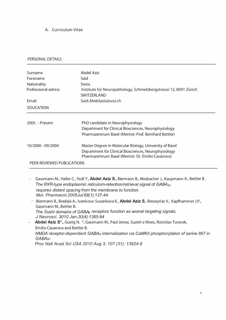
A. Curriculum Vitae
Surname Abdel Aziz Forename Said Nationality Swiss
Professional adress Institute for Neuropathology, Schmelzbergstrasse 12, 8091 Zürich SWITZERLAND
Email [email protected]
2005 - Present PhD candidate in Neurophysiology Department for Clinical Biosciences, Neurophysiology
Pharmazentrum Basel (Mentor: Prof. Bernhard Bettler)
10/2000 –09/2004 Master Degree in Molecular Biology, University of Basel Dep artment for Clinical Biosciences, Neurophysiology Pharmazentrum Basel (Mentor: Dr. Emilio Casanova)
EDUCATION
PERSONAL DETAILS
- Gassmann M., Haller C., Stoll Y., Abdel Aziz S., Biermann B., Mosbacher J., Kaupmann K., Bettler B . The RXR-type endoplasmic reticulum-retention/retrieval signal of GABAB1
requires distant spacing from the membrane to function. Mol. Pharmacol.2005Jul;68(1):137-44
- Biermann B., Bradaia A., Ivankova-Susankova K., Abdel Aziz S., Besseyrias V., Kapfhammer J.P.,
The Sushi domains of GABAB receptors function as axonal targeting signals.
- Abdel Aziz S*., Guetg N. *, Gassmann M., Paul Jenoe, Suzett e Moes, Rostislav Turecek, Emilio Casanova and Bettler B. NMDA receptor-dependent GABAB internalization via CaMKII phosphorylation of serine 867 in GABAB1
PEER-REVIEWED PUBLICATIONS
Gassmann M., Bettler B.
J Neurosci. 2010 Jan;30(4):1385-94-
Proc Natl Acad Sci USA 2010 Aug 3; 107 (31): 13924-9
II

B. Acknowledgements
III
I am deeply obliged to Prof. Dr. Bernhard Bettler for giving me the unique opportunity to do my Ph.D. in his laboratory and entrusting me with an exciting project. It was a great experience to benefit from the excellent insights in the scientific and daily life. Special thanks also to Dr. Martin Gassmann, it was a great honour to work and learn under his supervision. I benefited from his patience and from sharing his critical and supportive thinking in terms of research. I thank my collaborators Dr. Nicole Guetg, Jan Tchorz, Dr. Rotislav Turecek and Dr. Klara Ivankova-Susankova for assisting me to develop further this project. I would also thank Dr. Paul Jenoe and his group for allowing me to use their HPLC machine and the precious support and feedback I received during the analysis of my results and during the writing of my thesis. Thanks to Prof. Dr. Markus A. Ruegg and Prof. Dr. Martin Spiess for accepting to be members of my thesis committee. Thanks also to Prof. Dr. Mohammed Akaaboune for the fruitful and interesting discussions. Thanks to Prof. Dr. Markus Wymann, Prof. Dr. Brian Hemmings and Prof. Dr. Peter Parker for their helpful comments on my project. Great “thank you” to the former/ present lab mates from the Bettler, Brenner and Ruegg laboratories. It was a great privilege to work together with you and to enjoy your colleagueship. I appreciate your invaluable generosity. Explicit thanks to Dr. Audree Pinard for her indefatigueable, critical inputs to my thesis. Thanks to all the people on the floor and in the whole Bio- and Pharmazentrum, that helped me address the different questions during my Ph.D. Last but foremost I want to thank my family, my great parents and my beloved brothers, for their infinite love and support. I bow in front of you and pay you all my love and respect. And of course all my friends (in and outside the laboratories) for their help, support and love. All my love and respect to you too.