photo-induced foveal injury after viewing a solar eclipse
TRANSCRIPT
Photo-induced foveal injury afterviewing a solar eclipse
Fredrik P. Kallmark and Jan Ygge
St. Erik’s Eye Hospital, Department of Clinical Neuroscience, Karolinska Institute,
Stockholm, Sweden
ABSTRACT.
Purpose: To study the injury to and possible recovery of the visual function and
foveal morphology in patients with photo-induced foveal injury due to watching
the solar eclipse of August 11th, 1999 in Stockholm, Sweden.
Methods: Fifteen patients, all of whom viewed the solar eclipse, were followed
for 1 year, during which their visual symptoms were recorded and visual acuity
(VA) was tested, and ophthalmoscopy and scanning laser ophthalmoscopy were
performed.
Results: Photo-induced foveal injury gave rise to subjective visual disturbances,
reduced VA and morphological changes in the fovea. Central scotomas could still
be seen in all patients 1 year after the foveal injury.
Conclusion: Photo-induced foveal injury gave rise to subjective visual distur-
bances, reduced VA and morphological changes in the fovea. Scanning laser
ophthalmoscopy offers the possibility of detailed examination of small retinal
lesions, which can sometimes be difficult to localize with ophthalmoscopy.
Key words: microperimetry – scanning laser ophthalmoscope – fixation – visual acuity
Acta Ophthalmol. Scand. 2005: 83: 586–589Copyright # Acta Ophthalmol Scand 2005.
doi: 10.1111/j.1600-0420.2005.00511.x
Introduction
Solar retinopathy as a result of gazingtowards the sun has been describedpreviously (Wong et al. 2001; Chen& Lee 2004). The symptoms of thistype of injury are possible visual dis-turbances such as metamorphopsia,decreased visual acuity (VA) or scotoma(Rai et al. 1998; Awan et al. 2002).Biophysical analysis has revealed retinallesions and the minimum exposure tothe sun that would produce suchlesions (Sadun et al. 1984). A correla-tion between the funduscopic appear-ance and VA 2 weeks after solar injuryhas been shown (Atmaca et al. 1995),
whereas other investigators have foundno such correlation (Dhir et al. 1981).It has also been established with the useof the scanning laser ophthalmoscope(SLO) that solar-induced scotoma candramatically reduce reading perfor-mance (Ehrt et al. 1999). Very few stu-dies have described the relationshipbetween functional loss after solarinjury and the time required for possi-ble recovery (Ehrt et al. 1999).Furthermore, it is not known to whatextent solar-induced lesions can giverise to permanent damage to the photo-receptors (Atmaca et al. 1995). BecauseSLO is able to map the retinal functionexactly, this technique could be useful
for following retinal damage such as insolar retinopathy (Ehrt et al. 1999).The usefulness of the SLO techniquehas been demonstrated in studyingfixational patterns in normal subjects(Kallmark & Ygge, ‘Fixation patternin healthy subjects during microperi-metry with the scanning laser ophthal-moscope’; unpublished observation).The aims of the present study were toassess patients’ VA and the subjectivedisturbances resulting from solar-induced retinopathy and to performophthalmoscopic investigations of thefundus over a period of time. We alsoaimed to use the SLO to map solar-induced scotomas and evaluate thefixation pattern after such injury.
Methods
Subjects and clinical investigations
Fifteen patients (15 eyes; no patienthad gazed towards the sun with botheyes) came to the emergency room atSt. Erik’s Eye Hospital in Stockholm,Sweden within 1 week of the partialsolar eclipse on August 11th, 1999.Nine of the patients were female andsix were male. Their mean age was35.2 years (range 17–51 years). All thepatients had visual disturbances suchas reduced VA and central visual fieldloss. When asked, the patients esti-mated the time they had spent gazingat the sun to have been between 20 sec-onds and 5 mins. None of the patientsreported any ongoing eye disease andnone were using any medication. Onepatient had experienced solar retinopa-thy in the other eye several years
ACTA OPHTHALMOLOGICA SCANDINAVICA 2005
586
previously. Only the eyes with the above-mentioned symptoms are described here.
At the initial visit (visit 1) best cor-rected distance VA was measured usingthe traditional Snellen line chart, withrows of letters of decreasing size, and anophthalmoscopic examination was per-formed. At the follow-up visits at3 months (visit 2) and 12 months (visit3), the patients’ VA was tested andophthalmoscopic (through a 90 Dlens), and SLO examinations were per-formed while the pupil was dilated withone drop of 0.5% cyclopentolate. Anyophthalmoscopic findings of retinaloedema, lamellar macular hole and/ordistinct retinal pigment epithelium(RPE) disturbances (referred to aslesions later in the text) were noted.During each visit the patients were alsoasked to subjectively describe the visualdisturbance caused by the scotomas assevere, mild or no disturbance at all.
Instrumentation
Scanning laser ophthalmoscopy wasperformed with the Rodenstock SLO-101 using previously described methods(Iwas et al. 1986; Mainster et al. 1989;Timberlake et al. 1989; Van de Veldeet al. 1990a, 1990b; Weber 1990).Visual stimuli were produced in thelaser beam raster by means of a micro-computer and graphics board con-nected to an acousto-optic modulatorin the SLO. Any pattern produced bythe SLO computer and projected ontothe patient’s retina was displayed
simultaneously on a monitor. TheSLO provided a 32 � 22-degree imageof the fundus with a minimum resolu-tion of 3 minutes of arc (15 mm) formeasurement and positioning of thetargets. The SLO’s graphics capabilitiesallowed the investigator to determinethe retinal location of visual stimulidirectly on the retinal image in realtime. The SLO obtained retinal imagescontinuously with an infrared laser(780 nm) and scanned stimuli on theretina with a modulated visiblehelium-neon laser (633 nm). Theoperator displayed the stimuli directlyon the patient’s retina. The helium-neon laser was used as the light sourcefor background illumination, the fixa-tion target and for generating the sti-muli by acousto-optic modulation.
The variables used for manual staticvisual field testing with the SLO werethose commonly used in conventionalprojection perimetry (i.e. backgroundillumination: 10 Cd/m2; incrementaltest stimuli [brighter than background]of 7 � 7 arc minutes [corresponding toa slightly larger diameter of 0.12degree]), which approximates Goldmansize I, with a stimulus duration of200 ms. The patients looked into theSLO with the affected eye and an aver-age of 38 stimuli were presented in aradial grid pattern (Fig. 1) within anarea of approximately 5 degrees radiusfrom the foveola. We chose to presentthe stimulus in a radial grid patternbecause this allows for more precisemapping of the most central retinal
areas. Three different light intensitieswere used: 0 dB (if not seen, consideredas a dense scotoma); 10 dB (relative sco-toma) and 20 dB (minimal suprathres-hold scotoma; Fig. 1). A scotoma wasdefined as a negative response to a givenstimulus presentation with the SLO.
Data collection and analyses
The same examiner (FPK) performedall examinations, except during theinitial visit to the emergency roomwhere they were performed by the doc-tor on call. As the stimuli in the centralpart of the radial grid pattern werepresented close to one another, it waspresumed that the retinal areasbetween the stimulus and any possibleoverlap of the stimuli presented werevery small and therefore the most per-ipheral and seen stimuli were regardedas representing the outer limit of thescotoma. During each stimulus presen-tation the location of the fixation wasrecorded (in x and y co-ordinates rela-tive to the actual fixation cross-location). These fixation locationswere used for estimating the fixationpattern in each subject. The mean ofthe x and y co-ordinates in each subjectwere used as a ‘centre of gravity’ (CG)for the fixation locations. The distanceand angular location of each CG rela-tive to the fixation cross-location wascalculated in all patients. All calcula-tions and plotting were performedusing Origin Scientific Graphing andAnalysis Software, Version 7 (MicrocalInc., Northampton, MA, USA). Visualacuity was evaluated with Friedmannon-parametric repeated measuresANOVA test and fixation with Wilcoxonmatched-pairs signed-ranks test.Statistical significance was set to the95% confidence interval (p < 0.05).
Results
Subjective disturbances
The patients’ subjective estimation ofthe disturbances from the scotomashowed that all patients (15) wereexperiencing severe disturbance on theinitial visit. At 3 months (visit 2), eightwere experiencing severe disturbance,five had mild disturbance and two hadno disturbance at all. At 1 year (visit3), six had mild disturbance and ninehad no disturbance at all.
1818
1818
18
18
1811 11
1111
11
1111
11
11
111818
1 1
1 1
1
1
11
11
1
1 11
1
1818
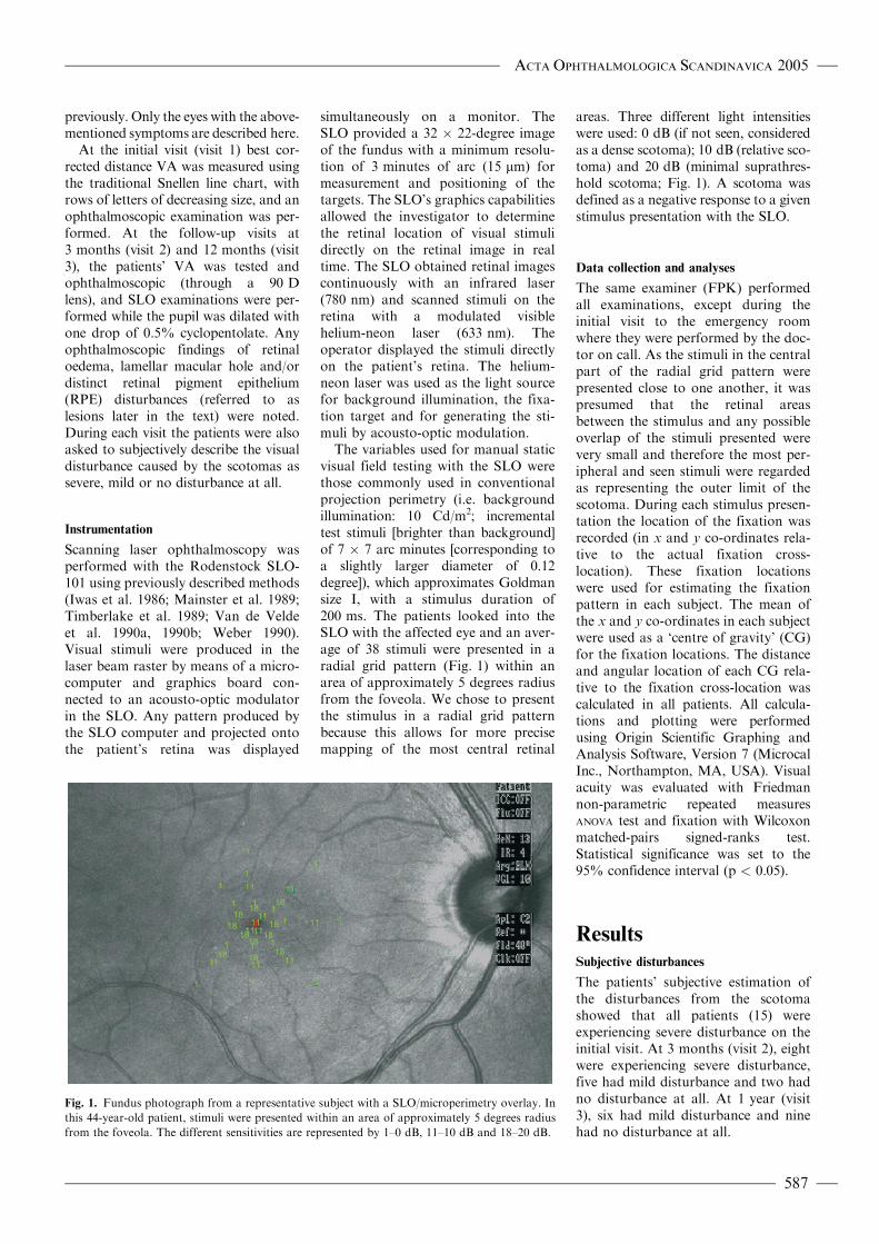
Fig. 1. Fundus photograph from a representative subject with a SLO/microperimetry overlay. In
this 44-year-old patient, stimuli were presented within an area of approximately 5 degrees radius
from the foveola. The different sensitivities are represented by 1–0 dB, 11–10 dB and 18–20 dB.
ACTA OPHTHALMOLOGICA SCANDINAVICA 2005
587
Visual acuity
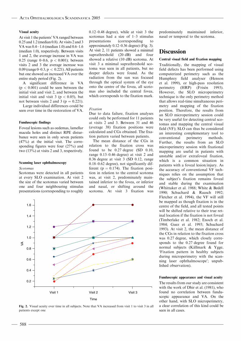
At visit 1 the patients’ VA ranged between0.25 and 1.2 (median 0.8).At visits 2 and 3VAwas 0.4�1.6 (median 1.0) and 0.6–1.6(median 1.0), respectively. Between visits1 and 2, the average increase in VA was0.25 (range 0–0.6, p < 0.001); betweenvisits 2 and 3 the average increase was0.09 (range 0–0.3, p < 0.221).All patientsbut one showed an increased VA over theentire study period (Fig. 2).
A significant difference in VA(p < 0.001) could be seen between theinitial visit and visit 2, and between theinitial visit and visit 3 (p < 0.05), butnot between visits 2 and 3 (p ¼ 0.221).
Large individual differences could beseen over time in the restoration of VA.
Funduscopic findings
Foveal lesions such as oedemas, lamellarmacula holes and distinct RPE distur-bance were seen in only seven patients(47%) at the initial visit. The corre-sponding figures were four (27%) andtwo (13%) at visits 2 and 3, respectively.
Scanning laser ophthalmoscopy
Scotomas
Scotomas were detected in all patientsat every SLO examination. At visit 2the size of the scotomas varied betweenone and four neighbouring stimuluspresentations (corresponding to roughly
0.12–0.48 degree), while at visit 3 thescotomas had a size of 1–3 stimuluspresentations (corresponding toapproximately 0.12–0.36 degree) (Fig. 3).At visit 2, 11 patients showed a minimalsuprathreshold (20 dB) and fourshowed a relative (10 dB) scotoma. Atvisit 3 a minimal suprathreshold sco-toma was seen in all patients, but nodeeper defects were found. As theradiation from the sun was focusedthrough the optical system of the eyeonto the centre of the fovea, all scoto-mas also included the central fovea,which corresponds to the fixation mark.
Fixation
Due to data failure, fixation analysescould only be performed for 11 patientsat visits 2 and 3. Between 31 and 46(average 38) fixation positions werecalculated and CGs obtained. The fixa-tion pattern varied between patients.
The mean distance of the CGs inrelation to the fixation cross wasfound to be 0.27 degree (SD 0.10,range 0.13–0.46 degree) at visit 2 and0.36 degree at visit 3 (SD 0.12, range0.18–0.62 degree), not significantly dif-ferent (p ¼ 0.174). The fixation posi-tion in relation to the central scotomawas, at visit 2, predominately main-tained inferior to the fovea, or inferiorand nasal, or shifting around thescotoma. At visit 3 fixation was
predominately maintained inferior,nasal or temporal to the scotoma.
Discussion
Central visual field and fixation mapping
Traditionally, the mapping of visualfield defects has been performed usingcomputerized perimetry such as theHumphrey field analyser (Hensonet al. 1999), or high-pass resolutionperimetry (HRP) (Frisen 1993).However, the SLO microperimetrytechnique is the only perimetry methodthat allows real-time simultaneous peri-metry and mapping of the fixationposition. Therefore, the results froman SLO microperimetry session couldbe very useful for detecting central sco-tomas and mapping the central visualfield (VF); SLO can thus be consideredan interesting complementary tool toconventional perimetry methods.Further, the results from an SLOmicroperimetry session with fixationalmapping are useful in patients withunstable and/or extrafoveal fixation,which is a common situation inpatients with a foveal lesion/injury. Asthe accuracy of conventional VF tech-niques relies on the assumption thatthe subject’s fixation remains fovealand stable during the examination(Whittaker et al. 1988; White & Bedell1990; Schuchard & Raasch 1992;Fletcher et al. 1994), the VF will stillbe mapped as though fixation is in thecentre of the field, and all tested pointswill be shifted relative to their true ret-inal location if the fixation is not foveal(Timberlake et al. 1982; Enoch et al.1984; Guez et al. 1993; Schuchard1993). At visit 2, the mean distance ofthe CGs in relation to the fixation crosswas 0.27 degree, which closely corre-sponds to the 0.27 degree found fornormal subjects (Kallmark & Ygge,‘Fixation pattern in healthy subjectsduring microperimetry with the scan-ning laser ophthalmoscope’; unpub-lished observation).
Funduscopic appearance and visual acuity
The results from our study are consistentwith the work of Dhir et al. (1981), whofound no correlation between fundu-scopic appearance and VA. On theother hand, with SLO microperimetry,a clear correlation of this kind could beseen in all cases.
1.6
1.4
1.2
1.0
0.8
VA
0.6
0.4
0.2
Visit 1 Visit 2 Visit 3
Time
Fig. 2. Visual acuity over time in all subjects. Note that VA increased from visit 1 to visit 3 in all
patients except one
ACTA OPHTHALMOLOGICA SCANDINAVICA 2005
588
Most of the improvement in VA wasobserved to occur between visits 1 and 2.Between visits 2 and 3, no statisticallysignificant improvement was noted,which also correlates well with the find-ings of other studies (Atmaca et al. 1995;Awan et al. 2002). However, a number ofeyes showed an improvement in VAbetween visits 2 and 3 and the lack ofsignificance was probably due to thesmall sample size. Subjectively, a decreasein the degree of visual disturbanceoccurred over the whole period, but40% of the patients experienced a milddisturbance after 1 year. Further, theSLO results revealed the presence of sco-tomas at visit 3, even when the patientsdid not experience any visual disturbance.The good visual prognosis in most casescan probably be attributed to the resis-tance of the foveal cone cells to photoche-mical damage (Hope-Ross et al. 1993).
The results of the present study alsogive a clear indication of the hazards oflooking into the sun and the impor-tance of providing adequate safetyinformation to the public before animpending eclipse. Prevention remainsthe mainstay of therapy.
ReferencesAtmacaLS, Idil A&CanD (1995): Early and late
visual prognosis in solar retinopathy. Graefes
Arch Clin Exp Ophthalmol 12: 801–804.
Awan AA, Khan T, Mohammad S & Arif AS
(2002): Eclipse retinopathy: follow-up of 36
cases after the April 1995 solar eclipse in
Pakistan. J Ayub Med Coll Abbottabad 14:
8–10.
Chen JC & Lee JR (2004): Solar retinopathy
and associated optical coherence tomogra-
phy findings. Clin Exp Optom 87: 390–393.
Dhir SP, Gupta A & Jain IS (1981): Eclipse
retinopathy. Br J Ophthalmol 65: 42–45.
Ehrt O, Tavcar I & Eckl-Titz G (1999):
Microperimetry and reading saccades in
retinopathia solaris. Follow-up with the
scanning laser ophthalmoscope.
Ophthalmologe 5: 325–331.
Enoch JM, O’Donnell J, Williams RA &
Essock EA (1984): Retinal boundaries and
visual function in gyrate atrophy. Arch
Ophthalmol 102: 1314–1316.
Fletcher DC, Schuchard RA, Livingstone CL,
Grane WG & Hu SY (1994): Scanning laser
ophthalmoscope macular perimetry and
applications for low vision rehabilitation clin-
icians. Low Vision Vision Rehab 2: 257–265.
Frisen L (1993): High-pass resolution perimetry.
A clinical review. Doc Ophthalmol 83: 1–25.
Guez JE, Le Gargasson JF, Rigaudiere F &
O’Regan JK (1993): Is there a systematic
location for the pseudo-fovea in patients
with central scotoma? Vision Res 9: 1271–
1279.
Henson DB, Artes PH & Chauhan BC (1999):
Diffuse loss of sensitivity in early glaucoma.
Invest Ophthalmol Vis Sci 40: 3147–3151.
Hope-Ross MW, Mahon GJ, Gardiner TA &
Archer DB (1993): Ultra structural findings
in solar retinopathy. Eye 7: 29–33.
Iwas A, Kitazawa Y & Ohno Y (1986): On
age-related norms of the visual field. Jpn J
Ophthalmol 32: 429.
Mainster MA, Timberlake GT, Webb RH &
HughesGW(1989): Scanning laserophthalmo-
scopy: clinical applications. Ophthalmology
89: 852–857.
Rai N, Thuladar L, Brandt F, Arden GB &
Berninger TA (1998): Solar retinopathy.
A study from Nepal and from Germany.
Doc Ophthalmol 95: 99–108.
Sadun AC, Sadun AA & Sadun LA (1984):
Solar retinopathy. A biophysical analysis.
Arch Ophthalmol 102: 1510–1512.
Schuchard RA (1993): Validity and interpreta-
tion of Amsler grid reports. Arch
Ophthalmol 111: 776–780.
Schuchard RA & Raasch TW (1992): Retinal
locus for fixation: pericentral fixation tar-
gets. Clin Vision Sci 7: 511–620.
Timberlake GT, Mainster MA, Webb RH,
Hughes GW & Trempe CL (1982): Retinal
localization of scotoma by scanning laser
ophthalmoscope. Invest Ophthalmol Vis
Sci 22: 91–97.
Timberlake GT, Van de Velde FJ & Jalkh AE
(1989): Clinical use of scanning laser
ophthalmoscope retinal function map in
macular disease. Laser Light Ophthalmol 4:
211–222.
Van de Velde F, Timberlake GT, Jalkh AE,
Katsumi O, Hirose T & Schepens CL
(1990a): Clinical scanning laser ophthalmo-
scope applications: an overview. In:
Nasemann JE & Burk ROW (eds).
Scanning Laser Ophthalmoscopy and
Tomography. Munich: Quintessenz 35–47.
Van de Velde F, Timberlake GT, Jalkh AE &
Schepens CL (1990b): La microperemetrie
statique avec l’ophtalmoscope a balayage
laser. Ophtalmologie 4: 291–294.
Weber J (1990): Eine neue Strategie fur die
automatisierte statische Perimetrie.
Fortschr Ophthalmol 87: 37–40.
White JM & Bedell HE (1990): The oculomo-
tor reference in humans with bilateral macu-
lar disease. Invest Ophthalmol Vis Sci 31:
1149–1161.
Whittaker SG, Budd JM & Cummings RW
(1988): Eccentric fixation with macular sco-
toma. Invest Ophthalmol Vis Sci 29:
268–278.
Wong SCK, Eke T & Zaikas NG (2001):
Eclipse burns: a prospective study of solar
retinopathy following the 1999 solar eclipse.
Lancet 357: 199–200.
Received on December 17th, 2003.
Accepted on April 19th, 2005.
Correspondence:
Dr Fredrik P. Kallmark
St Erik’s Eye Hospital
Polhemsgatan 50
S-112 82 Stockholm
Sweden
Tel: þ 46 8 672 3660
Fax: þ 46 8 672 3330
Email: [email protected]
11
A
B
1
1
11
11
11
1 1
1
1
1111 11
11111111
11
11
1111
11
11
1111
11
111111
11
111111
11
1111111111
111111111111
11
11
1111
11
111111
1111
11111111
11
11
11
111
1
1
1
1
1
1
1
11
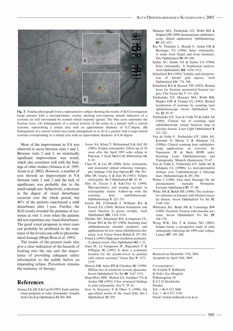
Fig. 3. Fundus photograph from a representative subject showing the results of SLO investigation
(large picture) with a microperimetry overlay showing non-response stimuli indicative of a
scotoma (in red) surrounded by normal retinal response (green). The blue cross represents the
fixation cross. (A) Enlargement of a central portion of the retina in a patient with a central
scotoma, representing a retinal area with an approximate diameter of 0.12 degree. (B)
Enlargement of a central retinal area (same enlargement as in A) in a patient with a large central
scotoma corresponding to a retinal area with an approximate diameter of 0.36 degree.
ACTA OPHTHALMOLOGICA SCANDINAVICA 2005
589