photocatalytic properties of zinc oxide...
TRANSCRIPT
PHOTOCATALYTIC PROPERTIES OF ZINC OXIDE NANOSTRUCTURES
SYNTHESIZED USING SYNERGISTIC PULSE LASER ABLATION
AND HYDROTHERMAL METHODS
KHALDOON NAJI ABBAS AL-GBURI
A thesis submitted in fulfilment for the
requirements of the award of the degree of
Doctor ofPhilosophy (Physics)
Faculty of Science
Universiti Teknologi Malaysia
mLY 2017

iii
Dedicated to my beloved family
To the most precious persons in my life, my parents, my wives, my brothers and
sisters, and my sweetheart beautiful son and daughters.

iv
ACKNOWLEDGEMENT
Thanks to ALLAH, the Most G racious, the Most Merciful, the Most
Bountiful who gave me the courage and patience to accomplish this research work.
Without his help and mercy, this would not have come into reality.
I would like to deeply express my gratitude for the help and support from my
Supervisor Prof. Dr. Noriah Bidin for their fascinating guidance, encouragement,
patience and valuable comments throughout the research work. I was fortunate to be
one of their graduate students. Their experience and creativity gave me great profit
for carving my future career.
I would like to acknowledge the Universiti Teknologi Malaysia (UTM) for
providing the facilities and support during this research. I wish also to thank Ministry
of High Education of Iraq for their continuous help and support for this research.
Special thanks go to all those who have contributed directly and indirectly to
the completion of this research and thesis. This includes UTM staff and my fellow
postgraduate students who provided me with help and company during my study
here.
Last, but not the least, my greatest thanks from my heart to my family for
giving the unlimited supports and patience to complete my study. I would never ever
forget their sacrifice that they have done for me. I appreciate the sacrifice of my
parents, brothers and sisters in helping me morally to finish my study, god bless you
all.
v
ABSTRACT
Mild growth conditions based novel techniques are essential for the controlled
synthesis of zinc oxide (ZnO) nanostructure (ZNS) with desired properties. Most of the
existing synthesis methods of ZNSs are limited by complicated growth conditions such as
high vacuum, expensive devices, high temperature, long growth time and requirement of
template. The freshwater pollution due to residual industrial organic dyes is presently being a
major environmental concern that needs efficient photocatalytic materials to overcome it. To
achieve these goals, this work synthesized good quality ZNS films using two different
methods and evaluated the photodegradation of industrial methylene blue (MB) dye by these
ZNS films under sunlight irradiation. Such films were grown on glass and silicon (Si)
substrates via a simple hydrothermal method in the absence of any catalyst. Prepared
samples were characterized using various techniques to determine their physical and optical
properties. The effects of growth time and temperature, substrate type, nutrient pH and
concentration on the morphology, size, crystallinity, and the emission properties of as-grown
ZNS films were evaluated. Results showed that the morphology of these ZNSs was strongly
influenced by the growth time, nutrient pH and concentration. A good quality ZNS was
achieved within 5 min whereby nutrient solution with lower pH was appropriate for the
growth of 1D ZNSs. However, higher pH values produced 3D flower-like ZNSs. Variation
of growth temperature from 90-110 oC allowed good size-control of ZNSs. Growth
conditions and substrate type dependent emission spectra were used to evaluate the optical
band-gap energy. To get better control of the ZNS growth and evolving morphology, a novel
catalyst-free and rapid preparation technique was adopted. In this method a mixture of
precursor solution and ZnO nanoparticles (ZNPs) colloid/or only ZNPs colloid as a nutrient
solution was used. Pulse laser ablation in liquid (PLAL) was combined with hydrothermal
(H) method to develop PLAL-H growth technique. A Q-switched Nd:YAG laser with
wavelength 532 nm, 8 ns pulse duration, and 10 Hz repetition rate was employed as the
irradiation source. Metallic zinc target of 1 mm thick was used to produce colloidal ZNPs.
The effects of varying growth time of 0.5, 5, 30 and 60 min and ablation energy of 200-400
mJ on the physical and optical properties of the grown ZNSs were examined. Four types of
ZNSs with varying sizes and shapes were obtained on Si substrate at 110 oC for 5 min
duration. Increasing ablation energy led to a substantial change of ZNS morphology and
promoted the structure quality together with photoluminescence emission intensity. ZNSs
synthesized under prolonged growth time of 60 min exhibited remarkable morphology
alteration from rod/flower-like ZNSs to ZNPs with higher crystallinity and enlarged band-
gap due to increase of nutrient pH of 10.5. Finally, the photocatalytic activities of the
optimal ZNS films were assessed via sunlight driven photodegradation of MB dye.
Experimental findings verified that the ZNPs prepared by PLAL-H technique possessed
excellent photocatalytic efficiency (97.4%) towards degradation of MB dye. The observed
boost in the photocatalytic activities was ascribed to the synergism of the improved surface
area and band-gap modification. It was established that the proposed novel PLAL-H growth
strategy is not only cost-effective but greatly useful for the rapid production of different
quality of ZNSs at low temperature in a controlled way. This may overcome the
shortcomings involving the effective exploitation of sunlight source towards practical
photocatalytic applications of ZNSs.
vi
ABSTRAK
Teknik novel berdasarkan keadaan pertumbuhan lambut adalah perlu untuk sintesis
terkawal struktur-nano (ZNS) zink oksida (ZnO) dengan sifat yang dikehendaki. Kebanyakan
kaedah sintesis ZNS sedia ada adalah terhad oleh keperluan pertumbuhan yang rumit seperti
keadaan vakum tinggi, peralatan yang mahal, suhu tinggi, tempoh pertumbuhan yang lama dan
keperluan terhadap templet. Pencemaran air tawar oleh sisa pewarna organik industri kini
merupakan masalah persekitaran utama yang memerlukan bahan foto pemangkin yang efisien
untuk mengatasinya. Untuk mencapai matlamat ini, kajian ini mensintesis filem ZNS yang
berkualiti dengan menggunakan dua kaedah yang berbeza dan menilai fotodigredasi pewarna biru
metalin (MB) industri oleh filem ZNS di bawah sinaran cahaya matahari. Filem tersebut
dihasilkan di atas substrat kaca dan wafer silikon (Si) melalui kaedah hidroterma mudah tanpa
pemangkin. Sampel yang disediakan dicirikan bagi menentukan ciri fizikal dan optik dengan
menggunakan pelbagai teknik. Kesan masa pertumbuhan dan suhu, jenis substrat, pH dan
kepekatan nutrien, terhadap morfologi, saiz, penghabluran, dan sifat pancaran filem ZNS telah
dinilai. Hasil kajian menunjukkan bahawa morfologi ZNS ini sangat dipengaruhi oleh masa
pertumbuhan, pH dan kepekatan nutrien. Kualiti ZNS yang baik telah dicapai dalam masa 5 minit
apabila cecair nutrien dengan pH lebih rendah adalah sesuai untuk pertumbuhan ZNS 1D. Walau
bagaimanapun, nilai pH yang lebih tinggi menghasilkan ZNS berbentuk bunga 3D. Perubahan
suhu pertumbuhan dalam lingkungan 90-110 oC membolehkan kawalan saiz ZNS yang baik.
Spektrum pancaran yang bergantung kepada keadaan pertumbuhan dan jenis substrat telah
digunakan untuk menilai jurang tenaga optik. Untuk mencapai kawalan yang lebih baik ke atas
pertumbuhan ZNS dan perkembangan morfologi, teknik penyediaan yang novel dan bebas
pemangkin serta pantas telah digunakan. Dalam kaedah ini, campuran cecair pelopor dan koloid
nanopartikel ZnO (ZNP)/atau hanya koloid ZNP telah digunakan sebagai cecair nutrien. Ablasi
laser denyut dalam cecair (PLAL) telah digabungkan dengan kaedah hidroterma (H) untuk
menghasilkan teknik pertumbuhan PLAL-H. Laser Nd:YAG pensuisan-Q dengan panjang
gelombang 532 nm, tempoh denyutan 8 ns, dan kadar ulangan 10 Hz telah digunakan sebagai
sumber sinaran. Sasaran logam zink dengan ketebalan 1 mm digunakan untuk menghasilkan
koloid ZNP. Kesan masa pertumbuhan yang berbeza-beza dari 0.5, 5, 30 dan 60 minit dan
tenaga ablasi 200-400 mJ terhadap ciri fizikal dan optik ZNS telah diperiksa. Empat jenis ZNS
dengan pelbagai saiz dan bentuk telah diperolehi pada substrat Si pada suhu 110 oC untuk tempoh
5 minit. Peningkatan tenaga ablasi membawa kepada perubahan ketara pada morfologi ZNS dan
mempromosikan kualiti struktur bersama-sama dengan keamatan pancaran
kefotopendarcahayaan. ZNS yang disintesis di bawah masa pertumbuhan selama 60 minit
mempamerkan transformasi morfologi luar biasa dari ZNS berbentuk rod/bunga ke ZNP dengan
penghabluran yang lebih tinggi dan nilai jurang yang lebih besar disebabkan oleh peningkatan pH
nutrien kepada 10.5. Akhir sekali, aktiviti fotopemangkin filem ZNS optimum telah dinilai
terhadap fotodigeradasi pewarna biru metalin (MB) berpacuan cahaya matahari. Hasil
eksperimen mengesahkan bahawa ZNP yang disediakan oleh teknik PLAL-H memiliki
kecekapan fotopemangkin yang sangat baik (97.4%) ke arah degradasi pewarna MB. Peningkatan
yang diperhatikan dalam aktiviti fotopemangkin telah disifatkan sebagai sinergi peningkatan
kawasan permukaan dan perubahan jurang jalur. Ternyata bahawa strategi pertumbuhan PLPAL-
H novel yang dicadangkan ini bukan sahaja menjimatkan kos tetapi berguna untuk pengeluaran
pantas secara terkawal ZNS dengan kualiti yang berbeza pada suhu rendah. Ini boleh mengatasi
kelemahan yang melibatkan keberkesanan penggunaan sumber cahaya matahari untuk aplikasi
praktikal fotopemangkin ZNS.

vii
TABLE OF CONTENTS
CHAPTER TITLE PAGE
DECLARATION ii
DEDICATION iii
ACKNOWLEDGEMENT iv
ABSTRACT v
ABSTRAK vi
TABLE OF CONTENTS vii
LIST OF TABLES xiii
LIST OF FIGURES xv
LIST OF ABBREVIATIONS xxviii
LIST OF SYMBOLS xxxi
LIST OF APPENDICES xxxiv
1 INTRODUCTION 1
1.1 Overview 1
1.2 Photocatalytic Application 5
1.3 Problem Statement 6
1.4 Research Objectives 8
1.5 Scope of Study 8
1.6 Significance of Study 10
1.7 Thesis Organization 10
2 LITERATURE REVIEW 12
2.1 Introduction 12
2.2 Properties ZnO Semiconductor 13
viii
2.2.1 Crystal Structures 13
2.2.2 Optical Properties of ZnO 14
2.2.3 Electrical Properties of ZnO 16
2.3 Native Point Defects of ZnO Structure 18
2.4 Growth of ZnO Nanostructure 20
2.4.1 Pulse Laser Ablation in Liquid Method 21
2.4.1.1 Growth Mechanism and Experimental
Setup of PLAL Method
23
2.4.1.2 Thermal Evaporation Mechanism 24
2.4.1.3 Growth Conditions of PLAL Method 27
2.4.2 Hydrothermal Growth Method 32
2.5 Controlled Mode of ZnO Nanostructure 38
2.5.1 Effects of Nutrient Concentration and Type on
ZnO Nanostructures Properties
41
2.5.2 Influence of Growth Temperature and Time on
ZnO Nanostructures Properties
42
2.5.3 Influence of Nutrient pH on ZnO Nanostructures
Properties
44
2.5.4 Influence of Seed Layer on ZnO Nanostructures
Properties
45
2.6 ZnO Nanostructures Thin Film Deposition 46
2.7 Application of ZnO Nanostructure Films for
Photocatalysis
47
2.7.1 ZnO Nanostructures for Photocataysis 49
2.7.2 Photocatalytic Mechanism of ZnO
Nanostructures
50
2.7.2.1 Charge Carrier Generation 50
2.7.2.2 Charge Carrier Trapping 51
2.7.2.3 Charge Carrier Recombination 53
2.7.2.4 Photocatalytic Degradation of Dye
Pollutants
53
2.7.3 Strategies for Improving Photocatalytic
Efficiency of ZnO Nanostructures
55

ix
2.7.3.1 Band-Gap Modification of ZnO 55
2.7.3.2 Photosensitization Process of ZnO
Surface
56
2.7.3.3 Architecture Adjustment of ZnO 56
2.7.3.4 Optimization of ZnO Surface Area 57
2.7.4 Methylene Blue as Organic Dye Pollutant 57
3 RESEARCH METHODOLOGY 60
3.1 Introduction 60
3.2 Methods for ZnO Nanostructures Synthesis 61
3.2.1 Raw Materials 61
3.2.2 Substrate Cleaning for ZnO Nanostructures Film
Deposition
62
3.2.3 Hydrothermal Method for ZnO Nanostructure
Growth
62
3.2.4 PLAL-H Technique for ZnO Nanostructures
Growth
65
3.2.4.1 Q-switched Nd:YAG Laser for
Ablation of Zn Target
66
3.2.4.2 PLAL Method for Colloidal ZnO
Nanoparticles Synthesis
69
3.2.4.3 PLAL-H Technique (Strategy I) for
ZnO Nanostructure Films
Deposition
72
3.2.4.4 PLAL-H Technique (Strategy II) for
ZnO Nanostructure Films Deposition
75
3.3 Characterization Techniques 77
3.3.1 Scanning Electron Microscope Imaging 77
3.3.2 Energy Dispersive X-ray Spectrometer 78
3.3.3 Transmission Electron Microscope Imaging 78
3.3.4 X- Ray Diffraction Measurement 79
3.3.5 Fourier Transform Infrared Spectrometer 82
3.3.6 Photoluminescence Spectrometer 83

x
3.3.7 UV-Vis-NIR Spectrophotometer 84
3.4 Photocatalytic Activity Evaluation of ZNSs-based Films 84
4 RESULTS AND DISCUSSION 86
4.1 Introduction 86
4.2 Properties of ZnO Nanostructures Film Grown by
Hydrothermal Method
87
4.2.1 Effects of Growth Time and Substrate on
Nanostructures
88
4.2.1.1 Morphology, Size and Stoichiometric
Ratio of Nanostructures
88
4.2.1.2 XRD Analysis of ZnO Nanostructure
Film
94
4.2.1.3 Optical Properties of ZnO
Nanostructure Films
97
4.2.2 Effect of Nutrient Solution ZnO Nanostructure
Properties
100
4.2.2.1 Morphology Analysis 100
4.2.2.2 Structural Properties 102
4.2.2.3 FTIR Analysis of ZnO Nanostructure
Films
103
4.2.2.4 Photoluminescence Properties 104
4.2.3 Effect of Nutrient pH on ZnO Nanostructure
Films
105
4.2.3.1 Morphology Analysis 105
4.2.3.2 Structural Analysis 107
4.2.3.3 Photoluminescence Properties of ZnO
Nanostructure Films
108
4.2.3.4 FTIR Spectra of ZnO Nanostructures
Film
109
4.2.4 Influence of Growth Temperature on ZnO
Nanostructure Films
110
4.2.4.1 Morphology Analysis 110
xi
4.2.5 Growth Mechanism of ZnO Nanostructures 111
4.3 ZnO Nanostructures Synthesis via PLAL-H Method 115
4.3.1 Characterization of ZnO Nanoparticles Prepared
via PLAL Method
116
4.3.1.1 Morphological Properties 116
4.3.1.2 Absorption Spectra of Colloidal ZnO
Nanoparticles
120
4.3.2 Synthesis ZnO Nanostructure Films by PLAL-H
Technique
121
4.3.2.1 Morphology Analysis of As-Grown
ZnO Nanostructure Films
123
4.3.2.2 Structural Properties of ZnO
Nanostructures
130
4.3.2.3 FTIR Analysis of ZnO Nanostructures 132
4.3.2.4 Absorption and Luminescence
Properties of ZnO Nanostructures
133
4.3.2.5 Growth Mechanism of ZnO
Nanostructures Obtained via PLAL-H
Method
138
4.3.3 Influence of Laser Ablation Energy on the ZnO
Nanostructures
142
4.3.3.1 Morphology Analysis of ZnO
Nanostructures
142
4.3.3.2 Structural Analysis of ZnO
Nanostructure Films
144
4.3.3.3 FTIR Analysis 145
4.3.3.4 Photoluminescence Properties of ZnO
Nanostructures
146
4.4 Influence of PLAL-H Growth Conditions on ZnO
Nanostructure Films
148
4.4.1 Morphology Analysis of As-Grown ZnO
Nanostructures
149
xii
4.4.2 Structural Properties of As-Grown ZnO
Nanostructures
153
4.4.3 FTIR Analysis 154
4.4.4 Absorption and Photoluminescence Properties of
As-Grown Samples
155
4.4.5 Growth Mechanism of As-Grown ZnO
Nanostructures
159
4.5 Photocatalytic Applications of As-Grown ZnO
Nanostructures
163
4.5.1 Photocatalytic Activity of As-Synthesized ZnO
Nanostructure Films
163
4.6 Synergism in PLAL and Hydrothermal Methods 177
5 CONCLUSION AND RECOMMENDATIONS 179
5.1 Conclusions 179
5.2 Recommendations for Future Work 183
REFERENCES 185
Appendix A 207-208

xiii
LIST OF TABLES
TABLE NO. TITLE PAGE
2.1 Basic characteristics of stable wurtzite ZnO
structure
1
2.2 Early literature review of synthesis ZNPs by pulse
laser ablation method
28
2.3 Different reaction based solution for achieving
diverse ZNSs using hydrothermal method
39
3.1 Growth parameters of hydrothermal method and
achieved morphologies
63
3.2 Specification of Q-switched Nd:YAG laser (BKF-
K9 model)
68
3.3 Growth parameters and conditions of PLAL-H
technique
74
4.1 Growth parameters of hydrothermal method and
morphology of achieved samples
87
4.2 Values of lattice constants, crystallite size, and
micro-strain of ZMNSs deposited on glass and Si
substrates for 3 hr duration
97
4.3 Growth conditions of PLAL-H technique (first
strategy) and achieved samples
122
4.4
Growth conditions of PLAL-H technique (second
strategy) and morphology of grown samples
149
4.5 Wavelength (λ, nm) and energy band-gap (Eg, eV)
of PL emission peaks obtained for ZNSs at
different nutrient pH values
156

xiv
4.6 Optimum photocatalytic ZNSs films grown by
PLAL-H and hydrothermal methods
164
4.7 Display of photocatalytic efficiency of de-
colorization of MB suspension under various
irradiation sources for various nanomaterials
prepared via different growth techniques
174
xv
LIST OF FIGURES
FIGURE NO. TITLE
PAGE
1.1 Overview of different structures and geometries at
nanoscale
2
1.2 SEM images of various ZNSs synthesized on Si
substrates using hydrothermal method: (a, b and c)
rod-like, (d) star-like, and (e and f)
flower-like
3
1.3 Pictures showing the freshwater pollution (a and
b) due to chemicals fallout and (c and d) its
influence on the living organism
5
2.1 Crystal structure of ZnO: (a and b) hexagonal
wurtzite structure unit and (c) cubic zinc-blende
unit
14
2.2 Room temperature PL spectra of ZNWs with
average diameters of 12 nm, 16 nm and 31 nm
revealing a prominent UV peak at 380 nm and a
broad visible peak
16
2.3 Energy level diagram for native defects in ZnO
with defect ionization energies in the range
of 1.6-3.01 eV
19
2.4 (a) Experimental setup for PLAL of solid target
and (b) ablation process of the target material
24
2.5 Schematic illustration of the evolution of the laser-
induced plasma in liquid leading to the growth of
nano/microstructure: (a) generation of Zn plasma
xvi
having high temperature and pressure on the top
of the Zn target immediately after single pulse
irradiation, (b-c) adiabatic and ultrasonic
expansion of the plasma and creation of Zn
clusters, and (d) creation of ZNSs
26
2.6 Schematic diagram displays the formation process
of different ZNSs dependent various laser fluence
31
2.7 TEM images of ZNSs prepared by PLAL at
different increasing laser ablation energy: (a, b
and c) ZnO morphology is varied from nanoflakes
at 20 mJ to (d) ZNPs at 70 mJ, and to (e and f)
short ZNRs at 120 mJ
32
2.8 Schematic representation of hydrothermal process:
(a) experimental setup and (b) growth mechanism
34
2.9 FESEM images of different ZNSs morphology
grown by hydrothermal method at 140 oC for
10 hr: (a) sphere, (b) rod, (c) sheet, and (d) wire
35
2.10 Schematic illustration of the growth mechanism
steps using hydrothermal treatment under 80 oC
for 12 hr: (A) primary growth units, (B) ZnO
nuclei, (C) ZnO crystallites, and (D) flower-like
ZNSs
37
2.11 Nutrient solution concentration dependent average
diameters and lengths of the ZNWs synthesized
via hydrothermal method
42
2.12 The growth time dependent (1-11 hr) UV-Vis
absorption spectra of ZnO structures prepared by
hydrothermal method
44
2.13 Schematic illustration the photogeneration of
oxidizing species in ZnO photocatalysis and
photocatalysis process by ZnO semiconductor
51
2.14 Schematic diagram revealing the molecular
structure of the MB dye
58
xvii
3.1 Schematic presentation of hydrothermal method:
(a) Oven, (b) autoclave glass (140 oC of volume
250 mL), (c) grown diverse ZNSs films with
various morphology such as (d) flower-like,
(e) rice-like, and (f) enclosed tube-like
63
3.2 The flowchart displaying the methodology ZNSs-
based films preparation by hydrothermal method,
various characterizations and their photocatalytic
application toward MB dye photodegradation
64
3.3 Schematic diagram of: (a) pulsed Nd:YAG laser
structure and (b) energy-level diagram for lasing
action in Nd3+
ion
67
3.4 Calibration of Q-switched Nd:YAG laser pulse
energy at 532 nm, 8 ns and 10 Hz in range of 40-
800 mJ/pulse
68
3.5 Depiction of PLAL method: (a) Photograph of
PLAL system experimental setup with Zn-target
under water, (b) schematic diagram of PLAL
method for colloidal ZNPs synthesis, (c) actual
ablation process and (d) the schematic diagrams of
the ablation process
70
3.6 Photograph showing the achieved laser ablated
products from Zn target immersed inside water
71
3.7 Flowchart displaying PLAL method based
synthesis of colloidal suspension of ZNPs and
their subsequent characterizations
72
3.8 Schematic presentation for the synthesis of ZNSs-
based film by PLAL-H technique (First stage): (a)
colloidal solution ZNPs preparation by PLAL
method (denoted as N2), and (b) preparation of
nutrient solution by mixing N1 and N2 subjected
to hydrothermal treatment
73
xviii
3.9 The flowchart showing the growth of ZNSs-based
films by PLAL-H technique using ZNPs colloid as
nutrient solution, samples characterizations, and
photocatalytic application
76
3.10 (a) Schematic presentation demonstrating the
XRD from regular lattice planes following the
Bragg’s law, and (b) constructive and destructive
interference patterns
80
3.11 Schematic diagrams showing the experimental
setup to evaluate the photocatalytic activity of
ZNSs films via the degradation of MB driven by
solar radiation
85
4.1 FESEM images of ZNSs (samples L1, L2, L3 and
L4) at different magnification grown for (A-a) 1
hr, (B-b) 2 hr, (C-c) 3 hr, and (D-d) 4 hr on glass
(left column) and Si (right column) substrates.
Inset in (c) is the tip of NR and red circle in (d)
destroyed NRs
89
4.2 Growth time duration dependent average length
and diameter of ZNRs synthesized on glass
substrate
90
4.3 Distribution (length and diameter) of ZNRs (fitted
to Gaussian) grown on glass substrate for the
duration of (a-a1) 1 hr, (b-b1) 2 hr, (c-c1) 3 hr,
and (d-d1) 4 hr. Similar behaviour was observed
for samples grown on Si substrate
91
4.4 ZMNSs (samples L5 and L6) grown on glass
substrate for 6 or 8 hr duration (a) FESEM images
and (b) XRD pattern
92
4.5 EDX spectra of ZMNSs grown on glass substrate
for 6 or 8 hr duration
93
4.6 EDX spectra of dense ZMNSs grown for 3 hr on:
(a) glass and (b) Si substrates
94
xix
4.7 XRD patterns of ZMNSs grown with 0.1 M
precursor solution at 110 °C for 1, 2 and 4 hr
duration on the: (a) Si substrate, (b) glass
substrate, and (c) substrate dependent comparison
for 3 hr growth duration
95
4.8 PL spectra of ZMNSs grown on glass and Si
substrates for time duration of: (a) 1 hr, (b) 2 hr,
(c) 3 hr, and (d) 4 hr. Dashed lines are the
Gaussian fits
99
4.9 Growth time duration dependent variations of the
PL peak positions for samples grown on glass and
Si substrates
100
4.10 FESEM images showing ZNs film (sample L7)
grown on Si substrate at 90 oC for 3 hr, pH 5.7 and
0.1 M: (a and b) enclosed tube- and solid rod-like
ZNs, (c) enclosed tube-like ZNs and (d) solid rod-
like ZNs
101
4.11 FESEM images showing ZNs film morphology
(sample L8) grown on Si substrate at 90 oC for 3
hr, pH 5.7 and 0.01 M: (a and b) rice-, star rice-
and flower rice-like, Insets dashed lines in image
(b) are the flower rice-like ZNs, (c) FESEM
magnified image of rice-, star rice- like ZNs, and
(d) FESEM magnified image of rice-like ZNs tip
102
4.12 XRD patterns of ZNSs films grown on Si
substrates for different precursor concentration of
0.01 to 0.1 M at 90 °C for 3 hr with pH 5.7
103
4.13 FTIR spectra of ZNSs films grown under various
precursor concentrations (0.01-0.10 M) for 90 oC
and 3 hr and pH 5.7
104
4.14 PL emission spectra of ZNSs films grown on Si
substrates at 90 oC for 3 hr with pH 5.7 for
varying nutrient molar concentration from 0.01 M
xx
to 0.1 M 105
4.15 FESEM images of high density ZNSs morphology
(sample L9) grown on Si substrate at 90 oC for
3 hr, 0.1 M and pH 10.5: (a) flower-like ZNSs
film and (b and c) FESEM images with closer
view of flower-like ZNSs
106
4.16 XRD patterns of as-prepared ZNSs films on Si
substrates deposited at 90 °C for 3 hr under
varying nutrient pH range of 5.7 to 10.5
107
4.17 PL spectra of as-prepared ZNSs films grown on Si
substrate at 90 oC for 3 hr with solution pH values
of 5.7 and 10.5
108
4.18 FTIR spectra of ZNSs films grown with different
pH of 5.7 to 10.5 at 90 oC for 3 hr duration
109
4.19 (a and b) FESEM images of as-synthesized ZNSs-
based film on Si substrate at 110 oC for 3 hr, 0.1
M and pH 10.5. Red circles reveal the formation
of multipods-like ZNs and (c) represent the high
magnification of FESEM image (dotted circle) of
flower-like ZNs
111
4.20 Schematic picture of a hexagonal ZnO crystal
revealing different planes with polar planes (top
and bottom ends) and others nonpolar planes
112
4.21 Schematic diagram of the growth mechanism for
urchin-, flower-, and multipods-like ZMNSs
114
4.22 (a) HRTEM image of the ZnO NPs produced via
laser ablation of the zinc target in water at 200 mJ,
10 Hz and 1000 pulses, and (b) size distribution of
the corresponding sample containing ZNPs. The
blue curve is the Gaussian fit (inset: the hexagonal
ZnO structure)
117
4.23 (a-b) HRTEM images of ZNPs grown at 200 mJ,
(c-d) corresponding single profile lattice
xxi
fringesof ZNP 118
4.24 (a) HRTEM image, and (b) histogram of size
distribution of the ZnO NPs produced via PLAL
technique with the zinc target at 400 mJ for
10 HZ and 1000 pulses. The blue curve is the
Gaussian fit
119
4.25 (a) HRTEM image (b) enlarged of square area of
ZNPs grown at 400 mJ, and (c and d)
corresponding single profile lattice fringes of
ZNPs. The red circle in (a) revealing the existence
of hexagonal shaped ZNP
120
4.26 UV-Vis spectra of as-grown ZNPs colloidal
solutions for different laser ablation energy (200
and 400 mJ)
121
4.27 FESEM images of sample S1 revealing four types
of morphologies grown on Si substrate at 110 oC
for 5 min with pH 10.5: (a) hollow flower-like, (b)
taper-like, and (c) flower- and multipod-like. The
ZNPs colloid solution is prepared at 200 mJ
124
4.28 ZnO nanotapers for four types of ZNSs grown on
Si substrate at 110 oC for 5 min and pH 10.5: (a)
lengths, and (b) diameters
125
4.29 FESEM images of ZNSs (sample S2) grown on Si
substrate at (110 oC, 0.5 min and pH 10.5) with
ZNPs colloid prepared at 200 mJ: (a and b)
flower- and multipod-like ZNs, (c and d) FESEM
images with high magnification of single flower-
like ZNs with hollow centre (red circle)
126
4.30 FESEM images of ZNSs (sample S3) morphology
grown on Si substrate at 110 oC for 60 min and pH
10.5. The ZNPs colloid prepared at 200 mJ: (a and
b) dispersive and collapsing taper-like and hollow
flower-like ZNSs-based film and (c and d)
xxii
FESEM magnified images of hollow flower-like
ZNSs
127
4.31 FESEM images of sample L10 consisted of
flower-like and multipod-like ZNSs deposited on
Si substrate at (110 °C for 5 min and pH 10.5) by
hydrothermal method: (A, B and C) flower-like
and multipod-like ZNSs, (a and b) ZnO nanotaper
with flat and sharp tips dependent flower-like and
multipod-like ZNSs, and (c) FESEM image with
high magnification of multipod-like ZNSs
129
4.32 Distribution of ZnO nanotaper dependent flower-
like and multipod-like ZNSs for samples S1 and
L10: (a) lengths, and (b) diameters
130
4.33 XRD patterns of the ZNSs grown on Si substrates
for samples: (a) S1 and S2 samples grown via
PLAL-H technique at different growth time
(0.5-5.0 min), and (b) S1 and L10 samples grown
using PLAL-H and hydrothermal technique
131
4.34 FTIR spectra of the as-grown S1, S2 and L10
ZNSs films
133
4.35 UV-Vis spectra of the as-grown ZNSs films for
S1, S2 and L10 samples
135
4.36 Room temperature PL spectra of the as-grown
ZNSs films for samples S1, S2 and L10
137
4.37 Schematic diagram illustrating the growth
mechanism of the diverse ZNSs morphologies
with various aspect ratios deposited on the Si
substrate via PLAL-H technique: (a) multipod-
like, flower-like, and hollow flower-like ZNSs and
(b) taper-like, multipod-like, flower-like, and
hollow flower-like ZNSs
141
4.38 (A) FESEM images of ZNSs (sample S4)
deposited on Si substarte through employ colloidal
xxiii
ZNPs prepared at (400 mJ), hydrotherml treatment
is carried out at 110 oC for 5 min and pH 10.5: (a)
FESEM magnification image of follower-like
ZNs, (B-b) insets dashed lines are clarified for
deformation and collapse of NRs dependent
flower-like ZNs, and (C, c, D and d) magnified
images are elucidated of ZNRs dependent flower-
like ZNs embedded in ZnO nanowall
143
4.39 Histogram distribution of ZNRs dependent flower-
like ZNs of samples S1 and S4 grown based on
using colloidal ZNPs prepared for various ablation
energy (200-400 mJ): (a) lengths and (b)
diameters
144
4.40 XRD patterns of as-prepared ZNSs films (samples
S1 and S4) grown on Si substrate using PLAL-H
technique at 110 oC for 5 min and pH 10.5.
Colloidal ZNPs prepared under various ablation
energy (200-400 mJ)
145
4.41 FTIR spectra of as-synthesized ZNSs films for
samples S1 and S4 grown on Si substrates via
PLAL-H technique at 110 oC for 5 min and pH
10.5. Colloidal ZNPs prepared under various
ablation energy (200-400 mJ)
146
4.42 PL spectra of as-prepared ZNSs films (samples S1
and S4) grown on Si substrates via PLAL-H
technique at 110 oC for 5 min and pH 10.5. ZNPs
colloids are prepared at different ablation energy
(a) 200 mJ, and (b) 400 mJ
148
4.43 FESEM images of as-prepared ZNSs-based films
(samples S5, S6 and S7) deposited on Si substrates
at 110 oC, inherent pH 7.6 and different growth
times (a-A) (5 min), (b-B) (30 min) and (c-C)
(60 min), respectively. (a-A) rod- and flower-like
xxiv
ZNs, (b-B) irregular shaped disordered NRs, and
(c-C) high density NRs comprising of NFs-like
ZNSs possessed high aspect ratio. Insets
(Figure 4.43(a-c)) are the closer view of ZNRs and
ZNFs. The colloidal ZNPs produced by PLAL
at (200 mJ)
150
4.44 Distribution of ZNRs dependent ZNSs grown on
Si substrate at 110 oC, pH 7.6 and growth time of
60 min for: (a) lengths and (b) diameters
fitted to the Gaussian
151
4.45 FESEM images of as-grown ZNSs-based films
(samples S8, S9 and S10) deposited on Si
substrates at 110 oC, pH 10.5 and different growth
times (a-A) (5 min), (b-B) (30 min) and (c-C) (60
min), respectively. (a-A) independent rod- and
tube-like ZNSs, (b-B) independent small spherical
clusters and NSs collapsed, and (c-C) high density
ZNPs. Inset in (a) and (C) displays the closer view
of NRs and NPs diameter distribution,
respectively. The colloidal ZNPs produced by
PLAL at (200 mJ)
152
4.46 XRD patterns of rod/flower-like ZNs and ZNPs
films, synthesized by PLAL-H technique under
110 oC for 60 min in the nutrient pH range
of 7.6-10.5
154
4.47 Nutrient pH (7.6-10.5) value dependent FTIR
spectra of ZNSs prepared using PLAL-H
technique at 110 oC for 1 hr duration
155
4.48 Nutrient pH dependent PL spectra of as-
synthesized ZNSs using PLAL-H method grown
at 110 oC for 60 min
157
4.49 UV-Vis absorption spectra of as-prepared ZNSs
(using PLAL-H technique under 110 oC for

xxv
60 min) for different nutrient pH 159
4.50 Schematic illustration of the growth mechanism
for different ZNSs by PLAL-H technique
depending on various nutrient pH values (I) 7.6
and (II) 10.5 at growth temperature of 110 oC and
growth time of 5, 30 and 60 min: (I-A) small
petal-like ZNRs/ZNFs and (I-B) distinct
ZNRs/ZNFs of higher density and aspect ratio;
(II-a) rod- and tube-like ZNSs, (II-b) deformation
and collapse of ZNSs and subsequent formation of
independent small spherical clusters, and (II-c)
ZNPs–based compact film
162
4.51 UV-Vis absorption spectra displaying the
temporal evolution of photocatalytic decolouration
of MB under solar irradiation for ZNSs grown by
PLAL-H technique at nutrient pH of: (a) 7.6 for
NRs/NFs-like ZNSs (sample S7) and (b) 10.5 for
NPs-like ZNSs (sample S10)
165
4.52 Solar irradiation duration dependent
photodegradation of MB dye for as-synthesized
ZNSs grown by PLAL-H technique at different
nutrient pH as well as photographs of
photodegradation of MB suspensions
(a) pH 10.5 and (b) pH 7.6
166
4.53 UV-Vis absorption spectra displaying the
temporal evolution of photocatalytic
decolorization of MB under solar irradiation for
ZNSs grown by PLAL-H technique dependent
ZNPs colloids prepared at various
ablation energy of: (a) 200 mJ (sample S1), and
(b) 400 mJ (sample S4)
169
4.54 Solar irradiation duration dependent
photodegradation of MB dye for as-synthesized
xxvi
ZNSs grown by PLAL-H technique dependent
ZNPs colloids prepared at different ablation
energy (200-400 mJ), as well as photographs of
MB photo-degradation (a) 400 mJ and (b) 200 mJ
169
4.55 UV-Vis absorption spectra exhibiting the temporal
evolution of photocatalytic decolorization of
MB under solar irradiation for ZNSs
films developed by traditional hydrothermal
method at 110 oC, pH 10.5, 0.1 M
and different growth times of: (a) 5 min
(sampleL10), and (b) 3 hr (sample L3)
170
4.56 Solar irradiation duration dependent
photodegradation of MB dye for as-synthesized
ZNSs developed by traditional hydrothermal
method at 110 oC, pH 10.5, 0.1 M and different
growth times of (5 to 3 hr), as well as
photographs of MB photo-degradation (a) 5 min
(sample L10), and (b) 3 hr (sample L3)
171
4.57 UV-Vis absorption spectra showing the temporal
evolution of photocatalytic decolourization of MB
under solar irradiation for ZNSs films grown by
direct hydrothermal method at 90 oC for 3 hr,
pH 5.7 and at various concentrations of: (a) 0.1 M
(sample L7), and (b) 0.01 M (sample L8)
172
4.58 Solar irradiation duration dependent
photodegradation of MB dye for as-synthesized
ZNSs developed by traditional hydrothermal
method at 90 oC for 3 hr and pH 5.7 under nutrient
concentrations of (0.1 M and 0.01 M),
as well as photographs of MB photo-degradation
of: (a) 0.1 M (sample L7), and (b) 0.01 M (sample
L8)
172

xxvii
4.59 Histogram shows the photocatalytic efficiency of
de-colorization of MB suspension under solar
irradiation at different days for divers ZNSs films
dependent growth techniques (hydrothermal and
PLAL-H methods)
173
xxviii
LIST OF ABBREVIATIONS
AOPs - Advanced Oxidation Processes
BSEs - Back-Scattered Electrons
CB - Conduction Band
CVD - Chemical Vapor Deposition
DW - Distilled Water
EDX - Energy Dispersive X-ray Diffraction
EDA - Ethylenediamine
FESEM - Field Emission Scanning Electron Microscopy
FTIR - Fourier-Transform Infrared
FTO - Fluorine-Doped Tin-Oxide-Coated Glass
IR - Infrared
ITO - Indium Tin Oxide
H - Hydrothermal
HRTEM - High Resolution Transmission Electron Microscopy
JCPDS - Joint Committee on Powder Diffraction Standards
MOCVD - Metal Organic Chemical Vapor Deposition
MBE - Molecular Beam Epitaxy
m-ZnO - Modified ZnO
NPs - Nanoparticles
NWs - Nanowires
NRs - Nanorods
NLs - Nanoleafs
NBs - Nanobelts
NCs - Nanocages
NFs - Nanoflowers
Nd:YAG - Neodymium-Doped Yttrium Aluminum Garnet
xxix
NIR - Near Infrared
NBE - Near Band Edge Emission
n-type - Donor Type
1D - One Dimension
Oi - Oxygen Interstitials
Vo - Oxygen Vacancies
OZn - Oxygen Antisites
PLD - Pulsed Laser Deposition
PLAL - Pulse Laser Ablation Under Liquid
PL - Photoluminescence
PVD - Physical Vapor Deposition
p-ZnO - Pure ZnO
PEs - Primary Electrons
QDs - Quantum Dots
QCEs - Quantum (Size) Confinement Effects
RT - Room Temperature
SEM - Scanning Electron Microscopy
SPR - Surface Plasmon Resonance
PLAL-H - Synergistic Effect of PLAL and Hydrothermal Methods
SEs - Secondary Electron
2D - Two Dimensions
3D - Three Dimensions
UV - Ultraviolet
Vis - Visible
VB - Valance Band
XRD - X-ray Diffraction
RF - Radio-Frequency
ZMNSs - ZnO Micro/Nanostructures
ZNSs - Zinc Oxide Nanostructures
ZNPs - Zinc Oxide Nanoparticles
ZNRs - Zinc Oxide Nanorods
ZNFs - Zinc Oxide Nanoflowers
0D - Zero Dimension

xxx
VZn - Zinc Vacancies
Zni - Zn Interstitials
Zno - Zn Antisites
xxxi
LIST OF SYMBOLS
α - Absorption Coefficient
Å - Angstrom
D - Average Crystallite Size
Atm - Atmosphere
NH3 - Ammonia
I-V - Current-Voltage
oC - Celsius (Degree Centigrade)
cm - Centimeter
k - Crystallite Shape-Dependent Scherrer’s Constant
Cmb - Concentration of MB After Irradiation Time (t).
Co - Cobalt
Cu - Copper
C - Carbon
CO2 - Carbon Dioxide
CdS - Cadmium Sulfide
d - Distance Between Atomic Layers
θ - Diffraction Angle
𝛿 - Dislocation Density
eV - Electron-Volt
e-h - Electron-Hole
Eg - Energy Band Gap
υ - Frequency
β (FWHM) - Full Width at Half-Maximum
6HCHO - Formaldehyde
F - Fluorine
Au - Gold

xxxii
hr - Hour
Hz - Hertz
H2O2 - Hydrogen Peroxide
H+ - Hydrogen (Cationic)
OH- - Hydroxide Ion
HO.2 - Hydroperoxyl Radical
(CH2)6N4 or HMT - Hexamethylenetetramine
H2O - Water
n - Order of Diffraction
Fe2O3 - Iron Oxide
Co - Initial Concentration of MB
J - Joule
mJ - Milli-Joule
kV - Kilo-Volt
K - Kelvin
kB - Boltzmann Constant
a and c - Lattice Constant
m - Meter
mm - Millimeter
µm - Micrometer
nm - Nanometer
min - Minute
hkl - Miller Indices
M - Molarity
C16H18N3SCl /MB - Methylene Blue
- Microstrain
N - Nitrogen
NiO - Nickel Oxide
O - Oxygen
pH - Potential of Hydrogen
Pa - Pascal
kPa - Kilopascal
GPa - Gigapascal
xxxiii
ps - Picosecond
ns - Nanosecond
h - Planck’s Constant
Pt - Platinum
% - Percentage Photodegradation Efficiency
N1 - Precursor Solution
mo - Rest Mass of the Electron (9.110 × 10⁻³¹
kg)
SiO2 - Silicon Dioxide (Silica)
Si - Silicon
Ag - Silver
t - Time
T - Temperature
SnO2 - Tin Dioxide
TiO2 - Titanium Dioxide
WO3 - Tungsten Oxide
λ - Wavelength
W - Watt
ZnO - Zinc Oxide
Zn - Zinc
N2 - ZNPs Colloid
Zn(OH)2 - Zinc Hydroxide
Zn(NO3)2.6H2O - Zinc Nitrate Hexahydrate
Zn(NO3)2 - Zinc Nitrate
ZrO2 - Zirconium Dioxide

xxxiv
LIST OF APPENDICES
APPENDIX TITLE PAGE
A List of Publications 183
1
CHAPTER 1
INTRODUCTION
1.1 Overview
Richard Feynman (1959) first introduced the concept of “nanotechnology” in
a seminar presentation entitled “There’s Plenty of Room at the Bottom” [1]. This
notion was further extended by E. Drexler in a celebrated book titled “engines of
creation: the coming era of nanotechnology” [1, 2]. Since then, several discoveries
and inventions related to nanoscience and nantechnology have made a significant
impact in terms of potential applications. Presently, nanoscience is the frontier
research topic in every frontier of science, technology and engineering. Intense
efforts are made to achieve new functional nanomaterials for efficient, economic and
environmental friendly applications. Nanotechnology involves materials or structures
at length scales ranged between sub-nanometer to few hundreds of nanometer. Over
the years, it is realized that properties of materials can be fine-tuned by changing the
dimension (size and shape) without altering the chemical composition.
It is proven that the properties materials at nanoscale are very different from
their bulk counterpart. The large surface area to volume ratio and the quantum
confinement or quantum size effects make low dimensional distinct compared to
bulk materials. For example, metals (e.g. Au and Ag) at nanoscale possess an
enhanced absorption and scattering properties for visible light due to the influence of
surface plasmon resonance (SPR) [3]. Whereas, semiconductors materials (e.g. ZnO
2
and TiO2) at lower dimensions (nanometer size) show emerging optical and
electronic structure properties due to quantum size effects. Several studies revealed
that the effect of quantum confinement in semiconductor nanostructures appears
more prominent at length scale comparable to exciton Bohr radius, where energy
levels become quantized [4-6].
Nanotechnology offers diverse prospective applications in the field of optics,
energy system, electronics, biomedicine, biology, environment, security, gas sensing
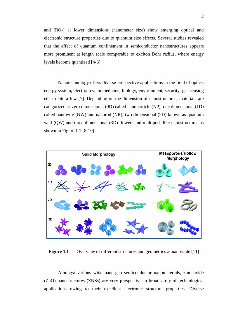
etc. to cite a few [7]. Depending on the dimension of nanostructures, materials are
categorized as zero dimensional (0D) called nanoparticle (NP), one dimensional (1D)
called nanowire (NW) and nanorod (NR); two dimensional (2D) known as quantum
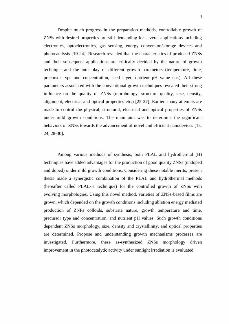
well (QW) and three dimensional (3D) flower- and multipod- like nanostructures as
shown in Figure 1.1 [8-10].
Figure 1.1 Overview of different structures and geometries at nanoscale [11]
Amongst various wide band-gap semiconductor nanomaterials, zinc oxide
(ZnO) nanostructures (ZNSs) are very prospective in broad array of technological
applications owing to their excellent electronic structure properties. Diverse
3
nanostructures of ZnO with unique features can easily be achieved using different
synthesis methods. These nanostructures possess outstanding optical properties
which are advantageous for the advancement of photovoltaic and optoelectronic
nanodevices. Furthermore, control of ZnO epitaxial layer quality together with native
and dopant point defects remains a vital issue for direct nanodevice production [12].
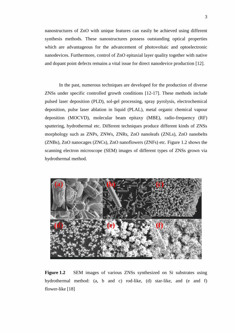
In the past, numerous techniques are developed for the production of diverse
ZNSs under specific controlled growth conditions [12-17]. These methods include
pulsed laser deposition (PLD), sol-gel processing, spray pyrolysis, electrochemical
deposition, pulse laser ablation in liquid (PLAL), metal organic chemical vapour
deposition (MOCVD), molecular beam epitaxy (MBE), radio-frequency (RF)
sputtering, hydrothermal etc. Different techniques produce different kinds of ZNSs
morphology such as ZNPs, ZNWs, ZNRs, ZnO nanoleafs (ZNLs), ZnO nanobelts
(ZNBs), ZnO nanocages (ZNCs), ZnO nanoflowers (ZNFs) etc. Figure 1.2 shows the
scanning electron microscope (SEM) images of different types of ZNSs grown via
hydrothermal method.
Figure 1.2 SEM images of various ZNSs synthesized on Si substrates using
hydrothermal method: (a, b and c) rod-like, (d) star-like, and (e and f)
flower-like [18]
(a)
(f) (e) (d)
(c) (b)
4
Despite much progress in the preparation methods, controllable growth of
ZNSs with desired properties are still demanding for several applications including
electronics, optoelectronics, gas sensing, energy conversion/storage devices and
photocatalysis [19-24]. Research revealed that the characteristics of produced ZNSs
and their subsequent applications are critically decided by the nature of growth
technique and the inter-play of different growth parameters (temperature, time,
precursor type and concentration, seed layer, nutrient pH value etc.). All these
parameters associated with the conventional growth techniques revealed their strong
influence on the quality of ZNSs (morphology, structure quality, size, density,
alignment, electrical and optical properties etc.) [25-27]. Earlier, many attempts are
made to control the physical, structural, electrical and optical properties of ZNSs
under mild growth conditions. The main aim was to determine the significant
behaviors of ZNSs towards the advancement of novel and efficient nanodevices [13,
24, 28-30].
Among various methods of synthesis, both PLAL and hydrothermal (H)
techniques have added advantages for the production of good quality ZNSs (undoped
and doped) under mild growth conditions. Considering these notable merits, present
thesis made a synergistic combination of the PLAL and hydrothermal methods
(hereafter called PLAL-H technique) for the controlled growth of ZNSs with
evolving morphologies. Using this novel method, varieties of ZNSs-based films are
grown, which depended on the growth conditions including ablation energy mediated
production of ZNPs colloids, substrate nature, growth temperature and time,
precursor type and concentration, and nutrient pH values. Such growth conditions
dependent ZNSs morphology, size, density and crystallinity, and optical properties
are determined. Propose and understanding growth mechanisms processes are
investigated. Furthermore, these as-synthesized ZNSs morphology driven
improvement in the photocatalytic activity under sunlight irradiation is evaluated.
5
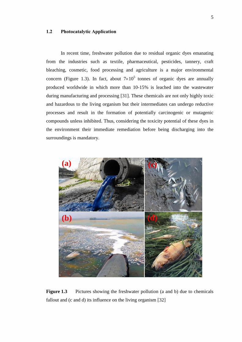
1.2 Photocatalytic Application
In recent time, freshwater pollution due to residual organic dyes emanating
from the industries such as textile, pharmaceutical, pesticides, tannery, craft
bleaching, cosmetic, food processing and agriculture is a major environmental
concern (Figure 1.3). In fact, about 7105 tonnes of organic dyes are annually
produced worldwide in which more than 10-15% is leached into the wastewater
during manufacturing and processing [31]. These chemicals are not only highly toxic
and hazardous to the living organism but their intermediates can undergo reductive
processes and result in the formation of potentially carcinogenic or mutagenic
compounds unless inhibited. Thus, considering the toxicity potential of these dyes in
the environment their immediate remediation before being discharging into the
surroundings is mandatory.
Figure 1.3 Pictures showing the freshwater pollution (a and b) due to chemicals
fallout and (c and d) its influence on the living organism [32]
(a)
(b)
(c)
(d)
6
In the past, diverse methods are developed to diminish the impact of such
toxic chemical pollutants on the non-human environment, aquatic systems, and
human life. Methods including adsorption, membrane separation and biological
treatments are limited due to their high operating cost and inefficient in removing
these pollutants. Currently, photocatalytic reactions based photodegradation
processes have been introduced as an effective and economic strategy to eliminate
such pollutants. In this regard, semiconductor nanomaterials (ZnO, SiO2 and TiO2)
revealed great prospects for photocatalytic applications to treat the environmental
pollutants. Categorically, heterogeneous photocatalysis-based ZNSs owing to their
unusual attributes such as diverse morphologies, ability to absorb a wide solar
spectrum, high chemical stabilizations, nontoxicity, abundance in nature and low cost
became attractive. Thus, this material is chosen for the effective remediation of toxic
environmental pollutants [19, 33, 34]. Many literature findings showed that the
optimization of physical properties of as-synthesized ZNSs in terms of large surface
area and modified bandgap can be useful for the increase of the number of active
sites and subsequent charge transfer useful for enhanced photocatalytic activity.
This in turn enhances the electron–hole pairs’ separation efficiency in the
photocatalytic reactions, resulting in the enhancement of the photocatalytic
efficiency [19, 31, 34-36].
1.3 Problem Statement
Despite the development of various syntheses methods of ZNSs a controlled
growth technique with mild conditions that produce evolving morphologies and
desirable properties is far from being achieved. Controlled growths of ZNSs are
significant for determining the overall properties as well as potential applications.
Most of the existing synthesis methods have complicated conditions such as high
vacuum, expensive devices, specialized laboratory, complex growth procedures, high
temperature, requirement of catalyst or template, and longer growth time. It is
necessary to develop a simple, fast, scalable, cost-effective and catalyst-free new
growth approach such as PLAL-H (combination of PLAL and hydrothermal
7
methods). This novel technique is expected to achieve better control on the growth of
ZNSs-based film, which is still lacking. A better understanding of the PLAL-H
growth mechanism that produces ZNSs with evolving morphologies in addition to
improved photocatalytic efficiency toward MB dye under sunlight irradiation is
prerequisite.
Modifications in the photocatalytic activity of ZNSs for practical applications
remain a major issue due to their wide band-gap nature and subsequent absorption in
the UV region only. On top, the wide band-gap nature of ZNSs allows only ~ 4-5%
of the solar spectrum in the UV-range for effective use as a renewable energy source,
which is the main limitation for photocatalytic application. Furthermore, studies on
superior photo-degradation ability of MB by un-doped ZNSs-based film under
sunlight irradiation are rarely performed. Earlier studies mainly focused on ZNSs-
based powder to achieve photodegradation of MB dye. These are not only expensive,
but required complex processing tools such as centrifugation. Consequently, it is
necessary to overcome the limitations associated with weak photocatalytic activities
of ZNSs where an enhancement in the harvesting (absorption) efficiency of the solar
spectra in the broad region (UV and visible (Vis) regions) and reduction of
processing cost is necessary. In this view, present thesis took a fair attempt to
develop a new PLAL-H growth technique for overcoming all the above mentioned
limitations associated with the synthesis of ZNSs using conventional methods as well
as the photocatalytic application of ZNSs driven by sunlight irradiation.
8
1.4 Research Objectives
Based on the aforementioned problem statement and research background on
ZNSs the following objectives are set:
1. To develop a new PLAL-H technique with mild growth conditions for the
controlled synthesis of ZNSs-based film having evolving morphologies.
2. To determine the influence of growth parameters on the morphology, size,
density, structure, composition, optical properties and photocatalytic
activities of the synthesized ZNSs-based film.
3. To evaluate the photocatalytic activity of the as-grown ZNSs-based films in
terms of photo-degradation ability of MB under sunlight irradiation.
1.5 Scope of the Study
Present study includes the synthesis, characterization and determination of
photocatalytic properties of ZNSs-based films. Amongst all the metal oxides
semiconductors ZnO is preferred for various applications because of their distinct
physical and chemical attributes including wide direct band gap, large exciton
binding energy, excellent stability, environmental friendliness, low cost and easy
availability. These fascinating properties are supported by ability of ZnO to easy
formation of different nanostructures (ZNSs) gained high surface area to volume
ratio and effective quantum confinement compared to bulk structure. Intense
researches revealed the possibility of tuning the electronic band structure of ZnO via
controlled synthesis of ZNSs useful for light-emitting diodes, sensors, catalysts, field
emitters, biosensors, solar cell and photocatalytic application. Preparation of
ZNSs-based films with controlled morphologies will be achieved via newly proposed
PLAL-H growth technique as well as using conventional hydrothermal route to
authenticate its ability for producing high quality ZNSs with diverse morphologies.
The growth conditions optimization by varying different growth parameters would
9
be the major focus. The impact of various processing parameters including laser
energy, nutrient type, nutrient pH, nutrient concentration, growth temperature and
time on the ZNSs growth mechanism will be determined. Such NSs properties such
as morphology, density, orientation, aspect ratio, crystal size and crystallinity are
found to be strongly depended on the synthesis technique and processing conditions.
Current newly proposed simple PLAL-H technique is capable of controlling
the morphology, aspect ratio, density, structure, optical and photocatalytic properties
of ZNSs. This technique is cost-effective and can produce good quality ZNSs at short
growth time and low temperature (under mild growth conditions) without requiring
any catalysts. Moreover, more than three ZNSs can be grown simultaneously.
Such ZNSs with diverse morphologies, sizes and crystallinity are essential for the
development of ZNSs based optoelectronic devices which are cheap and efficient.
Using the proposed systematic characterization method it is possible to determine the
purity, size, emission and absorption properties, band gap, photocatalytic activity and
morphology of ZNSs as well as other nanomaterials required for broad array of
applications. The photocatalytic performance of the optimal ZNSs samples under
sunlight irradiation will be measured in terms of photo-degradation of MB dye.
As-prepared samples are thoroughly characterized using various imaging and
spectroscopic techniques. The experimental results on as-synthesized ZNSs are
compared with similar existing findings for better understanding of the growth
mechanisms. Samples structures and morphology (surface morphology, size, density,
crystallinity, and elemental traces) are determined using field emission scanning
electron microscopy (FESEM), X-ray diffraction (XRD) measurement, high
resolution transmission electron microscopy (HRTEM), Energy dispersive X-ray
diffraction (EDX) and Fourier-transform infrared (FTIR) spectroscopy. The optical
properties of ZNSs samples are determined via photoluminescence (PL)
spectroscopy and UV-Vis absorption spectroscopy.
10
1.6 Significance of the Study
Nowadays the pollution of freshwater due to the residual organic
contaminants (dyes) in the form of chemical waste being the major environmental
concern needs remediation. These chemical pollutants are highly toxic and harmful
for the entire eco-system. Therefore, degradation of these organic contaminants is
essential to circumvent the human health risks. In this view, ZNSs-based films grown
via innovative PLAL-H route can be superior in terms of photocatalytic efficiency
toward pollutant MB dye under sunlight irradiation. This study is expected to
contribute towards the development of high performing ZNSs with excellent
photocatalytic activity under sunlight irradiation which is a free, clean, and
inexhaustible irradiation source. The capacity to use ZNSs-based films for useful
exploitation of sunlight source to achieve enhanced photocatalytic action would
certainly be beneficial in terms of economy and environment. Furthermore, PLAL-H
technique can be extended for producing other semiconductor NSs morphologies.
Present PLAL-H technique may constitute a basis for controlled manipulation of
nanomaterials desirable for high performance nanodevices.
1.7 Thesis Organization
This thesis is composed into five chapters as follows:
Chapter 1 presents a brief background of the research to justify the
importance of ZNSs and need for further studies. It clearly shows the research gap to
set out the precise objectives to be accomplished. It includes the problem statement,
research objectives, scope, and significance.
Chapter 2 describes the comprehensive literature survey to show the existing
research gaps and the relevant findings on the cited topic made so far. It describes the
11
electrical, optical and structural properties of ZnO nanostructure. It explains the
growth mechanism of ZNSs associated with the hydrothermal and PLAL synthesis
techniques. Influence of growth technique and growth conditions on the morphology
of ZNSs films are discussed. The mechanism of photocatalytic activity of ZNSs is
underscored. The major parameters of different growth techniques that can achieve
enhanced photocatalytic efficiency of ZNSs film are explained in depth.
Chapter 3 highlights the detailed research methodology in terms of synthesis
techniques used and the adopted steps towards photocatalytic applications. The
background information of major experimental techniques for collecting the data and
analysis related to as-grown ZNSs samples are emphasized.
Chapter 4 presents detailed results, analysis and discussion. Findings from
two different synthesis methods such as hydrothermal and PLAL-H are evaluated in
terms of optimization of growth conditions to produce various catalyst-free ZNSs
films at low temperature and growth time compared with previous studies. The
remarkable features of the synergistic PLAL-H technique for controlled preparation
of ZNSs-based film are explained. Photocatalytic performance of obtained optimal
ZNSs samples under sunlight irradiation is assessed using photodegradation of MB
dye. Dependence of photodegradation efficiency of MB dyes on morphology and
structure evolution of as-grown ZNSs films as well as ability of these ZNSs film as
photocatalysis to overcome the shortcomings for beneficial exploiting of sunlight
source is attributed.
Chapter 5 concludes the thesis and provides some future outlook in terms of
recommendations. The successful accomplishments of the proposed research
objectives are demonstrated.
185
REFERENCES
1. Corbett, J., P. McKeown, G. Peggs and R. Whatmore. Nanotechnology:
international developments and emerging products. CIRP Annals-
Manufacturing Technology. 2000. 49(2): 523-545.
2. Drexler, K. E. and M. Minsky. Engines of creation: Fourth Estate London.
1990.
3. Louis, C. and O. Pluchery. Gold nanoparticles for physics, chemistry and
biology: World Scientific. 2012.
4. Senger, R. and K. Bajaj. Optical properties of confined polaronic excitons in
spherical ionic quantum dots. Physical Review B. 2003. 68(4): 045313-1-
045313-19.
5. Bawendi, M. G., P. Carroll, W. L. Wilson and L. Brus. Luminescence
properties of CdSe quantum crystallites: Resonance between interior and
surface localized states. The Journal of Chemical Physics. 1992. 96(2): 946-
954.
6. Reimann, S. M. and M. Manninen. Electronic structure of quantum dots.
Reviews of Modern Physics. 2002. 74(4): 1283-1298.
7. Djurišić, A. B. and Y. H. Leung. Optical properties of ZnO nanostructures.
Small. 2006. 2(8‐9): 944-961.
8. Cao, G. Nanostructure and nanomaterials synthesis, properties and
applications: World Scientific. 2004.
9. Wang, N., Y. Yang and G. Yang. Great blue-shift of luminescence of ZnO
nanoparticle array constructed from ZnO quantum dots. Nanoscale Research
Letters. 2011. 6(1): 1-6.
10. Vanalakar, S. A., V. L. Patil, N. S. Harale, S. A. Vhanalakar, M. G. Gang, J.
Y. Kim, P. S. Patil and J. H. Kim. Controlled growth of ZnO nanorod arrays
186
via wet chemical route for NO2 gas sensor applications. Sensors and
Actuators B: Chemical. 2015. 221: 1195-1201.
11. Wu, Z., S. Yang and W. Wu. Shape control of inorganic nanoparticles from
solution. Nanoscale. 2016. 8(3): 1237-1259.
12. Willander, M. Zinc Oxide Nanostructures: Advances and Applications: CRC
Press. 2014.
13. Baruah, S. and J. Dutta. Hydrothermal growth of ZnO nanostructures. Science
and Technology of Advanced Materials. 2009. 10(1): 013001-1-013001-18.
14. Shi, F. and C. Xue. Morphology and growth mechanism of multileg ZnO
nanostructures by chemical vapor deposition. CrystEngComm. 2012. 14(12):
4173-4175.
15. Talebian, N., M. R. Nilforoushan and N. Maleki. Ultraviolet to visible-light
range photocatalytic activity of ZnO films prepared using sol-gel method:
The influence of solvent. Thin Solid Films. 2013. 527: 50-58.
16. Feng, J. J., Q. C. Liao, A. J. Wang and J. R. Chen. Mannite supported
hydrothermal synthesis of hollow flower-like ZnO structures for
photocatalytic applications. CrystEngComm. 2011. 13(12): 4202-4210.
17. Zhu, B., X. Zhao, F. Su, G. Li, X. Wu, J. Wu, R. Wu and J. Liu. Structural
and optical properties of ZnO thin films on glass substrate grown by laser-
ablating Zn target in oxygen atmosphere. Physica B: Condensed Matter.
2007. 396(1): 95-101.
18. Amin, G., M. Asif, A. Zainelabdin, S. Zaman, O. Nur and M. Willander.
Influence of pH, precursor concentration, growth time, and temperature on
the morphology of ZnO nanostructures grown by the hydrothermal method.
Journal of Nanomaterials. 2011. 2011: 269692-5- 269692-9.
19. Kuriakose, S., B. Satpati and S. Mohapatra. Enhanced photocatalytic activity
of Co doped ZnO nanodisks and nanorods prepared by a facile wet chemical
method. Physical Chemistry Chemical Physics. 2014. 16(25): 12741-12749.
20. Zhou, Y., D. Li, X. Zhang, J. Chen and S. Zhang. Facile synthesis of ZnO
micro-nanostructures with controllable morphology and their applications in
dye-sensitized solar cells. Applied Surface Science. 2012. 261: 759-763.
21. Öztürk, S., N. Kılınç, N. Taşaltin and Z. Öztürk. A comparative study on the
NO2 gas sensing properties of ZnO thin films, nanowires and nanorods. Thin
Solid Films. 2011. 520(3): 932-938.
187
22. Sun, X., Q. Li, J. Jiang and Y. Mao. Morphology-tunable synthesis of ZnO
nanoforest and its photoelectrochemical performance. Nanoscale. 2014.
6(15): 8769-8780.
23. Peng, C., J. Guo, W. Yang, C. Shi, M. Liu, Y. Zheng, J. Xu, P. Chen, T.
Huang and Y. Yang. Synthesis of three-dimensional flower-like hierarchical
ZnO nanostructure and its enhanced acetone gas sensing properties. Journal
of Alloys and Compounds. 2016. 654: 371-378.
24. Ansari, S. A., M. M. Khan, S. Kalathil, A. Nisar, J. Lee and M. H. Cho.
Oxygen vacancy induced band gap narrowing of ZnO nanostructures by an
electrochemically active biofilm. Nanoscale. 2013. 5(19): 9238-9246.
25. Schmidt-Mende, L. and J. L. MacManus-Driscoll. ZnO–nanostructures,
defects, and devices. Materials Today. 2007. 10(5): 40-48.
26. Riedel, W., Y. Fu, Ü. Aksünger, J. Kavalakkatt, C. H. Fischer, M. C. Lux-
Steiner and S. Gledhill. ZnO and ZnS nanodots deposited by spray methods:
A versatile tool for nucleation control in electrochemical ZnO nanorod array
growth. Thin Solid Films. 2015. 589: 327-330.
27. Hynek, J., . Kalousek, R. ou elka, P. Bezdi ka, P. Dzik, J. . Rathousk , J.
Demel and K. Lang. High photocatalytic activity of transparent films
composed of ZnO nanosheets. Langmuir. 2014. 30(1): 380-386.
28. Heo, S. N., K. Y. Park, Y. J. Seo, F. Ahmed, M. Anwar and B. H. Koo. Effect
of solution concentration on the functional properties of ZnO nanostructures:
Role of Hexamethylenetetramine. Electronic Materials Letters. 2013. 9(3):
261-265.
29. Dedova, T., M. Krunks, I. O. Acik, D. Klauson, O. Volobujeva and A. Mere.
Hierarchical nanostructures of ZnO obtained by spray pyrolysis. Materials
Chemistry and Physics. 2013. 141(1): 69-75.
30. Tang, J. F., H. H. Su, Y. M. Lu and S. Y. Chu. Controlled growth of ZnO
nanoflowers on nanowall and nanorod networks via a hydrothermal method.
CrystEngComm. 2014. 17(3): 592-597.
31. Lam, S. M., J. C. Sin, A. Z. Abdullah and A. R. Mohamed. Degradation of
wastewaters containing organic dyes photocatalysed by zinc oxide: a review.
Desalination and Water Treatment. 2012. 41(1-3): 131-169.
32. Rana, S. Environmental pollution: health and toxicology: Alpha Science
International Limited. 2006.
188
33. Hoffmann, M. R., S. T. Martin, W. Choi and D. W. Bahnemann.
Environmental applications of semiconductor photocatalysis. Chemical
Reviews. 1995. 95(1): 69-96.
34. Lee, K. M., C. W. Lai, K. S. Ngai and J. C. Juan. Recent developments of
zinc oxide based photocatalyst in water treatment technology: a review.
Water Research. 2016. 88: 428-448.
35. Sun, J. H., S. Y. Dong, J. L. Feng, X. J. Yin and X. C. Zhao. Enhanced
sunlight photocatalytic performance of Sn-doped ZnO for Methylene Blue
degradation. Journal of Molecular Catalysis A: Chemical. 2011. 335(1): 145-
150.
36. Mohan, R., K. Krishnamoorthy and S. J. Kim. Enhanced photocatalytic
activity of Cu-doped ZnO nanorods. Solid State Communications. 2012.
152(5): 375-380.
37. Fierro, J. L. G. Metal oxides: chemistry and applications: CRC press. 2005
38. Yano, M., K. Koike, S. Sasa and M. Inoue. Zinc Oxide, Bulk, Thin Films and
Nanostructures. Elsevier Science. 2006: 372-414.
39. Noriega, R., J. Rivnay, L. Goris, D. Kälblein, H. Klauk, K. Kern, L. M.
Thompson, A. C. Palke, J. F. Stebbins and J. R. Jokisaari. Probing the
electrical properties of highly-doped Al: ZnO nanowire ensembles. Journal of
Applied Physics. 2010. 107(7): 074312-1-074312-8.
40. Choi, H. H. Synthesis and characterization of tailored zinc oxide
nanostructures and their engineered nanocomposites. Thesis Doctor
Philosophy. University of Florida. 2004.
41. Huh, P., F. Yan, L. Li, M. Kim, R. Mosurkal, L. A. Samuelson and J. Kumar.
Simple fabrication of zinc oxide nanostructures. Journal of Materials
Chemistry. 2008. 18(6): 637-639.
42. Xiaowei, S. and Y. Yang. ZnO Nanostructures and Their Applications: CRC
Press. 2016.
43. Wang, Z. L. Zinc oxide nanostructures: growth, properties and applications.
Journal of Physics: Condensed Matter. 2004. 16(25): R829.
44. Chen, Z., Z. Shan, S. Li, C. Liang and S. X. Mao. A novel and simple growth
route towards ultra-fine ZnO nanowires. Journal of Crystal growth. 2004.
265(3): 482-486.
189
45. Li, S., X. Zhang, B. Yan and T. Yu. Growth mechanism and diameter control
of well-aligned small-diameter ZnO nanowire arrays synthesized by a
catalyst-free thermal evaporation method. Nanotechnology. 2009. 20(49):
495604-1-495604-9.
46. Özgür, Ü., Y. I. Alivov, C. Liu, A. Teke, M. Reshchikov, S. Doğan, .
Avrutin, S. J. Cho and H. Morkoc. A comprehensive review of ZnO materials
and devices. Journal of Applied Physics. 2005. 98(4): 041301-1-041301-5.
47. Gil, B. and A. V. Kavokin. Giant exciton-light coupling in ZnO quantum
dots. Applied Physics Letters. 2002. 81(4): 748-750.
48. Wong, E. M. and P. C. Searson. ZnO quantum particle thin films fabricated
by electrophoretic deposition. Applied Physics Letters. 1999. 74(20): 2939-
2941.
49. Gorla, C., W. Mayo, S. Liang and Y. Lu. Structure and interface-controlled
growth kinetics of ZnAl2O4 formed at the (1120) ZnO/(0112) Al2O3 interface.
Journal of Applied Physics. 2000. 87(8): 3736-3743.
50. Abbas, K. N., N. Bidin and R. S. Sabry. Controllable ZnO Nanostructures
Evolution via Synergistic Pulsed Laser Ablation and Hydrothermal Methods.
Ceramics International. 2016. 42: 13535-13546.
51. Bera, A. and D. Basak. Role of defects in the anomalous photoconductivity in
ZnO nanowires. Applied Physics Letters. 2009. 94(16): 163119-1-163119-4.
52. Janotti, A. and C. G. Van de Walle. Fundamentals of zinc oxide as a
semiconductor. Reports on Progress in Physics. 2009. 72(12): 126501-1-
126501-29.
53. Epie, E. and W. Chu. Ionoluminescence study of Zn− and O− implanted ZnO
crystals: An additional perspective. Applied Surface Science. 2016. 371: 28-
34.
54. Fang, Y., Y. Wang, L. Gu, R. Lu and J. Sha. Effect of the defect on
photoluminescence property of Al-coated ZnO nanostructures. Optics
Express. 2013. 21(3): 3492-3500.
55. Liang, Y. C. and H. Zhong. Self-catalytic crystal growth, formation
mechanism, and optical properties of indium tin oxide nanostructures.
Nanoscale Research Letters. 2013. 8(1): 1-10.
56. Eason, R. Pulsed laser deposition of thin films: applications-led growth of
functional materials: John Wiley and Sons. 2007.
190
57. Xiao, Q., J. Zhang, C. Xiao and X. Tan. Photocatalytic decolorization of
methylene blue over Zn1-xCoxO under visible light irradiation. Materials
Science and Engineering: B. 2007. 142(2): 121-125.
58. Fan, Z. and J. G. Lu. Zinc oxide nanostructures: synthesis and properties.
Journal of Nanoscience and Nanotechnology. 2005. 5(10): 1561-1573.
59. Zeng, H., X. W. Du, S. C. Singh, S. A. Kulinich, S. Yang, J. He and W. Cai.
Nanomaterials via laser ablation/irradiation in liquid: a review. Advanced
Functional Materials. 2012. 22(7): 1333-1353.
60. Kumar, R., G. Kumar and A. Umar. Pulse laser deposited nanostructured
ZnO thin films: A review. Journal of Nanoscience and Nanotechnology.
2014. 14(2): 1911-1930.
61. Ishikawa, Y., Y. Shimizu, T. Sasaki and N. Koshizaki. Preparation of zinc
oxide nanorods using pulsed laser ablation in water media at high
temperature. Journal of Colloid and Interface Science. 2006. 300(2): 612-
615.
62. Fazio, E., A. Mezzasalma, G. Mondio, F. Neri and R. Saija. ZnO
nanostructures produced by laser ablation in water: optical and structural
properties. Applied Surface Science. 2013. 272: 30-35.
63. Zeng, H., S. Yang and W. Cai. Reshaping formation and luminescence
evolution of ZnO quantum dots by laser-induced fragmentation in liquid. The
Journal of Physical Chemistry C. 2011. 115(12): 5038-5043.
64. Osuwa, J. and P. Anusionwu. Some advances and prospects in
nanotechnology: a review. Asian Journal of Information Technology. 2011.
10: 96-100.
65. Zeng, H., Z. Li, W. Cai, B. Cao, P. Liu and S. Yang. Microstructure control
of Zn/ZnO core/shell nanoparticles and their temperature-dependent blue
emissions. The Journal of Physical Chemistry B. 2007. 111(51): 14311-
14317.
66. Yang, G. Laser ablation in liquids: principles and applications in the
preparation of nanomaterials: CRC Press. 2012.
67. Badawy, M. I., F. Mahmoud, A. A. Abdel-Khalek, T. A. Gad-Allah and A.
Abdel Samad. Solar photocatalytic activity of sol-gel prepared Ag-doped
ZnO thin films. Desalination and Water Treatment. 2014. 52(13-15): 2601-
2608.
191
68. Russo, R., X. Mao, C. Liu and J. Gonzalez. Laser assisted plasma
spectrochemistry: laser ablation. Journal of Analytical Atomic Spectrometry.
2004. 19(9): 1084-1089.
69. Mei, T. and Y. Hu. Synthesis, Self-assembly and Optoelectronic Properties of
Monodisperse ZnO Quantum Dots: Intech Open Access Publisher. 2011.
70. Phipps, C. Laser ablation and its applications, ed. Vol. 129: Springer. 2007.
71. Guillén, G. G., M. M. Palma, B. Krishnan, D. Avellaneda, G. Castillo, T. D.
Roy and S. Shaji. Structure and morphologies of ZnO nanoparticles
synthesized by pulsed laser ablation in liquid: Effects of temperature and
energy fluence. Materials Chemistry and Physics. 2015. 162: 561-570.
72. Yan, Z. and D. B. Chrisey. Pulsed laser ablation in liquid for micro-
/nanostructure generation. Journal of Photochemistry and Photobiology C:
Photochemistry Reviews. 2012. 13(3): 204-223.
73. Yang, G. Laser ablation in liquids: applications in the synthesis of
nanocrystals. Progress in Materials Science. 2007. 52(4): 648-698.
74. Premkumar, T., P. Manoravi, B. Panigrahi and K. Baskar. Particulate assisted
growth of ZnO nanorods and microrods by pulsed laser deposition. Applied
Surface Science. 2009. 255(15): 6819-6822.
75. Zhang, X., H. Zeng and W. Cai. Laser power effect on morphology and
photoluminescence of ZnO nanostructures by laser ablation in water.
Materials Letters. 2009. 63(2): 191-193.
76. Thareja, R. and S. Shukla. Synthesis and characterization of zinc oxide
nanoparticles by laser ablation of zinc in liquid. Applied Surface Science.
2007. 253(22): 8889-8895.
77. Tripathi, S., R. Choudhary, A. Tripathi, V. Baranwa, A. Pandey, J. Gerlach,
C. Dar and D. Kanjilal. Studies of effect of deposition parameters on the ZnO
films prepared by PLD. Nuclear Instruments and Methods in Physics
Research Section B: Beam Interactions with Materials and Atoms. 2008.
266(8): 1533-1536.
78. Svetlichnyi, V., A. Shabalina, I. Lapin, D. Goncharova and A. Nemoykina.
ZnO nanoparticles obtained by pulsed laser ablation and their composite with
cotton fabric: Preparation and study of antibacterial activity. Applied Surface
Science. 2016. 372: 20-29.

192
79. Camarda, P., R. Schneider, R. Popescu, L. Vaccaro, F. Messina, G.
Buscarino, S. Agnello, F. Gelardi and M. Cannas. Effect of thermal annealing
on the luminescence of defective ZnO nanoparticles synthesized by pulsed
laser ablation in water. Physica Status Solidi (C). 2016. 13(10-12): 890-894.
80. Hamad, A., L. Li and Z. Liu. Comparison of characteristics of selected
metallic and metal oxide nanoparticles produced by picosecond laser ablation
at 532 and 1064 nm wavelengths. Applied Physics A. 2016. 122(10): 904-1-
904-15.
81. Goto, T., M. Honda, S. A. Kulinich, Y. Shimizu and T. Ito. Defects in ZnO
nanoparticles laser-ablated in water-ethanol mixtures at different pressures.
Japanese Journal of Applied Physics. 2015. 54(7): 070305-1-070305-6.
82. Hussain, M. Z., R. Khan, R. Ali and Y. Khan. Optical properties of laser
ablated ZnO nanoparticles prepared with Tween‐80. Materials Letters. 2014.
122: 147-150.
83. Solati, E., L. Dejam and D. Dorranian. Effect of laser pulse energy and
wavelength on the structure, morphology and optical properties of ZnO
nanoparticles. Optics and Laser Technology. 2014. 58: 26-32.
84. Soliman, W., N. Takada, N. Koshizaki and K. Sasaki. Structure and size
control of ZnO nanoparticles by applying high pressure to ambient liquid in
liquid-phase laser ablation. Applied Physics A. 2013. 110(4): 779-783.
85. Kim, K. K., D. Kim, S. K. Kim, S. M. Park and J. K. Song. Formation of ZnO
nanoparticles by laser ablation in neat water. Chemical Physics Letters. 2011.
511(1): 116-120.
86. Zeng, J. N., J. K. Low, Z. M. Ren, T. Liew and Y. F. Lu. Effect of deposition
conditions on optical and electrical properties of ZnO films prepared by
pulsed laser deposition. Applied Surface Science. 2002. 197: 362-367.
87. Fan, X., J. Lian, Z. Guo and H. Lu. ZnO thin film formation on Si (111) by
laser ablation of Zn target in oxygen atmosphere. Journal of Crystal Growth.
2005. 279(3): 447-453.
88. Sasaki, T., Y. Shimizu and N. Koshizaki. Preparation of metal oxide-based
nanomaterials using nanosecond pulsed laser ablation in liquids. Journal of
Photochemistry and Photobiology A: Chemistry. 2006. 182(3): 335-341.
89. Feng, Z. C. Handbook of Zinc Oxide and Related Materials: Volume Two,
Devices and Nano-Engineering, ed. Vol. 2: CRC Press. 2012.

193
90. Chen, M. Z., W. S. Chen, S. C. Jeng, S. H. Yang and Y. F. Chung. Liquid
crystal alignment on zinc oxide nanowire arrays for LCDs applications.
Optics Express. 2013. 21(24): 29277-29282.
91. Shen, G., P. C. Chen, K. Ryu and C. Zhou. Devices and chemical sensing
applications of metal oxide nanowires. Journal of Materials Chemistry. 2009.
19(7): 828-839.
92. Lee, C. Y., S. Y. Li, P. Lin and T. Y. Tseng. ZnO nanowires hydrothermally
grown on PET polymer substrates and their characteristics. Journal of
Nanoscience and Nanotechnology. 2005. 5(7): 1088-1094.
93. Byrappa, K. and M. Yoshimura. Handbook of Hydrotermal Technology. A
Technology for Crystal Growth and Materials Processing. Byrappa, M.
Yoshimura-Noyes Publications, Park Ridge, NJ. 2001.
94. Motevalizadeh, L., Z. Heidary and M. E. Abrishami. Facile template-free
hydrothermal synthesis and microstrain measurement of ZnO nanorods.
Bulletin of Materials Science. 2014. 37(3): 397-405.
95. Guo, W., T. Liu, L. Huang, H. Zhang, Q. Zhou and W. Zeng. HMT assisted
hydrothermal synthesis of various ZnO nanostructures: Structure, growth and
gas sensor properties. Physica E: Low-Dimensional Systems and
Nanostructures. 2011. 44(3): 680-685.
96. Öztürk, S., N. Kılınç, İ. Torun, A. Kösemen, Y. Şahin and Z. Z. Öztürk.
Hydrogen sensing properties of ZnO nanorods: Effects of annealing,
temperature and electrode structure. International Journal of Hydrogen
Energy. 2014. 39(10): 5194-5201.
97. Sambath, K., M. Saroja, M. Venkatachalam, K. Rajendran and K.
Jagatheeswaran. Hydrothermal Synthesis of Flower-Like ZnO Nanostructures
and their Optical Properties. Advanced Materials Research. 2013. 678: 91-96.
98. Zhou, Q., W. Chen, S. Peng and W. Zeng. Hydrothermal synthesis and
acetylene sensing properties of variety low dimensional zinc oxide
nanostructures. The Scientific World Journal. 2014: 489170-1-489170-8.
99. Wang, H., J. Xie, K. Yan and M. Duan. Growth mechanism of different
morphologies of ZnO crystals prepared by hydrothermal method. Journal of
Materials Science and Technology. 2011. 27(2): 153-158.
194
100. Chakraborty, S. and P. Kumbhakar. Observation of bandgap narrowing effect
and photoluminescence emission characteristics of chemically synthesized Co
doped ZnO nanosheets. Indian Journal of Physics. 2014. 88(3): 251-257.
101. Kim, H. and K. Yong. A highly efficient light capturing 2D (nanosheet)-1D
(nanorod) combined hierarchical ZnO nanostructure for efficient quantum dot
sensitized solar cells. Physical Chemistry Chemical Physics. 2013. 15(6):
2109-2116.
102. Zhang, Y., H. Wang, H. Jiang and X. Wang. Water induced protonation of
amine-terminated micelles for direct syntheses of ZnO quantum dots and
their cytotoxicity towards cancer. Nanoscale. 2012. 4(11): 3530-3535.
103. Li, H., S. Jiao, S. Gao, H. Li and L. Li. Dynamically controlled synthesis of
different ZnO nanostructures by a surfactant-free hydrothermal method.
CrystEngComm. 2014. 16(38): 9069-9074.
104. Yang, L., P. W. May, L. Yin and T. B. Scott. Growth of self-assembled ZnO
nanoleaf from aqueous solution by pulsed laser ablation. Nanotechnology.
2007. 18(21): 215602-1-215602-8.
105. Son, N. T., J.-S. Noh and I.-H. Lee. Low-temperature solution syntheses of
hexagonal ZnO nanorods and morphology-controlled nanostructures.
Chemical Physics Letters. 2016. 646: 185-189.
106. Guo, Y., S. Lin, X. Li and Y. Liu. Amino acids assisted hydrothermal
synthesis of hierarchically structured ZnO with enhanced photocatalytic
activities. Applied Surface Science. 2016. 384: 83-91.
107. Harish, S., M. Navaneethan, J. Archana, A. Silambarasan, S. Ponnusamy, C.
Muthamizhchelvan and Y. Hayakawa. Controlled synthesis of organic ligand
passivated ZnO nanostructures and their photocatalytic activity under visible
light irradiation. Dalton Transactions. 2015. 44(22): 10490-10498.
108. Zhou, Y., C. Liu, X. Zhong, H. Wu, M. Li and L. Wang. Simple
hydrothermal preparation of new type of sea urchin-like hierarchical ZnO
micro/nanostructures and their formation mechanism. Ceramics
International. 2014. 40(7): 10415-10421.
109. Shi, R., P. Yang, X. Dong, Q. Ma and A. Zhang. Growth of flower-like ZnO
on ZnO nanorod arrays created on zinc substrate through low-temperature
hydrothermal synthesis. Applied Surface Science. 2013. 264: 162-170.
195
110. Chen, P., L. Gu, X. Xue, Y. Song, L. Zhu and X. Cao. Facile synthesis of
highly uniform ZnO multipods as the supports of Au and Ag nanoparticles.
Materials Chemistry and Physics. 2010. 122(1): 41-48.
111. Mun, D. H., S. Bak, J. S. Ha, H. J. Lee, J. Lee, S. Lee and Y. Moon. Effects
of precursor concentration on the properties of ZnO nanowires grown on (1–
102) R-Plane sapphire substrates by hydrothermal synthesis. Journal of
Nanoscience and Nanotechnology. 2014. 14(8): 5970-5975.
112. Pawar, R. C., H. Kim and C. S. Lee. Defect-controlled growth of ZnO
nanostructures using its different zinc precursors and their application for
effective photodegradation. Current Applied Physics. 2014. 14(4): 621-629.
113. Song, J. and S. Lim. Effect of seed layer on the growth of ZnO nanorods. The
Journal of Physical Chemistry C. 2007. 111(2): 596-600.
114. Zhang, X., J. Qin, Y. Xue, P. Yu, B. Zhang, L. Wang and R. Liu. Effect of
aspect ratio and surface defects on the photocatalytic activity of ZnO
nanorods. Scientific Reports. 2014. 4: 4596-1-4596-8.
115. Yang, B., X. Zuo, X. Yang, L. Zhou and G. Li. Effects of pH values on
crystal growth and photoluminescence properties of ZnO hexagonal rods with
cones. Materials Letters. 2014. 130: 123-126.
116. Wahab, R., S. Ansari, Y. S. Kim, M. Song and H. S. Shin. The role of pH
variation on the growth of zinc oxide nanostructures. Applied Surface
Science. 2009. 255(9): 4891-4896.
117. Samaele, N., P. Amornpitoksuk and S. Suwanboon. Effect of pH on the
morphology and optical properties of modified ZnO particles by SDS via a
precipitation method. Powder Technology. 2010. 203(2): 243-247.
118. Tong, Y., Y. Liu, L. Dong, D. Zhao, J. Zhang, Y. Lu, D. Shen and X. Fan.
Growth of ZnO nanostructures with different morphologies by using
hydrothermal technique. The Journal of Physical Chemistry B. 2006.
110(41): 20263-20267.
119. Giri, P., S. Dhara and R. Chakraborty. Effect of ZnO seed layer on the
catalytic growth of vertically aligned ZnO nanorod arrays. Materials
Chemistry and Physics. 2010. 122(1): 18-22.
120. Peiris, T. N., H. Alessa, J. S. Sagu, I. A. Bhatti, P. Isherwood and K. U.
Wijayantha. Effect of ZnO seed layer thickness on hierarchical ZnO nanorod

196
growth on flexible substrates for application in dye-sensitised solar cells.
Journal of Nanoparticle Research. 2013. 15(12): 1-10.
121. Kozuka, Y., A. Tsukazaki and M. Kawasaki. Challenges and opportunities of
ZnO-related single crystalline heterostructures. Applied Physics Reviews.
2014. 1(1): 011303-1-011303-18.
122. Hai-Bo, F., Z. Xin-Liang, W. Si-Cheng, L. Zhi-Gang and Y. He-Bao. Zn/O
ratio and oxygen chemical state of nanocrystalline ZnO films grown at
different temperatures. Chinese Physics B. 2012. 21(3): 038101-1- 038101-5.
123. Weissler, G. Methods of experimental physics. ed. Volume 14. Vacuum
Physics and Technology: Academic Press, Inc., New York, NY. 1979.
124. Tsoutsouva, M., C. Panagopoulos and M. Kompitsas. Laser energy density,
structure and properties of pulsed-laser deposited zinc oxide films. Applied
Surface Science. 2011. 257(14): 6314-6319.
125. Sakthivel, S., B. Neppolian, M. Shankar, B. Arabindoo, M. Palanichamy and
V. Murugesan. Solar photocatalytic degradation of azo dye: comparison of
photocatalytic efficiency of ZnO and TiO2. Solar Energy Materials and Solar
Cells. 2003. 77(1): 65-82.
126. Kansal, S., M. Singh and D. Sud. Studies on photodegradation of two
commercial dyes in aqueous phase using different photocatalysts. Journal of
hazardous materials. 2007. 141(3): 581-590.
127. Panthi, G., M. Park, H. Y. Kim, S. Y. Lee and S. J. Park. Electrospun ZnO
hybrid nanofibers for photodegradation of wastewater containing organic
dyes: a review. Journal of Industrial and Engineering Chemistry. 2015. 21:
26-35.
128. Zheng, L., Y. Zheng, C. Chen, Y. Zhan, X. Lin, Q. Zheng, K. Wei and J. Zhu.
Network structured SnO2/ZnO heterojunction nanocatalyst with high
photocatalytic activity. Inorganic Chemistry. 2009. 48(5): 1819-1825.
129. Marci, G., V. Augugliaro, M. J. Lopez-Munoz, C. Martin, L. Palmisano, V.
Rives, M. Schiavello, R. J. Tilley and A. M. Venezia. Preparation
characterization and photocatalytic activity of polycrystalline ZnO/TiO2
systems. 2. Surface, bulk characterization, and 4-nitrophenol
photodegradation in liquid-solid regime. The Journal of Physical Chemistry
B. 2001. 105(5): 1033-1040.

197
130. Zhang, Z., C. Shao, X. Li, C. Wang, M. Zhang and Y. Liu. Electrospun
nanofibers of p-type NiO/n-type ZnO heterojunctions with enhanced
photocatalytic activity. ACS Applied Materials and Interfaces. 2010. 2(10):
2915-2923.
131. Yu, C., K. Yang, Y. Xie, Q. Fan, C. Y. Jimmy, Q. Shu and C. Wang. Novel
hollow Pt-ZnO nanocomposite microspheres with hierarchical structure and
enhanced photocatalytic activity and stability. Nanoscale. 2013. 5(5): 2142-
2151.
132. Grätzel, M. Dye-sensitized solar cells. Journal of Photochemistry and
Photobiology C: Photochemistry Reviews. 2003. 4(2): 145-153.
133. Fageria, P., S. Gangopadhyay and S. Pande. Synthesis of ZnO/Au and
ZnO/Ag nanoparticles and their photocatalytic application using UV and
visible light. RSC Advances. 2014. 4(48): 24962-24972.
134. Ma, S., R. Li, C. Lv, W. Xu and X. Gou. Facile synthesis of ZnO nanorod
arrays and hierarchical nanostructures for photocatalysis and gas sensor
applications. Journal of Hazardous Materials. 2011. 192(2): 730-740.
135. Yan, H., J. Hou, Z. Fu, B. Yang, P. Yang, K. Liu, M. Wen, Y. Chen, S. Fu
and F. Li. Growth and photocatalytic properties of one-dimensional ZnO
nanostructures prepared by thermal evaporation. Materials Research Bulletin.
2009. 44(10): 1954-1958.
136. Rauf, M. A., M. A. Meetani, A. Khaleel and A. Ahmed. Photocatalytic
degradation of methylene blue using a mixed catalyst and product analysis by
LC/MS. Chemical Engineering Journal. 2010. 157(2): 373-378.
137. Cenens, J. and R. Schoonheydt. Visible spectroscopy of methylene blue on
hectorite, laponite B, and barasym in aqueous suspension. Clays and Clay
Minerals. 1988. 36(3): 214-224.
138. Aguiar, J., B. Bezerra, A. Siqueira, D. Barrera, K. Sapag, D. Azevedo, S.
Lucena and I. Silva Jr. Improvement in the adsorption of anionic and cationic
dyes from aqueous solutions: A comparative study using aluminium pillared
clays and activated carbon. Separation Science and Technology. 2014. 49(5):
741-751.
139. Misra, M., P. Kapur, M. K. Nayak and M. Singla. Synthesis and visible
photocatalytic activities of a Au@ Ag@ ZnO triple layer core-shell
nanostructure. New Journal of Chemistry. 2014. 38(9): 4197-4203.
198
140. Roozbehi, M., P. Sangpour, A. Khademi and A. Z. Moshfegh. The effect of
substrate surface roughness on ZnO nanostructures growth. Applied Surface
Science. 2011. 257(8): 3291-3297.
141. Eichhorn, M. Laser physics: from principles to practical work in the lab:
Springer Science and Business Media. 2014.
142. Sennaroglu, A. Solid-state lasers and applications, ed. Vol. 119: CRC press.
2006.
143. Kim, H. J., J. A. Jeon, J. Y. Choi, J. Kim, J. T. Hong, H. Seo, D. G. Lee, K. J.
Lee and M. K. Son. Proposal of optimal process parameters for
polymethylmethacryl plastic adhesion using a pulsed Nd: YAG laser. Optical
Engineering. 2009. 48(8): 084301-084301-7.
144. Makala, R. S., K. Jagannadham and B. C. Sales. Pulsed laser deposition of
Bi2Te3-based thermoelectric thin films. Journal of Applied Physics. 2003.
94(6): 3907-3918.
145. Egerton, R. F. Physical principles of electron microscopy: an introduction to
TEM, SEM, and AEM: Springer Science and Business Media. 2006.
146. Aneesh, P. Growth and characterization of nanostructured wide band gap
semiconductors for optoelectronic applications. Thesis Doctor Philosophy.
Cochin University of Science and Technology. 2010.
147. Bogner, A., P. H. Jouneau, G. Thollet, D. Basset and C. Gauthier. A history
of scanning electron microscopy developments: towards “wet-STEM”
imaging. Micron. 2007. 38(4): 390-401.
148. Bowen, D. K. and B. K. Tanner. High resolution X-ray diffractometry and
topography: CRC press. 2005.
149. Hofmann, S. Auger-and X-ray photoelectron spectroscopy in materials
science: a user-oriented guide, ed. Vol. 49: Springer Science and Business
Media. 2012.
150. Gondal, M., Q. Drmosh, Z. Yamani and T. Saleh. Synthesis of ZnO2
nanoparticles by laser ablation in liquid and their annealing transformation
into ZnO nanoparticles. Applied Surface Science. 2009. 256(1): 298-304.
151. Tiemann, M., F. Marlow, J. Hartikainen, Ö. Weiss and M. Lindén. Ripening
effects in ZnS nanoparticle growth. The Journal of Physical Chemistry C.
2008. 112(5): 1463-1467.
199
152. Sabry, R. and O. AbdulAzeez. Hydrothermal growth of ZnO nano rods
without catalysts in a single step. Manufacturing Letters. 2014. 2(2): 69-73.
153. Larkin, P. Infrared and Raman spectroscopy; principles and spectral
interpretation: Elsevier Science. 2011.
154. Adachi, S. Optical constants of crystalline and amorphous semiconductors:
numerical data and graphical information: Springer Science and Business
Media. 1999.
155. Chandrinou, C., N. Boukos, C. Stogios and A. Travlos. PL study of oxygen
defect formation in ZnO nanorods. Microelectronics Journal. 2009. 40(2):
296-298.
156. Amma, D. S. D., V. Vaidyan and P. Manoj. Structural, electrical and optical
studies on chemically deposited tin oxide films from inorganic precursors.
Materials Chemistry and Physics. 2005. 93(1): 194-201.
157. Moulahi, A. and F. Sediri. ZnO nanoswords and nanopills: Hydrothermal
synthesis, characterization and optical properties. Ceramics International.
2014. 40(1): 943-950.
158. Ren, C., B. Yang, M. Wu, J. Xu, Z. Fu, T. Guo, Y. Zhao and C. Zhu.
Synthesis of Ag/ZnO nanorods array with enhanced photocatalytic
performance. Journal of Hazardous Materials. 2010. 182(1): 123-129.
159. Wakano, T., N. Fujimura, Y. Morinaga, N. Abe, A. Ashida and T. Ito.
Magnetic and magneto-transport properties of ZnO: Ni films. Physica E:
Low-Dimensional Systems and Nanostructures. 2001. 10(1): 260-264.
160. Cao, B., Z. Liu, H. Xu, H. Gong, D. Nakamura, K. Sakai, M. Higashihata and
T. Okada. Catalyst/dopant-free growth of ZnO nanobelts with different
optical properties from nanowires grown via a catalyst-assisted method.
CrystEngComm. 2011. 13(12): 4282-4287.
161. Chee, C. Y., K. Nadarajah, M. K. Siddiqui and Y. Wong. Optical and
structural characterization of solution processed zinc oxide nanorods via
hydrothermal method. Ceramics International. 2014. 40(7): 9997-10004.
162. Tan, S. T., B. Chen, X. Sun, W. Fan, H. Kwok, X. Zhang and S. Chua.
Blueshift of optical band gap in ZnO thin films grown by metal-organic
chemical-vapor deposition. Journal of Applied Physics. 2016. 98: 013505-1-
013505-5.
200
163. Cho, D.-H., J. H. Kim, B. M. Moon, Y. D. Jo and S. M. Koo. Control of a-
and c-plane preferential orientations of ZnO thin films. Applied Surface
Science. 2009. 255(6): 3480-3484.
164. Awad, M., A. Ahmed, V. Khavrus and E. Ibrahim. Tuning the morphology of
ZnO nanostructure by in doping and the associated variation in electrical and
optical properties. Ceramics International. 2015. 41(8): 10116-10124.
165. Rajan, S. T., B. Subramanian, A. N. Kumar, M. Jayachandran and M. R. Rao.
Fabrication of nanowires of Al-doped ZnO using nanoparticle assisted pulsed
laser deposition (NAPLD) for device applications. Journal of Alloys and
Compounds. 2014. 584: 611-616.
166. Lyu, S. C., Y. Zhang, H. Ruh, H. J. Lee, H. W. Shim, E. K. Suh and C. J.
Lee. Low temperature growth and photoluminescence of well-aligned zinc
oxide nanowires. Chemical Physics Letters. 2002. 363(1): 134-138.
167. Huang, M. H., Y. Wu, H. Feick, N. Tran, E. Weber and P. Yang. Catalytic
growth of zinc oxide nanowires by vapor transport. Advanced Materials.
2001. 13(2): 113-116.
168. Hur, T. B., Y. H. Hwang and H. K. Kim. Quantum confinement in Volmer-
Weber-type self-assembled ZnO nanocrystals. Applied Physics Letters. 2005.
86(19): 193113-1-193113-9.
169. Vinodkumar, R., I. Navas, K. Porsezian, V. Ganesan, N. Unnikrishnan and V.
M. Pillai. Structural, spectroscopic and electrical studies of nanostructured
porous ZnO thin films prepared by pulsed laser deposition. Spectrochimica
Acta Part A: Molecular and Biomolecular Spectroscopy. 2014. 118: 724-732.
170. Cao, B., X. Teng, S. H. Heo, Y. Li, S. O. Cho, G. Li and W. Cai. Different
ZnO nanostructures fabricated by a seed-layer assisted electrochemical route
and their photoluminescence and field emission properties. The Journal of
Physical Chemistry C. 2007. 111(6): 2470-2476.
171. Van Dijken, A., E. Meulenkamp, D. Vanmaekelbergh and A. Meijerink. The
luminescence of nanocrystalline ZnO particles: the mechanism of the
ultraviolet and visible emission. Journal of Luminescence. 2000. 87: 454-456.
172. Liu, B. and H. C. Zeng. Direct growth of enclosed ZnO nanotubes. Nano
Research. 2009. 2(3): 201-209.
173. Lee, Y. J., T. L. Sounart, J. Liu, E. D. Spoerke, B. B. McKenzie, J. W. Hsu
and J. A. Voigt. Tunable arrays of ZnO nanorods and nanoneedles via seed
201
layer and solution chemistry. Crystal Growth and Design. 2008. 8(6): 2036-
2040.
174. Ekthammathat, N., S. Thongtem, T. Thongtem and A. Phuruangrat.
Characterization and antibacterial activity of nanostructured ZnO thin films
synthesized through a hydrothermal method. Powder Technology. 2014. 254:
199-205.
175. Gendron-Badou, A. C., T. Coradin, J. Maquet, F. Fröhlich and J. Livage.
Spectroscopic characterization of biogenic silica. Journal of Non-Crystalline
Solids. 2003. 316(2): 331-337.
176. Ivanova, T., A. Harizanova, T. Koutzarova and B. Vertruyen. Study of ZnO
sol-gel films: effect of annealing. Materials Letters. 2010. 64(10): 1147-1149.
177. Zhai, J., X. Tao, Y. Pu, X. F. Zeng and J. F. Chen. Core/shell structured
ZnO/SiO2 nanoparticles: preparation, characterization and photocatalytic
property. Applied Surface Science. 2010. 257(2): 393-397.
178. Cun, T., C. Dong and Q. Huang. Ionothermal precipitation of highly
dispersive ZnO nanoparticles with improved photocatalytic performance.
Applied Surface Science. 2016. 384: 73-82.
179. Zak, A. K., M. E. Abrishami, W. A. Majid, R. Yousefi and S. Hosseini.
Effects of annealing temperature on some structural and optical properties of
ZnO nanoparticles prepared by a modified sol-gel combustion method.
Ceramics International. 2011. 37(1): 393-398.
180. Suwanboon, S., P. Amornpitoksuk, P. Bangrak and C. Randorn. Physical and
chemical properties of multifunctional ZnO nanostructures prepared by
precipitation and hydrothermal methods. Ceramics International. 2014.
40(1): 975-983.
181. Ko, Y. H. and J. S. Yu. Tunable growth of urchin-shaped ZnO nanostructures
on patterned transparent substrates. CrystEngComm. 2012. 14(18): 5824-
5829.
182. Govender, K., D. S. Boyle, P. B. Kenway and P. O'Brien. Understanding the
factors that govern the deposition and morphology of thin films of ZnO from
aqueous solution. Journal of Materials Chemistry. 2004. 14(16): 2575-2591.
183. Xu, S. and Z. L. Wang. One-dimensional ZnO nanostructures: solution
growth and functional properties. Nano Research. 2011. 4(11): 1013-1098.
202
184. Liu, Y., C. Li, J. Wang, X. Fan, G. Yuan, S. Xu, M. Xu, J. Zhang and Y.
Zhao. Field emission properties of ZnO nanorod arrays by few seed layers
assisted growth. Applied Surface Science. 2015. 331: 497-503.
185. Hou, Z., Y. Wang, L. Shen, H. Guo, G. Wang, Y. Li, S. Zhou, Q. Zhang and
Q. Jiang. Synthesis of dumbbell-like ZnO microcrystals via a simple solution
route. Nanoscale Research Letters. 2012. 7(1): 1-7.
186. Ngom, B., T. Mpahane, N. Manyala, O. Nemraoui, U. Buttner, J. Kana, A.
Fasasi, M. Maaza and A. Beye. Structural and optical properties of nano-
structured tungsten-doped ZnO thin films grown by pulsed laser deposition.
Applied Surface Science. 2009. 255(7): 4153-4158.
187. Zhao, J., L. Hu, Z. Wang, J. Sun and Z. Wang. ZnO thin films on Si (111)
grown by pulsed laser deposition from metallic Zn target. Applied Surface
Science. 2006. 253(2): 841-845.
188. Ramakrishna, G. and H. N. Ghosh. Effect of particle size on the reactivity of
quantum size ZnO nanoparticles and charge-transfer dynamics with adsorbed
catechols. Langmuir. 2003. 19(7): 3006-3012.
189. Li, Q., V. Kumar, Y. Li, H. Zhang, T. J. Marks and R. P. Chang. Fabrication
of ZnO nanorods and nanotubes in aqueous solutions. Chemistry of
Materials. 2005. 17(5): 1001-1006.
190. Qu, J., C. Y. Cao, Y. L. Hong, C. Q. Chen, P. P. Zhu, W. G. Song and Z. Y.
Wu. New hierarchical zinc silicate nanostructures and their application in
lead ion adsorption. Journal of Materials Chemistry. 2012. 22(8): 3562-3567.
191. Xu, H., C. Wei, C. Li, G. Fan, Z. Deng, M. Li and X. Li. Sulfuric acid
leaching of zinc silicate ore under pressure. Hydrometallurgy. 2010. 105(1):
186-190.
192. Zamiri, R., A. Rebelo, G. Zamiri, A. Adnani, A. Kuashal, M. S. Belsley and
J. Ferreira. Far-infrared optical constants of ZnO and ZnO/Ag nanostructures.
RSC Advances. 2014. 4(40): 20902-20908.
193. Pholnak, C., C. Sirisathitkul, S. Suwanboon and D. J. Harding. Effects of
precursor concentration and reaction time on sonochemically synthesized
ZnO nanoparticles. Materials Research. 2014. 17(2): 405-411.
194. Stuart, B. Infrared spectroscopy: Wiley and Sons, Inc., 2005.
195. Trezza, M. A. Hydration study of ordinary Portland cement in the presence of
zinc ions. Materials Research. 2007. 10(4): 331-334.
203
196. Latthe, S. S., S. Liu, C. Terashima, K. Nakata and A. Fujishima. Transparent,
Adherent, and Photocatalytic SiO2-TiO2 Coatings on Polycarbonate for Self-
Cleaning Applications. Coatings. 2014. 4(3): 497-507.
197. Wang, Y., J. Yang, H. Jia, M. Yu and H. Jin. Self-assembled urchin-like ZnO
nanostructures fabricated by electrodeposition-hydrothermal method. Journal
of Alloys and Compounds. 2016. 665: 62-68.
198. Mahmood, K., S. B. Park and H. J. Sung. Enhanced photoluminescence,
Raman spectra and field-emission behavior of indium-doped ZnO
nanostructures. Journal of Materials Chemistry C. 2013. 1(18): 3138-3149.
199. Zhao, C., A. Chen, X. Ji, Y. Zhu, X. Gui, F. Huang and Z. Tang. Growth of
vertically aligned ZnO nanowire arrays on ZnO single crystals. Materials
Letters. 2015. 154: 40-43.
200. Liqiang, J., Q. Yichun, W. Baiqi, L. Shudan, J. Baojiang, Y. Libin, F. Wei, F.
Honggang and S. Jiazhong. Review of photoluminescence performance of
nano-sized semiconductor materials and its relationships with photocatalytic
activity. Solar Energy Materials and Solar Cells. 2006. 90(12): 1773-1787.
201. Panigrahy, B., M. Aslam, D. S. Misra, M. Ghosh and D. Bahadur. Defect‐
related emissions and magnetization properties of ZnO nanorods. Advanced
Functional Materials. 2010. 20(7): 1161-1165.
202. Sugunan, A., H. C. Warad, M. Boman and J. Dutta. Zinc oxide nanowires in
chemical bath on seeded substrates: role of hexamine. Journal of Sol-Gel
Science and Technology. 2006. 39(1): 49-56.
203. Usui, H., Y. Shimizu, T. Sasaki and N. Koshizaki. Photoluminescence of
ZnO nanoparticles prepared by laser ablation in different surfactant solutions.
The Journal of Physical Chemistry B. 2005. 109(1): 120-124.
204. Zhang, C. and J. Lin. Defect-related luminescent materials: synthesis,
emission properties and applications. Chemical Society Reviews. 2012.
41(23): 7938-7961.
205. Rashid, T. R., D. T. Phan and G. S. Chung. A flexible hydrogen sensor based
on Pd nanoparticles decorated ZnO nanorods grown on polyimide tape.
Sensors and Actuators B: Chemical. 2013. 185: 777-784.
206. Ahn, M. W., K. S. Park, J. H. Heo, J. G. Park, D. W. Kim, K. J. Choi, J. H.
Lee and S. H. Hong. Gas sensing properties of defect-controlled ZnO-

204
nanowire gas sensor. Applied Physics Letters. 2008. 93(26): 263103-1-
263103-3.
207. Yang, C., Q. Li, L. Tang, K. Xin, A. Bai and Y. Yu. Synthesis, photocatalytic
activity, and photogenerated hydroxyl radicals of monodisperse colloidal
ZnO nanospheres. Applied Surface Science. 2015. 357: 1928-1938.
208. Srivastava, V., D. Gusain and Y. C. Sharma. Synthesis, characterization and
application of zinc oxide nanoparticles (n-ZnO). Ceramics International.
2013. 39(8): 9803-9808.
209. Lin, K. F., H. M. Cheng, H. C. Hsu, L. J. Lin and W. F. Hsieh. Band gap
variation of size-controlled ZnO quantum dots synthesized by sol-gel method.
Chemical Physics Letters. 2005. 409(4): 208-211.
210. Zhang, T., Y. Zeng, H. Fan, L. Wang, R. Wang, W. Fu and H. Yang.
Synthesis, optical and gas sensitive properties of large-scale aggregative
flowerlike ZnO nanostructures via simple route hydrothermal process.
Journal of Physics D: Applied Physics. 2009. 42(4): 045103-1-045103-8.
211. Zhang, C., M. Shao, F. Ning, S. Xu, Z. Li, M. Wei, D. G. Evans and X. Duan.
Au nanoparticles sensitized ZnO nanorod@ nanoplatelet core-shell arrays for
enhanced photoelectrochemical water splitting. Nano Energy. 2015. 12: 231-
239.
212. Chang, Y. M., P. H. Kao, M. C. Liu, C. M. Lin, H. Y. Lee and J. Y. Juang.
Fabrication and optoelectronic properties of core-shell biomimetic ZnO/Si
nanoball arrays. RSC Advances. 2012. 2(29): 11089-11094.
213. Liu, T., Y. Li, H. Zhang, M. Wang, X. Fei, S. Duo, Y. Chen, J. Pan and W.
Wang. Tartaric acid assisted hydrothermal synthesis of different flower-like
ZnO hierarchical architectures with tunable optical and oxygen vacancy-
induced photocatalytic properties. Applied Surface Science. 2015. 357: 516-
529.
214. Jitianu, M. and D. V. Goia. Zinc oxide colloids with controlled size, shape,
and structure. Journal of Colloid and Interface Science. 2007. 309(1): 78-85.
215. Al-Asedy, H. J., A. M. Al-Azawi, N. Bidin and K. N. Abbs. ZnO QDs
Deposited on Si by Sol-Gel Method: Role of Annealing Temperature on
Structural and Optical Properties. Modern Applied Science. 2016. 10(4): 12-
20.
205
216. Degen, A. and M. Kosec. Effect of pH and impurities on the surface charge
of zinc oxide in aqueous solution. Journal of the European Ceramic Society.
2000. 20(6): 667-673.
217. Cross, R., M. De Souza and E. S. Narayanan. A low temperature combination
method for the production of ZnO nanowires. Nanotechnology. 2005. 16(10):
2188-20192.
218. Dong, B., X. Yu, Z. Dong, X. Yang and Y. Wu. Facile synthesis of ZnO
nanoparticles for the photocatalytic degradation of methylene blue. Journal of
Sol-Gel Science and Technology. 2016: 1-10.
219. Mimouni, R., A. Souissi, A. Madouri, K. Boubaker and M. Amlouk. High
photocatalytic efficiency and stability of chromium-indium codoped ZnO thin
films under sunlight irradiation for water purification development purposes.
Current Applied Physics. 2017. 17(8): 1058-1065.
220. Singh, J., K. Sahu, S. Kuriakose, N. Tripathi, D. Avasthi and S. Mohapatra.
Synthesis of nanostructured TiO2 thin films with highly enhanced
photocatalytic activity by atom beam sputtering. Advanced Materials Letters.
2017. 8: 107-113.
221. Bensouici, F., M. Bououdina, A. Dakhel, R. Tala-Ighil, M. Tounane, A.
Iratni, T. Souier, S. Liu and W. Cai. Optical, structural and photocatalysis
properties of Cu-doped TiO2 thin films. Applied Surface Science. 2017. 395:
110-116.
222. Poungchan, G., B. Ksapabutr and M. Panapoy. One-step synthesis of flower-
like carbon-doped ZrO2 for visible-light-responsive photocatalyst. Materials
and Design. 2016. 89: 137-145.
223. Helal, A., F. A. Harraz, A. A. Ismail, T. M. Sami and I. Ibrahim.
Hydrothermal synthesis of novel heterostructured Fe2O3/Bi2S3 nanorods with
enhanced photocatalytic activity under visible light. Applied Catalysis B:
Environmental. 2017. 213: 18-27.
224. Chandran, D., L. S. Nair, S. Balachandran, K. R. Babu and M. Deepa.
Band gap narrowing and photocatalytic studies of Nd3+
ion-doped SnO2
nanoparticles using solar energy. Bulletin of Materials Science. 2016. 39(1):
27-33.
225. Abbas, N., G. N. Shao, S. M. Imran, M. S. Haider and H. T. Kim.
Inexpensive synthesis of a high-performance Fe3O4-SiO2-TiO2 photocatalyst:
206
Magnetic recovery and reuse. Frontiers of Chemical Science and
Engineering. 2016. 10(3): 405-416.
226. Peng, Z. and H. Yang. Designer platinum nanoparticles: Control of shape,
composition in alloy, nanostructure and electrocatalytic property. Nano
Today. 2009. 4(2): 143-164.
227. Samavati, A., Z. Othaman, S. Ghoshal and M. Mustafa. Effects of annealing
temperature on shape transformation and optical properties of germanium
quantum dots. Chinese Physics B. 2015. 24(2): 028103-1-028103-6.
228. Du, J., R. Zhao and Y. Xue. Effects of sizes of nano-copper oxide on the
equilibrium constant and thermodynamic properties for the reaction in
nanosystem. The Journal of Chemical Thermodynamics. 2012. 45(1): 48-52.
229. Sheela, T., Y. A. Nayaka, R. Viswanatha, S. Basavanna and T. Venkatesha.
Kinetics and thermodynamics studies on the adsorption of Zn (II), Cd (II) and
Hg (II) from aqueous solution using zinc oxide nanoparticles. Powder
Technology. 2012. 217: 163-170.