photometry ii anu
TRANSCRIPT
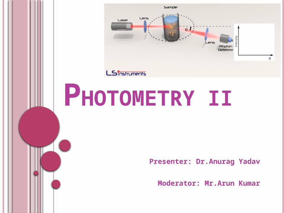
PHOTOMETRY II
Presenter: Dr.Anurag Yadav
Moderator: Mr.Arun Kumar

CONTENT :
Nephelometry.
Turbidimetry.
Reflectance photometry.

NEPHELOMETRY AND TURBIDIMETRY
These are the analytical techniques used to measure scattered
light.
Principle of nephelometry – intensity of light scattered by a
suspension is measured at 90 degrees angle.
Intensity of scattered light α concentration of
suspension
Principle of turbidimetry- measurement of decrease in light
transmitted through a turbid solution is measured .

FACTORS INFLUENCING LIGHT SCATTER
1. Particle size
2. Concentration of particles
3. Molecular weight of particles
4. Wavelength dependence
5. Effect of polarization of incident light
6. Distance of observation

LIGHT SCATTERING
3 types
1. Wavelength of light > particle size - RAYLEIGH
light symmetrically scattered around the particle –
RAYLEIGH
eg – Ig, Albumin

LIGHT SCATTERING
2. Wavelength of light < particle size - MIE THEORY
light appears scattered forward due to destruction out of
phase background scatter- MIE THEORY
Particle size – 7000- 40,000nm like in RBC and bacteria .

LIGHT SCATTERING 3. wavelength of light = particle size – RAYLEIGH DEBYE
SCATTER
light scattered is more in forward than in backward direction –
RAYLEIGH DEBYE SCATTER
Application
Light scatter analysis is used for Ag- Ab reactions with size 250 –
1500nm its Rayleigh Debye scatter and blank scatter by Rayleigh.
RAYLAIGH DEBYE
Wavelength dependence of light scattering:
Intensity of light scattered is inversely proportional to
the wavelength of incident light.
Scattered light intensity is inversely related to
distance from the particle to detector.
Concentration and molecular weight:
From equation, it direct relationship of light scattering
to the conc & molecular weight of particle.

INSTRUMENTATION OF NEPHELOMETER
1. Light source- quartz halogen lamp,mercury arc lamps, xenon
lamps,lasers.
Lasers:
stable, collimated intense light beams,
Reduces stray light, background scatter
2. Collimating optics
3. Sample cell
4. Collection optics –light scattering optics
- detector filter
- detector(PMD)

INSTRUMENTATION OF NEPHELOMETER

INSTRUMENTATION OF TURBIDIMETER:

LIMITATIONS Antigen excess
Ag -Ab reactions are complex and appear to result in a
mixture of aggregate sizes .
Turbidity ↑→ adding Ag to Ab & then ↓ →marking the
beginning of antigen excess.

LIMITATIONS Matrix effects
Particles, solvent and serum macromolecules scatter light.
Lipoprotein and chylomicrons in lipemic samples→
background interference
This is avoided by rate measurements with elimination of
initial sample blank
Large particles: suspended dust → background interference
Filtering all buffers, diluted antisera before analysis.

APPLICATIONS Quantify AA , proteins , vitamins , glycogen , and antibiotics
in blood.
Quantification of urine, csf protein( conc is less) by
immunonephalometry
Quantification of urine ALB, ASO, CRP, U.MAU →
Immunoturbidimetry

DIFFERENCE BETWEEN NEPHELOMETRY AND
TURBIDIMETRY
1. Mercury arc lamp
2. Rectangular cuvette used
3. Scattered light is measured
4. Measured at 90 deg
5. PMT is detector
1. Tu / Du lamp is used
2. Semi octagonal cuvette
3. Light transmitted is measured
4. Measured in straight line
5. Photocell is detector
Nephalometry Turbidimetry

REFLECTANCE SPECTROPHOTOMETRY
Beam of light is directed at a flat reaction
surface & the reflected light is quantified.
Reaction mixture in a carrier is illuminated with
diffuse light, & the intensity of the reflected light
from the chromogen is compared with the
intensity of the light reflected from a reference
surface.

The reflected light intensity is non linear in
relation to conc of analyte.
DR = log ( Ro/Rtest)
Kubelka-Munk or Clapper-Williams transformation
equation used to convert the data into linear
format.

INSTRUMENTATION :
Components are same as Absorbance photometry.
except that the geometry of the system is modified so that the light source & the detector are on one side of the sample.

USES:
Used as quantitative measurement of surface reactions such as dipstick or Dry film chemistry system.

REFERENCE Clinical chemistry: Kaplan Clinical chemistry: TIETZ


IMMUNONEPHELOMETRY Principle o Ag +Ab form small aggregates that scatter light – turbid
appearance o These agg to form large matrix as seen in immunoppt assays
like double diff or radial immunodiff .o Light scatter intensity α amt of ppt in Ab excesso Agg from primary reaction –seconds to minutes o Secondary reaction- takes hours o Light scatter assay measure early 2nd order reaction bet Ag and
Ab
Agg formation enhanced by addition of solu polyethylene glycol of conc 2% to 4%
IMMUNONEPHELOMETRY
Monoclonal Ab reagents Polyclonal Ab need monitoring of titre specificity
and affinity . This is overcome by use of monoclonal Ab

IMMUNONEPHELOMETRY Sample req and preparation – serum urine CSF Reagents Instrumentation Common pitfalls Ab excess high background scatter interference by coloured solu Mixing insufficient Limitations Diff to determine if ppt is in Ag or Ab excess

LIGHT SCATTER INHIBITION IMMUNOASSAY
NINIA 1ST described for progesterone by CAMBIASO ET AL 1974 Principle – ppt from antihapten is inhibited by adding free
hapten Used for rapid analysis of drugs in mg/l like phenytoin ,
phenobarbital , theophylline .

LIGHT SCATTER INHIBITION IMMUNOASSAY
Sample req and prep – serum Reagents Instrumentation Common pitfalls Reaction should be in antigen excess.

ADDITIONAL ASSAY MODIFICATION 1. Particle enhanced light scatter Type of agglutination procedure Ag or Ab coupled with inert carrier particles like
polystyrene latex beads Fast signal transmission and economy of reagents Eg latex fixation test for detection of RF
2. Monoclonal Ab reagents Polyclonal Ab need monitoring of titre specificity and
affinity . This is overcome by use of monoclonal Ab .