photon sources for brachytherapy - indico€¦ · iodine-125 i-125 28 kev 59,4 days 0,025 mm (4.5...
TRANSCRIPT

Photon Sources for Photon Sources for
BrachytherapyBrachytherapy
Alex RijndersEurope HospitalsBrussels, Belgium

Topics of this lecture:
� Terminology
� Sources and source types
� Definition of some relevant physical quantities
PHOTON SOURCES for PHOTON SOURCES for
BRACHYTHERAPYBRACHYTHERAPY

Brachytherapy
Brachytherapy is a treatment method in which sealed radioactive source(s) are used to deliver radiation dose at a short distance.
A high dose can be delivered locally to the tumor with a rapid dose fall-of to the surrounding healthy tissue

AL
PL
AL
EL
EL
s
s 1/2 s
PL
PL
Tube
Needle
Wire
Seed Ribbon
Source Train
Stepping source
AL Active Length ; PL Physical Length ; EL Equivalent Active Length
EL = n x s (Number of seeds x distance between source centres)Condition: equal activity
Sealed sources


Temporary ImplantsTemporary Implants
LDR
0 1 2 3 4 5 6 7 8 9 10 11 12 13 14 15 16 17 18 19 20 21 22 23 24
Hours
Dose R
ate
PDR
0 1 2 3 4 5 6 7 8 9 10 11 12 13 14 15 16 17 18 19 20 21 22 23 24
Dose R
ate
Low Dose Rate:● Continuous
irradiation
● 0.40 – 2 Gy/h
Pulsed Dose Rate:● mimic low dose rate ● short pulses, same average dose rate
High Dose Rate:● >0.2 Gy/min ● One/a few fractions
LDR
PDR
HDR
HDR
0 1 2 3 4 5 6 7 8 9 10 11 12 13 14 15 16 17 18 19 20 21 22 23 24
Dose R
ate

Important milestonesImportant milestones
Early 1900s: use of Radium for BT
1930s: Manchester System
End 1950s: artificial isotopes (60Co – 137Cs)
1960s: 192Ir wire sources – Manual afterloading techniques – Paris System
1970s-1990s: Remote Afterloading Devices
2000s: Imaging Assisted Brachytherapy
2010s : Improved dose calculation algorithms

SOURCES in BRACHYTHERAPYSOURCES in BRACHYTHERAPYRadium needles and tubes (original design)Radium needles and tubes (original design)

Radium sources : UnitsRadium sources : Units
226Ra source strength specification in terms of contents:
mass, expressed in milligrams (mg Ra)
Drawbacks of 226Ra associated with safety(possible Radon-leaks, too long half time)
End 1950’s : Replacement by artificially produced sources such as 137Cs, of which the strength was expressed as radium-equivalent
=> mg Ra-equivalent

1950s ARTIFICIEL ISOTOPES1950s ARTIFICIEL ISOTOPESReplacementReplacement of Radiumof Radium
Example of a 2 cm length tube source, 137Cs

RReplacement of Radiumeplacement of Radium
Other new sources and source types were developed: leading to a need for other source strength specification, based on “contents”, activity, # of disintegrations per time unit
Definition of Curie:1 Ci (3.7 x 10¹⁰ s-1) = activity of 1 g 226Raactually 1g 226Ra = 3.655 x 10¹⁰ S-1 or .988 Ci
SI-units: 1 Bq = 1 disintegration per secMBq, GBq

- Number of nuclei prone to decay:
N(t) = N0 ���� e-λλλλt
- Activity:
A(t) = -∆∆∆∆N(t) / ∆∆∆∆t ≈≈≈≈ N(t)
- Half life of the source:
A(t = T½) = ½ ���� A0
- Decay constant:
λλλλ = ln(2) / T½ = 0.693 / T½
Radioactivity, decay Radioactivity, decay
calculationcalculation

� Reference Air Kerma Rate (µµµµGy/s)
� Kerma rate to air, in air, at reference distance 1 m, corrected for air attenuation and scattering (ICRU 38 and 58)
RK
•
Specification of source Specification of source
strength for dosimetrystrength for dosimetry
Practical units: µµµµGy/h (LDR),
mGy/h or µµµµGy/s (HDR)

Activity – MBq (mCi)
Air Kerma Rate -
µGy/s (Exposure Rate – C/kg.s)
Kδ
R2
refapp
)(Γ
KdA
•
=
( )Γδ K : air kerma rate constant (source construction, encapsulation and ene
dref : reference distance (1m)

137Cs: from 3 to 3.31 192Ir: from 4.0 to 5.0

Jack Venselaar
SOURCES in BRACHYTHERAPYSOURCES in BRACHYTHERAPY
Examples of source test certificates
Example of TPS output
Reference Air Kerma Rate is the reference
unit for the clinical physicist

Important milestonesImportant milestones
Early 1900s: use of Radium for BT
1930s: Manchester System
End 1950s: artificial isotopes (60Co – 137Cs)
1960s: 192Ir wire sources – Manual afterloading techniques – Paris System
1970s-1990s: Remote Afterloading Devices
2000s: Imaging Assisted Brachytherapy
2010s : Improved dose calculation algorithms

1960s MANUAL AFTERLOADING1960s MANUAL AFTERLOADING

Interstitial cathetersInterstitial catheters

Fletcher-Suit-DelclosVaginal cylinder
Ring applicator

Important milestonesImportant milestones
Early 1900s: use of Radium for BT
1930s: Manchester System
End 1950s: artificial isotopes (60Co – 137Cs)
1960s: 192Ir wire sources – Manual afterloading techniques – Paris System
1970s-1990s: Remote Afterloading Devices
2000s: Imaging Assisted Brachytherapy
2010s : Improved dose calculation algorithms

Cs afterloader (LDR)Cs afterloader (LDR)
137Cs pellets – Free composition of trains of active/inactive pellets

IrIr afterloadersafterloaders (HDR(HDR--
PDR)PDR)
192Ir stepping source, HDR or PDR

SOURCES in BRACHYTHERAPYSOURCES in BRACHYTHERAPY
Example of tip of a pulsed dose rate (PDR) source, Ir-192,
welded to the end of a drive cable


Advantage of HDR technology
� One single source (costs)
� Half life 74 days => usable for 3-4 months (costs)
� Afterloader system => radiation protection
� Stepping source technology allows dose optimisation => optimisation algorithms
� Short irradiation time (10-15 minutes)

Stepping sourceStepping source

Advanced Optimisation Technology Advanced Optimisation Technology
(example :SWIFT(example :SWIFTTM)TM)

Permanent ImplantsPermanent Implants
Radioactive sources
remain in the patient
and decay ● Relative short half life
● Low energy (radiation
protection)
Permanent Implant 125
I
0.000
0.100
0.200
0.300
0.400
0.500
0.600
0.700
0.800
0.900
1.000
0 30 60 90 120 150 180 210 240 270 300 330 360
Days
Do
se
Ra
te
0.000
0.100
0.200
0.300
0.400
0.500
0.600
0.700
0.800
0.900
1.000
To
tal D
os
e

Permanent ImplantsPermanent Implants
e.g., for prostate, brain
these sources should combine a relative short half life with low energy:
=> half life determines dose rate/radiobiology
=> patient should be able to continue life as usual
Examples:
I-125 (60 days; 28 keV)
Pd-103 (17 days; 21 keV)

Cross-Sectional drawings of sources with a Rod, Wire, or Cylinder internal core design; (a) Amersham 6711 OncoSeed, (b) Syncor PharmaSeed, (c) UroMed Symmetra, (d)SourceTech Medical 125Implant, (e) Med-Tec I-Plant, (f) International Brachytherapy, Inc. InterSource125, (g) Best Medical Model 2301 (h) Amersham 6702, (i) UroCor ProstaSeed, (j)Imagyn IsoSTAR, (k) Mentor's IoGold, (l) DraxImage BrachySeed.
From: Heintz et al, Comparison of I-125 sources used for permanent interstitial implants. Med Phys 2001
Some examples of seeds:

Special presentations of Special presentations of
sourcessources
“Rapidstrand®” seed ribbon technique with
the 125I sources connected in a suture
“Isocord®”: comparable technique with the 125I sources connected in a bio-absorbable suture
And there are many more …
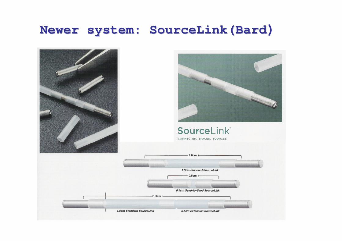
NewerNewer system: system: SourceLinkSourceLink(Bard(Bard)

Afterloader for Afterloader for 125125I seedsI seeds

Important milestonesImportant milestones
Early 1900s: use of Radium for BT
1930s: Manchester System
End 1950s: artificial isotopes (60Co – 137Cs)
1960s: 192Ir wire sources – Manual afterloading techniques – Paris System
1970s-1990s: Remote Afterloading Devices
2000s: Imaging Assisted Brachytherapy
2010s : Improved dose calculation algorithms

MOTIVATION
Apply also to modern
brachytherapy
Apply also to (modern)
brachytherapy
� ‘Modern Radiotherapy’ seems to be driven
by significant developments in EBT
� 3D Conformal Radiotherapy
� Stereotactic Radiotherapy: High Precision
� Intensity Modulated Radiotherapy: Dose Shaping
� Imaging for GTV/PTV, OAR (structure segmentation)
� Computerized treatment plan optimization
� Image guided RT
From Poetter et al
=> Evaluate potential of Brachytherapy based on modern technology
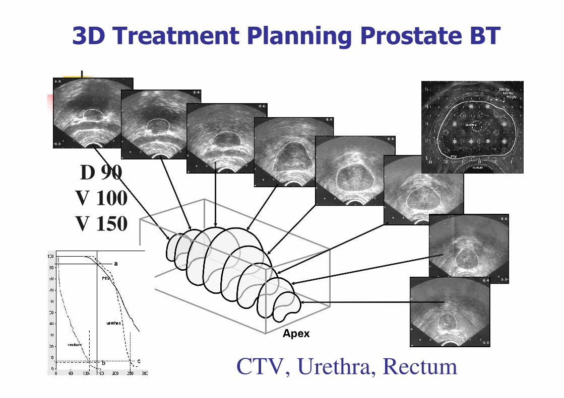
3D Treatment Planning Prostate BT
D 90
V 100
V 150
CTV, Urethra, Rectum

- CT
- MRI
- US
- PET
- Functional…
=> the role of ‘imaging’in RT increases rapidly
� Better understanding of anatomy
� Better understanding of pathology
� More appropriate contouring
Multi modalityImaging

Examples of CT/MRI compatible Examples of CT/MRI compatible
applicatorsapplicators

• Long Half Life=> Economical
• High Specific activity: activity per unit of mass
=> physical size of source)
• Low mean energy of radiation
=> less penetration in tissue
• Small half value layer in lead or concrete
=> radiation protection
Ideal source/isotope Ideal source/isotope (hospital use)(hospital use)

Physical properties of nuclides
λ=ln2 / T½
IsotopeAverage photon energy*
Half-life T½Half value layer in lead
Treatment room wall
Cobalt-60 Co-60 1,25 MeV 5,26 years 12 mm(Concrete)typical values
Caesium-137 Cs-137 662 KeV 30,1 years 6,5 mm
Iridium-192 Ir-192 380 KeV 73,8 days 3,0 mm (40 cm)
Ytterbium-169 Yb-169 93 KeV 32,0 days 0,23 mm
Thulium-170 Tu-170 66 KeV 128.6 days 0,17 mm (12 cm)
Iodine-125 I-125 28 KeV 59,4 days 0,025 mm (4.5 cm)
Palladium-103 Pd-103 21 KeV 17,0 days 0,01 mm (1 cm)
Caesium-131 Cs-131 30 KeV 9,7 days -
* Approximate values, depending on the source make and filtration

⇒ Radioprotection:
� Reduced Mean Energy (<= 100 KeV)
� Yb-169, Tu-170, I-125, Pd-103
⇒ Increased half-life (source exchange)
� Co-60
« New » Isotopes

X-ray source tip detail
Miniature x-ray source inserted into a flexible cooling catheter� High vacuum x-ray tube technology � 50 kV max. operating potential� Water cooled� Fully disposable device
X-Ray Tube HV Cable
Cooling connections
HV connection
miniature
x-ray
source
Electronic BT sourcesElectronic BT sources
Xoft Inc.
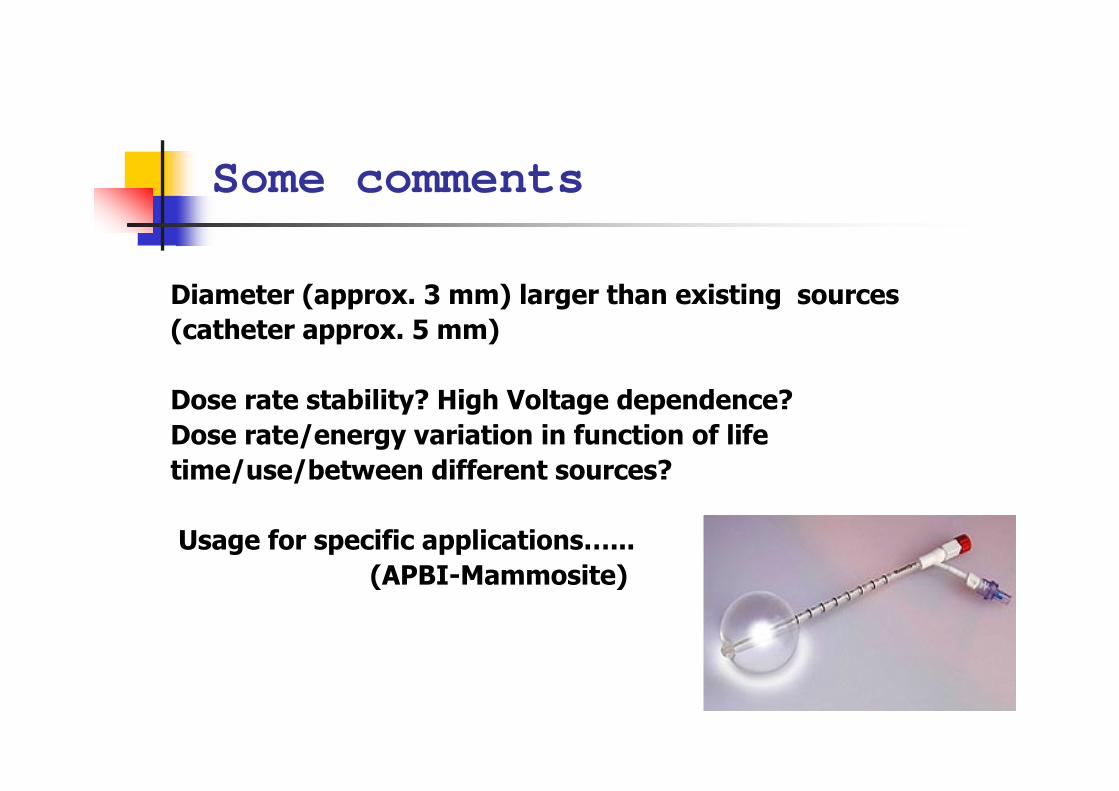
Diameter (approx. 3 mm) larger than existing sources
(catheter approx. 5 mm)
Dose rate stability? High Voltage dependence?
Dose rate/energy variation in function of life
time/use/between different sources?
Usage for specific applications…...
(APBI-Mammosite)
Some comments


Training in Brachytherapy
� As treatment techniques and delivery systems become more complex
� Need for better formed/trained staff
� ! Few centres specialised in “ high end BT “(certainly in Western Europe)

Brachytherapie ���� Teletherapie
Investments
€ 300.000 € 2.400.000
(+ € 13.000 / source) (+ maintenance)
Workload ++ Workload +++

References / more reading
� ICRU, International Commission on Radiation Units and Measurements, Dose and volume specification for reporting intracavitary therapy in gynecology, ICRU Report 38, 1985.
� ICRU, International Commission on Radiation Units and Measurements. “Dose and volume specification for reporting interstitial therapy”. ICRU Report 58, 1997.
� The GEC ESTRO Handbook of Brachytherapy, Editors: Alain Gerbaulet, Richard Pötter, Jean-Jacques Mazeron, Erik van Limbergen,
available at the ESTRO web site: www.estro.org� A Practical Guide to Quality Control of Brachytherapy
Equipment, ESTRO Physics Booklet No. 8, Editors: Jack Venselaar, José Pérez-Calatayud,
available at the ESTRO web site: www.estro.orghttp://www.estro.org/school/articles/publications/publications

Still more reading
� Nath, R., Anderson, L.L., Meli, J.A., Olch, A.J., Stitt, J.A. and Williamson, J.F. “Code of practice for brachytherapy physics: Report of the AAPM Radiation Therapy Committee Task Group No. 56”. Med. Phys. 24, 1557-1598, 1997.
� Radiation Oncology Physics: a Handbook for Teachers and Students, Editor E.B.Podgorsak, available at the IAEA web site: www.iaea.org
� Dosimétrie en Curiethérapie, A. Dutreix, G. Marinello and A. Wambersie, Masson, Paris, 1982
� The Physics of Modern Brachytherapy for Oncology, D. Baltas, L. Sakelliou and N. Zamboglou, Taylor and Francis, New York London, 2007
