photophysical properties supporting information ... · potassium carbonate, potassium iodide, ,...
TRANSCRIPT
Supporting Information
Bimodal self-assembly of an amphiphilic gelator to hydrogel-nanocatalyst and organogel of different morphologies and photophysical properties
Papri Sutar and Tapas Kumar Maji*
Molecular Materials Laboratory, Chemistry and Physics of Materials Unit, Jawaharlal
Nehru Centre for Advanced Scientific Research, Jakkur, Bangalore 560 064, India,
Email: [email protected]
1. Experimental Section.
1.1 Materials and Methods: 1,3,5-tris(bromomethyl)benzene, methyl 4-hydroxybenzoate,
4′-chloro-2,2′:6′,2′′-terpyridine, 1,3-diaminopropane, triphenylphosphine (PPh3),
trichloroisocyanuric acid (TCIC) were purchased from Sigma-Aldrich chemical Co. Ltd.
Potassium carbonate, potassium iodide, , malononitrile and all solvents were purchased from
Spectrochem. For doing catalysis all substrates, benzaldehyde, 4-nitrobenzaldehyde, 4-
chlorobenzaldehyde, p-tolualdehyde and p-anisaldehyde, were purchased from Sigma-
Aldrich chemical Co. Ltd. All solvents were pre-dried using standard procedures before
using. For UV-Vis experiments spectroscopic grade solvents were purchased from
Spectrochem. 1H NMR is recorded on a Bruker AV-400 spectrometer with chemical shifts
recorded as ppm and all spectra were calibrated against TMS. UV-Vis spectra were recorded
in a Perkin-Elmer lamda 900 spectrometer. Fluorescence studies were accomplished using
Perkin Elmer Ls 55 Lumeniscence spectrometer. Infrared spectral studies were carried out by
making samples with KBr pellets using Bruker FT-IR spectrometer. Powder X-ray diffraction
studies were recorded on a Bruker D8 discover instrument using Cu-Kα radiation.
Electronic Supplementary Material (ESI) for ChemComm.This journal is © The Royal Society of Chemistry 2016
Morphology studies were carried out using Lica-S440I field emission scanning electron
microscopy (FESEM) by placing samples on silicon wafer under vacuum with accelerating
voltage of 10 kV. Transmission electron microscopy (TEM) analysis was performed using
JOEL JEM-3010 with accelerating voltage of 300kV. For this analysis the xerogel was
dispersed in ethanol and then drop casted on a carbon coated copper grid.
1.2 Synthesis of 4,4′,4″-[1,3,5-phenyl-tri(methoxy)]-tris-benzoic acid: methyl 4-
hydroxybenzoate (1.065 g, 7 mmol), potassium carbonate ( 2.89 g, 21 mmol), potassium
iodide (85 mg, 0.518 mmol) were suspended in dry N, N-dimethylformamide (DMF). The
reaction mixture was refluxed at 100 °C for 2 hours maintaining the inert condition. 1,3,5-
tris(bromomethyl)benzene (500 mg, 1.4 mmol) was dissolved in 20 ml dry DMF and was
dropwise added to the above heated reaction mixture. The mixture was subsequently stirred
and heated at 100 °C for 4 hours. After cooling the reaction mixture to room temperature, 100
ml of distilled water was added and the precipitate formed was collected by filtration. The
precipitate was washed several times with cold distilled water and air dried. A white solid
was obtained with 85% yield. The product was taken in a round bottom flux. 40 ml methanol
and 6 g of NaOH in 20 ml of distilled water was added into it. The reaction mixture was
refluxed at 50 °C for 12 hours. After cooling to room temperature, the solution was placed in
a ice bath and acidified to pH 2 with 3N HCl. The precipitate was then collected, washed
several times with distilled water and air dried. A brownish solid was obtained with 98%
yield. 1H-NMR (400 MHz, DMSO-d6) δ: 7.89 (d, 6H, ArH), 7.53 (s, 3H, ArH), 7.11 (d, 6H,
ArH), 5.22 (s, 6H, CH2). Selected FTIR data (KBr, cm-1): 3451 (b), 3078-2879 (s), 2673 (s),
2550 (s), 1680 (sh), 1603 (sh), 1510 (m), 1428 (m), 1248 (sh), 1169 (sh), 1009 (m), 940 (s),
846 (m), 771 (m), 695 (s), 648 (s), 609 (s), 564 (s), 504 (s).
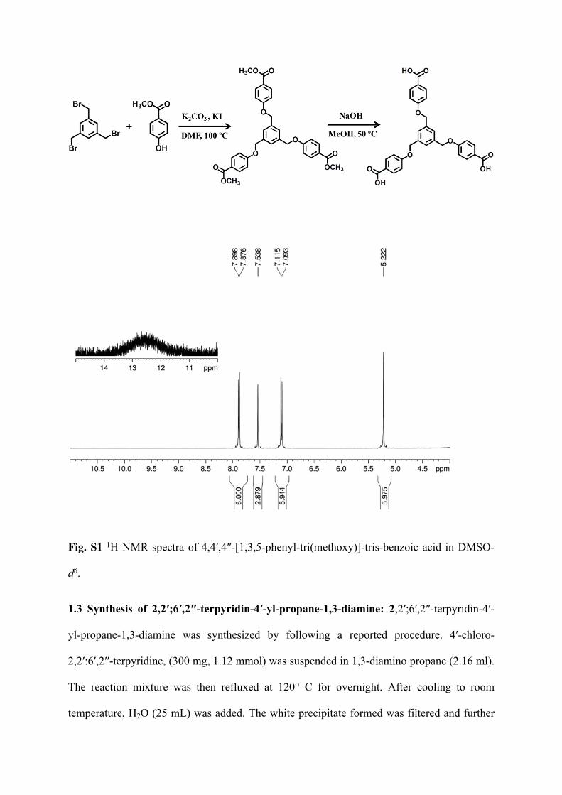
Fig. S1 1H NMR spectra of 4,4′,4″-[1,3,5-phenyl-tri(methoxy)]-tris-benzoic acid in DMSO-
d6.
1.3 Synthesis of 2,2′;6′,2″-terpyridin-4′-yl-propane-1,3-diamine: 2,2′;6′,2″-terpyridin-4′-
yl-propane-1,3-diamine was synthesized by following a reported procedure. 4′-chloro-
2,2′:6′,2′′-terpyridine, (300 mg, 1.12 mmol) was suspended in 1,3-diamino propane (2.16 ml).
The reaction mixture was then refluxed at 120° C for overnight. After cooling to room
temperature, H2O (25 mL) was added. The white precipitate formed was filtered and further
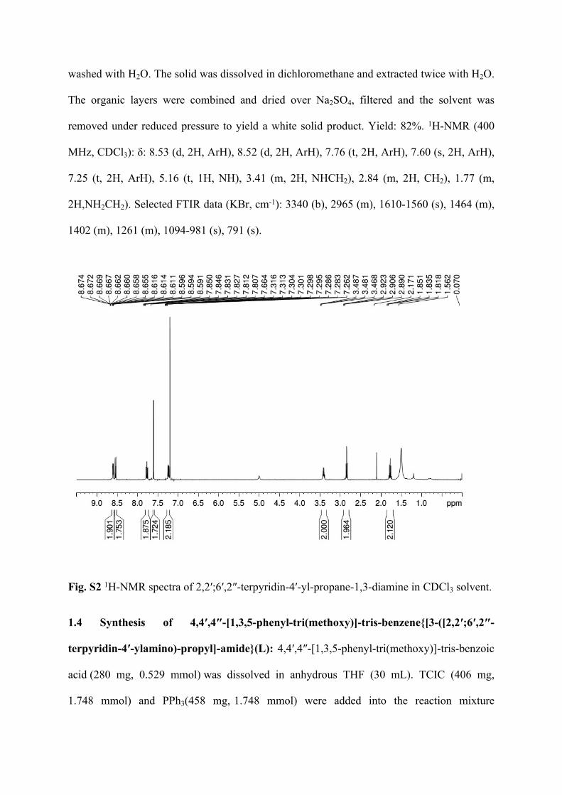
washed with H2O. The solid was dissolved in dichloromethane and extracted twice with H2O.
The organic layers were combined and dried over Na2SO4, filtered and the solvent was
removed under reduced pressure to yield a white solid product. Yield: 82%. 1H-NMR (400
MHz, CDCl3): δ: 8.53 (d, 2H, ArH), 8.52 (d, 2H, ArH), 7.76 (t, 2H, ArH), 7.60 (s, 2H, ArH),
7.25 (t, 2H, ArH), 5.16 (t, 1H, NH), 3.41 (m, 2H, NHCH2), 2.84 (m, 2H, CH2), 1.77 (m,
2H,NH2CH2). Selected FTIR data (KBr, cm-1): 3340 (b), 2965 (m), 1610-1560 (s), 1464 (m),
1402 (m), 1261 (m), 1094-981 (s), 791 (s).
Fig. S2 1H-NMR spectra of 2,2′;6′,2″-terpyridin-4′-yl-propane-1,3-diamine in CDCl3 solvent.
1.4 Synthesis of 4,4′,4″-[1,3,5-phenyl-tri(methoxy)]-tris-benzene{[3-([2,2′;6′,2″-
terpyridin-4′-ylamino)-propyl]-amide}(L): 4,4′,4″-[1,3,5-phenyl-tri(methoxy)]-tris-benzoic
acid (280 mg, 0.529 mmol) was dissolved in anhydrous THF (30 mL). TCIC (406 mg,
1.748 mmol) and PPh3(458 mg, 1.748 mmol) were added into the reaction mixture
and stirred at 0 ºC for 40 min under inert condition. 2,2′;6′,2″-terpyridin-4′-yl-propane-
1,3-diamine (532 mg, 1.748 mmol) was dissolved in anhydrous THF ( 20 ml) and Et3N
(484 µl, 3.496 mmol) was added into it. This reaction mixture was drop-wise added
into 4,4′,4″-[1,3,5-phenyl-tri(methoxy)]-tris-benzoic acid / TCIC/ PPh3 solution at 0 ºC and
stirred for 45 minutes. After that the reaction mixture was stirred at room temperature
for 3 hrs. Precipitate was collected by filtration and washed several times with CHCl3 and
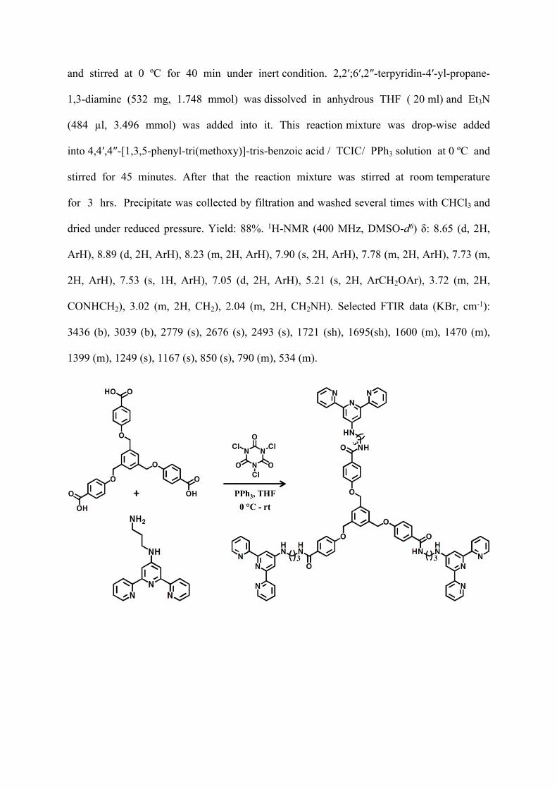
dried under reduced pressure. Yield: 88%. 1H-NMR (400 MHz, DMSO-d6) δ: 8.65 (d, 2H,
ArH), 8.89 (d, 2H, ArH), 8.23 (m, 2H, ArH), 7.90 (s, 2H, ArH), 7.78 (m, 2H, ArH), 7.73 (m,
2H, ArH), 7.53 (s, 1H, ArH), 7.05 (d, 2H, ArH), 5.21 (s, 2H, ArCH2OAr), 3.72 (m, 2H,
CONHCH2), 3.02 (m, 2H, CH2), 2.04 (m, 2H, CH2NH). Selected FTIR data (KBr, cm-1):
3436 (b), 3039 (b), 2779 (s), 2676 (s), 2493 (s), 1721 (sh), 1695(sh), 1600 (m), 1470 (m),
1399 (m), 1249 (s), 1167 (s), 850 (s), 790 (m), 534 (m).
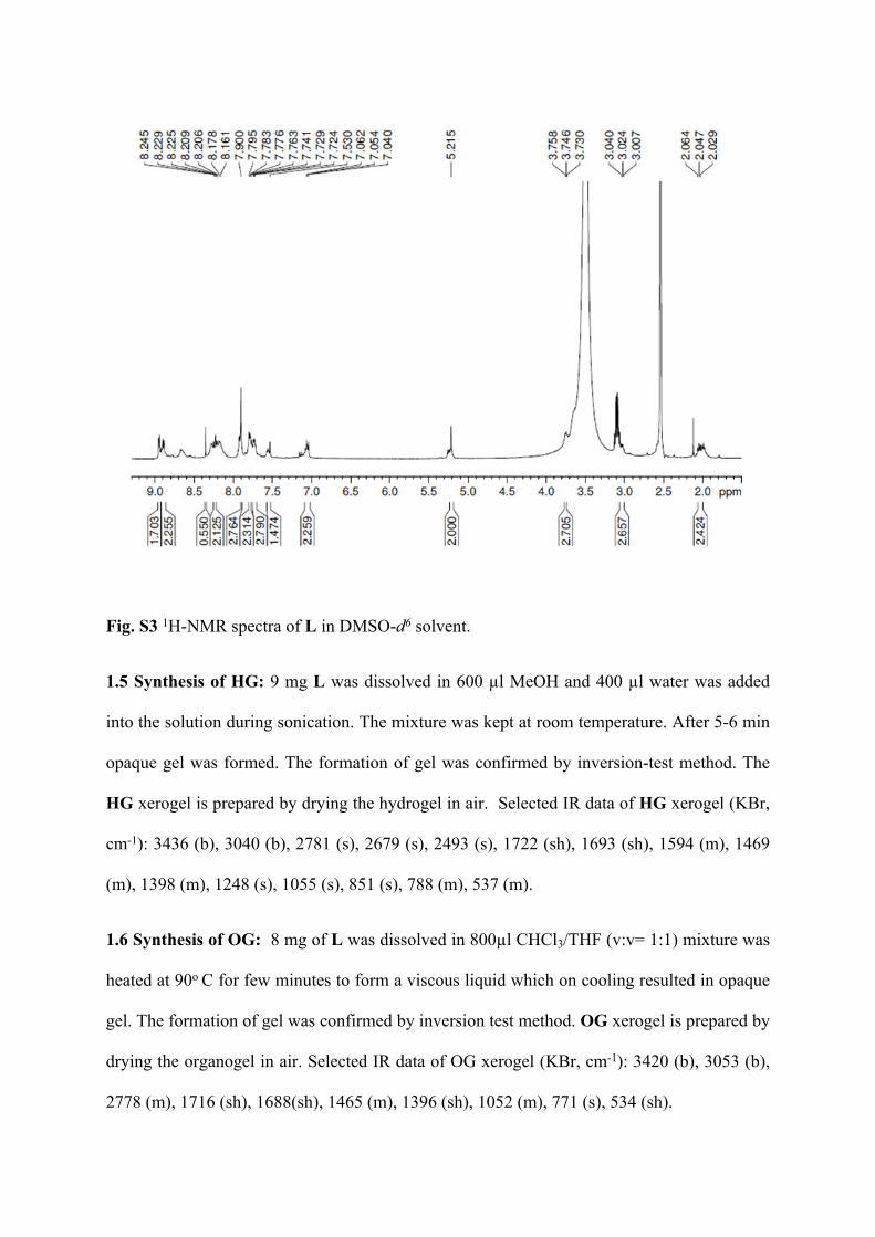
Fig. S3 1H-NMR spectra of L in DMSO-d6 solvent.
1.5 Synthesis of HG: 9 mg L was dissolved in 600 µl MeOH and 400 µl water was added
into the solution during sonication. The mixture was kept at room temperature. After 5-6 min
opaque gel was formed. The formation of gel was confirmed by inversion-test method. The
HG xerogel is prepared by drying the hydrogel in air. Selected IR data of HG xerogel (KBr,
cm-1): 3436 (b), 3040 (b), 2781 (s), 2679 (s), 2493 (s), 1722 (sh), 1693 (sh), 1594 (m), 1469
(m), 1398 (m), 1248 (s), 1055 (s), 851 (s), 788 (m), 537 (m).
1.6 Synthesis of OG: 8 mg of L was dissolved in 800µl CHCl3/THF (v:v= 1:1) mixture was
heated at 90o C for few minutes to form a viscous liquid which on cooling resulted in opaque
gel. The formation of gel was confirmed by inversion test method. OG xerogel is prepared by
drying the organogel in air. Selected IR data of OG xerogel (KBr, cm-1): 3420 (b), 3053 (b),
2778 (m), 1716 (sh), 1688(sh), 1465 (m), 1396 (sh), 1052 (m), 771 (s), 534 (sh).
1.7 General procedure for the catalytic reactions: Benzaldehyde derivatives (1 mmol) and
melanonitrile (1 mmol) were taken in a Schlenk tube and 15 ml dry THF was added into it
under inert atmosphere. The reaction mixture was stirred at room temperature for 5 minutes
and HG xerogel (1 mol %) was added into it. After that the reaction mixture was refluxed at
40 °C under nitrogen atmosphere. The reaction mixture was cooled down to room
temperature and filtered to recover the catalyst. The filtrate was concentrated and analysed
using GC-MS analyser and 1H NMR spectroscopy.
Fig. S4 (a) Absorption spectra of 2,2′;6′,2″-terpyridin-4′-yl-propane-1,3-diamine (1×10⁻⁴ M)
in CHCl3, (b) emission spectra of of 2,2′;6′,2″-terpyridin-4′-yl-propane-1,3-diamine (1×10⁻⁴
M) in CHCl3.
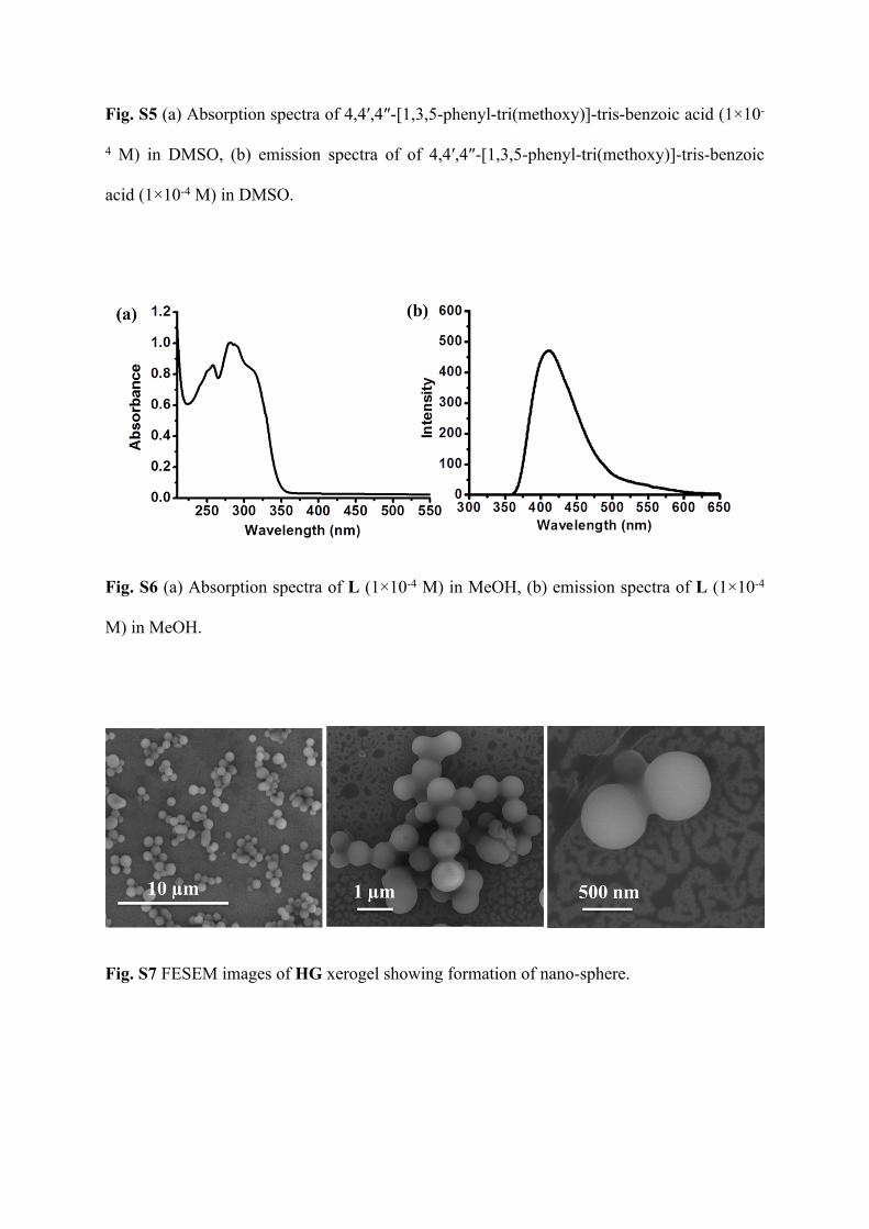
Fig. S5 (a) Absorption spectra of 4,4′,4″-[1,3,5-phenyl-tri(methoxy)]-tris-benzoic acid (1×10-
4 M) in DMSO, (b) emission spectra of of 4,4′,4″-[1,3,5-phenyl-tri(methoxy)]-tris-benzoic
acid (1×10⁻⁴ M) in DMSO.
Fig. S6 (a) Absorption spectra of L (1×10⁻⁴ M) in MeOH, (b) emission spectra of L (1×10⁻⁴
M) in MeOH.
Fig. S7 FESEM images of HG xerogel showing formation of nano-sphere.
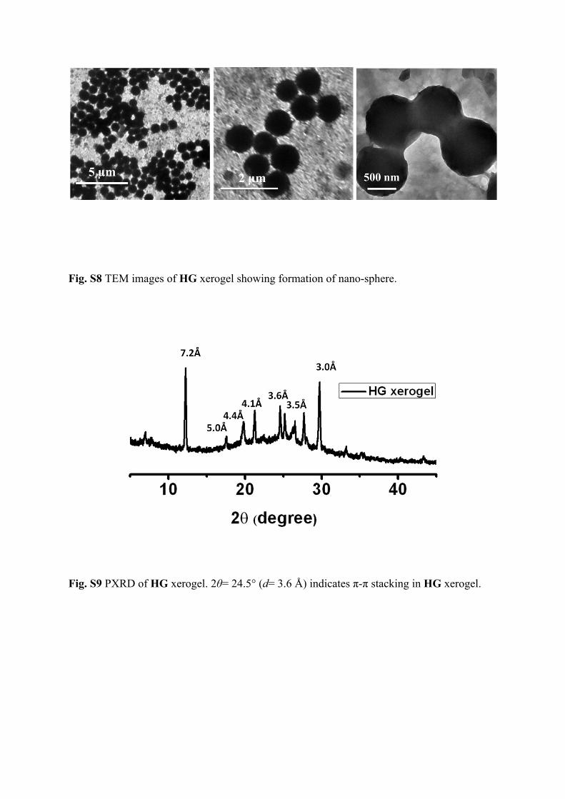
Fig. S8 TEM images of HG xerogel showing formation of nano-sphere.
Fig. S9 PXRD of HG xerogel. 2θ= 24.5° (d= 3.6 Å) indicates π-π stacking in HG xerogel.
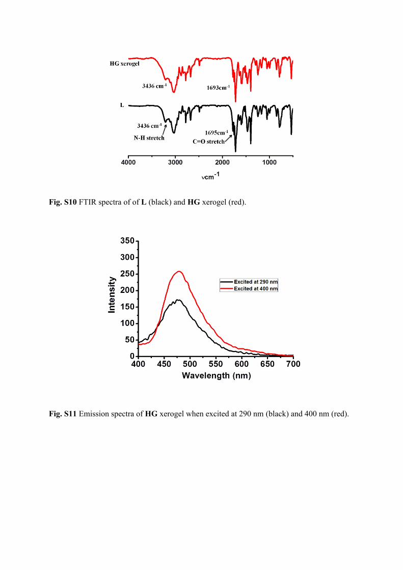
Fig. S10 FTIR spectra of of L (black) and HG xerogel (red).
Fig. S11 Emission spectra of HG xerogel when excited at 290 nm (black) and 400 nm (red).
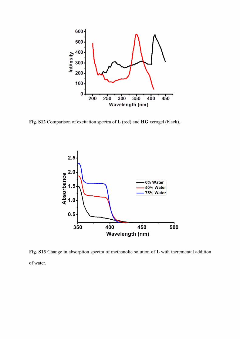
Fig. S12 Comparison of excitation spectra of L (red) and HG xerogel (black).
Fig. S13 Change in absorption spectra of methanolic solution of L with incremental addition
of water.
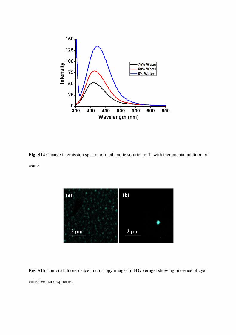
Fig. S14 Change in emission spectra of methanolic solution of L with incremental addition of
water.
Fig. S15 Confocal fluorescence microscopy images of HG xerogel showing presence of cyan
emissive nano-spheres.
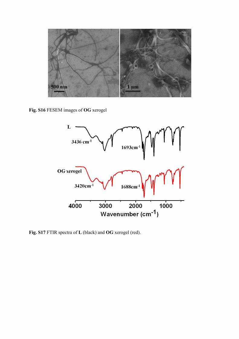
Fig. S16 FESEM images of OG xerogel
Fig. S17 FTIR spectra of L (black) and OG xerogel (red).
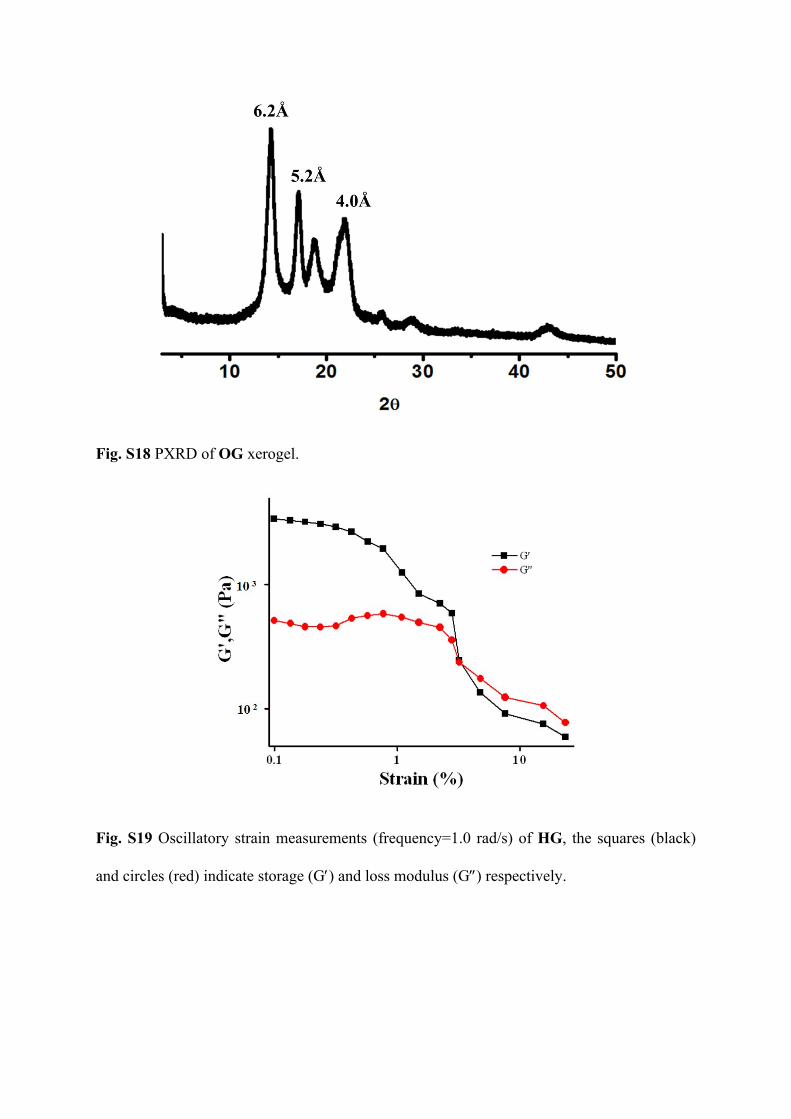
Fig. S18 PXRD of OG xerogel.
Fig. S19 Oscillatory strain measurements (frequency=1.0 rad/s) of HG, the squares (black)
and circles (red) indicate storage (G) and loss modulus (G) respectively.
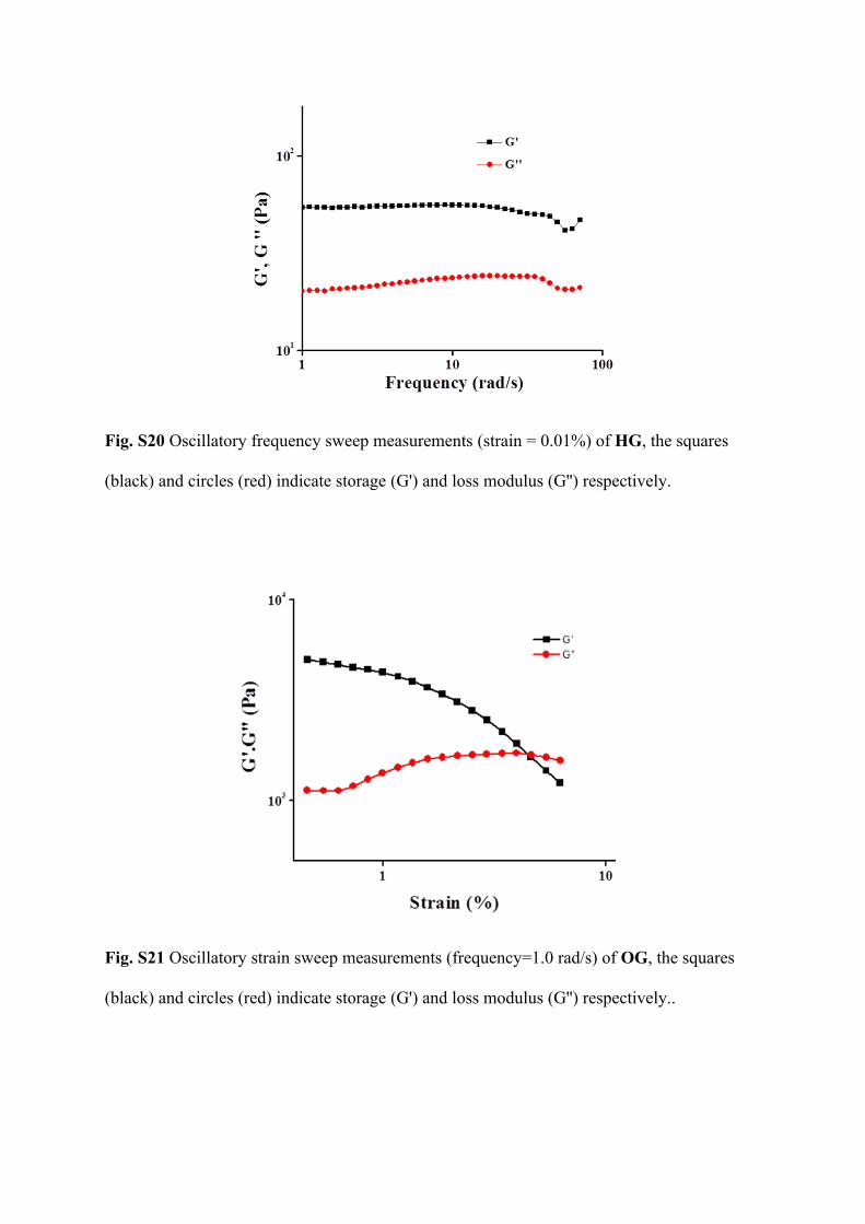
Fig. S20 Oscillatory frequency sweep measurements (strain = 0.01%) of HG, the squares
(black) and circles (red) indicate storage (G') and loss modulus (G'') respectively.
Fig. S21 Oscillatory strain sweep measurements (frequency=1.0 rad/s) of OG, the squares
(black) and circles (red) indicate storage (G') and loss modulus (G'') respectively..
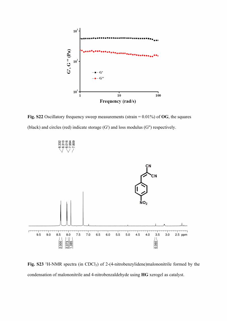
Fig. S22 Oscillatory frequency sweep measurements (strain = 0.01%) of OG, the squares
(black) and circles (red) indicate storage (G') and loss modulus (G'') respectively.
Fig. S23 1H-NMR spectra (in CDCl3) of 2-(4-nitrobenzylidene)malononitrile formed by the
condensation of malononitrile and 4-nitrobenzaldehyde using HG xerogel as catalyst.
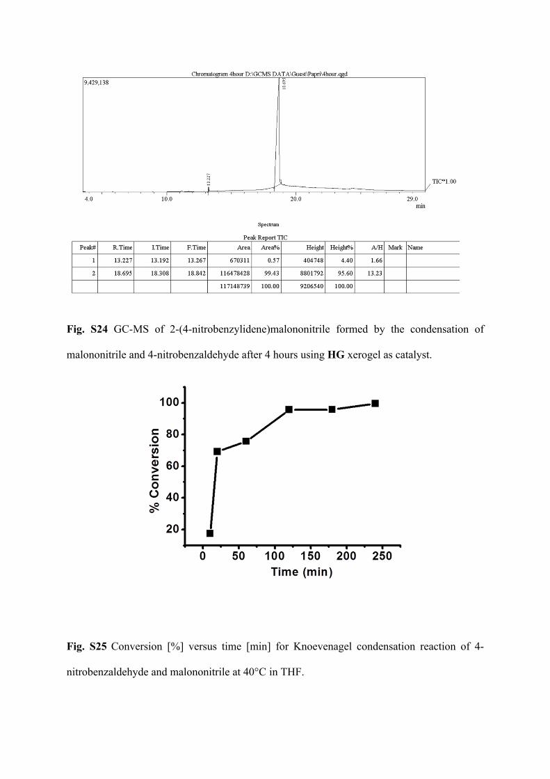
Fig. S24 GC-MS of 2-(4-nitrobenzylidene)malononitrile formed by the condensation of
malononitrile and 4-nitrobenzaldehyde after 4 hours using HG xerogel as catalyst.
Fig. S25 Conversion [%] versus time [min] for Knoevenagel condensation reaction of 4-
nitrobenzaldehyde and malononitrile at 40°C in THF.
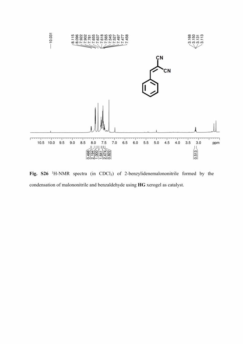
Fig. S26 1H-NMR spectra (in CDCl3) of 2-benzylidenemalononitrile formed by the
condensation of malononitrile and benzaldehyde using HG xerogel as catalyst.
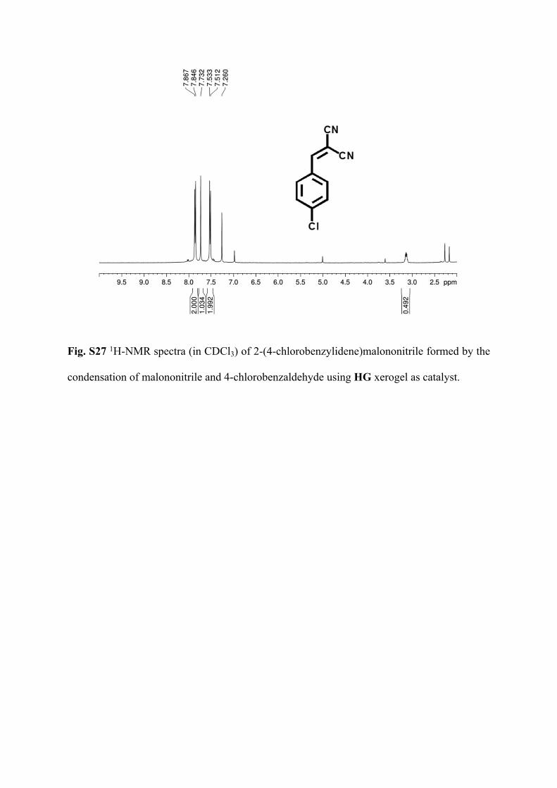
Fig. S27 1H-NMR spectra (in CDCl3) of 2-(4-chlorobenzylidene)malononitrile formed by the
condensation of malononitrile and 4-chlorobenzaldehyde using HG xerogel as catalyst.
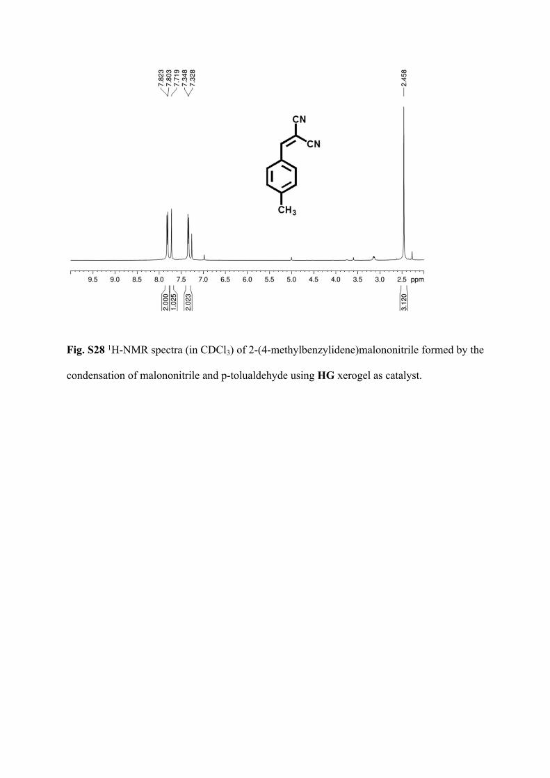
Fig. S28 1H-NMR spectra (in CDCl3) of 2-(4-methylbenzylidene)malononitrile formed by the
condensation of malononitrile and p-tolualdehyde using HG xerogel as catalyst.
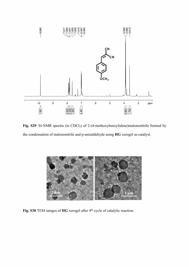
Fig. S29 1H-NMR spectra (in CDCl3) of 2-(4-methoxybenzylidene)malononitrile formed by
the condensation of malononitrile and p-anisaldehyde using HG xerogel as catalyst.
Fig. S30 TEM iamges of HG xerogel after 4th cycle of catalytic reaction.
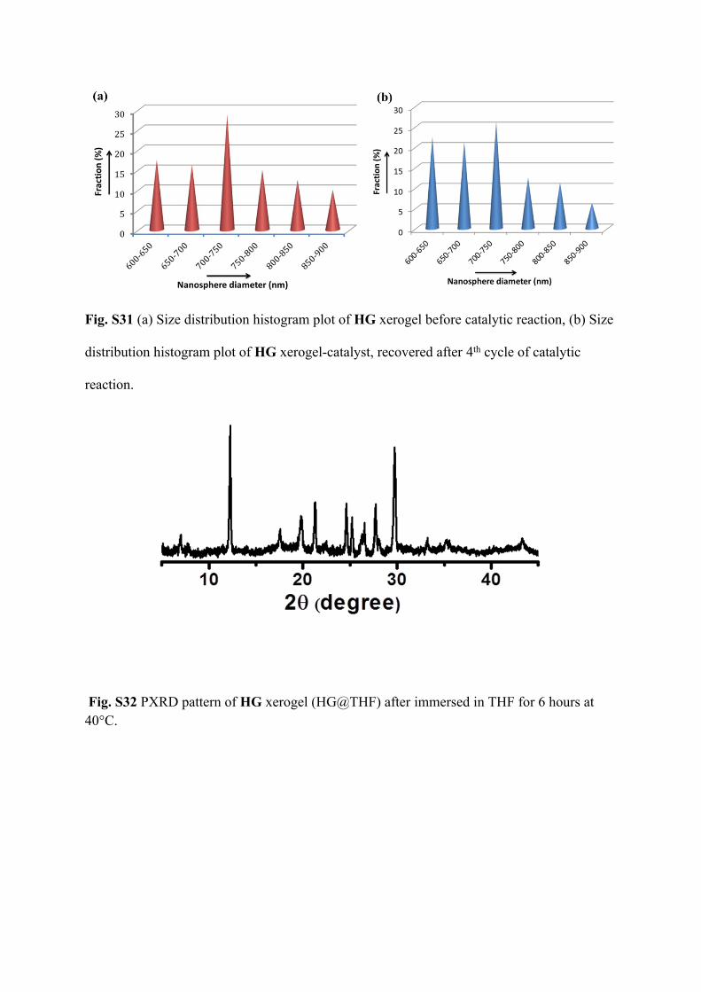
Fig. S31 (a) Size distribution histogram plot of HG xerogel before catalytic reaction, (b) Size
distribution histogram plot of HG xerogel-catalyst, recovered after 4th cycle of catalytic
reaction.
Fig. S32 PXRD pattern of HG xerogel (HG@THF) after immersed in THF for 6 hours at 40°C.
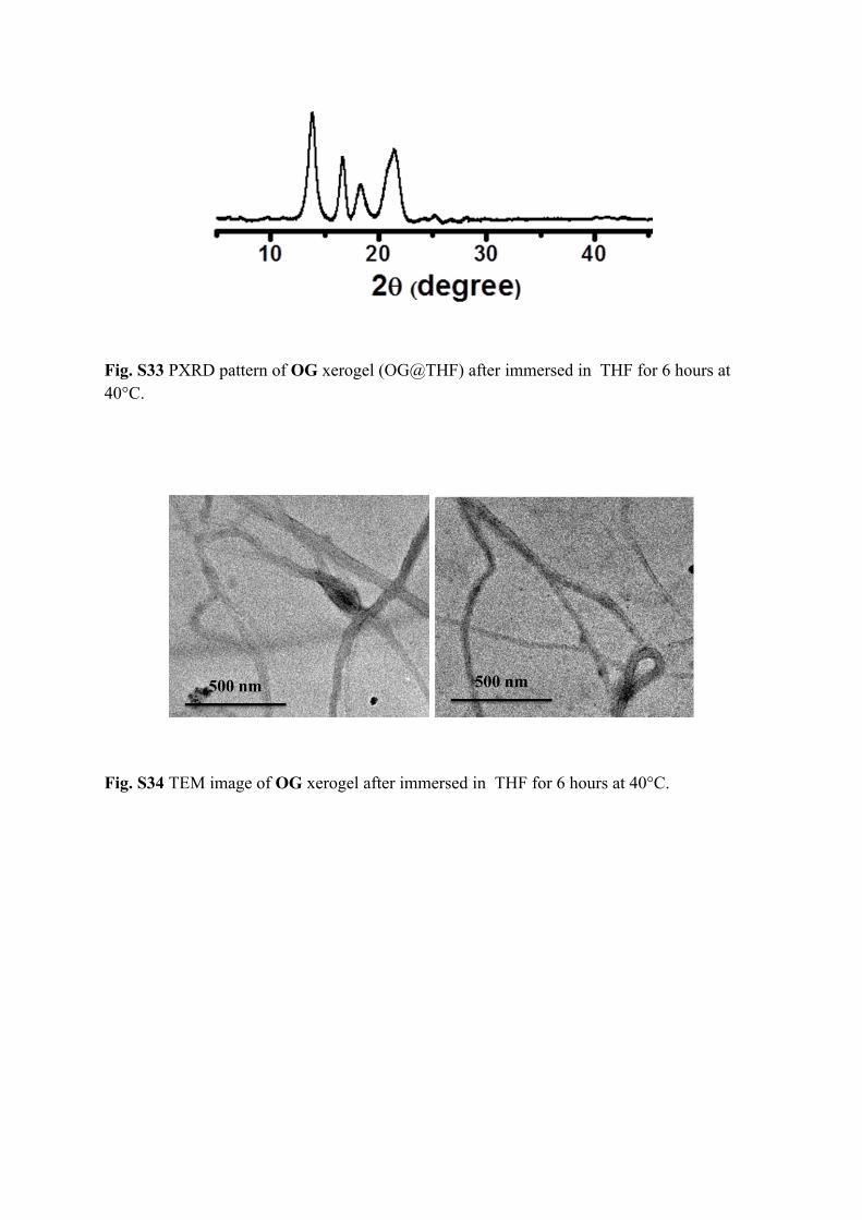
Fig. S33 PXRD pattern of OG xerogel (OG@THF) after immersed in THF for 6 hours at 40°C.
Fig. S34 TEM image of OG xerogel after immersed in THF for 6 hours at 40°C.